Article

Brain Frontal-Lobe Misery Perfusion in COVID-19 ICU Survivors: An MRI Pilot Study

1
Department of Biomedical Engineering, Johns Hopkins University School of Engineering, Baltimore, MD 21218, USA
2
Department of Surgery, Johns Hopkins University School of Medicine, Baltimore, MD 21287, USA
3
Department of Biomedical Engineering, Johns Hopkins University School of Medicine, Baltimore, MD 21287, USA
4
The Russell H. Morgan Department of Radiology & Radiological Science, Johns Hopkins University School of Medicine, 600 N. Wolfe Street, Park 324, Baltimore, MD 21287, USA
5
Department of Medicine, Johns Hopkins University School of Medicine, Baltimore, MD 21287, USA
6
F. M. Kirby Research Center for Functional Brain Imaging, Kennedy Krieger Research Institute, Baltimore, MD 21205, USA
7
Department of Neurology, Neurosurgery, Surgery, Anesthesiology, and Critical Care Medicine, Johns Hopkins University School of Medicine, Baltimore, MD 21287, USA
*
Author to whom correspondence should be addressed.
Brain Sci. 2024, 14(1), 94; https://doi.org/10.3390/brainsci14010094
Submission received: 29 December 2023 / Revised: 13 January 2024 / Accepted: 16 January 2024 / Published: 18 January 2024
(This article belongs to the Special Issue Perfusion and Functional MRI in Basic and Clinical Neuroscience)

Abstract

:
Post-acute COVID-19 syndrome (PCS) is highly prevalent. Critically ill patients requiring intensive care unit (ICU) admission are at a higher risk of developing PCS. The mechanisms underlying PCS are still under investigation and may involve microvascular damage in the brain. Cerebral misery perfusion, characterized by reduced cerebral blood flow (CBF) and elevated oxygen extraction fraction (OEF) in affected brain areas, has been demonstrated in cerebrovascular diseases such as carotid occlusion and stroke. This pilot study aimed to examine whether COVID-19 ICU survivors exhibited regional misery perfusion, indicating cerebral microvascular damage. In total, 7 COVID-19 ICU survivors (4 female, 20–77 years old) and 19 age- and sex-matched healthy controls (12 female, 22–77 years old) were studied. The average interval between ICU admission and the MRI scan was 118.6 ± 30.3 days. The regional OEF was measured using a recently developed technique, accelerated T2-relaxation-under-phase-contrast MRI, while the regional CBF was assessed using pseudo-continuous arterial spin labeling. COVID-19 ICU survivors exhibited elevated OEF (β = 5.21 ± 2.48%, p = 0.047) and reduced relative CBF (β = −0.083 ± 0.025, p = 0.003) in the frontal lobe compared to healthy controls. In conclusion, misery perfusion was observed in the frontal lobe of COVID-19 ICU survivors, suggesting microvascular damage in this critical brain area for high-level cognitive functions that are known to manifest deficits in PCS. Physiological biomarkers such as OEF and CBF may provide new tools to improve the understanding and treatment of PCS.

1. Introduction

Coronavirus disease 2019 (COVID-19), caused by the infection of severe acute respiratory syndrome coronavirus 2 (SARS-CoV-2), has presented unprecedented challenges to global healthcare systems. Although the mortality rate of COVID-19 has significantly decreased with the evolution of SARS-CoV-2 and advances in COVID-19 vaccines and treatments, post-acute COVID-19 syndrome (PCS) is highly prevalent and has garnered increasing attention [1]. PCS is characterized by signs and symptoms that develop during and after SARS-CoV-2 infection and persist for over 12 weeks [2]. It is estimated that approximately 40% of patients diagnosed with COVID-19 suffer from neurological and neuropsychiatric symptoms of PCS, including fatigue, brain fog, sleep disturbances, memory issues, etc. [3]. Notably, reports indicate that COVID-19 intensive care unit (ICU) survivors, namely patients who were critically ill due to COVID-19 and required ICU admission, are at a higher risk of developing long-term neurological deficits and cognitive impairments than individuals with mild symptoms [4]. The mechanisms underlying PCS are still under investigation and have been suggested to involve cerebral microvascular damage among some proposed mechanisms [1,5,6]. Cerebral microvascular damage may disrupt oxygen delivery and utilization in the brain, leading to cognitive dysfunctions such as brain fog.
Misery perfusion, characterized by elevated oxygen extraction fraction (OEF) and diminished cerebral blood flow (CBF) in affected brain areas, has been demonstrated as a reliable hallmark in cerebrovascular diseases. For example, in chronic major cerebral artery occlusive diseases, misery perfusion in the hemisphere ipsilateral to the occlusion is a strong and independent predictor of future ischemic stroke risk [7,8]. In ischemic stroke, misery perfusion signifies salvageable penumbra in the hyperacute to the acute stage [9,10,11]. In cerebral small vessel diseases, misery perfusion has been observed in the centrum semiovale among lacunar stroke patients with severe white-matter lesions [12].
Therefore, we hypothesize that COVID-19 ICU survivors, who are among the most severe cases of COVID-19, will exhibit long-term misery perfusion in certain brain regions, indicating cerebral microvascular damage. In this pilot study, we utilized a recently developed MRI technique, the accelerated T2-relaxation-under-phase-contrast (aTRUPC) method [13], to evaluate the regional OEF and employed pseudo-continuous arterial spin labeling (pCASL) [14,15,16] to assess the regional CBF in COVID-19 ICU survivors, in comparison with healthy controls.

2. Materials and Methods

2.1. Participants

The study protocol was approved by the Johns Hopkins University Institutional Review Board (IRB00276578, approved on 19 May 2021 and IRB00048412, approved on 18 October 2021). Each subject provided written informed consent before participating in this study.
COVID-19 ICU survivors were enrolled between October 2021 and January 2023. The inclusion criteria were age > 18 years and a history of ICU admission due to COVID-19. The exclusion criteria included a history of diagnosed cognitive impairment and contraindications for MRI.
Healthy adults were recruited between April 2022 and July 2023 to serve as the control group. The inclusion criteria comprised general good health based on self-report, no history of hospitalization related to COVID-19, and the absence of COVID-19 symptoms or positive test results for COVID-19 at the time of the MRI scan. Subjects were excluded if they had cognitive impairment or contraindications for MRI.

2.2. MRI Experiments

All participants were scanned on a 3T Siemens Prisma scanner (Siemens Healthineers, Erlangen, Germany), using a 32-channel head coil for receiving and the body coil for transmission.
aTRUPC MRI was utilized to measure the regional OEF in each subject. Briefly, aTRUPC uses phase-contrast complex subtraction to isolate pure blood signal in major cerebral veins and employs T2 preparation with varying effective echo times (eTEs) to quantify the blood T2, which can be converted to venous oxygenation (Yv) through a calibration model [13,17]. Subsequently, OEF can be calculated as the difference between Yv and arterial oxygenation (Ya), which was measured with a pulse oximeter on the subject’s finger outside of the MRI scanner room. In this study, aTRUPC had the following parameters [13]: 2D single-slice in the mid-sagittal plane, field of view (FOV) = 200 × 200 mm2, slice thickness = 10 mm, reconstructed in-plane resolution = 0.8 × 0.8 mm2, 3 eTEs for T2 preparation (0, 40 and 80 ms), velocity encoding = 15 cm/s, GRAPPA factor = 2, 4 averages, and total scan time = 2.1 min.
Each subject also underwent a pCASL scan to assess the regional CBF, using the following parameters [14]: labeling duration = 1800 ms, post-labeling delay = 2200 ms, FoV = 220 × 220 × 144 mm3, voxel size = 3.4 × 3.4 × 4.0 mm3, repetition time (TR) = 4320 ms, echo time = 12.6 ms, and scan time = 4.7 min. For quantification purposes, an M0 scan was acquired with TR = 10,000 ms and scan duration = 1.5 min.
In addition, 3D T1-weighted magnetization-prepared rapid gradient echo (MPRAGE) was acquired for brain parcellation, using the following parameters: FoV = 256 × 256 × 176 mm3, voxel size = 1.0 × 1.0 × 1.0 mm3, TR = 2530 ms, inversion time = 1100 ms, and scan time = 6 min.

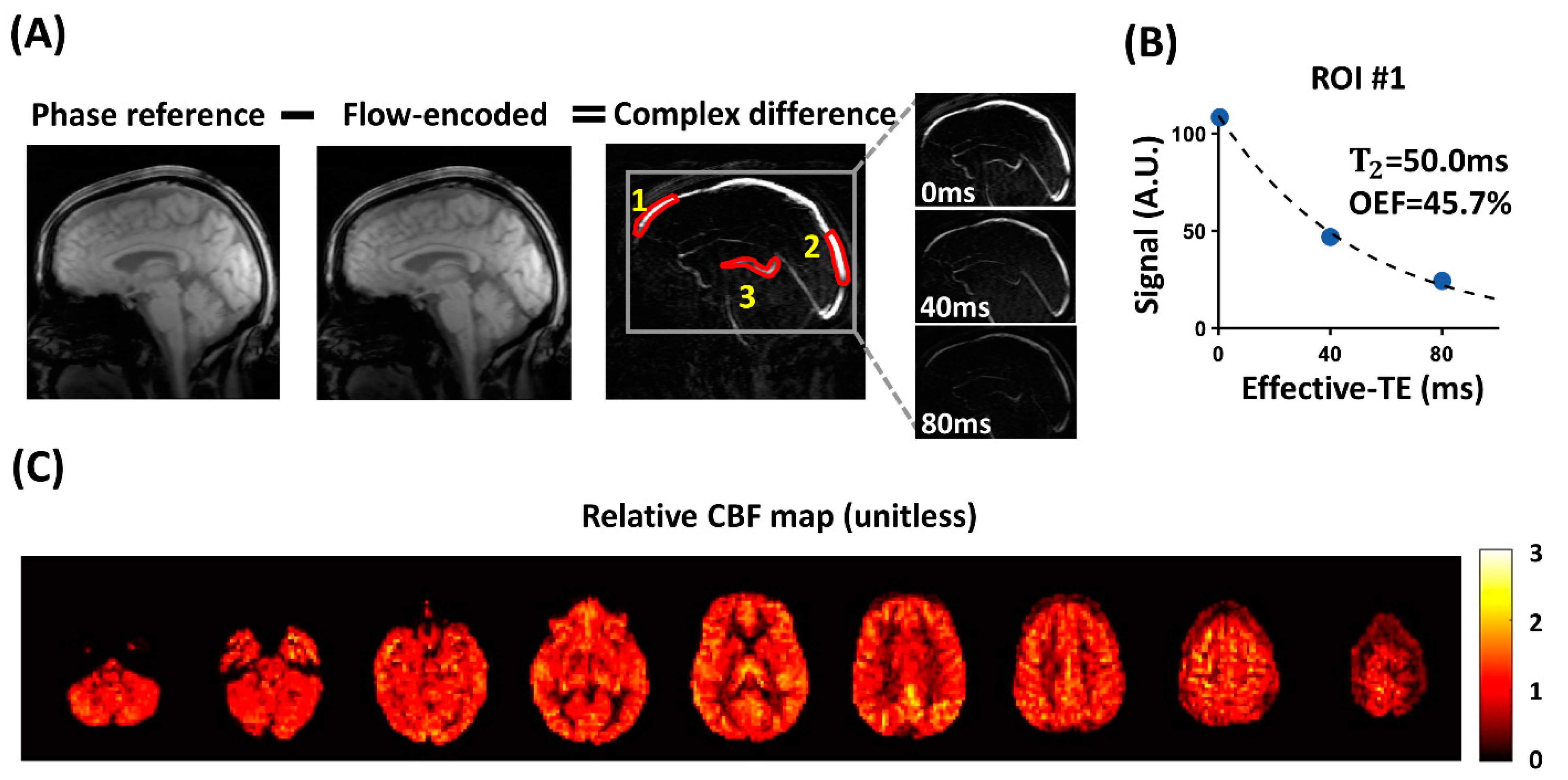
2.3. aTRUPC Data Processing

The aTRUPC data were processed to derive Yv values in major cerebral veins [13,18] using in-house MATLAB (R2022b, MathWorks, Natick, MA, USA) scripts. The Statistical Parametric Mapping tool (SPM12, release date: 13th January 2020, University College London, London, UK) was utilized for motion correction across images. Complex subtraction was conducted between phase-reference images and flow-encoded images to generate complex difference (CD) images (Figure 1A).

2.3.1. Iterative Maxwell and Eddy-Current Correction

Phase errors (Δε) induced by Maxwell concomitant field and eddy-current effects result in residual tissue signals in the CD images, which may introduce bias into the T2 quantification and thus need to be corrected [13]. When there are no Maxwell concomitant field or eddy-current effects, the phase difference (Δφ) between the phase-reference and flow-encoded images should be zero for static tissue voxels. Therefore, Δε can be estimated from the measured Δφ of static tissue voxels. To automatically compute a static tissue mask and perform the phase correction, we employed the following iterative algorithm:
Step 1: A brain mask was derived from the phase-reference image, and an initial vessel mask was obtained from the CD image at eTE = 0 ms, both using Otsu’s method [19]. Note that this initial vessel mask may contain non-vessel voxels due to residual tissue signals in the CD image.
Step 2: A static tissue mask was generated by subtracting the vessel mask from the brain mask. Δφ within the static tissue mask was fitted to a hyperplane [13,20]:
Δ φ = c 1 + c 2 X + c 3 Y + c 4 X Y + c 5 X 2 + c 6 Y 2
Step 3: The phase error Δε was estimated for each voxel as the corresponding values on the fitted hyperplane. Subsequently, Δε was removed from the complex subtraction procedure, resulting in corrected CD images:
C D c o r r e c t e d = I p h a s e r e f e r e n c e · e i Δ ε I f l o w e n c o d e d
As the residual tissue signals were largely eliminated in CDcorrected, we recomputed the vessel mask using CDcorrected and repeated Step 2 and Step 3. Based on our experience, five iterations were sufficient to achieve convergence for this iterative algorithm.

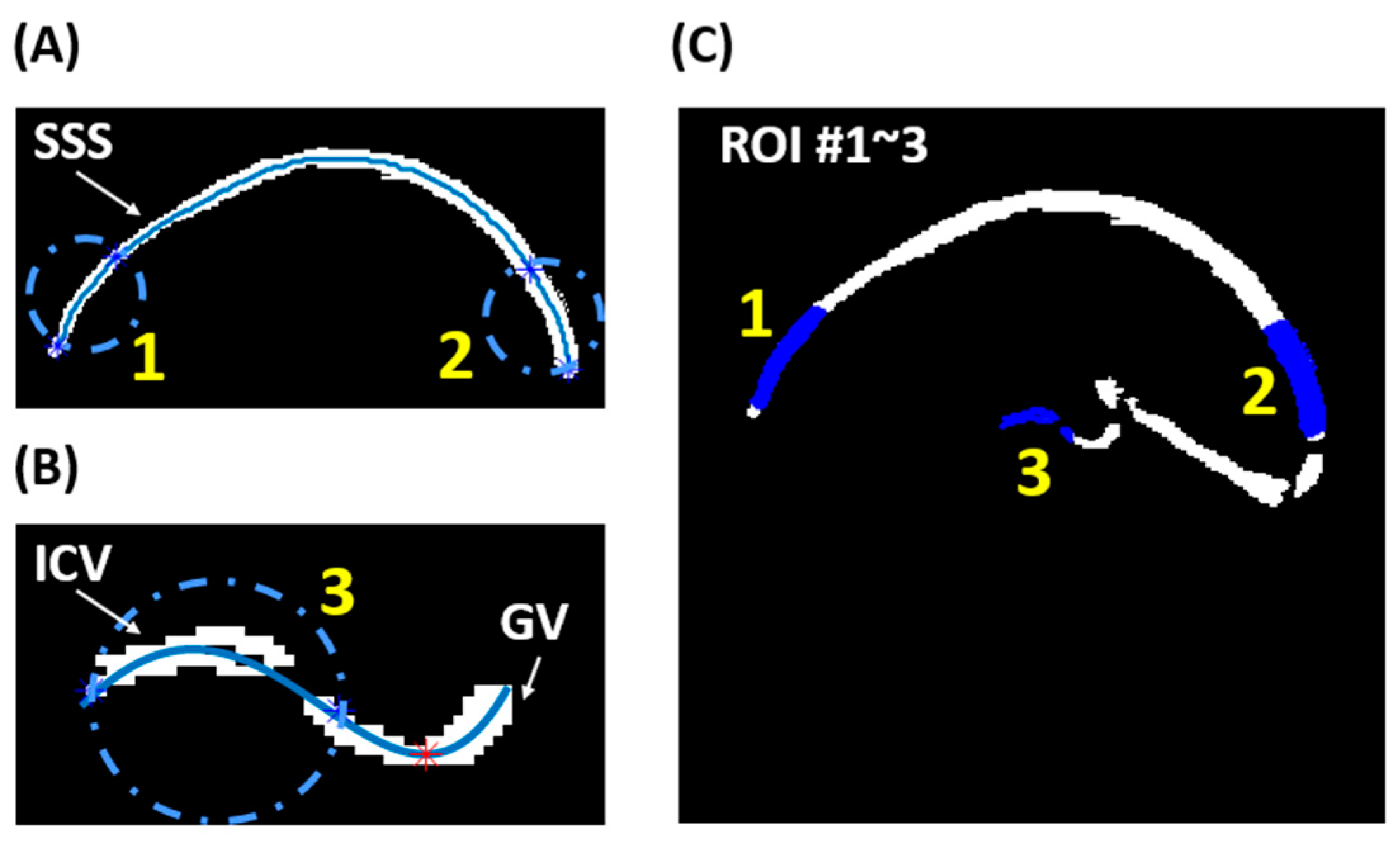
2.3.2. Semi-Automatic ROI Analysis

For quantification purposes, three regions of interest (ROIs) were determined using a semi-automatic algorithm: ROI #1 and #2 were positioned near the frontal and posterior ends of the superior sagittal sinus (SSS), respectively, while ROI #3 included the internal cerebral veins (ICVs). The locations of the three ROIs are illustrated in Figure 1.
To minimize rater dependence in selecting the ROIs, we developed a semi-automatic algorithm. Initially, a preliminary vessel mask was manually drawn on CDcorrected at eTE = 0 ms to encompass major cerebral veins, including the SSS, straight sinus, the vein of Galen (GV), and ICVs. The final vessel mask was obtained by applying a threshold within this preliminary vessel mask [13].
For ROI #1 and #2, we extracted the skeleton of the SSS segment of the final vessel mask, as shown in Figure 2A. The anteriormost and posteriormost 30 mm segments along the SSS skeleton were selected as ROI #1 and #2, respectively.
For ROI #3, we extracted the skeleton of the ICV and GV segments from the final vessel mask, as depicted in Figure 2B. To distinguish the ICV from GV, we smoothed the skeleton using polynomial curve fitting and identified the lowest point of the smoothed curve (red asterisk in Figure 2B). ROI #3 was selected as the skeleton segment located 10 mm anterior to the red asterisk. Figure 2C presents an overlay of the final three ROIs on the final vessel mask.
Signals within each ROI were spatially averaged and fitted as a monoexponential function of eTEs to yield the blood T2, which was subsequently translated to Yv using a published calibration model that has been validated against the gold-standard 15O-PET method [17,21]. The T2 − Yv conversion factored in the hematocrit level, which was assumed to be 0.4 for females and 0.42 for males. Subsequently, the OEF of each ROI was calculated as OEF = (Ya − Yv)/Ya × 100%, where Ya represents the arterial oxygenation measured with a pulse oximeter on the subject’s finger outside of the MRI scanner room.

2.4. pCASL Data Processing

The pCASL data were processed using the open access ASL-MRICloud tool (Version 5) [22] to obtain voxel-wise maps of CBF. To pair with aTRUPC regional OEF measurements, we derived the average CBF values from the three selected ROIs based on T1-MPRAGE segmentations using the MRICloud platform [23]. According to the general venous drainage territories, the frontal SSS mainly drains the frontal gray matter (GM) and the posterior SSS receives drainage from most of the neocortical GM, whereas the ICVs mainly drain the basal ganglia and thalami (BGT) [24]. Therefore, to pair with the OEF ROIs, CBF ROI #1 covered the frontal GM, CBF ROI #2 included the GM of all brain lobes, and CBF ROI #3 covered the BGT. It is known that the quantification of absolute CBF in pCASL can be influenced by several methodological factors such as variations in labeling efficiency, arterial transit time, and blood/tissue T1 [14,25,26]. Therefore, this study focused on the comparison of relative CBF (rCBF) values that are regional CBF values normalized using whole-brain averaged CBF.

2.5. Statistical Analysis

All statistical analyses were conducted using MATLAB (R2022b). The Shapiro–Wilk test was employed to assess the normality of the data. Data with a normal distribution were presented as mean ± standard deviation, whereas data not following a normal distribution were presented as median and range. A two-sample t-test, Wilcoxon rank-sum test, or Fisher’s exact test was utilized to compare the characteristics between COVID-19 ICU survivors and control subjects, as deemed appropriate.
Linear regression analyses were employed to compare the OEF and rCBF values of each ROI between the COVID-19 ICU survivors and the control group, with age as a covariate. We also repeated the regression analyses after adding sex as an additional covariate.
In all analyses, a two-tailed p-value < 0.05 was considered statistically significant.

3. Results

Table 1 lists the characteristics of COVID-19 ICU survivors and healthy controls, showing no significant differences in sex (p = 1.00), age (p = 0.71), or Ya (p = 0.72) between the two groups. Regarding COVID-19 ICU survivors, the median time from symptom onset to ICU admission was 1 day (ranging from 1 to 8 days), with an average ICU stay of 20.0 ± 18.2 days and an average length of hospital stay of 29.1 ± 25.1 days. Two patients were intubated during their ICU stay. The average interval between ICU admission and the MRI scan was 118.6 ± 30.3 days. A subset of patients reported post-acute COVID-19 symptoms, including coughing (N = 3), shortness of breath (N = 4), and fatigue (N = 3).
Figure 1 shows representative aTRUPC OEF and pCASL CBF data of a COVID-19 ICU survivor (female, 56 years old). Table 2 lists the ROI OEF and rCBF values of COVID-19 ICU survivors and healthy controls.
Table 3 summarizes the results of the linear regression analyses. We found that COVID-19 ICU survivors had significantly elevated OEF in the frontal SSS (p = 0.047) and reduced rCBF in the frontal GM (p = 0.003). No significant differences in OEF or rCBF were found in other ROIs (p > 0.2). In addition, OEF in the frontal SSS increased with age (β = 0.11 ± 0.05%/year, p = 0.03). rCBF in the frontal GM decreased with age (β = −0.0018 ± 0.00051/year, p = 0.002), while rCBF in BGT increased with age (β = 0.0031 ± 0.00064/year, p = 0.0001). We repeated the regression analyses after adding sex as an additional covariate. We found that sex did not have a significant effect (p > 0.1), and the findings concerning OEF and CBF remained consistent (Supplemental Material Table S1), although the group difference in OEF in the frontal SSS became marginally significant (p = 0.054).

4. Discussion

In this pilot study, we observed that, compared to healthy controls, COVID-19 ICU survivors exhibited significantly increased OEF and decreased rCBF in the frontal lobe, consistent with the “misery perfusion” pattern.
The frontal lobe, including the prefrontal cortex, plays a critical role in high-level cognitive functions such as memory, attention, and executive function [27]. Studies investigating the neurocognitive sequelae of COVID-19 have reported impaired memory, attention, language, and executive functions among post-infection patients [28,29,30]. Specifically, in hospitalized COVID-19 patients, the most severely affected cognitive domains appear to be memory and executive function [30]. In our study, we observed misery perfusion in the frontal lobe in COVID-19 ICU survivors with an average interval of 118.6 ± 30.3 days after ICU admission. This finding suggests persistent microvascular damage in the frontal lobe of these patients, which may disrupt the delivery of oxygen and nutrients to brain tissues, thereby contributing to deficits in cognitive functions.
Cerebral microvascular damage associated with COVID-19 has been well documented in both post-mortem and in vivo studies [5,31,32,33], yet the underlying mechanisms remain unclear. For example, in a recent autopsy study examining the brains of patients who died of COVID-19, Lee et al. demonstrated multifocal vascular damage characterized by the leakage of serum proteins into the brain parenchyma, accompanied by widespread endothelial cell activation [5]. An in vivo MRI study also suggested blood–brain barrier breakdown in COVID-19 ICU survivors [34]. It is worth noting that autopsy studies have either found no presence of the SARS-CoV-2 virus in the brain or detected it at very low levels, suggesting that the neurovascular injury is more likely a result of cytotoxicity targeting endothelial cells induced by antibodies or cytokines, rather than the direct SARS-CoV-2 infection of the endothelium [5,35,36].
Several studies have reported post-acute physiological or metabolic abnormalities in the frontal lobe or other brain regions among patients following COVID-19 infections. Research utilizing 18-fluorodeoxyglucose positron emission tomography (18F-FDG PET) has identified impaired glucose metabolism in various brain regions, including the frontal lobe, temporal lobe, thalamus, brainstem, and cerebellum in patients with PCS [30,37,38]. Investigations employing pCASL have revealed hypoperfusion in the frontal lobe [39,40], temporal lobe [40], subcortical nuclei, and limbic structures [41] among post-COVID-19 patients. A recent study showed increased global OEF in PCS patients compared to healthy controls, with higher OEF associated with slower gait speed [42]. To the best of our knowledge, our study is the first to investigate post-acute alterations in the regional OEF among patients following COVID-19 infections. The elevated OEF we observed in the frontal lobe likely represents a compensatory response to reduced blood flow.
In addition, we found significant age effects on OEF and rCBF in this study (Table 3). The observed positive association between age and OEF in the frontal SSS aligned with several previous studies indicating an increase in OEF with age [18,43,44,45]. Regarding CBF, it has been consistently reported that the CBF of cortical GM decreased with age [46,47,48]. The literature on subcortical GM yields mixed results. While some studies report insignificant age-related changes in the CBF of subcortical GM [48], others show an increase in the CBF of subcortical GM with age [49,50]. In our study, we observed a negative association between age and rCBF in the frontal GM, contrasting with a positive association between age and rCBF in the BGT. Since rCBF was computed as the ratio of ROI CBF to whole-brain averaged CBF, our results indicated that the rate of age-related CBF decline is faster in the frontal GM compared to the whole-brain average, while the decrease rate is slower (or possibly absent) in the BGT. These findings suggest that age effects on brain physiology vary across different brain regions.
The conventional gold standard for measuring OEF and CBF in humans is PET with 15O-labeled radiotracers [51,52]. However, the broad clinical applications of this 15O-PET method have been impeded by radiation exposure, complex logistics, and the need for an on-site cyclotron to produce the 15O isotope, which has a short half-life of approximately 2 min [51,52,53]. In this pilot study, we utilized aTRUPC MRI to evaluate the regional OEF and employed pCASL to measure the regional CBF. The accuracy of aTRUPC in OEF measurement has been validated against gold-standard blood gas oximetry [54]. Several studies have revealed significant correlations between pCASL and 15O-PET in CBF measurement, although bias in absolute CBF values may exist [55,56]. Since both aTRUPC and pCASL are non-invasive; do not involve any ionizing radiation, nor do they require any contrast agents; and can be performed on standard clinical MRI scanners, they have great potential for broad clinical applications. Therefore, the findings of this work may be easily translated to other studies.
Our study had several limitations. Firstly, the sample size of COVID-19 ICU survivors was small. Therefore, this study should be viewed as a pilot study, and our findings are intended to generate hypotheses for future large-scale studies in PCS patients.
Secondly, we did not conduct standardized cognitive tests on all participants because the primary goal of this pilot study was to assess whether COVID-19 ICU survivors exhibited alterations in brain perfusion and oxygen extraction. Consequently, we were unable to examine the associations of OEF or CBF measures with cognitive dysfunctions, which are known to frequently occur in PCS patients [28,29,30]. Several 18F-FDG PET studies have reported associations between cerebral glucose hypometabolism and cognitive dysfunctions in PCS patients. For example, Hosp et al. found that the expression of a frontoparietal hypometabolism pattern was associated with impaired cognition evaluated using the Montreal Cognitive Assessment [30]. Guedj et al. reported that the decreased metabolism of the brainstem and cerebellum was correlated with an increased number of functional complaints [38]. On the other hand, Blazhenets et al. showed that the reversal of the frontoparietal hypometabolism pattern was accompanied by a significant improvement in cognition [57]. Since the brain’s glucose metabolism is closely related to its oxygen metabolism [58], which is determined by brain perfusion and oxygen extraction, it is possible that the OEF and CBF measures may be correlated with patients’ cognitive performance and may be useful in monitoring the progression of PCS or patients’ recovery from COVID-19. Evaluating such correlations will be the goal of our future studies.
Thirdly, it has been reported that ICU admission itself may lead to long-lasting complications in ICU survivors, known as post-ICU syndrome [59,60]. Since our control group did not experience ICU admission, it was not possible for us to differentiate COVID-19-related effects from those associated with post-ICU syndrome. Future studies should involve subjects with a history of ICU admission due to non-COVID diseases.

5. Conclusions

This pilot study provided preliminary evidence indicating that COVID-19 ICU survivors exhibited misery perfusion in the frontal lobe. Physiological parameters such as OEF and CBF may offer new tools to improve our understanding of long-term alterations in brain physiology and function after severe COVID-19 infection.

Supplementary Materials

The following supporting information can be downloaded at: https://www.mdpi.com/article/10.3390/brainsci14010094/s1, Table S1. Results of linear regression analyses for regional OEF and rCBF, with sex as an additional covariate.

Author Contributions

Conceptualization, S.-M.C., H.L. and D.J.; methodology, J.S., D.J., H.L., S.-M.C., S.K. and H.R.; software, J.S., D.J. and H.L.; formal analysis, J.S., D.J. and S.K.; investigation, J.S., S.K., H.R., W.S., K.H., G.P.P., E.J., C.X., Z.H. and D.J.; resources, S.-M.C., H.L., D.L, S.Y. and D.J.; data curation, D.J. and S.K.; writing—original draft preparation, J.S. and D.J.; writing—review and editing, J.S., D.J., H.L., S.-M.C., D.L. and S.Y.; visualization, J.S.; supervision, H.L., S.-M.C. and D.J.; funding acquisition, H.L., S.-M.C. and D.J. All authors have read and agreed to the published version of the manuscript.

Funding

This work was supported by the National Institutes of Health: R21 AG079098, R01 AG085299, R01 AG064792, U01 NS100588, RF1 AG071515, R01 NS106711, R01 NS106702, and P41 EB031771.

Institutional Review Board Statement

The study protocol was approved by the Johns Hopkins University Institutional Review Board (IRB00276578, approved on 19 May 2021 and IRB00048412, approved on 18 October 2021).

Informed Consent Statement

Informed consent was obtained from all subjects involved in the study.

Data Availability Statement

Data will be made available on request due to privacy restrictions. The source code of this work is available at: https://github.com/NeuroFunctionLab/aTRUPC_processing (accessed on 16 December 2023).

Acknowledgments

The authors thank Jessica Briscoe from The Johns Hopkins University School of Medicine for her assistance in data collection from the COVID-19 ICU survivors.

Conflicts of Interest

The authors declare that they have no conflicts of interest.

References

  1. Nalbandian, A.; Sehgal, K.; Gupta, A.; Madhavan, M.V.; McGroder, C.; Stevens, J.S.; Cook, J.R.; Nordvig, A.S.; Shalev, D.; Sehrawat, T.S.; et al. Post-acute COVID-19 syndrome. Nat. Med. 2021, 27, 601–615. [Google Scholar] [CrossRef]
  2. Venkatesan, P. NICE guideline on long COVID. Lancet Respir. Med. 2021, 9, 129. [Google Scholar] [CrossRef] [PubMed]
  3. Premraj, L.; Kannapadi, N.V.; Briggs, J.; Seal, S.M.; Battaglini, D.; Fanning, J.; Suen, J.; Robba, C.; Fraser, J.; Cho, S.M. Mid and long-term neurological and neuropsychiatric manifestations of post-COVID-19 syndrome: A meta-analysis. J. Neurol. Sci. 2022, 434, 120162. [Google Scholar] [CrossRef] [PubMed]
  4. Mattioli, F.; Piva, S.; Stampatori, C.; Righetti, F.; Mega, I.; Peli, E.; Sala, E.; Tomasi, C.; Indelicato, A.M.; Latronico, N.; et al. Neurologic and cognitive sequelae after SARS-CoV2 infection: Different impairment for ICU patients. J. Neurol. Sci. 2022, 432, 120061. [Google Scholar] [CrossRef] [PubMed]
  5. Lee, M.H.; Perl, D.P.; Steiner, J.; Pasternack, N.; Li, W.; Maric, D.; Safavi, F.; Horkayne-Szakaly, I.; Jones, R.; Stram, M.N.; et al. Neurovascular injury with complement activation and inflammation in COVID-19. Brain 2022, 145, 2555–2568. [Google Scholar] [CrossRef] [PubMed]
  6. Leng, A.; Shah, M.; Ahmad, S.A.; Premraj, L.; Wildi, K.; Li Bassi, G.; Pardo, C.A.; Choi, A.; Cho, S.M. Pathogenesis Underlying Neurological Manifestations of Long COVID Syndrome and Potential Therapeutics. Cells 2023, 12, 816. [Google Scholar] [CrossRef]
  7. Grubb, R.L., Jr.; Derdeyn, C.P.; Fritsch, S.M.; Carpenter, D.A.; Yundt, K.D.; Videen, T.O.; Spitznagel, E.L.; Powers, W.J. Importance of hemodynamic factors in the prognosis of symptomatic carotid occlusion. JAMA 1998, 280, 1055–1060. [Google Scholar] [CrossRef] [PubMed]
  8. Yamauchi, H.; Higashi, T.; Kagawa, S.; Nishii, R.; Kudo, T.; Sugimoto, K.; Okazawa, H.; Fukuyama, H. Is misery perfusion still a predictor of stroke in symptomatic major cerebral artery disease? Brain 2012, 135, 2515–2526. [Google Scholar] [CrossRef]
  9. Stone, A.J.; Harston, G.W.J.; Carone, D.; Okell, T.W.; Kennedy, J.; Blockley, N.P. Prospects for investigating brain oxygenation in acute stroke: Experience with a non-contrast quantitative BOLD based approach. Hum. Brain Mapp. 2019, 40, 2853–2866. [Google Scholar] [CrossRef]
  10. Fan, A.P.; Khalil, A.A.; Fiebach, J.B.; Zaharchuk, G.; Villringer, A.; Villringer, K.; Gauthier, C.J. Elevated brain oxygen extraction fraction measured by MRI susceptibility relates to perfusion status in acute ischemic stroke. J. Cereb. Blood Flow Metab. 2020, 40, 539–551. [Google Scholar] [CrossRef]
  11. An, H.; Ford, A.L.; Chen, Y.; Zhu, H.; Ponisio, R.; Kumar, G.; Shanechi, A.M.; Khoury, N.; Vo, K.D.; Williams, J.; et al. Defining the ischemic penumbra using magnetic resonance oxygen metabolic index. Stroke 2015, 46, 982–988. [Google Scholar] [CrossRef] [PubMed]
  12. Nezu, T.; Yokota, C.; Uehara, T.; Yamauchi, M.; Fukushima, K.; Toyoda, K.; Matsumoto, M.; Iide, H.; Minematsu, K. Preserved acetazolamide reactivity in lacunar patients with severe white-matter lesions: 15O-labeled gas and H2O positron emission tomography studies. J. Cereb. Blood Flow Met. 2012, 32, 844–850. [Google Scholar] [CrossRef]
  13. Jiang, D.; Lu, H.; Parkinson, C.; Su, P.; Wei, Z.; Pan, L.; Tekes, A.; Huisman, T.; Golden, W.C.; Liu, P. Vessel-specific quantification of neonatal cerebral venous oxygenation. Magn. Reson. Med. 2019, 82, 1129–1139. [Google Scholar] [CrossRef]
  14. Alsop, D.C.; Detre, J.A.; Golay, X.; Gunther, M.; Hendrikse, J.; Hernandez-Garcia, L.; Lu, H.; MacIntosh, B.J.; Parkes, L.M.; Smits, M.; et al. Recommended implementation of arterial spin-labeled perfusion MRI for clinical applications: A consensus of the ISMRM perfusion study group and the European consortium for ASL in dementia. Magn. Reson. Med. 2015, 73, 102–116. [Google Scholar] [CrossRef] [PubMed]
  15. Dai, W.; Garcia, D.; de Bazelaire, C.; Alsop, D.C. Continuous flow-driven inversion for arterial spin labeling using pulsed radio frequency and gradient fields. Magn. Reson. Med. 2008, 60, 1488–1497. [Google Scholar] [CrossRef]
  16. Zhao, L.; Vidorreta, M.; Soman, S.; Detre, J.A.; Alsop, D.C. Improving the robustness of pseudo-continuous arterial spin labeling to off-resonance and pulsatile flow velocity. Magn. Reson. Med. 2017, 78, 1342–1351. [Google Scholar] [CrossRef]
  17. Lu, H.Z.; Xu, F.; Grgac, K.; Liu, P.Y.; Qin, Q.; van Zijl, P. Calibration and validation of TRUST MRI for the estimation of cerebral blood oxygenation. Magn. Reson. Med. 2012, 67, 42–49. [Google Scholar] [CrossRef]
  18. Jiang, D.; Liu, P.; Lin, Z.; Hazel, K.; Pottanat, G.; Lucke, E.; Moghekar, A.; Pillai, J.J.; Lu, H. MRI assessment of cerebral oxygen extraction fraction in the medial temporal lobe. Neuroimage 2023, 266, 119829. [Google Scholar] [CrossRef]
  19. Otsu, N. A Threshold Selection Method from Gray-Level Histograms. IEEE Trans. Syst. Man. Cybern. 1979, 9, 62–66. [Google Scholar] [CrossRef]
  20. Bernstein, M.A.; Zhou, X.H.J.; Polzin, J.A.; King, K.F.; Ganin, A.; Pelc, N.J.; Glover, G.H. Concomitant gradient terms in phase contrast MR: Analysis and correction. Magn. Reson. Med. 1998, 39, 300–308. [Google Scholar] [CrossRef]
  21. Jiang, D.R.; Deng, S.W.; Franklin, C.G.; O’Boyle, M.; Zhang, W.; Heyl, B.L.; Pan, L.; Jerabek, P.A.; Fox, P.T.; Lu, H.Z. Validation of T2-based oxygen extraction fraction measurement with 15O positron emission tomography. Magn. Reson. Med. 2021, 85, 304–311. [Google Scholar] [CrossRef] [PubMed]
  22. Li, Y.A.-O.; Liu, P.; Li, Y.; Fan, H.; Su, P.; Peng, S.A.-O.; Park, D.C.; Rodrigue, K.M.; Jiang, H.; Faria, A.V.; et al. ASL-MRICloud: An online tool for the processing of ASL MRI data. NMR Biomed. 2019, 32, e4051. [Google Scholar] [CrossRef] [PubMed]
  23. Mori, S.; Wu, D.; Ceritoglu, C.; Li, Y.; Kolasny, A.; Vaillant, M.A.; Faria, A.V.; Oishi, K.; Miller, M.I. MRICloud: Delivering High-Throughput MRI Neuroinformatics as Cloud-Based Software as a Service. Comput. Sci. Eng. 2016, 18, 21–35. [Google Scholar] [CrossRef]
  24. Rhoton, A.L., Jr. The cerebral veins. Neurosurgery 2002, 51, S159–S205. [Google Scholar] [CrossRef] [PubMed]
  25. Chen, Z.; Zhang, X.; Yuan, C.; Zhao, X.; van Osch, M.J.P. Measuring the labeling efficiency of pseudocontinuous arterial spin labeling. Magn. Reason Med. 2017, 77, 1841–1852. [Google Scholar] [CrossRef] [PubMed]
  26. Chen, Z.; Zhao, X.; Zhang, X.; Guo, R.; Teeuwisse, W.M.; Zhang, B.; Koken, P.; Smink, J.; Yuan, C.; van Osch, M.J.P. Simultaneous measurement of brain perfusion and labeling efficiency in a single pseudo-continuous arterial spin labeling scan. Magn. Reson. Med. 2018, 79, 1922–1930. [Google Scholar] [CrossRef]
  27. Friedman, N.P.; Robbins, T.W. The role of prefrontal cortex in cognitive control and executive function. Neuropsychopharmacology 2022, 47, 72–89. [Google Scholar] [CrossRef]
  28. Becker, J.H.; Lin, J.J.; Doernberg, M.; Stone, K.; Navis, A.; Festa, J.R.; Wisnivesky, J.P. Assessment of Cognitive Function in Patients After COVID-19 Infection. JAMA Netw. Open 2021, 4, e2130645. [Google Scholar] [CrossRef]
  29. Zhao, S.; Shibata, K.; Hellyer, P.J.; Trender, W.; Manohar, S.; Hampshire, A.; Husain, M. Rapid vigilance and episodic memory decrements in COVID-19 survivors. Brain Commun. 2022, 4, fcab295. [Google Scholar] [CrossRef]
  30. Hosp, J.A.; Dressing, A.; Blazhenets, G.; Bormann, T.; Rau, A.; Schwabenland, M.; Thurow, J.; Wagner, D.; Waller, C.; Niesen, W.D.; et al. Cognitive impairment and altered cerebral glucose metabolism in the subacute stage of COVID-19. Brain 2021, 144, 1263–1276. [Google Scholar] [CrossRef]
  31. Paniz-Mondolfi, A.; Bryce, C.; Grimes, Z.; Gordon, R.E.; Reidy, J.; Lednicky, J.; Sordillo, E.M.; Fowkes, M. Central nervous system involvement by severe acute respiratory syndrome coronavirus-2 (SARS-CoV-2). J. Med. Virol. 2020, 92, 699–702. [Google Scholar] [CrossRef]
  32. Franceschi, A.M.; Arora, R.; Wilson, R.; Giliberto, L.; Libman, R.B.; Castillo, M. Neurovascular Complications in COVID-19 Infection: Case Series. AJNR Am. J. Neuroradiol. 2020, 41, 1632–1640. [Google Scholar] [CrossRef]
  33. Agarwal, S.; Jain, R.; Dogra, S.; Krieger, P.; Lewis, A.; Nguyen, V.; Melmed, K.; Galetta, S. Cerebral Microbleeds and Leukoencephalopathy in Critically Ill Patients With COVID-19. Stroke 2020, 51, 2649–2655. [Google Scholar] [CrossRef]
  34. Shi, W.; Jiang, D.; Rando, H.; Khanduja, S.; Lin, Z.; Hazel, K.; Pottanat, G.; Jones, E.; Xu, C.; Lin, D.; et al. Blood-brain barrier breakdown in COVID-19 ICU survivors: An MRI pilot study. NeuroImmune Pharm. Ther. 2023, 2, 333–338. [Google Scholar] [CrossRef] [PubMed]
  35. Matschke, J.; Lutgehetmann, M.; Hagel, C.; Sperhake, J.P.; Schroder, A.S.; Edler, C.; Mushumba, H.; Fitzek, A.; Allweiss, L.; Dandri, M.; et al. Neuropathology of patients with COVID-19 in Germany: A post-mortem case series. Lancet Neurol. 2020, 19, 919–929. [Google Scholar] [CrossRef] [PubMed]
  36. Thakur, K.T.; Miller, E.H.; Glendinning, M.D.; Al-Dalahmah, O.; Banu, M.A.; Boehme, A.K.; Boubour, A.L.; Bruce, S.S.; Chong, A.M.; Claassen, J.; et al. COVID-19 neuropathology at Columbia University Irving Medical Center/New York Presbyterian Hospital. Brain 2021, 144, 2696–2708. [Google Scholar] [CrossRef]
  37. Meyer, P.T.; Hellwig, S.; Blazhenets, G.; Hosp, J.A. Molecular Imaging Findings on Acute and Long-Term Effects of COVID-19 on the Brain: A Systematic Review. J. Nucl. Med. 2022, 63, 971–980. [Google Scholar] [CrossRef] [PubMed]
  38. Guedj, E.; Campion, J.Y.; Dudouet, P.; Kaphan, E.; Bregeon, F.; Tissot-Dupont, H.; Guis, S.; Barthelemy, F.; Habert, P.; Ceccaldi, M.; et al. 18F-FDG brain PET hypometabolism in patients with long COVID. Eur. J. Nucl. Med. Mol. Imaging 2021, 48, 2823–2833. [Google Scholar] [CrossRef]
  39. Ajcevic, M.; Iscra, K.; Furlanis, G.; Michelutti, M.; Miladinovic, A.; Buoite Stella, A.; Ukmar, M.; Cova, M.A.; Accardo, A.; Manganotti, P. Cerebral hypoperfusion in post-COVID-19 cognitively impaired subjects revealed by arterial spin labeling MRI. Sci. Rep. 2023, 13, 5808. [Google Scholar] [CrossRef]
  40. Helms, J.; Kremer, S.; Merdji, H.; Clere-Jehl, R.; Schenck, M.; Kummerlen, C.; Collange, O.; Boulay, C.; Fafi-Kremer, S.; Ohana, M.; et al. Neurologic Features in Severe SARS-CoV-2 Infection. N. Engl. J. Med. 2020, 382, 2268–2270. [Google Scholar] [CrossRef]
  41. Qin, Y.; Wu, J.; Chen, T.; Li, J.; Zhang, G.; Wu, D.; Zhou, Y.; Zheng, N.; Cai, A.; Ning, Q.; et al. Long-term microstructure and cerebral blood flow changes in patients recovered from COVID-19 without neurological manifestations. J. Clin. Investig. 2021, 131, e147329. [Google Scholar] [CrossRef] [PubMed]
  42. Liu, P.; Emst, T.; Liang, H.; Jiang, D.; Cunningham, E.; Lin, Z.; Ryan, M.C.; Lu, H.; Wilson, E.; Levine, A.; et al. Lower Cerebral Venous Oxygenation in Post-acute Sequelae of COVID-19. In Proceedings of the 30th Annual Meeting of ISMRM, London, UK, 7–12 May 2022; p. 2207. [Google Scholar]
  43. Lin, Z.; Lim, C.; Jiang, D.; Soldan, A.; Pettigrew, C.; Oishi, K.; Zhu, Y.; Moghekar, A.; Liu, P.; Albert, M.; et al. Longitudinal changes in brain oxygen extraction fraction (OEF) in older adults: Relationship to markers of vascular and Alzheimer’s pathology. Alzheimer’s Dement. 2023, 19, 569–577. [Google Scholar] [CrossRef] [PubMed]
  44. Catchlove, S.J.; Macpherson, H.; Hughes, M.E.; Chen, Y.; Parrish, T.B.; Pipingas, A. An investigation of cerebral oxygen utilization, blood flow and cognition in healthy aging. PLoS ONE 2018, 13, e0197055. [Google Scholar] [CrossRef]
  45. Aanerud, J.; Borghammer, P.; Chakravarty, M.M.; Vang, K.; Rodell, A.B.; Jonsdottir, K.Y.; Moller, A.; Ashkanian, M.; Vafaee, M.S.; Iversen, P.; et al. Brain energy metabolism and blood flow differences in healthy aging. J. Cereb. Blood Flow Metab. 2012, 32, 1177–1187. [Google Scholar] [CrossRef] [PubMed]
  46. Staffaroni, A.M.; Cobigo, Y.; Elahi, F.M.; Casaletto, K.B.; Walters, S.M.; Wolf, A.; Lindbergh, C.A.; Rosen, H.J.; Kramer, J.H. A longitudinal characterization of perfusion in the aging brain and associations with cognition and neural structure. Hum. Brain Mapp. 2019, 40, 3522–3533. [Google Scholar] [CrossRef] [PubMed]
  47. Juttukonda, M.R.; Li, B.; Almaktoum, R.; Stephens, K.A.; Yochim, K.M.; Yacoub, E.; Buckner, R.L.; Salat, D.H. Characterizing cerebral hemodynamics across the adult lifespan with arterial spin labeling MRI data from the Human Connectome Project-Aging. Neuroimage 2021, 230, 117807. [Google Scholar] [CrossRef]
  48. Chen, J.J.; Rosas, H.D.; Salat, D.H. Age-associated reductions in cerebral blood flow are independent from regional atrophy. Neuroimage 2011, 55, 468–478. [Google Scholar] [CrossRef]
  49. Pagani, M.; Salmaso, D.; Jonsson, C.; Hatherly, R.; Jacobsson, H.; Larsson, S.A.; Wägner, A. Regional cerebral blood flow as assessed by principal component analysis and 99mTc-HMPAO SPET in healthy subjects at rest: Normal distribution and effect of age and gender. Eur. J. Nucl. Med. Mol. Imaging 2002, 29, 67–75. [Google Scholar]
  50. Lee, C.; Lopez, O.L.; Becker, J.T.; Raji, C.; Dai, W.; Kuller, L.H.; Gach, H.M. Imaging cerebral blood flow in the cognitively normal aging brain with arterial spin labeling: Implications for imaging of neurodegenerative disease. J. Neuroimaging 2009, 19, 344–352. [Google Scholar] [CrossRef]
  51. Mintun, M.A.; Raichle, M.E.; Martin, W.R.; Herscovitch, P. Brain oxygen utilization measured with O-15 radiotracers and positron emission tomography. J. Nucl. Med. 1984, 25, 177–187. [Google Scholar]
  52. Fox, P.T.; Raichle, M.E. Focal physiological uncoupling of cerebral blood flow and oxidative metabolism during somatosensory stimulation in human subjects. Proc. Natl. Acad. Sci. USA 1986, 83, 1140–1144. [Google Scholar] [CrossRef] [PubMed]
  53. Frackowiak, R.S.; Pozzilli, C.; Legg, N.J.; Du Boulay, G.H.; Marshall, J.; Lenzi, G.L.; Jones, T. Regional cerebral oxygen supply and utilization in dementia. A clinical and physiological study with oxygen-15 and positron tomography. Brain 1981, 104, 753–778. [Google Scholar] [CrossRef] [PubMed]
  54. Jiang, D.; Koehler, R.C.; Liu, X.; Kulikowicz, E.; Lee, J.K.; Lu, H.; Liu, P. Quantitative validation of MRI mapping of cerebral venous oxygenation with direct blood sampling: A graded-O(2) study in piglets. Magn. Reason. Med 2021, 86, 1445–1453. [Google Scholar] [CrossRef]
  55. Puig, O.; Henriksen, O.M.; Vestergaard, M.B.; Hansen, A.E.; Andersen, F.L.; Ladefoged, C.N.; Rostrup, E.; Larsson, H.B.; Lindberg, U.; Law, I. Comparison of simultaneous arterial spin labeling MRI and 15O-H2O PET measurements of regional cerebral blood flow in rest and altered perfusion states. J. Cereb. Blood Flow Metab. 2020, 40, 1621–1633. [Google Scholar] [CrossRef]
  56. Fan, A.P.; Jahanian, H.; Holdsworth, S.J.; Zaharchuk, G. Comparison of cerebral blood flow measurement with [15O]-water positron emission tomography and arterial spin labeling magnetic resonance imaging: A systematic review. J. Cereb. Blood Flow Metab. 2016, 36, 842–861. [Google Scholar] [CrossRef]
  57. Blazhenets, G.; Schroeter, N.; Bormann, T.; Thurow, J.; Wagner, D.; Frings, L.; Weiller, C.; Meyer, P.T.; Dressing, A.; Hosp, J.A. Slow but Evident Recovery from Neocortical Dysfunction and Cognitive Impairment in a Series of Chronic COVID-19 Patients. J. Nucl. Med. 2021, 62, 910–915. [Google Scholar] [CrossRef]
  58. Hyder, F.; Herman, P.; Bailey, C.J.; Moller, A.; Globinsky, R.; Fulbright, R.K.; Rothman, D.L.; Gjedde, A. Uniform distributions of glucose oxidation and oxygen extraction in gray matter of normal human brain: No evidence of regional differences of aerobic glycolysis. J. Cereb. Blood Flow Metab. 2016, 36, 903–916. [Google Scholar] [CrossRef]
  59. Desai, S.V.; Law, T.J.; Needham, D.M. Long-term complications of critical care. Crit. Care Med. 2011, 39, 371–379. [Google Scholar] [CrossRef]
  60. Pandharipande, P.P.; Girard, T.D.; Jackson, J.C.; Morandi, A.; Thompson, J.L.; Pun, B.T.; Brummel, N.E.; Hughes, C.G.; Vasilevskis, E.E.; Shintani, A.K.; et al. Long-term cognitive impairment after critical illness. N. Engl. J. Med. 2013, 369, 1306–1316. [Google Scholar] [CrossRef]
Figure 1. Representative aTRUPC and pCASL data of a COVID-19 ICU survivor: (A) aTRUPC uses phase-contrast complex subtraction between phase-reference and flow-encoded images to yield complex difference (CD) images, in which the static tissues are canceled out, leaving only the blood signals in the vessels. Zoom-in views of the CD images are presented for all eTEs. The red contours on the CD image delineate the ROIs for OEF quantification. (B) Scatterplot showing venous signal of ROI #1 as a function of effective TEs. Fitted venous blood T2 and the corresponding OEF values are also shown. (C) The relative CBF map plotted using pCASL in this patient. Abbreviations: aTRUPC: accelerated T2 relaxation under phase contrast; pCASL: pseudo-continuous arterial spin labeling; COVID-19: coronavirus disease 2019; ICU: intensive care unit; CD: complex difference; eTE: effective echo time; ROI: regions of interest; OEF: oxygen extraction fraction; CBF: cerebral blood flow.
Brainsci 14 00094 g001
Figure 2. Semi-automatic ROI delineation: (A) ROI selection on the SSS; (B) ROI selection for ICV; (C) locations of the three ROIs on the final vessel mask.
Brainsci 14 00094 g002
Table 1. Demographical and clinical characteristics of the participants.
COVID-19 ICU SurvivorsControlp-Value
N719N/A
Sex (Female) *4 (57.1%)12 (63.2%)1.00 a
Age (years) 50 (20–77)64 (22–77)0.71 b
Ya (%) 97.7 ± 1.597.5 ± 1.00.72 c
Length of hospital stay 29.1 ± 25.1NANA
Time from symptom onset to ICU admission (days) 1 (1–8)NANA
Duration of stay in ICU (days) 20.2 ± 18.2NANA
Time from ICU admission to MRI (days) 118.6 ± 30.3NANA
Intubation *2 (28.6%)NANA
Post-acute COVID-19 symptoms
    Coughing *3 (42.9%)NANA
    Shortness of breath *4 (57.1%)NANA
    Fatigue *3 (42.9%)NANA
* Data are numbers of participants, followed by percentages in parentheses. Data are medians, with ranges in parentheses. Data are means ± standard deviations. a Fisher’s exact test. b Wilcoxon rank-sum test. c Two-sample t-test. Abbreviations: COVID-19: coronavirus disease 2019; ICU: intensive care unit; Ya: arterial oxygenation.
Table 2. Regional OEF and rCBF values of COVID-19 ICU survivors and healthy controls.
Variable ROICOVID-19 ICU SurvivorsControl
OEF (%)frontal SSS38.3 ± 9.033.5 ± 4.7
posterior SSS38.2 ± 8.435.4 ± 5.1
ICV32.5 ± 11.527.9 ± 7.7
rCBF (unitless)frontal GM1.20 ± 0.071.27 ± 0.07
GM of all lobes1.22 ± 0.041.23 ± 0.03
BGT0.96 ± 0.150.95 ± 0.08
Data are means ± standard deviation. Abbreviations: OEF: oxygen extraction fraction; rCBF: relative cerebral blood flow; COVID-19: coronavirus disease 2019; ICU: intensive care unit; SSS: superior sagittal sinus; ICV: internal cerebral veins; GM: gray matter; BGT: basal ganglia and thalami.
Table 3. Results of linear regression analyses for regional OEF and rCBF.
ModelDependent VariableIndependent VariablesCoefficient ± Standard Errorp-Value
OEF1OEF in frontal SSSGroup5.21 ± 2.48%0.047
Age0.11 ± 0.050%/year0.03
OEF2OEF in posterior SSSGroup3.06 ± 2.65%0.26
Age0.078 ± 0.053%/year0.16
OEF3OEF in ICVGroup4.90 ± 3.93%0.23
Age0.068 ± 0.079%/year0.40
rCBF1rCBF in frontal GMGroup−0.083 ± 0.0250.003
Age−0.0018 ± 0.00051/year0.002
rCBF2rCBF in GM of all lobesGroup−0.018 ± 0.0150.24
Age−0.00056 ± 0.00030/year0.08
rCBF3rCBF in BGTGroup0.027 ± 0.0320.41
Age0.0031 ± 0.00064/year0.0001
Group = 1 for COVID-19 ICU survivors and Group = 0 for healthy controls. Abbreviations: OEF: oxygen extraction fraction; rCBF: relative cerebral blood flow; SSS: superior sagittal sinus; ICV: internal cerebral veins; GM: gray matter; BGT: basal ganglia and thalami; COVID-19: coronavirus disease 2019; ICU: intensive care unit.
Disclaimer/Publisher’s Note: The statements, opinions and data contained in all publications are solely those of the individual author(s) and contributor(s) and not of MDPI and/or the editor(s). MDPI and/or the editor(s) disclaim responsibility for any injury to people or property resulting from any ideas, methods, instructions or products referred to in the content.

