Article

Correlates of Theta and Gamma Activity during Visuospatial Incidental/Intentional Encoding and Retrieval Indicate Differences in Processing in Young and Elderly Healthy Participants

by
Mariana Lizeth Junco-Muñoz
1,2,
Oliva Mejía-Rodríguez
3,
José Miguel Cervantes-Alfaro
4,
Adriana del Carmen Téllez-Anguiano
5,
Miguel Ángel López-Vázquez
6 and
María Esther Olvera-Cortés
1,*
1
Laboratorio de Neurofisiología Clínica y Experimental, Centro de Investigación Biomédica de Michoacán, Instituto Mexicano del Seguro Social, Morelia 58060, Michoacán, Mexico
2
Facultad de Psicología, Universidad Michoacana de San Nicolás de Hidalgo, Morelia 58194, Michoacán, Mexico
3
División de Investigación Clínica, Centro de Investigación Biomédica de Michoacán, Instituto Mexicano del Seguro Social, Morelia 58060, Michoacán, Mexico
4
Laboratorio de Neurociencias, División de Estudios de Posgrado, Facultad de Ciencias Médicas “Dr. Ignacio Chávez”, Universidad Michoacana de San Nicolás de Hidalgo, Morelia 58194, Michoacán, Mexico
5
Division of Graduate Studies an Research, Tecnológico Nacional de México, Morelia 58120, Michoacán, Mexico
6
Laboratorio de Neuroplasticidad, División de Neurociencias, Centro de Investigación Biomédica de Michoacán, Instituto Mexicano del Seguro Social, Morelia 58060, Michoacán, Mexico
*
Author to whom correspondence should be addressed.
Brain Sci. 2024, 14(8), 786; https://doi.org/10.3390/brainsci14080786
Submission received: 11 June 2024 / Revised: 23 July 2024 / Accepted: 30 July 2024 / Published: 2 August 2024
(This article belongs to the Section Cognitive Social and Affective Neuroscience)

Abstract

:
Incidental visuospatial learning acquired under incidental conditions is more vulnerable to aging than in the intentional case. The theta and gamma correlates of the coding and retrieval of episodic memory change during aging. Based on the vulnerability of incidental coding to aging, different theta and gamma correlates could occur under the incidental versus intentional coding and retrieval of visuospatial information. Theta and gamma EEG was recorded from the frontotemporal regions, and incidental/intentional visuospatial learning was evaluated in young (25–60 years old) and elderly (60–85 years old) participants. The EEG recorded during encoding and retrieval was compared between incidental low-demand, incidental high-demand, and intentional conditions through an ANCOVA considering the patient’s gender, IQ, and years of schooling as covariates. Older adults exhibited worse performances, especially in place–object associations. After the intentional study, older participants showed a further increase in false-positive errors. Higher power at the theta and gamma bands was observed for frontotemporal derivations in older participants for both encoding and retrieval. Under retrieval, only young participants had lower power in terms of errors compared with correct responses. In conclusion, the different patterns of power and coherence support incidental and intentional visuospatial encoding and retrieval in young and elderly individuals. The correlates of power with behavior are sensitive to age and performance.

1. Introduction

Aging is characterized by a decline in several cognitive capabilities, with high variability in different cognitive domains. Behavior flexibility, executive function, declarative memory, and visuospatial learning and memory abilities are among the most vulnerable [1]. One of the factors that strongly influences the impact of aging on memory is the intentionality to acquire information. Compared with intentional encoding, incidental encoding, defined as the acquisition of information without an intended effort, is more vulnerable to aging [2,3,4]. In addition, discrete item information is preserved, but the association of items is impaired in aged people in terms of face–name (e.g., name–face associations were impaired compared with only names or only faces), word–color, and cue–context associations, among others [2,3,5]. Additionally, the combination of these attributes (incidental, visuospatial, and item association) results in greater difficulties in mature adults (45–65 years old) compared with young adults (18–40 years old) [6]. It has been proposed that incidental and intentional memory processes take place in the temporal lobe structures, possibly through different circuits. The temporal lobe is recognized as a crucial structure in the processing of declarative memory for both encoding and retrieval [4]. Neuroimaging studies have shown increased activity in the medial temporal lobe (MTL) during encoding in both intentional and incidental tasks for novel compared with familiar items [7,8]. Additionally, the subsequent effect of memory retrieval is observed in MTL activation during encoding, and increased activation was observed in fMRI for unfamiliar information compared with familiar information in the anterior temporal–medial cortex [9,10]. In addition, several studies have reported increased activity in the anterior temporal–medial region during successful retrieval, whereas no changes were found in other works [9,10,11,12]. It has been suggested that the activation of the anterior MTL occurs when relational information is being processed, whereas posterior activation takes place with familiarity processing [8,12,13]. Additionally, neuroimaging studies have shown the differential participation of the left inferior frontal cortex and the right dorsolateral prefrontal cortex, being more activated under intentional than incidental memory, whereas hippocampal activation was related to successful retrieval but not with the intentionality of remembering [4]. Thus, at present, it is not clear which networks are involved with respect to the intentionality of memory. Declarative memory has been related to changes in power and coherence in the theta and gamma bands expressed in the frontotemporal circuits [14,15,16]. Early works by Klimesch and coworkers showed an evoked increase in theta power associated with the successful encoding of memory in the temporal areas [17], as well as an induced reduction in theta power through the encoding process, which was also observed in the frontal areas [18]. In addition, theta synchronization between the frontal and parietal–temporal posterior regions has been observed during success memory encoding [19]. A subsequent memory effect on EEG oscillations with increased theta and reduced alpha evoked power has been observed under the encoding of semantic memory. Moreover, a retrieval-related increase in theta power and theta–gamma expression during successful memory encoding has been reported [18,20,21]. With regard to incidental associative memory, increased theta power in the left frontal areas during successful word–word and word–color association encoding was observed. In addition, the coherence values were higher at the theta frequency in the right and left frontal–posterior areas when participants correctly associated word–color information [22]. As with memory, the activity of the brain changes with age. The main changes in EEG activity with aging reported under rest include a general reduction in power but a global increase in the theta/alpha ratio, possibly due to a general increase in slow-frequency (delta and theta) activity [23,24]. Thus, changes in EEG activity related to memory performance coexist with changes related to normal aging and with pathology-related changes. This increases the difficulty in elucidating whether changes in EEG are a reflection of different but efficient modes of memory processing or indicative of a pathological aging process. Lopez-Loeza et al. [6] evaluated the incidental visuospatial memory performance of young and mature adult participants (18–25 years old and 45–65 years old, respectively) and observed a reduced span but similar efficiency in mature adults under incidental conditions. Meanwhile, under intentional conditions, the span was similar, but mature adults exhibited reduced accuracy in placing the objects into the observed positions. The authors observed increased theta activity in the frontotemporal areas in mature adults compared with young participants. When the authors grouped the participants by performance efficiency and compared their theta activity, the less efficient group showed higher frontal theta and gamma activity compared with the efficient group. The main age-related difference in EEG activity was observed at gamma frequencies, whereas theta and gamma were principally related to the efficiency at frontal derivations. From the above, we hypothesized that older participants would be deficient in incidental/intentional visuospatial coding and retrieval, and these deficiencies would be related to differences in the theta and gamma patterns recorded during the test. To test this hypothesis, in the present work, visuospatial incidental/intentional learning and memory were evaluated and compared between young and old participants. Additionally, an incidental high-demand condition was tested to determine whether incidental visuospatial memory showed a marked decline in healthy participants. Moreover, the theta and gamma activity were analyzed to assess whether performance and age interact during incidental and intentional processing and between encoding and retrieval.

2. Materials and Methods

Forty-nine participants from two age groups were included in this study: one young group (28 participants, 11 women, 25–45 years old) and one elderly group (20 participants, 13 women, 60–87 years old). The participants were invited from the “Unidad de Medicina Familiar # 83” of the “Instituto Mexicano del Seguro Social”, and informed written consent was provided by each participant. All procedures were performed in accordance with the National Ethics and Research Committee of the Instituto Mexicano del Seguro Social (CNCI: R-2014-785-058) and in accordance with the Declaration of Helsinki.

2.1. Inclusion Criteria

All participants had normal or corrected visual acuity. Beck’s depression inventory was applied to young participants and the Yesavage inventory to elderly participants [25]. The Montreal Cognitive Assessment (MoCA), adapted to the Mexican population [26], was applied to all participants. In addition, the PC version of the Raven test and the inverted figures and squares WAIS subscales were applied; the latter served to obtain the intelligence quotient (normal low, normal high and superior) and perceptual reasoning index, respectively [27]. The Raven test for PC also enabled the elderly participants to become familiar with the use of the laptop and mouse. For older adults who could not manipulate the mouse due to physical conditions, a facilitator was responsible for pressing the corresponding button, and the participant carried out a movement similar to the one that would be performed when using a mouse. Additional demographic data were obtained, including the participants’ years of schooling, general health state, and use of prescription drugs.

2.2. Exclusion Criteria

Participants with a history of cerebral damage, the use of prescribed drugs with action on the central nervous system, hypertensive diseases, and/or uncontrolled metabolic diseases were excluded from the study. Participants with scores of less than 18 on the MoCA Test Spanish 7.1BLIND, less than 50 in the Raven test, higher than 11 on Beck’s inventory, and higher than 10 on Yesavage’s inventory were excluded from this study. Finally, 3 participants who were unable to control their movementsthus affecting the EEG recordings (tics, jaw movements, head movements) were excluded.

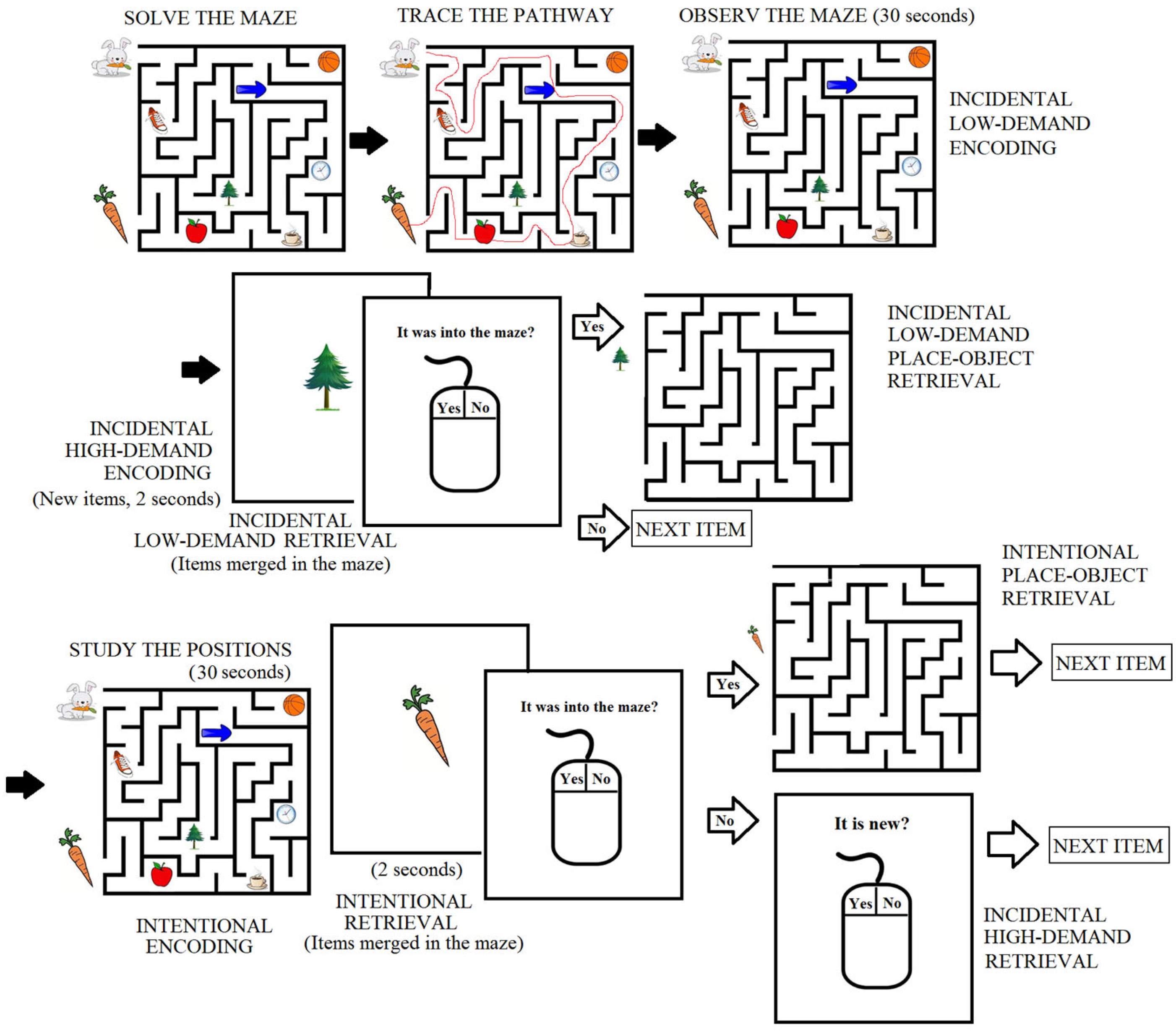
2.3. Incidental/Intentional Visuospatial Task

The task was performed in an isolated room where the participants sat in front of a laptop monitor (70 cm distance to the eyes). Once the electrodes were placed, instructions for the procedure were given to the participants. The visuospatial task was realized based on that reported by Lopez-Loeza et al. [6], with some modifications, using the software developed by Tellez-Anguiano et al. [28].
The task started with the instruction to observe a fixation central cross on the screen for 60 s (baseline condition), and then a 60-s recording was taken with the eyes closed. Then, the image of a maze with seven objects embedded in it and two objects at the start and end points of the correct pathway was presented to the participants (Figure 1), with the instruction to “mentally solve the maze and indicate when finished”. Once the participant had solved the maze, the instruction was given to trace the pathway (using the mouse; data not analyzed), followed by the instruction to observe the maze (without any other insight) for 30 s (corresponding to the incidental low-demand encoding condition). After this, an image of one object (one that was in the maze or one new object) appeared in the center of the screen for two seconds, followed by the question, was this object in the maze? If the participant answered “no” (by clicking with the mouse), another object was presented on the screen for 2 s, and the question was presented again. If the participant answered “Yes”, the image of the maze without objects appeared on the screen, and the participant had to drag the item to the position remembered using the mouse. This part of the task was completed with the presentation of 8 objects embedded in the maze, interspersed with 7 new objects. The time at which the participants responded to each item was registered by the software and used to mark a point, from which an EEG sample of 4 s was analyzed. This period was considered as the incidental low-demand retrieval condition regarding the 8 objects that were present in the maze. In addition, the two seconds during which a new object was observed were considered and analyzed as the incidental high-demand encoding condition, because the participants had a shorter period of time (two seconds) to observe each object than in the incidental low-demand encoding. In addition, the participants were required to determine whether the item was new or familiar at that time, and they were not notified that this information would be asked for later. Once the 8 objects had merged in the maze and the 7 interspersed objects were observed, the maze with the eight objects was presented again to the participants, with the instruction to study the objects’ positions (for a 30-s period). This was considered the intentional encoding condition. Once the study time had elapsed, an object appeared on the screen for 2 s, followed by the question, was this object in the maze? If the participant answered “Yes”, the image of the maze was presented on the screen, and the participant had to drag the cursor to the position remembered. The period of 4 s (for each of the 8 studied objects) before the time of the response was analyzed and considered the intentional retrieval condition. If the participant answered “No”, another question was presented—did you see this object before, or is it a new object? Thus, the participants had to discriminate between the seven objects previously presented as new in the first incidental test and 7 new objects that were interspersed. The period of 4 s before the participant’s response was analyzed as the incidental high-demand retrieval condition. This part continued until the participant responded to the 7 objects used for contrast in the first test (incidental low) and the seven new objects interspersed with the 8 studied objects. Finally, a baseline open-eye EEG was recorded for 60 s; this was the baseline record included in this study, because the first record was obtained when the elderly participants were highly anxious compared with the young participants.
The responses of the participants were stored by the software and analyzed to obtain the number of false-positive errors (when the participant responded “yes” for objects that were not in the maze or were not new), false-negative errors (when the participant answered “no” for objects that were in the maze or were new), and total errors (the two types summed) for each behavioral subtest (incidental low demand, incidental high demand, and intentional). In addition, the number of object errors (objects that were not in the maze but were placed in it by the participants), place errors (objects that were or were not in the maze but were placed in the incorrect position by the participants), and correct object–place associations (correct objects placed in the correct positions) were compared for the incidental low-demand and intentional conditions. All behavioral variables described were compared between the groups with the Mann–Whitney U test, and intragroup comparisons were performed to assess the differences between the incidental and intentional acquisition of the information at each age through Wilcoxon’s paired test.

2.4. EEG Records

EEG records for the frontotemporal areas (Fp1, Fp2, F3, F4, F7, F8, T3, T4, T5, and T6), according to the 10–20 international system, referring to short-circuited and grounded ear lobes, were obtained. Electrodes to register blinks and cardiac pulse potentials were also placed. The EEG signals were amplified (Neuro-Data Acquisition System Mod. 15 Grass for 36 channels). The records were acquired and stored with Gamma software for Mod. 15 (Grass telefactor), with the filters set at 1–300 Hz and a sample frequency of 1024 Hz, and stored on a PC to be analyzed offline. EEG recording started simultaneously with the memory test, and the test software marked the time of each behavioral condition, the time for which each object was displayed, and the responses of the participants [28].
The encoding and retrieval EEG records were sampled and analyzed separately, each divided into behavioral conditions as described in the behavioral task section. Baseline records were taken and compared with the encoding and retrieval stages, including incidental low-demand, incidental high-demand, and intentional. Moreover, correct and incorrect responses were compared only for retrieval, as the encoding process was obtained from a continuous record. The length of the records depended on the behavioral condition as described above: 30 s for incidental low-demand encoding and intentional encoding; 4 s for incidental low-demand, incidental high-demand, and intentional retrieval; and two seconds for incidental high-demand encoding.
The raw EEG records were visually inspected, and artifacts were eliminated using the eeglab12_0_1_0b utility from MATLAB. Then, samples corresponding to each behavioral condition were obtained and subjected to a Fourier Transform (samples of one second with 50% overlapped windows, using the periodogram method with MATLAB scripts) to obtain the mean spectrum power of the theta (4–8 Hz) and gamma (low gamma, 30–50 Hz) bands. Coherence values were computed as the magnitude square coherence using Welch’s averaged periodogram method. Normalized power (natural logarithm) and coherence values were obtained in bins of 1 Hz of the theta and gamma bands and were compared between groups and conditions with an ANCOVA, with the participants’ gender, intellectual quotient (IQ), and years of schooling as covariates. Partial correlation coefficients were obtained between the behavioral variables and their corresponding EEG power values, considering gender as an independent partial factor and the IQ and years of study as dependent partial factors. When significant correlations were found (p < 0.01), the analysis was repeated, including the age as a partial correlate. If the initial significant correlation was lost, we considered the correlation to be age-dependent; when the correlation value did not change after age adjustment, we considered both age and efficiency to be significantly correlated with the EEG power.

3. Results

Descriptive data from the samples are shown in Table 1. Younger participants had significantly higher IQ values and more years of schooling than the elderly group. The young participants presented values in the normal high and superior ranges (2 and 3), whereas the older group presented normal low and high values (1 and 2). In addition, the elderly group had lower values in the MoCA test, whereas no differences were observed in the WAIS test.

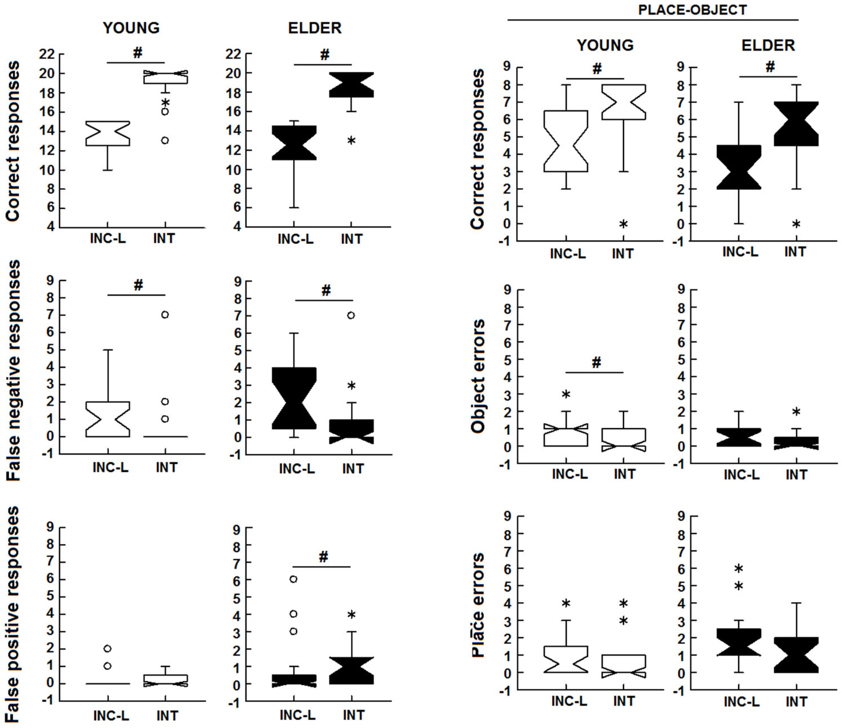
3.1. Incidental/Intentional Visuospatial Test

In the incidental low-demand condition, no differences were observed in the number of correct responses (U1df = 256.500, p = 0.103), total errors (U1df = 200.500, p = 0.090), and false-negative (U1df = 221.000, p = 0.207) and false-positive (U1df = 236.500, p = 0.161) errors. In the comparison of place–object associations, the elderly participants obtained fewer correct responses (U1df = 379.5, p = 0.035, dCohen = 0.630), more place recall errors (U1df = 167.5, p = 0.014, dCohen = 0.722), and more false-positive errors (U1df = 218.500, p = 0.025, dCohen = 0.378) than the younger group. No differences in object errors were observed (Figure 2).
In the incidental high-demand condition, the elderly group exhibited more total (U1df = 124.000, p = 0.001, dCohen = 1.067), false-negative (U1df = 187.000, p = 0.045, dCohen = 0.585), and false-positive (U1df = 140.000, p = 0.001, dCohen = 0.933) errors and had a smaller number of correct responses (U1df = 425.500, p = 0.002, dCohen = 0.978) than the younger group.
In the intentional test, the elderly group exhibited more total (U1df = 174.501, p = 0.016, dCohen = 0.672) and false-positive (U1df = 186.500, p = 0.023, dCohen = 0.588) errors than the younger group and had a smaller number of correct responses (U1df = 385.500, p = 0.016, dCohen = 0.672), whereas no difference in false-negative errors was observed (U1df = 246.500, p = 0.341). In the place–object associations, the elderly participants exhibited more place (U1df = 177.500, p = 0.016 dCohen = 0.651, Figure 2) and false-positive errors (U1df = 138.000, p < 0.001, dCohen = 0.949) and had a smaller number of correct responses (U1df = 377.500, p = 0.035, dCohen = 0.616). No differences were observed in the false-negative errors and object errors (U1df = 246.000, p = 0.334 and U1df = 296.000, p = 0.675, respectively). In summary, the elderly participants were less efficient in terms of their incidental low-demand (for place–object associations), incidental high-demand, and intentional performance on the visuospatial task.
To assess whether the young and elderly participants showed a similar increase in performance after the intentional study of the items, an intragroup comparison of the scores obtained between the incidental (low-demand) and intentional conditions was performed. The number of correct responses increased under the intentional compared with the incidental low-demand test for both young (z = 4.655, p < 0.001, dCohen = 3.7) and elderly (z = 3.831, p < 0.001, dCohen = 3.321) participants. The young (z = −2.591, p = 0.010, dCohen = 1.123) but not the elderly participants (z = −1.792, p = 0.073, dCohen = 0.875, whereas both groups had a reduced number of false-negative errors (z = −3.256, p = 0.001, dCohen = 1.561; z = −2.454, p = 0.014, dCohen = 1.313). No changes between the incidental and intentional numbers of false-positive errors occurred in the younger participants, but an increase was observed in the elderly participants (z = 2.069, p = 0.039, dCohen = 1.043) (Figure 3). Thus, the elderly group was unable to further reduce its total errors after the intentional study of the positions due to the increase in the number of false-positive errors. Finally, the learning of place–object associations increased under intentional effort for young (z = 3.939, p < 0.001, dCohen = 2.229) and elderly participants (z = 3.587, p < 0.001, dCohen = 2.686). Additionally, the young participants, but not the elderly ones, showed reduced object errors (z = −2.428, p = 0.015, dCohen = 1.032) after the intentional study (Figure 3).

3.2. EEG Activity

3.2.1. Theta and Gamma Activity during Encoding

Significant differences were observed in the mean power for all derivations recorded for both the theta and gamma bands, in which the elderly participants showed higher power values than the younger participants (Table 2, main effect). Additionally, the interaction of the group and behavioral condition was significant for all derivations and for the two bands (interaction statistic results, Table 3).
The paired comparisons of the theta band showed a general pattern in which the EEG power of the young participants had lower values under the incidental low-demand, incidental high-demand, and intentional conditions compared with the baseline record; meanwhile, the elderly participants had similar power values throughout all behavioral conditions. In addition, the EEG power of the young participants was lower than the power of the elderly participants in all test conditions, with the exception of the baseline record.
In the incidental high-demand condition, a significant and negative correlation occurred between the correct responses and the T6 power, but this correlation was lost after age adjustment, indicating a strong age relation. Finally, no significant correlation between power and behavior in the intentional encoding was observed for the theta band (Table 4). In summary, more power in the temporal (T3, T4, T6) and frontal (Fp1, Fp2) derivations occurred in less efficient participants, but this was more strongly related to aging than efficiency. Additionally, significant correlations were observed only for incidental and not for intentional encoding.
In the gamma band, under incidental low-demand encoding, the greater power in T3, T4, and F4 was related to the minor number of correct responses and correct place–object associations. However, the positive correlations between the false-negative errors and T3 power and between the place–object false-negative errors and T4 power were significant. All correlations lost their significance after age adjustment, and the r values were significantly reduced (Table 4). In the incidental high-demand condition, the greater power in Fp1, Fp2, and F3 was related to the minor number of correct responses, whereas the greater power in Fp1, Fp2, and T6 was related to the greater number of false-positive errors. All correlations were lost after age adjustment, and the reduction in the r values was significant. However, positive correlations between the F4, T3, and T4 power and the number of correct responses, as well as negative correlations between the F4 and T3 power and false-positive errors, were observed after age adjustment. Thus, in the gamma band, higher power at the frontal (Fp1, Fp2, F3) regions was related to a smaller number of correct responses and was also dependent on age, whereas the power at F4, T3, and T4 initially showed a negative correlation with correct responses and with false-positive errors but showed a positive correlation after age adjustment, probably due to the performance dependence. Under intentional conditions, only the greater power in T3 and T6 was related to more place errors, but this was dependent on age (Table 4).

3.2.2. Theta and Gamma Activity at Retrieval

Theta and Gamma Power

The EEG power from the retrieval periods was analyzed in each behavioral condition and separated into correct and incorrect responses. The analysis revealed a significant group effect for each derivation, similar to that observed in the encoding condition; the power was higher in the older group in both the theta and gamma bands (Table 2). In addition, a significant effect of the interaction of the group and behavioral condition was observed for each derivation in the two bands (Table 3). Whereas the theta power of the elderly participants did not change in magnitude throughout the different behavioral conditions or between correct and incorrect responses, the EEG power of the younger participants was lower for the retrieval of incorrect responses compared with correct responses and with the incorrect responses of the elderly participants. Specifically, the theta power recorded under the incidental low-demand condition was significantly lower for error responses than for correct responses in F3, F8, T3, T4, and T6. Meanwhile, in the incidental high-demand condition, the theta power was lower for all recorded derivations, with the exception of Fp2. Finally, in the intentional retrieval, a similar pattern was observed for all derivations (Figure 4).
In the gamma band power recorded during retrieval, reduced gamma power occurred in regard to errors compared with correct responses and the baseline record, in all derivations and tasks, in the younger but not in the older group. Additionally, as a result of the power reduction in the error responses in the young participants, the power was significantly higher for the older group, whereas, regarding the correct responses, no differences between the groups occurred (Figure 5).

Power Behavior Correlations

In the incidental low-demand condition, the correlations were significant and positive for the false-positive errors and T4 theta power; however, after age adjustment, positive and significant correlations were also observed for the F8 and T3 power. Additionally, significant and positive correlations were observed for Fp1, F3, F4, F7, F8, T3, and T4 with place errors. After age adjustment, the F3, F7, F8, and T4 correlations were significantly reduced, although those of F4 and T3 remained significant, indicating the strong effect of the performance on the theta power modulation. No significant correlations were observed between the power in incidental high-demand retrieval EEG and behavior. Finally, in the intentional retrieval condition, there were significant and negative correlations between the power in Fp2, F3, F7, T3, and T5 and the number of correct place–object association responses, all of which significantly changed after age adjustment and lacked significance, indicating strong modulation by age (Table 5).
In the gamma band, the power–behavior correlations were significant only under the incidental low-demand condition, in which the number of false-positive errors was higher when the power was also higher in the F7 and F8 derivations after age adjustment, indicating a performance relation between power and behavior. Additionally, positive correlations between the gamma power and the number of place errors in Fp2, F4, F7, and F8 were observed, and these all remained after the age adjustment, with the exception of that of Fp2, which was significantly reduced. Thus, all were better related to the efficiency of processing, with the exception of Fp2, which could have been related to aging. Finally, a negative correlation between the power in F3 and F4 and correct place–object responses, which remained after age adjustment, was observed (Table 5).

4. Discussion

The presented results are in line with the proposed increased vulnerability of incidental, relational, and visuospatial information with aging [2,4,6,29,30]. Our results show a reduced ability to perform place–object associations with aging under incidental low-demand conditions, which was possibly due to the increase in place and false-positive errors in the elderly participants. Meanwhile, under the intentional condition, only the place–object false-positive errors were related to age. Koster et al. [31] compared the incidental versus intentional acquisition of memory for objects in young adults and two groups of children (mean of 7 and 10 years old). The authors observed that all groups had better performance under the intentional compared with the incidental condition in terms of the features of the objects observed; thus, an increased number of false alarms under intentional conditions appears to be a hallmark sign of aging. Although they did not observe an increased rate of false alarms/hits at these ages under the intentional compared with the incidental condition, previous works have reported an increased rate of false alarms [2,32]. This increase in false alarms was considered a deficit in recollection, combined with the overexpression of familiarity, possibly as a result of the lack of distinction between the newly learned information and the memory resources, leading to increased familiarity. Moreover, this deficit could be related to reduced hippocampal-entorhinal functional connectivity, based on the suggested role of the hippocampus in fast episodic memory encoding and recollection. Meanwhile, the hippocampus-associated cortex encodes familiarity and is less affected by aging [33,34,35].
These results did not show differences with regard to incidental versus intentional encoding for either the theta or gamma power. Similarly, in the study by Lopez-Loeza et al. [6], which compared younger and mature adults, the theta power of young participants did not show differences between incidental and intentional types of encoding. The authors showed that mature adults had increased theta power compared with the baseline records for incidental memory and intentional learning at frontal recording sites and only for intentional learning at temporoparietal sites. However, when the authors grouped the participants by accuracy (high efficiency and low efficiency), the less efficient group showed minor theta and gamma power at baseline and in incidental conditions, which increased in intentional conditions at the same derivations. Meanwhile, the efficient group did not show changes throughout the conditions (they started with high baseline power, and it remained high throughout all conditions). In this study, minor theta and gamma power in the incidental low-demand, incidental high-demand, and intentional conditions compared with the baseline records was observed for both encoding and retrieval errors, only for young participants. However, Koster et al. [31], reported an increase in the theta power of intentional compared with incidental encoding at the frontal and parietal electrodes. However, in Koster’s work, the theta power was measured as the phase-locked activity evoked by the stimulus. Additionally, different studies have shown an increase in the power of theta when comparing subsequent remembered and subsequent forgotten items, with the activity recorded during encoding and retrieval [17,22,36]. However, reductions in theta power have also been observed in other studies. Specifically, intracranial records in epileptic patients showed decreased high-frequency theta activity with cognitive processing, whereas increases in power were observed at lower frequencies in the so-called slow theta range (<4 Hz) [37]. In addition, early works by Klimesch et al. showed reduced power in the theta band, related to better performance in a memory task for word lists [24]. In this work, the authors evaluated the tonic changes in EEG through a continuous test in four-second epochs (similar to our study), and the results were explained as changes in induced power, which could reflect the default network state and were modulated by the stimuli but not phase-locked with them [17,24,38,39]. In another study, Nicolas et al. [38] reported a reduction in theta activity in the fronto-central regions during the retrieval of episodic information, as evaluated after weeks or months of encoding. Moreover, a theta power decrease during encoding compared with pre-stimulus in young participants was observed by Rondina et al. [39], who also reported increased theta power between encoding (the first phase of the study) and retrieval (test phase), despite a general theta power decrease from the baseline to the task. However, the theta range analyzed in Rondina’s study was 2–7 Hz. In the present work, the elderly participants had low efficiency and greater theta power both during encoding and retrieval, whereas the young participants had a reduction in theta power compared with the baseline records under the encoding and retrieval of incidental or intentional memory, but they showed a greater reduction when errors were made, in line with the mentioned studies. In addition, during retrieval, the theta power mirrored the accuracy of execution only for the young participants (higher power occurred for correct responses compared with errors). In the present work, the epochs obtained during encoding were taken from continuous recordings without a specific signal of the processing of information (when the participants observed the maze without other instructions). Thus, the changes in the tonic power measured during an extended period in which encoding occurred are quite similar to those reported by Klimesch et al. as the induced power, whereas the retrieval occurred after the presentation of an object and prior to the emission of a response in a period of four seconds in which the processing and decision-making were performed. This is in line with the assumption that increased theta power is observed as an event-related response due to a mechanism designed to increase the noise/signal ratio, reflecting the selectivity of information processing [17,18].
Under encoding, increased theta activity and power was correlated with the behavioral variables only for the incidental conditions in the frontotemporal areas, but the significance was lost after age adjustment. Under retrieval, higher theta power occurred in the left frontotemporal areas, which was related to aging, whereas, under incidental low-demand conditions, the theta power was more closely related to the efficiency than the age. High power at F8, T3, T4, and F4 was related to the greater number of errors after age adjustment; thus, the theta power at these derivations was related to both aging and the accuracy of processing under incidental conditions. These results are in line with the hypothesis of the overactivation or recruitment of additional resources to achieve a similar function in elderly adults compared with young adults [40,41], and with the reported increase in theta activity related to aging [23]. Several studies have associated cognitive interference with theta activity expression, principally through the performance of interference paradigms, in which the prefrontal theta expression reflects the interference level [42]. Additionally, in the context of episodic memory, increased theta activity has been related to higher levels of interference during memory retrieval in healthy young humans [43]. In this sense, the impaired inhibition of interferent information with aging is proposed in inhibitory deficit theory [1], which states that deficits in attention, language, and memory with aging are due to a decline in inhibitory function, producing difficulties in the suppression of irrelevant information or competing representations [42,44,45]. In this study, the higher levels of theta activity observed in the older participants could be related to higher levels of interference from information irrelevant to the task during encoding, whereas, during retrieval, additional interference from other memory representations could occur. In accordance with this, the successful inhibition of interference was associated with a reduction in theta activity in the frontoparietal areas in young humans’ EEG [43]. This is in line with the reduced theta power recorded in the young participants in memory-related conditions compared with their own baselines and with the theta power of older participants, under both encoding and retrieval, in this study. Moreover, the higher theta activity seen during correct responses compared with errors in young participants could be related to theta memory processing. Thus, these results support the dual modulation of the theta power by age (with higher interference) and memory processing, and they highlight the importance of dissociating the two effects in the study of the EEG correlates of memory processes.
The expression of gamma power during encoding was more strongly related to age than performance (the correlations observed under incidental low-load and intentional conditions were lost after age adjustment); however, the gamma power was also related to efficiency. The expression of gamma power could be related to the cortical representations of the stimuli, which, initially unfamiliar, are observed and compared with the previously observed items. In this regard, the oscillatory activity induced in the gamma band at frequencies higher than 20 Hz, observed after the presentation of an unfamiliar stimulus, has been related to the cortical representation of the semantic and perceptual characteristics of the stimuli, particularly with indirect memory (incidental) [20,46]. Additionally, it has been reported that the gamma activity decreases with the repetition of stimuli (e.g., faces), being higher for new stimuli than for familiar stimuli [46,47]. In accordance with this, under retrieval, the greater gamma power for Fp2, F4, F7, and F8 was related to the greater numbers of false-positive and place errors, and the lower power at F3 and F4 was related to the greater number of correct place–object responses. Additionally, this is in accordance with the increased gamma power during encoding but reduced during retrieval (only in young participants and possibly altered in elderly participants). It is also in line with the better performance observed only under incidental conditions in this study, supporting the relevance of gamma activity for incidental processing [20,46]. Moreover, increased gamma power under retrieval errors could be related to a deficient process, resulting in the perception of previously seen items as new items. With regard to the changes in gamma activity associated with aging, it was previously reported that mature adults (45–60 years old) show increased gamma power related to age and performance in the frontotemporoparietal areas compared with younger adults (18–40 years old) [6]. Similar results were observed by Barr et al. [48] when comparing working-memory-related gamma activity between younger (19–29 years old) and mature (30–60 years old) adults. The power increase was correlated with age but not with performance in an n-back test, in which the two groups had similar performance. These results extend the observed increase in gamma activity in mature adults (45–60 years old), reported by Lopez-Loeza et al. [6], to older adults at all sites recorded.

5. Conclusions

In conclusion, these results indicate that incidental and intentional visuospatial relational memory is more vulnerable to changes related to aging. The EEG power of the frontotemporal theta and gamma bands underlying this type of memory changes with aging as a general effect, but also changes in relation to task performance. The greater theta power in young participants might be related to inhibitory processes, whereas, in the elderly, it may be associated with compensatory mechanisms. In addition, it was observed that the EEG power was responsive to errors only in the young participants. Meanwhile, the gamma activity increases in the elderly participants could be related to increased familiarity and reduced recall. Changes in power are related to both aging and visuospatial processing; thus, it is important to separate each influence on EEG activity recorded in cognitive evaluations when studying aging.

Author Contributions

Conceptualization, M.Á.L.-V., M.E.O.-C. and M.L.J.-M.; methodology, M.L.J.-M., M.E.O.-C. and O.M.-R.; software, A.d.C.T.-A.; validation, M.L.J.-M. and J.M.C.-A.; formal analysis, M.L.J.-M., M.Á.L.-V. and M.E.O.-C.X; investigation, M.E.O.-C., M.L.J.-M. and M.Á.L.-V.; resources, M.E.O.-C. and J.M.C.-A.; data curation, M.E.O.-C. and M.L.J.-M.; writing—original draft preparation, M.L.J.-M. and M.E.O.-C.; writing—review and editing, M.Á.L.-V. and J.M.C.-A. All authors have read and agreed to the published version of the manuscript.

Funding

This research received no external funding.

Institutional Review Board Statement

This study was conducted in accordance with the Declaration of Helsinki and approved by the National Ethics and Research Committee of the Instituto Mexicano del Seguro Social, Number CNCI: R-2014-785-058, 28 July 2014.

Informed Consent Statement

Written informed consent has been obtained from the patient(s) to publish this paper.

Data Availability Statement

The data presented in this study are available on request from the corresponding author due to to the authorization must be aproved by the Instituto Mexicano del Seguro Social.

Acknowledgments

Mariana Lizeth Junco Muñoz is a doctoral student from the Programa de Doctorado en Ciencias Biomédicas, Universidad Nacional Autónoma de México (UNAM), and has received CONAHCYT fellowship 664893.

Conflicts of Interest

The authors declare no conflicts of interest.

References

  1. Hasher, L.; Zacks, R.T. Working Memory, Comprehension, and Aging: A Review and a New View. Psychol. Learn. Motiv. 1988, 22, 193–225. [Google Scholar]
  2. Naveh-Benjamin, M. Adult age differences in memory performance: Tests of an associative deficit hypothesis. J. Exp. Psychol. Learn. Mem. Cogn. 2000, 26, 1170–1187. [Google Scholar] [CrossRef] [PubMed]
  3. Naveh-Benjamin, M.; Shing, Y.L.; Kilb, A.; Werkle-Bergner, M.; Lindenberger, U.; Li, S.C. Adult age differences in memory for name-face associations: The effects of intentional and incidental learning. Memory 2009, 17, 220–232. [Google Scholar] [CrossRef] [PubMed]
  4. Rugg, M.D.; Fletcher, P.C.; Frith, C.D.; Frackowiak, R.S.; Dolan, R.J. Brain regions supporting intentional and incidental memory: A PET study. Neuroreport 1997, 8, 1283–1287. [Google Scholar] [CrossRef] [PubMed]
  5. Caine, K.E.; Nichols, T.A.; Fisk, A.D.; Rogers, W.A.; Meyer, B. Age-related differences in learning incidental, environmental information. Exp. Aging Res. 2011, 37, 17–45. [Google Scholar] [CrossRef] [PubMed]
  6. Lopez-Loeza, E.; Rangel-Argueta, A.R.; Lopez-Vazquez, M.A.; Cervantes, M.; Olvera-Cortes, M.E. Differences in EEG power in young and mature healthy adults during an incidental/spatial learning task are related to age and execution efficiency. Age 2016, 38, 37. [Google Scholar] [CrossRef] [PubMed]
  7. Squire, L.R. The organization and neural substrates of human memory. Int. J. Neurol. 1987, 21–22, 218–222. [Google Scholar]
  8. Wagner, A.D.; Schacter, D.L.; Rotte, M.; Koutstaal, W.; Maril, A.; Dale, A.M.; Rosen, B.R.; Buckner, R.L. Building memories: Remembering and forgetting of verbal experiences as predicted by brain activity. Science 1998, 281, 1188–1191. [Google Scholar] [CrossRef] [PubMed]
  9. Gabrieli, J.D.; Brewer, J.B.; Desmond, J.E.; Glover, G.H. Separate neural bases of two fundamental memory processes in the human medial temporal lobe. Science 1997, 276, 264–266. [Google Scholar] [CrossRef] [PubMed]
  10. Schacter, D.L.; Wagner, A.D. Medial temporal lobe activations in fMRI and PET studies of episodic encoding and retrieval. Hippocampus 1999, 9, 7–24. [Google Scholar] [CrossRef]
  11. Davachi, L.; Mitchell, J.P.; Wagner, A.D. Multiple routes to memory: Distinct medial temporal lobe processes build item and source memories. Proc. Natl. Acad. Sci. USA 2003, 100, 2157–2162. [Google Scholar] [CrossRef] [PubMed]
  12. Schacter, D.L.; Curran, T.; Reiman, E.M.; Chen, K.; Bandy, D.J.; Frost, J.T. Medial temporal lobe activation during episodic encoding and retrieval: A PET study. Hippocampus 1999, 9, 575–581. [Google Scholar] [CrossRef]
  13. Dolan, R.J.; Fletcher, P.F. Encoding and retrieval in human medial temporal lobes: An empirical investigation using functional magnetic resonance imaging (fMRI). Hippocampus 1999, 9, 25–34. [Google Scholar] [CrossRef]
  14. Fuentemilla, L.; Barnes, G.R.; Duzel, E.; Levine, B. Theta oscillations orchestrate medial temporal lobe and neocortex in remembering autobiographical memories. Neuroimage 2014, 85 Pt 2, 730–737. [Google Scholar] [CrossRef] [PubMed]
  15. Herweg, N.A.; Apitz, T.; Leicht, G.; Mulert, C.; Fuentemilla, L.; Bunzeck, N. Theta-Alpha Oscillations Bind the Hippocampus, Prefrontal Cortex, and Striatum during Recollection: Evidence from Simultaneous EEG-fMRI. J. Neurosci. 2016, 36, 3579–3587. [Google Scholar] [CrossRef] [PubMed]
  16. Nyhus, E.; Curran, T. Functional role of gamma and theta oscillations in episodic memory. Neurosci. Biobehav. Rev. 2010, 34, 1023–1035. [Google Scholar] [CrossRef] [PubMed]
  17. Klimesch, W.; Vogt, F.; Doppelmayr, M. Interindividual differences in alpha and theta power reflect memory performance. Intelligence 1999, 27, 347–362. [Google Scholar] [CrossRef]
  18. Klimesch, W.; Doppelmayr, M.; Schimke, H.; Ripper, B. Theta synchronization and alpha desynchronization in a memory task. Psychophysiology 1997, 34, 169–176. [Google Scholar] [CrossRef] [PubMed]
  19. Weiss, S.; Muller, H.M.; Rappelsberger, P. Theta synchronization predicts efficient memory encoding of concrete and abstract nouns. Neuroreport 2000, 11, 2357–2361. [Google Scholar] [CrossRef] [PubMed]
  20. Gruber, T.; Muller, M.M. Oscillatory brain activity in the human EEG during indirect and direct memory tasks. Brain Res. 2006, 1097, 194–204. [Google Scholar] [CrossRef] [PubMed]
  21. Klimesch, W.; Doppelmayr, M.; Yonelinas, A.; Kroll, N.E.; Lazzara, M.; Rohm, D.; Gruber, W. Theta synchronization during episodic retrieval: Neural correlates of conscious awareness. Brain Res. Cogn. Brain Res. 2001, 12, 33–38. [Google Scholar] [CrossRef] [PubMed]
  22. Summerfield, C.; Mangels, J.A. Coherent theta-band EEG activity predicts item-context binding during encoding. Neuroimage 2005, 24, 692–703. [Google Scholar] [CrossRef] [PubMed]
  23. Klass, D.W.; Brenner, R.P. Electroencephalography of the elderly. J. Clin. Neurophysiol. 1995, 12, 116–131. [Google Scholar] [CrossRef] [PubMed]
  24. Klimesch, W. EEG alpha and theta oscillations reflect cognitive and memory performance: A review and analysis. Brain Res. Brain Res. Rev. 1999, 29, 169–195. [Google Scholar] [CrossRef] [PubMed]
  25. Jurado, S.; Villegas, E.M.; Mendez, L.; Rodríguez, F.; Loperena, V.; Varela, R. La estandarización del inventario de depresión de Beck para los residentes de la ciudad de México. Salud Ment. 1998, 21, 26–31. [Google Scholar]
  26. Ledesma, L.; García, P.; Salvador, J. Test MOCA, Versión Mexicana 7.2. Volume 2014. 2014. Available online: www.mocatest.org (accessed on 1 January 2014).
  27. Pons, J.I.; Pabón, L.F.; Matías-Carrelo, L.; Rodríguez, M.; Hernández, E.R.; Rodríguez, J.M.; Herrans, L.L.; Yang, J. Estudios de validez de la Escala de Inteligencia Wechsler para Adultos Versión III, Puerto Rico (EIWA-III). Rev. Puertorriqueña Psicol. 2008, 19, 75–111. [Google Scholar]
  28. Lara-Ramos, J.A.; Zavala-Santoyo, F.L.; Tellez-Anguiano, A.C.; Diaz-Huerta, J.L.; Olvera-Cortes, M.E. Computational interface to automate cognitive memory tests and synchronize with measured EEG signals. In Proceedings of the 2019 16th International Conference on Electrical Engineering, Computing Science and Automatic Control, CCE 2019, Mexico City, Mexico, 11–13 September 2019; Volume CCE 2019. IEEE: Mexico City, Mexico, 2019. [Google Scholar]
  29. Cansino, S.; Hernandez-Ramos, E.; Estrada-Manilla, C.; Torres-Trejo, F.; Martinez-Galindo, J.G.; Ayala-Hernandez, M.; Gomez-Fernandez, T.; Osorio, D.; Cedillo-Tinoco, M.; Garces-Flores, L.; et al. The decline of verbal and visuospatial working memory across the adult life span. Age 2013, 35, 2283–2302. [Google Scholar] [CrossRef] [PubMed]
  30. Old, S.R.; Naveh-Benjamin, M. Differential effects of age on item and associative measures of memory: A meta-analysis. Psychol. Aging 2008, 23, 104–118. [Google Scholar] [CrossRef] [PubMed]
  31. Koster, M.; Haese, A.; Czernochowski, D. Neuronal oscillations reveal the processes underlying intentional compared to incidental learning in children and young adults. PLoS ONE 2017, 12, e0182540. [Google Scholar] [CrossRef] [PubMed]
  32. Memon, A.; Bartlett, J.; Rose, R.; Gray, C. The aging eyewitness: Effects of age on face, delay, and source-memory ability. J. Gerontol. B Psychol. Sci. Soc. Sci. 2003, 58, P338–P345. [Google Scholar] [CrossRef] [PubMed]
  33. Eichenbaum, H. Hippocampus: Cognitive processes and neural representations that underlie declarative memory. Neuron 2004, 44, 109–120. [Google Scholar] [CrossRef] [PubMed]
  34. Yonelinas, A.P.; Kroll, N.E.; Quamme, J.R.; Lazzara, M.M.; Sauve, M.J.; Widaman, K.F.; Knight, R.T. Effects of extensive temporal lobe damage or mild hypoxia on recollection and familiarity. Nat. Neurosci. 2002, 5, 1236–1241. [Google Scholar] [CrossRef] [PubMed]
  35. Yonelinas, A.P.; Levy, B.J. Dissociating familiarity from recollection in human recognition memory: Different rates of forgetting over short retention intervals. Psychon. Bull. Rev. 2002, 9, 575–582. [Google Scholar] [CrossRef] [PubMed]
  36. Osipova, D.; Takashima, A.; Oostenveld, R.; Fernandez, G.; Maris, E.; Jensen, O. Theta and gamma oscillations predict encoding and retrieval of declarative memory. J. Neurosci. 2006, 26, 7523–7531. [Google Scholar] [CrossRef] [PubMed]
  37. Lega, B.C.; Jacobs, J.; Kahana, M. Human hippocampal theta oscillations and the formation of episodic memories. Hippocampus 2012, 22, 748–761. [Google Scholar] [CrossRef] [PubMed]
  38. Nicolas, B.; Sala-Padro, J.; Cucurell, D.; Santurino, M.; Falip, M.; Fuentemilla, L. Theta rhythm supports hippocampus-dependent integrative encoding in schematic/semantic memory networks. Neuroimage 2021, 226, 117558. [Google Scholar] [CrossRef] [PubMed]
  39. Rondina, R., 2nd; Olsen, R.K.; McQuiggan, D.A.; Fatima, Z.; Li, L.; Oziel, E.; Meltzer, J.A.; Ryan, J.D. Age-related changes to oscillatory dynamics in hippocampal and neocortical networks. Neurobiol. Learn. Mem. 2016, 134 Pt A, 15–30. [Google Scholar] [CrossRef] [PubMed]
  40. Cabeza, R. Hemispheric asymmetry reduction in older adults: The HAROLD model. Psychol. Aging 2002, 17, 85–100. [Google Scholar] [CrossRef] [PubMed]
  41. Cabeza, R.; Anderson, N.D.; Locantore, J.K.; McIntosh, A.R. Aging gracefully: Compensatory brain activity in high-performing older adults. Neuroimage 2002, 17, 1394–1402. [Google Scholar] [CrossRef] [PubMed]
  42. Ferreira, C.S.; Maraver, M.J.; Hanslmayr, S.; Bajo, T. Theta oscillations show impaired interference detection in older adults during selective memory retrieval. Sci. Rep. 2019, 9, 9977. [Google Scholar] [CrossRef] [PubMed]
  43. Staudigl, T.; Hanslmayr, S.; Bauml, K.H. Theta oscillations reflect the dynamics of interference in episodic memory retrieval. J. Neurosci. 2010, 30, 11356–11362. [Google Scholar] [CrossRef]
  44. Amer, T.; Ngo, K.W.J.; Weeks, J.C.; Hasher, L. Spontaneous Distractor Reactivation with Age: Evidence for Bound Target-Distractor Representations in Memory. Psychol. Sci. 2020, 31, 1315–1324. [Google Scholar] [CrossRef] [PubMed]
  45. Biss, R.K.; Campbell, K.L.; Hasher, L. Interference from previous distraction disrupts older adults’ memory. J. Gerontol. B Psychol. Sci. Soc. Sci. 2013, 68, 558–561. [Google Scholar] [CrossRef] [PubMed]
  46. Gruber, T.; Muller, M.M. Oscillatory brain activity dissociates between associative stimulus content in a repetition priming task in the human EEG. Cereb. Cortex 2005, 15, 109–116. [Google Scholar] [CrossRef] [PubMed]
  47. Zion-Golumbic, E.; Kutas, M.; Bentin, S. Neural dynamics associated with semantic and episodic memory for faces: Evidence from multiple frequency bands. J. Cogn. Neurosci. 2010, 22, 263–277. [Google Scholar] [CrossRef] [PubMed]
  48. Barr, M.S.; Radhu, N.; Guglietti, C.L.; Zomorrodi, R.; Rajji, T.K.; Ritvo, P.; Daskalakis, Z.J. Age-related differences in working memory evoked gamma oscillations. Brain Res. 2014, 1576, 43–51. [Google Scholar] [CrossRef] [PubMed]
Figure 1. Diagrammatic representation of the incidental/intentional visuospatial test.
Brainsci 14 00786 g001
Figure 2. Box plots showing the intergroup comparisons of the scores obtained by the participants in the incidental low-demand, the incidental high-demand, and the intentional visuospatial memory test. +, young vs. elderly participants, other symbols ° and * are values out of range. Mann–Whitney U test: p < 0.05.
Brainsci 14 00786 g002
Figure 3. Box plots showing the intragroup comparisons of the scores obtained by the participants under the incidental low-demand and the intentional visuospatial conditions. #, incidental vs. intentional scores, other symbols ° and * are values out of range. Mann–Whitney U test: p < 0.05.
Brainsci 14 00786 g003
Figure 4. Intergroup and intragroup comparisons of the theta band EEG absolute power values (natural logarithm) obtained during retrieval in baseline (BAS), incidental low-demand (INC-L), incidental high-demand (INC-H), and intentional (INT) conditions. Comparisons between age, conditions, and responses (correct vs. errors C, E, respectively) are shown. Values are mean ± EEM. *, difference from baseline; +, difference between young and old participants; (line), difference between correct and error responses. p < 0.01.
Brainsci 14 00786 g004
Figure 5. Intergroup and intragroup comparisons of the gamma band EEG absolute power values (natural logarithm) registered in retrieval, incidental low-demand (INC-L), incidental high-demand (INC-H), and intentional (INT) conditions. Comparisons between age and condition and between correct responses (C) and errors (E) are shown. Values are mean ± EEM. *, difference from baseline; +, difference between young and old participants; (line), difference between correct and error responses. p < 0.01.
Brainsci 14 00786 g005
Table 1. Descriptive data of the participants.
VariableYoung
(n = 28, 17 Women)
Elderly
(n = 20, 13 Women)
t-Test/dfp
Age 33.321 (±6.608)69.900 (±7.96)−16.826, 36.1<0.001
Years of schooling 15.500 (±3.097)11.20 (±4.538)3.671, 31.30.001
WAIS100.143 (±11.906)97.600 (±13.240)0.684, 10.498
MoCA19.24 (±1.261)18.250 (±0.55)4.527, 1<0.001
U test/df
Raven2.5 (1–3)1.5 (1–3)159, 10.006
For the age, years of schooling, WAIS, and MoCA results, which had a normal distribution, the mean ± standard deviation are presented, and the t-test and degrees of freedom (df) are reported. For the Raven category, the median and interquartile range are presented, and a paired comparison was performed with the Mann–Whitney U test.
Table 2. Intergroup comparisons of EEG power of theta and gamma bands, recorded during encoding and retrieval.
EncodingRetrieval
YoungElderlyFdCohenYoungElderlyFdCohen
Theta
PF17.015 (0.173)8.81 (0.139)40.4271.1377.750 (0.141)8.769 (0.111)34.3020.806
F36.861 (0.170)8.643 (0.125)40.0241.1327.410 (0.122)8.498 (0.091)49.9430.973
F76.513 (0.185)8.507 (0.100)51.7501.2877.080 (0.123)8.57 (0.073)74.9171.191
T36.195 (0.183)7.904 (0.089)44.8541.1986.895 (0.151)7.765 (0.074)19.8530.613
T56.296 (0.199)7.882 (0.070)35.8441.0716.887 (0.136)7.801 (0.089)41.7480.889
FP27.015 (0.180)8.833 (0.140)38.3111.1077.645 (0.131)8.747 (0.098)41.5080.887
F46.853 (0.170)8.541 (0.145)32.0631.0137.34 (0.120)8.326 (0.082)37.6460.844
F86.317 (0.156)8.200 (0.108)52.8271.3006.787 (0.119)8.154 (0.071)73.4751.180
T45.921 (0.184)7.887 (0.096)45.4371.2006.563 (0.129)7.706 (0.071)37.1630.839
T66.356 (0.153)7.952 (0.069)69.9531.4966.882 (0.115)7.825 (0.080)61.4971.079
Gamma
PF14.773 (0.215)7.719 (0.124)101.6231.8035.904 (0.183)7.747 (0.094)75.6921.197
F34.700 (0.208)7.365 (0.117)88.2011.6505.708 (0.181)7.430 (0.091)63.7581.099
F74.607 (0.213)7.148 (0.119)65.9411.4525.571 (0.178)7.411 (0.100)67.3501.130
T35.013 (0.221)7.393 (0.109)63.0231.4206.109 (0.183)7.512 (0.071)37.0270.873
T54.515 (0.218)7.179 (0.113)90.5881.7025.634 (0.187)7.289 (0.068)72.6071.173
FP24.675 (0.221)7.400 (0.122)92.7781.7235.817 (0.179)7.562 (0.095)65.7311.116
F44.734 (0.212)7.091 (0.112)52.6541.2985.706 (0.179)7.247 (0.086)44.4880.918
F84.532 (0.209)6.815 (0.117)61.4331.4025.435 (0.170)7.110 (0.090)58.6201.054
T44.862 (0.232)7.482 (0.130)49.2791.2565.724 (0.181)7.551 (0.094)43.7100.910
T64.401 (0.203)7.499 (0.146)124.9972.0005.51 (0.174)7.539 (0.080)127.361.553
The means (SEM) of the power values are expressed as a natural logarithm, with the F and p values from the ANCOVA for group effects and with gender, IQ, and years of study as covariables; size effect (dCohen) values are also shown (p < 0.001) for all comparisons.
Table 3. Statistical results of the interaction by group and behavioral condition for encoding and retrieval regarding the theta and gamma band power.
EncodingRetrieval
FpdCohenFpdCohen
Theta
PF15.4650.0010.7593.3180.0040.592
F35.4590.0010.7594.875<0.0010.717
F74.7300.0040.7076.007<0.0010.796
T35.7890.0010.7825.233<0.0010.743
T53.9740.0100.6483.5440.0020.612
FP24.8710.0030.7173.1260.0060.574
F45.3600.0020.7523.2760.0040.588
F85.0480.0020.7308.260<0.0010.934
T44.0160.0090.6514.699<0.0010.704
T68.485<0.0010.9466.052<0.0010.799
Gamma
PF115.810<0.0011.29213.572<0.0011.197
F322.304<0.0011.53419.619<0.0011.143
F717.060<0.0011.34215.561<0.0011.281
T316.885<0.0011.33513.298<0.0011.185
T517.031<0.0011.34114.232<0.0011.226
FP212.216<0.0011.13512.059<0.0011.128
F415.423<0.0011.27613.847<0.0011.209
F814.950<0.0011.25614.677<0.0011.245
T49.512<0.0011.0027.524<0.0010.891
T615.445<0.0011.27719.599<0.0011.438
The F and p values from the interaction between the group and behavioral condition were obtained in the ANCOVA with gender, CI, and years of study as covariables; the size effect (dCohen) values are also shown.
Table 4. Correlations between theta and gamma power and behavioral variables during the coding of information.
ThetaGamma
Incidental
Low-Demand
Age-adjustedAge-adjusted
r, pr, pqCohen r, pr, pqCohen
CRT3
T4
−0.373, 0.032
−0.354, 0.034
−0.257, 0.097
−0.254, 0.099
0.129
0.110
T3
T4
−0.321, 0.033
−0.309, 0.034
−0.191, 0.109
−0.213, 0.102
0.139
0.103
FNEFp1
Fp2
0.350, 0.034
0.342, 0.036
0.235, 0.123
0.238, 0.119
0.126
0.114
T30.299, 0.0480.147, 0.2220.160
P-O CR F4
T3
T4
−0.289, 0.041
−0.376, 0.011
−0.304, 0.037
−0.158, 0.182
−0.226, 0.055
−0.191, 0.144
0.138
0.165
0.121
P-O E
P-O FNE
T3
T4
0.338, 0.024
0.294, 0.044
0.170, 0.154
0.170, 0.196
0.180
0.131
Incidental
High-Demand
CR


FPE


T6


−0.438, 0.017


0.003, 0.981


0.473


Fp1
Fp2
F3
F4
T3
T4

Fp1
Fp2
F4
T3
T6
−0.393,0.027
−0.409, 0.020
−0.364, 0.048
−0.249, 0.160
−0.242, 0.195
−0.158, 0.367

0.369, 0.040
0.404, 0.021
0.126, 0.487
0.070, 0.713
0.371, 0.042
0.067, 0.613
0.057, 0.652
0.182, 0.114
0.257, 0.027
0.274, 0.032
0.292, 0.025

0.023, 0.862
0.056, 0.654
−0.264, 0.022
−0.337, 0.006
0.007, 0.954
0.482
0.491
0.566
0.517
0.528
0.460

0.410
0.372
0.397
0.421
0.397
Intentional
PE T3
T6
0.332, 0.027
0.294, 0.047
0.163, 0.184
0.105, 0.348
0.181
0.198
Behavioral Variable
Only significant correlations between the theta or gamma power and behavior are presented. Bold Pearson’s r and p values indicate correlations that changed after age adjustment (i.e., that were lacking or emerged). Bold Cohen’s q values indicate significant changes in r values. Cohen’s q indicates significant effect. Abbreviations: CR, correct responses; TE, total errors; FPE, false-positive error; P-O CR, place–object correct responses; PE, place errors.
Table 5. Correlations between theta and gamma power and behavioral variables during the retrieval of information.
ThetaGamma
Incidental
Low-Demand
Age-adjustedAge-adjusted
r, pr, pqCohen r, pr, pqCohen
FPEF8
T3
T4
0.359, 0.079
0.384, 0.059
0.393, 0.044
0.335, 0.026
0.367, 0.042
0.381, 0.042
0.027
0.020
0.014
F7
F8
0.257, 0.087
0.252, 0.105
0.236, 0.050
0.226, 0.029
0.022
0.028
PEFp1
F3
F4
F7
F8
T3
T4
0.415, 0.046
0.440, 0.023
0.474, 0.015
0.420, 0.042
0.401, 0.046
0.518, 0.007
0.429, 0.025
0.355, 0.087
0.354, 0.054
0.403, 0.034
0.300, 0.099
0.255, 0.105
0.419, 0.017
0.362, 0.055
0.070
0.102
0.088
0.138
0.164
0.127
0.079
Fp2
F4
F7
F8
0.350, 0.038
0.280, 0.022
0.374, 0.007
0.372, 0.010
0.255, 0.072
0.205, 0.036
0.293, 0.010
0.267, 0.007
0.105
0.080
0.091
0.117
P-O CR F3
F4
−0.295, 0.033
−0.324, 0.025
−0.273, 0.051
−0.292, 0.046
0.024
0.035
Intentional
P-O CRFp2
F3
F7
T3
T5
−0.338, 0.047
−0.356, 0.037
−0.358, 0.025
−0.396, 0.017
−0.336, 0.047
−0.111, 0.473
−0.141, 0.370
−0.119, 0.397
−0.237, 0.138
−0.103, 0.500
0.240
0.230
0.255
0.177
0.246
Behavioral Variable
Bold r and p values indicate correlations that changed after age adjustment (i.e., that were lacking or emerged). Bold Cohen’s q values indicate significant changes in r values. Abbreviations: P-O CR, place–object correct responses; PE, place errors. No significant correlations between the gamma power and behavioral variables were observed for incidental high-demand or intentional conditions.
Disclaimer/Publisher’s Note: The statements, opinions and data contained in all publications are solely those of the individual author(s) and contributor(s) and not of MDPI and/or the editor(s). MDPI and/or the editor(s) disclaim responsibility for any injury to people or property resulting from any ideas, methods, instructions or products referred to in the content.

