1. Introduction
Inflammation is the first immune response to infection, and the mechanism involved in inflammation is complex. When harmful substances such as bacteria or viruses enter the body, immune cells detect them and secrete various inflammatory mediators [
1]. Nitric oxide (NO), a free radical inorganic signaling molecule, is involved in many physiological processes, including the regulation of blood pressure, immune response and neural communication. Excessive release of NO can cause the development of various diseases, including chronic inflammation. NO is synthesized from L-arginine by NO synthases (NOS), which are divided into constitutive NOS and inducible NOS (iNOS) [
2]. In particular, it is reported that iNOS is expressed in various cells such as smooth muscle cells, hepatocytes, macrophages and monocytes following the stimulation of various inflammatory cytokines. Induction of iNOS can result in excessive tissue damage, genetic mutations, nerve damage, increased vascular permeability, and most of all, inflammation [
3].
Reactive oxygen species (ROS), often induced by iNOS, are normal cellular metabolites produced during the oxidation response of mitochondrial respiration, playing an important role in activating signaling pathways in various intracellular and extracellular environmental conditions [
4]. Although an appropriate amount of ROS has a positive effect on removing invading pathogens and recovering from wounds, excessive ROS interferes with oxidation–reduction homeostasis and affects a variety of diseases, including inflammatory-related metabolic syndromes, cardiovascular disease and cancer [
5,
6,
7]. In particular, ROS in the intestines activates C-Jun N-terminal kinase (JNK), protein kinase C (PKC) and nuclear factor kappa light chain enhancer of activated B cells (NF-κB), resulting in damage to the intestinal barrier as well as disruption of microbial balance. In addition, ROS has been reported to activate NF-κB directly through other stimuli such as tumor necrosis factor-α (TNF-α), interleukin-1β (IL-1β) and IL-6 [
8,
9].
The endoplasmic reticulum (ER) has significant roles in the synthesis, folding and maturation of proteins. Failure of ER homeostasis leads to ER stress, unfolded protein response (UPR) and apoptosis [
10,
11]. Malnutrition, hypoxia, oxidized lipids and loss of calcium homeostasis can increase ROS production, which eventually induces ER stress [
12]. ER stress activates unfolded protein response (UPR) signaling with its three sensors consisting of inositol-requiring enzyme 1 (IRE1), protein kinase RNA-like endoplasmic reticulum kinase (PERK) and activating transcription factor 6 (ATF6). Upon UPR activation, ER luminal chaperone binding immunoglobulin protein (BiP) binds to three ER stress sensors in the resting state, dissociates from the ER stress sensors and binds to unfolded proteins. Upon BiP release from ER stress sensors, IRE1, ATF6 and PERK undergo post-translational modifications and start the transcription and translation programs that define UPR. When ER stress increases, activated PERK signaling decreases protein synthesis through phosphorylation of eukaryotic initiation factor 2 α (eIF2α) and eventually promotes the expression of cytosine-cytosine-adenosine-adenosine-thymidine (CCAAT)/enhancer-binding homologous protein (CHOP), a major regulator of apoptosis. X-box-binding protein 1 (XBP1) is a downstream signal transduction molecule of IRE1, which processes full-length XBP1 (XBP1u) mRNA to produce a spliced form of the XBP1 protein (XBP1s). XBP1 encodes enzymes and genes used for the degradation of inappropriately folded proteins [
13]. Recently, increased ER stress and ROS production were observed in several diseases, suggesting ROS as a crucial regulator of ER stress, although the molecular link between ROS, and components of the ER stress machinery are still obscure.
Ginseng has been used in Asian countries as a conventional herbal medicine that positively affects vasodilation and has anti-inflammatory, antioxidant and anticancer effects [
14,
15]. Raw ginseng can be processed to form red ginseng or white ginseng to enhance its efficacy and preservation properties. Black ginseng (BG) is a new style of processed ginseng manufactured from white ginseng with nine steaming cycles, which turns the ginseng black in color. It has been reported that a frequent heatingimproves antioxidant activity [
16]. BG has been found to contain relatively lower polar ginsenosides compared to the white and red ginseng, and 19 ginsenosides were discovered such as Rb1, Rb2, Rc, Rd, Re, Rf, Rg1, Rg6, F4, Rk3, Rh4, 20(S)-, 20(R)-Rg3, 20(S)-, 20(R)-Rs3, Rk1, Rg5, Rs4 and Rs5 using high performance liquid chromatography-evaporative light scattering detector (HPLC-ELSD), Our previous study confirmed and identified composition and amounts of ginsenosides of BG, such as Rg1, Re, Rf, Rh1(S and R), Rh2 (S and R), Rb1, Rc, F1, Rb2, Rb3, F2, Rg3 (S and R), PPT (S and R), K, Rh2 (S and R), PPD and total ginsenosides in the leaf and roots. However, few detailed mechanisms associated with anti-inflammatory and antioxidant activities of BG have been described.
In this study, we explored the effect of BG on the inflammation, oxidation and ER stress in genetically engineered mouse embryonic fibroblast (MEF) cells in vitro and in a nonalcoholic fatty liver disease (NAFLD) mouse model in vivo as a metabolic syndrome related to inflammation.
2. Materials and Methods
2.1. Reagents and Chemicals
Korean ginseng was purchased from the Korea Ginseng Agricultural Cooperative Association (Gyeonggi-do, Korea) and prepared using standard production processes. Washed ginseng leaves and roots were briefly put in a strainer, and the processes of steaming and drying were repeated 9 times while providing water at 65 °C to 95 °C, followed by grinding. Extraction was performed for 24 h with 80% methanol to create a mixture 10 times the original weight. The extract was filtered, concentrated in a rotating evaporator, and freeze dried. BG extract was dissolved in 100% DMSO and used at 50 mg/mL to use in the study.
The interleukin-1β (IL-1β, JW CreaGene Co., Seongnam, Korea), tumor necrosis factor-α (TNF-α, JW Creagene Co., Seongnam, Korea), interferon-γ (IFN-γ, R&D Systems Inc., Minneapolis, MN, USA) and lipopolysaccharides (LPS, InvivoGen, San Diego, CA, USA) were purchased as indicated.
2.2. Cell Culture
The MEF cells were a generous gift from David Ron (University of Cambridge, Cambridge, UK). Human intestinal epithelial Caco-2 cells were purchased from the American Type Culture Collection (USA). Cells were cultured in Dulbecco’s modified Eagle’s medium (Gibco, USA) added with 1% penicillin–streptomycin (Sigma-Aldrich Co., Saint Louis, MO, USA) and 10% heat-inactivated fetal bovine serum (Gibco, Grand Island, NY, USA) at 37 °C under an atmosphere of 5% CO2.
2.3. Cell Viability
Cell viability was performed using the water-soluble tetrazolium (WST) assay kit (EZ Cytox, Daeil Lab Service Co. Ltd., Seoul, Korea) according to the manufacturer’s instructions. Wild-type (wt) MEF cells were seeded in 96-well cell culture plates (Hyundai Micro Co., Seoul, Korea) at 2.5 × 10⁴ cells/well, and Caco-2 cells were seeded in 96-well cell culture plates at 5 × 10⁴ cells/well. The cells were pretreated with BG (0, 1, 10, 50, 100, 250, 500 µg/mL) for 24 h or 48 h. Cytotoxicity was quantified after 4 h by measuring the absorbance at 450 nm with a microplate reader (xMark™ microplate absorbance spectrophotometer, Bio-Rad Inc., Hercules, CA, USA). The survival rate of cells at the indicated treatment concentration is expressed as a percentage (%) based on the survival rate of the untreated control group.
2.4. Zebrafish Husbandry and Embryos Collection
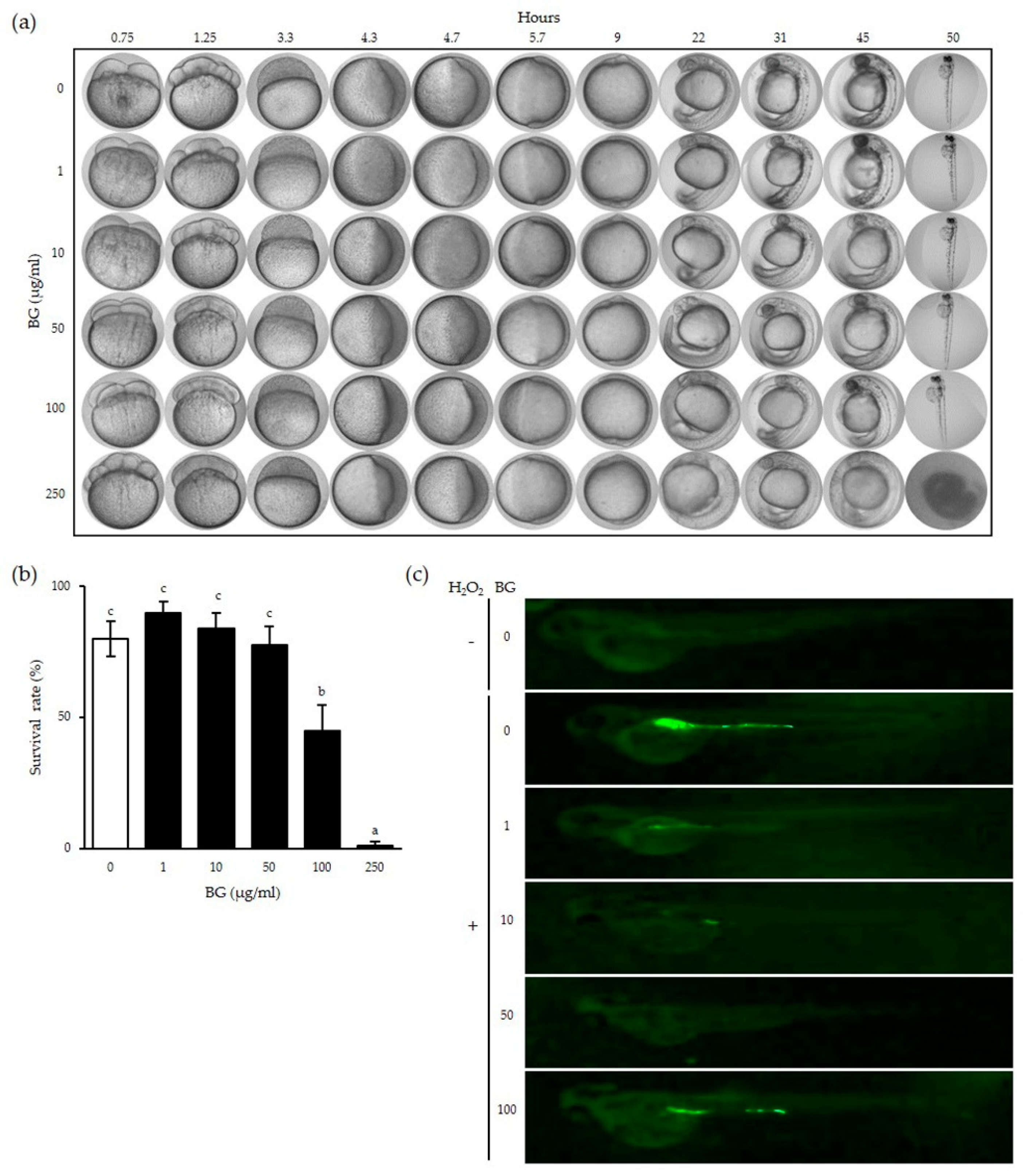
Zebrafish (Danio rerio) embryos were supplied by the Zebrafish Center for Disease Modeling (ZCDM, Korea) and maintained in a temperature-controlled room at 28 °C with a 14:10 h day/night cycle. Zebrafish were fed with brine shrimp 4 times per day. Experiments were performed following the Animal Research Guidelines at Chungnam National University (CNU-01027). At 0.75 to 50 h post fertilization (hpf), zebrafish embryos were isolated in 24-well cell culture plates (Hyundai Micro Co., Seoul, Korea) at 10 embryos/well with egg water supplemented with 0.1% methylene blue, then treated with BG (0, 1, 10, 50, 100, 250 µg/mL) for various times as indicated. Egg water supplemented with BG was changed every 24 h. Embryos were examined using an optical microscope (DM2000, Leica co., Wentzler, Germany), and zebrafish larvae were examined using a stereo microscope (SZ2-ILST, Olympus, Tokyo, Japan) to assess embryo viability. All experiments on the zebrafish were performed according to the protocol approved by the Animal Care and Use Committee of Chungnam National University (CNU-01027).
2.5. Animals and Experimental Diets
Four-week-old male ICR mice were purchased from Orient Bio Co. Ltd. (Gyeonggi-do, Korea) and housed in a light-controlled room (12:12 h day/night cycle) at 20–25 °C. After 1 week of quarantine, the mice were fed either a normal diet (Research diet, NJ, USA) with normal drinking water (NC) or a 45% high-fat diet (Research diet, NJ, USA) with 10% fructose in the drinking water (high-fat/high-fructose diet, HC). After NAFLD was induced, mice were randomly divided into 5 groups: normal diet (NC,
n = 10), HC diet (HC,
n = 9), HC with 0.5% BG (low dose of BG (LB),
n = 9), HC with 1% BG (medium dose of BG (MB),
n = 9) and HC with 2% BG (high dose of BG (HB),
n = 9). The diets supplemented with BG (0.5%, 1%, 2%) were fed to mice for 8 weeks. The detailed experimental design is shown in
Table 1. All animal experiments were approved by the Committee of Animal Care and Experimentation of Gachon University (GIAUAC-R2017019) and were carried out in accordance with the National Institutes of Health Guide for the Care and Use of Laboratory Animals (NIH Publications No. 8023, revised 1978).
2.6. NO Measurement
Nitric oxide (NO) assay was performed according to the manufacturer’s instructions using the Griess reagent system (Promega Co., Madison, WI, USA). MEF cell lines were seeded in 24-well cell culture plates at 5 × 10⁴ cells/well and pretreated with BG (0, 1, 10, 50, 100, 250 µg/mL) for 24 h, then subsequently a cytokine cocktail (CT: 50 ng/mL TNF-α + 50 ng/mL IFN-γ + 25 ng/mL IL-1β + 10 µg/mL LPS) stimulation for an additional 24 h. Absorbance was quantified at 540 nm on a microplate reader (xMark™ Microplate Absorbance Spectrophotometer, Bio-Rad Inc., Hercules, CA, USA).
2.7. ROS Measurement
MEF cell lines were seeded in 96-well cell culture plates (SPL Life Sciences Co., Pocheon, Korea) at 1.5 × 10⁴ cells/well and pretreated with BG (0, 1, 10, 50, 100, 250 µg/mL) for 23 h, then subsequently stimulated with 2 mM H2O2 for 1 h to induce ROS production. Then 10 μM 2′,7′-dichlorofluorescein diacetate (DCFH-DA, Sigma-Aldrich Co., Saint Louis, MO, USA) was added to the cells. After 1 h incubation at 37 °C in the dark, the florescence of DCFH-DA was measured at an excitation wavelength of 485 nm and an emission wavelength of 535 nm by using a fluorescence multimode detector (DTX800, Beckman Coulter, Inc., Brea, CA, USA).
For ROS measurement in vivo, zebrafish embryos at 8 hpf were isolated in 24-well plates at 10 embryos/well with egg water supplemented with 0.1% methylene blue, treated with BG (0, 1, 10, 50, 100, 250 µg/mL) for 1 h, and 5 mM H2O2 was added for an additional 24 h to induce ROS. After incubation, the embryos were washed with egg water and grown to 2-day post fertilization (dpf). At 2 dpf, the eggs were treated with egg water containing DCFH-DA (20 μg/mL), incubated for 1 h at 28 °C in the dark, and then washed with egg water. Images of stained embryos were observed using a digital microscope (Dino-Lite Digital Microscope, ANMO Electronics Co., New Taipei City, Taiwan).
2.8. RNA Extraction, RT-PCR and Real-Time qPCR
The liver and intestine tissues of mice were homogenized using a gentleMACS™ Dissociator (Miltenyi Biotec Co., Bergisch Gladbach, Germany) with TRI reagent (MRC Inc., Cincinnati, OH, USA) for RNA extraction. Then, chloroform (Junsei Co., Tokyo, Japan) was added and the homogenate was centrifuged at 12,000×
g for 15 min at 4 °C. The supernatant was collected, isopropanol (Duksan Co., Ansan-si, Korea) was added, and centrifuged at 12,000 × g for 8 min at 20 °C. The supernatant was removed, and the RNA concentration from the pellet was quantified using a NanoDrop spectrophotometer (Thermo Scientific Inc., Waltham, MA, USA). The cDNA was synthesized using the RT Kit (Biofact Co., Daejeon, Korea). In order to quantify the mRNA expression of binding immunoglobulin protein (BiP), CCAAT/enhancer-binding homologous protein (CHOP) and interleukin-6 (IL-6), reverse transcription polymerase chain reaction (RT-PCR) was performed using 2 × Taq Basic PCR Master Mix according to the manufacturer’s instructions (Biofact Co., Korea). The samples were loaded on to 2% agarose gel for electrophoresis and observed using a gel documentation system (AE-9000 E-Graph, ATTO Co., Tokyo, Japan) under UV light. For real-time quantitative PCR, cDNA was synthesized with SYBR Green Master Mix (Takara Bio, Otsu, Japan) and the mRNA expression was quantified using ABI QuantStudio 3 (Applied Biosystems, Foster City, CA, USA). Primers used in RT-PCR and the real-time qPCR experiments are shown in
Table 2.
2.9. Enzyme-Linked Immunosorbent Measurement (ELISA)
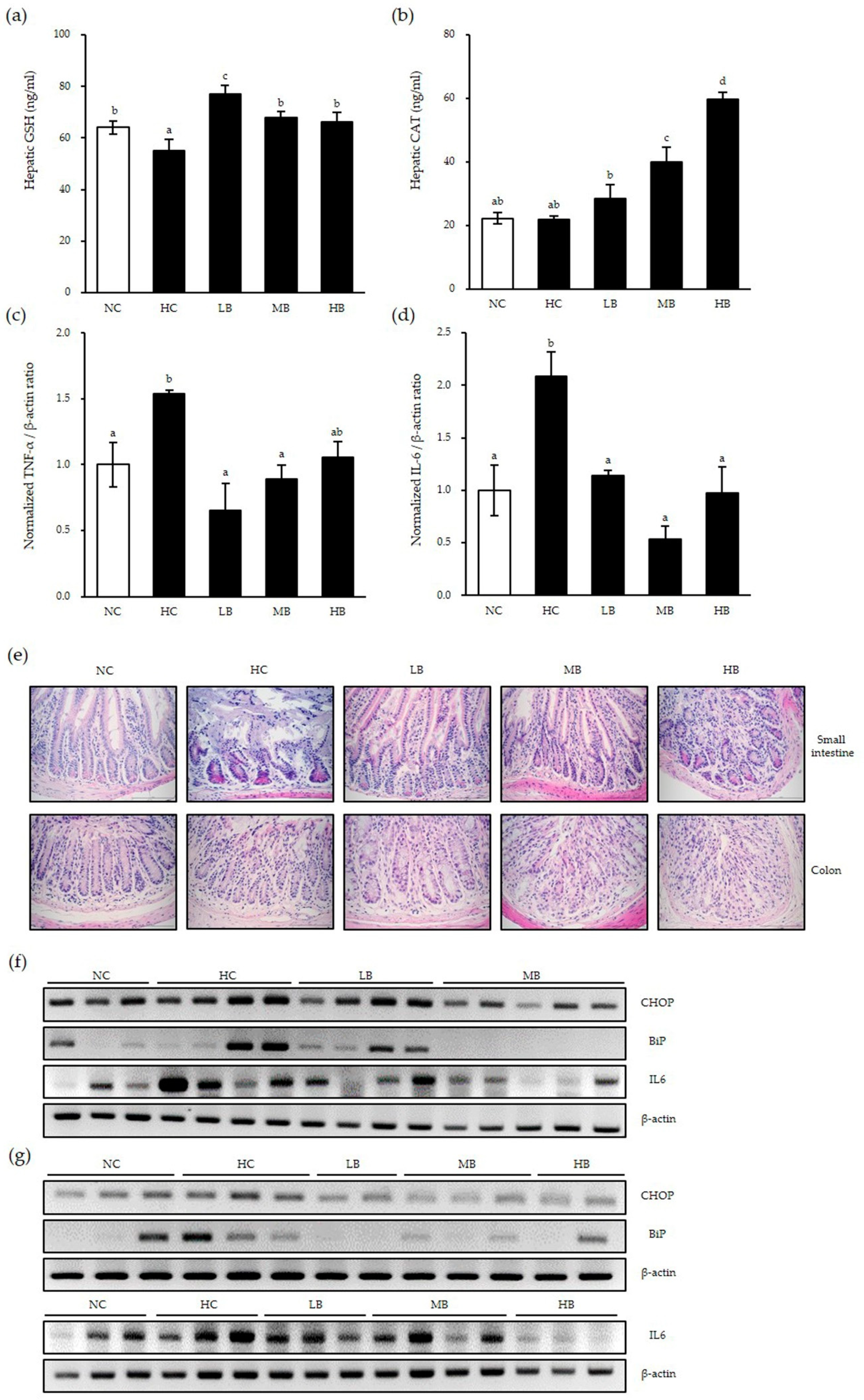
The liver tissues of mice were homogenized using a glass homogenizer with phosphate buffered saline (PBS) and ultrasonicated to further break the cell membranes. Then, homogenate was centrifuged at 1500× g for 15 min. The supernatant was collected to perform an ELISA assay for glutathione (GSH) and catalase (CAT) according to the manufacturer’s instructions (BlueGene Biotech, Shanghai, China).
2.10. Western Blotting
MEF cells were pretreated with BG for 24 h followed by CT treatment for an additional 4 h. Cell lysate was extracted radio immunoprecipitation assay buffer (RIPA buffer, Thermo Scientific Inc., Illinois, USA) on ice and centrifuged at 20,000× g for 20 min at 4 °C. Protein concentration was measured using a bicinchoninic acid (BCA) protein assay kit (Thermo Scientific Inc., USA) and samples were loaded onto 10% sodium dodecyl sulfate (SDS) polyacrylamide gel for electrophoresis and then transferred to nitrocellulose membrane (Bio-Rad Inc., Hercules, CA, USA). After blocking for 1 h with 5% nonfat milk in tris-buffered saline with 0.1% Tween 20 (TBST) buffer at room temperature, anti-iNOS rabbit antibodies (1:1000 dilution; Cell Signaling Technology, Illinois, USA) or anti-glyceraldehyde-3-phosphate dehydrogenase (GAPDH) mouse antibodies (1:1000 dilution; Thermo Scientific Inc., Illinois, USA) were added to the membrane and maintained overnight at 4 °C. Then, the secondary antibodies (1:10,000 dilution; Thermo Scientific Inc., Middlesex County, MA, USA) were added to the membranes for 1 h at room temperature. Enhanced chemiluminescence (West Femto Maximum Sensitivity Substrate Kit, Thermo Scientific Inc., Illinois, USA) and the ChemiDoc system (AE-9100 EZ Capture, ATTO Co., Tokyo, Japan) were used for detection.
2.11. Tissue Histology
The small intestine and colon tissues of mice were fixed in a 10% formaldehyde solution, processed into paraffin blocks using standard methods and stained with hematoxylin and eosin (H&E). The tissue sections were stained with H&E (T&P Bio, Gyeonggi-do, Korea) and all stained tissues were taken with an optical microscope (CX31, Olympus, Tokyo, Japan).
2.12. Statistical Analysis
Data were analyzed using SPSS 24.0 (SPSS Inc., Chicago, IL, USA) software. All experiments were conducted in triplicates. Results are expressed as mean ± standard deviation. Statistical analysis of comparisons between two groups was performed using Student’s t-test, while one-way analysis of variance (ANOVA) with Duncan’s multiple range test was used in the analysis of two or more groups. Statistical significance was assessed at a p < 0.05.
4. Discussion
Previous studies have shown that steaming ginseng nine times improves its preservative properties and efficacy [
15]. This method resulted in increased radical scavenging activity due to the release of phenolic compounds and Maillard reaction products during the heat treatment process. This process makes BG contain relatively lower polar ginsenosides compared to the white and red ginseng, and 19 ginsenosides such as Rb1, Rb2, Rc, Rd, Re, Rf, Rg1, Rg6, F4, Rk3, Rh4, 20(S)-, 20(R)-Rg3, 20(S)-, 20(R)-Rs3, Rk1, Rg5, Rs4 and Rs5 were discovered. In addition, our previous study confirmed and identified 22 different types of ginsenosides of BG and their amounts such as Rg1, Re, Rf, Rh1 (S and R), Rh2 (S and R), Rb1, Rc, F1, Rb2, Rb3, F2, Rg3 (S and R), PPT (S and R), K, Rh2 (S and R), PPD and total ginsenosides in the leaf (22.94 mg/g) and roots (21.00 mg/g) [
17,
18].
Antioxidant and anti-inflammatory effects of BG have been demonstrated in many studies [
24,
25,
26]. With these bioactive components, we showed reduction of lipid accumulation in the liver. However, there are few reports on the association of ER stress with the antioxidant and anti-inflammatory effects of BG. Herein, we used MEF cells knocked out of the ER pathway to determine how these functions are regulated under ER stress.
In this study, we showed that cytokine-induced ROS production and NO production were reduced significantly by BG treatment
Our data showed that H2O2-induced ROS production decreased with BG treatment in zebrafish. However, we believe that the reason the ROS expression level returned to its original state when using a BG concentration of 100 μg/mL is because a concentration of 100 μg/mL might be toxic to zebrafish.
In this study, we observed that BG enhanced the activity levels of antioxidant enzymes in the liver of NAFLD mice. We also observed that the expression levels of the ER stress factor BiP, the apoptosis factor CHOP, and the inflammatory factor IL-6 in the small and large intestines were higher in the HC group than in the NC group, and BG treatment of NAFLD mice reduced inflammation and ER stress significantly. Histological analysis of intestinal tissues showed that HFD-induced lymphocyte infiltration, an indicator of inflammation, was reduced by BG supplementation.

 ,
, 


