Article

Cell-Free Protein Synthesis with Fungal Lysates for the Rapid Production of Unspecific Peroxygenases

1
Institute of Biotechnology, Brandenburg University of Technology Cottbus-Senftenberg, Universitätsplatz 1, 01968 Senftenberg, Germany
2
Fraunhofer Institute for Cell Therapy and Immunology, Branch Bioanalytics and Bioprocesses, Am Mühlenberg 13, 14476 Potsdam-Golm, Germany
3
Department of Bio- and Environmental Sciences, TU Dresden-International Institute Zittau, Markt 23, 02763 Zittau, Germany
*
Author to whom correspondence should be addressed.
Antioxidants 2022, 11(2), 284; https://doi.org/10.3390/antiox11020284
Submission received: 14 January 2022 / Revised: 27 January 2022 / Accepted: 28 January 2022 / Published: 30 January 2022
(This article belongs to the Special Issue Dream Peroxygenases)

Abstract

:
Unspecific peroxygenases (UPOs, EC 1.11.2.1) are fungal biocatalysts that have attracted considerable interest for application in chemical syntheses due to their ability to selectively incorporate peroxide-oxygen into non-activated hydrocarbons. However, the number of available and characterized UPOs is limited, as it is difficult to produce these enzymes in homologous or hetero-logous expression systems. In the present study, we introduce a third approach for the expression of UPOs: cell-free protein synthesis using lysates from filamentous fungi. Biomass of Neurospora crassa and Aspergillus niger, respectively, was lysed by French press and tested for translational activity with a luciferase reporter enzyme. The upo1 gene from Cyclocybe (Agrocybe) aegerita (encoding the main peroxygenase, AaeUPO) was cell-free expressed with both lysates, reaching activities of up to 105 U L−1 within 24 h (measured with veratryl alcohol as substrate). The cell-free expressed enzyme (cfAaeUPO) was successfully tested in a substrate screening that included prototypical UPO substrates, as well as several pharmaceuticals. The determined activities and catalytic performance were comparable to that of the wild-type enzyme (wtAaeUPO). The results presented here suggest that cell-free expression could become a valuable tool to gain easier access to the immense pool of putative UPO genes and to expand the spectrum of these sought-after biocatalysts.

Graphical Abstract

1. Introduction

Over the last two decades, biocatalysis has increasingly developed into a mature and widely applicable technology for chemical synthesis and production [1]. A number of bio-catalytic approaches have been developed for the environmentally friendly and effective synthesis of a range of chiral products, such as pharmaceuticals, agrochemicals, and fine chemicals. However, further development of these biocatalytic processes on an industrial scale will depend crucially on the widespread availability of efficient and robust enzymes [2,3].
Unspecific peroxygenases (EC 1.11.2.1) are enzymes found in numerous fungi and are ideally suited for the synthesis of specialty chemicals. As extracellular enzymes, they are inherently stable and self-sufficient, as evidenced by their ease of activation by hydrogen peroxide and independence of electron transport proteins, electron donors, or cofactors other than heme-stabilizing magnesium ions [4,5]. They have become ‘dream catalysts’ in organic synthesis because of their ability to selectively incorporate peroxidic oxy-gen into non-activated hydrocarbons, causing hydroxylations and epoxidations that are difficult to achieve by chemical methods [6,7,8,9]. Essential for this type of reaction is the heme cofactor that is coordinated proximally by the thiol of a cysteine residue. Due to their P450-like catalytic mechanism and reaction spectrum, UPOs can be used for the conversion of pharmaceuticals and drugs, e.g., of volixibat (N-dealkylation) [10], cyclophosphamide (aliphatic hydroxylation) [11], propranolol (aromatic hydroxylation) [12], testosterone (epoxidation) [13], corticosteroids (side-chain removal by C-C scission) [14] and clopidogrel (sulfoxidation and epoxidation) [15].
A crucial drawback in the application of UPOs (representing a diverse superfamily of proteins) and the main reason why their scope is still very narrow is their low avail-ability, limited to a few representatives [16]. Contrary to their undemanding nature in catalysis, UPOs are demanding when it comes to their expression. To date, there are only a few reports demonstrating the efficient production and purification of UPOs using wild-type fungi [13,14,17,18,19,20]. To circumvent the disadvantages of homologous production, such as low production rates, long production times, or high concentrations of contaminating proteins, various attempts have been made to express UPOs heterologously in established hosts. An overview of recombinantly expressed UPOs is given in the recent review of Kinner and colleagues [21]. Another peroxygenase from Hypoxylon that has been recently produced in Pichia pastoris (syn. Komagataella phaffii) can be added to this list [22].
However, even when using quite different host organisms, such as Saccharomyces cerevisiae, P. pastoris, Aspergillus spp., or Escherichia coli, it has not yet been possible to establish a universal expression system for UPOs. Overall, the number of available UPOs today is less than 20, which is in stark contrast to the number of putative UPO genes in fungal genomes, which is in the range of several thousand [20]. To close the gap between theoretically and practically available UPOs and to gain access to new catalytically promising candidates, a third option (besides homologous and heterologous expression) was chosen in this study: the production of UPOs by cell-free protein synthesis (CFPS).
There are examples of functional heme-containing enzymes that were cell-free expressed using lysates from E. coli: manganese peroxidase [23,24], a P450 monooxygenase [25], cytochrome c oxidase [26], and horseradish peroxidase [27]. This demonstrates the general potential of CFPS for the production of enzymes with such a complex prosthetic group; however, similar results have not yet been published regarding UPOs.
In general, CFPS can be considered a powerful and versatile tool for the rapid discovery of new biocatalyst candidates or improved biocatalyst variants [28]. Optimized systems are also of interest for the preparative production of proteins with yields in the milligram-per-milliliter range. There are CFPS systems based on bacterial, plant, archaeal, fungal, and animal cells. The most relevant and well-studied lysates used for CFPS are from E. coli, Spodoptera frugiperda (insect), S. cerevisiae (yeast), Chinese hamster ovary (CHO), rabbit reticulocyte, wheat germ, and HeLa cells [29]. To date, filamentous fungi just play a minor role in CFPS. In some published work, lysates from Neurospora crassa have been employed to evaluate the potential for the production of cell-free expressed proteins using reporter proteins or for in vitro studies as such, for example, to investigate the impact of codon usage [30,31,32,33]. Regarding the genus Aspergillus, there is only one publication dealing with the use of a lysate from A. nidulans in CFPS [34]. No studies have been published on A. niger in the context of CFPS.
N. crassa and A. niger are filamentous fungi from the phylum Ascomycota, but belong to different classes (Sordariomycetes and Eurotiomycetes, respectively). Based on bioinformatic analyses, both organisms carry putative UPO genes in their genomes [20]. There are good reasons for using filamentous fungi in CFPS, such as their innate competence for post-translational modifications, their ability to secrete proteins, and also their low requirements for the growth medium, which makes the preparation of lysates inexpensive.
In this article, we report the preparation of a functional UPO by a novel cell-free synthesis approach using lysates from N. crassa and A. niger. The cell-free UPO was tested in a substrate screening that included typical UPO substrates as well as some pharmaceuticals, and its biocatalytic activity was compared with that of its homologous counterpart (wild-type UPO).

2. Materials and Methods

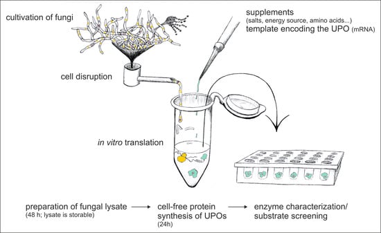
2.1. Lysate Preparation

2.1.1. Cultivation

Neurospora crassa (DSM 1257) was cultured in 500-mL Erlenmeyer flasks, each containing 200 mL of HA complete medium (10 g L−1 glucose and 10 g L−1 yeast extract) [35]. Each flask was inoculated with one drop of spore suspension (~107 spores mL−1 in glycerol) and incubated for 48 h on a rotary shaker (Multitron 2, Infors AG, Bottmingen, Switzerland) at 34 °C and 120 rpm.
Aspergillus niger (DSM 11167) was cultivated in 500-mL Erlenmeyer flasks, each containing 100 mL of 2HA-MS medium [36], but without antifoaming agent. After inoculation with one drop each of spore suspension (~3 × 10⁸ spores mL−1 in glycerol), the flasks were incubated on a rotary shaker (Multitron 2, Infors AG, Bottmingen, Switzerland) at 30 °C and 150 rpm for 24 h.

2.1.2. Preparation of Cell Lysates

Mycelium was harvested by vacuum filtration and washed twice with 50 mL of 4 °C cold mannitol buffer A [37]. Hereafter, the mycelium was resuspended in 1.5 mL of cold lysis buffer A [37] per g fresh weight. Cell disruption was performed by French press (HTU Digi-F-Press, G. Heinemann Ultraschall- und Labortechnik, Schwäbisch Gmünd, Germany) at 4 °C and 5000 psi (N. crassa) and 10,000 psi (A. niger), respectively. The raw lysate was centrifuged at 6500× g and 4 °C for 5 min. The supernatant was placed in a clean tube and clarified again under the same conditions; then the top 90% of the supernatant was collected without cell debris. All steps were carried out as quickly as possible and the samples were kept on ice in between. Lysis efficiency was monitored microscopi-cally.
To remove endogenous mRNA, micrococcal nuclease was added to the supernatant and incubated for 10 min at room temperature [37]. If not used immediately, aliquots (100–500 µL) of the lysate were snap-frozen in liquid nitrogen and stored at −80 °C.

2.2. Cell-Free Protein Synthesis

2.2.1. Preparation of mRNA Templates

The synthetic gene of Cyclocybe (Agrocybe) aegerita UPO (AaeUPO; UniProtKB/Swiss-Prot: B9W4V6) was purchased from Invitrogen (Thermo Fisher Scientific) with a codon optimized sequence for S. cerevisiae containing a C-terminal six histidine peptide, cloned into a pYES2 vector. For run-off transcription, the plasmid was linearized with BstEII-HF (New England Biolabs GmbH, Frankfurt am Main, Germany) and column purified by using the Monarch PCR & DNA cleanup kit (New England Biolabs GmbH, Frankfurt am Main, Germany). In vitro transcription was done with the HiScribe™ T7 ARCA mRNA Kit (with tailing) (New England Biolabs GmbH, Frankfurt am Main, Germany) using 1 µg of the linearized plasmid as template to get mRNA with 5′-Cap and poly(A) tail. Synthesized mRNA was column purified using the Monarch RNA cleanup kit from New England Biolabs GmbH (Frankfurt am Main, Germany), checked for quality by gel electrophoresis, and quantified photometrically at 260 nm.

2.2.2. Cell-Free Protein Synthesis

For the in vitro translation, a translation mix was freshly prepared containing 80 mM HEPES-KOH pH 7.4, 4 mM ATP, 0.4 mM GTP, 8 mM dithiothreitol, 80 mM creatine phosphate, 200 mM potassium acetate, 4.8 mM magnesium acetate, 0.24 mg mL−1 tRNA from yeast, amino acid mix (0.08 mM each) and 0.24 U µL−1 creatine phosphokinase. The cell-free reaction mixture consisted of 25% translation mix, mRNA (typically 28 nM) in DEPC water, and 50% fungal lysate. No-template controls contained only DEPC water instead of mRNA. Typical reactions were carried out in a volume of 20 µL in 1.5-mL reaction tubes (Eppendorf, Germany) in an incubator at 18 °C. In case of the reporter firefly luciferase, 28 nM mRNA (purchased from Promega, Walldorf, Germany) were used for CFPS reactions.

2.3. Analytics

2.3.1. Enzymatic Assays

For measuring the activity of the reporter firefly luciferase, the ONE-Glo™ Luciferase Assay System (Promega, Walldorf, Germany) was used. 10 µL of the CFPS reaction were added to 40 µL of Luciferase Assay Buffer containing Luciferase Assay Reagent in a white 96-well plate. Prior to measurement, the plate was shaken for 60 s at 300 rpm in the plate reader. Light emission resulting from the luciferase-catalyzed oxidation of the substrate luciferin was detected with a CLARIOstar Plus plate reader (BMG Labtech, Ortenberg, Germany).
The activity of functional cell-free UPO was routinely tested with an LC-MS-based propranolol assay. The assay reaction (100 µL total volume) was initiated by adding 90 µL of a reaction mixture (1 mM propranolol, 1 mM hydrogen peroxide, 5 mM ascorbate, 20 mM potassium phosphate buffer, pH 7.0) to 10 µL of cell-free reaction mixture. The reactions were incubated at 25 °C and 800 rpm on a thermal shaker (TurboShaker 3500, Scienova GmbH, Jena, Germany) for 5 min and stopped by adding 100 μL of ice-cold ace-tonitrile (−20 °C). Subsequently, the samples were centrifuged at 14,000× g for 10 min, the supernatants were transferred to HPLC vials and analyzed by liquid chromatography-mass spectrometry (LC-MS). The UPO activity is given in propranolol units, where one unit (1 U) corresponds to the oxygenation of 1 μmol of propranolol to 5-hydroxypropra-nolol (5-OHP) in 1 min. In order to quantify the formation of 5-OHP, 4-hydroxypropra-nolol (Sigma Aldrich, Schnelldorf, Germany) was used for the calibration (as internal standard).
For the comparative study of UPO activities, we measured the conversion of the typi-cal UPO substrates veratryl alcohol, 2,2′-azino-bis(3-ethylbenzothiazoline-6-sulfonic acid) diammonium salt (ABTS), 5-nitrobenzodioxole (NBD), and naphthalene as well as of the pharmaceutical substrates propranolol, diclofenac, and clopidogrel. The CFPS reaction mixtures were sonicated (3 × 10 s pulses with 30 s pauses in between; amplitude 10%; Sonopuls, Bandelin, Berlin, Germany) to release the enzyme from the microsomes. The lysed samples were centrifuged at 16,000× g and 4 °C for 10 min and the supernatants were used for the reactions. Homologous AaeUPO (wtAaeUPO) was produced and purified as described elsewhere [17]. Both enzyme preparations were diluted to 100 U L−1 (based on the oxidation of veratryl alcohol to veratraldehyde; ε310 = 9300 M−1cm−1). The formation of the ABTS cation radical (ε420 = 36,000 M−1 cm−1), 5-nitrocatechol (ε425 = 9700 M−1 cm−1) and 1-naphthtol (ε303 = 9700 M−1 cm−1) were measured photometrically (CLARIOstar Plus, BMG Labtech, Ortenberg, Germany). The comparative conversion of the pharmaceutical substrates was followed analogously to the propranolol assay, and the products were quantified by LC-MS as described below. Additional calibrations were performed with the authentic standards 4′-hydroxydiclofenac and 2-oxoclopidogrel.

2.3.2. High-Resolution Mass Spectrometry

Chromatographic separations in LC-MS experiments were performed on a Thermo Scientific Vanquish Flex Quaternary UHPLC system (Thermo Fisher Scientific, Waltham, MA, USA) using a Kinetex® column (C18, 2.6 µm, 100 Å, 150 × 2.1 mm, Phenomenex, Torrance, CA, USA). The injection volume was 1 µL, and the column was eluted at a flow rate of 0.5 mL min−1 and 40 °C with two mobile phases: A (diH2O, 0.1% formic acid) and B (acetonitrile, 0.1% formic acid) and following gradient: 0 min 10% B; 0.5 min, 10% B; 5 min, 80% B; 7 min, 80% B; 7.1 min, 10% B; 10 min, 10% B.
MS spectra were obtained using the Thermo Scientific Q Exactive Focus quadrupole-Orbitrap mass spectrometer (Thermo Electron, Waltham, MA, USA) coupled with a heated electrospray ionization source in positive mode. The tune operating parameters were as follows: rate of sheath gas flow and auxiliary gas flow—40 and 15 (arbitrary units), respectively; spray voltage—4.0 kV; temperature of capillary and sample heater—260 °C and 400 °C, respectively; high-resolution MS was operated at full scan positive mode with a mass range of m/z 150–1500 at a resolution of 70,000 (m/z 200).

2.3.3. Western Blot

If needed, the CFPS samples were fractionated by centrifugation at 16,000× g and 4 °C for 10 min prior to Western blot analyses. The supernatant was transferred to a new 1.5-mL reaction tube, and the pellet was resuspended in 20 mM HEPES buffer pH 7.0 with the same volume as the supernatant.
Protein samples (10 µL) were mixed with Laemmli sample buffer, heated at 95 °C for 5 min, and separated on Bolt 10% SDS-PAGE by using a mini gel tank (Life Technologies, Carlsbad, CA, USA). Subsequently, the proteins were transferred to a polyvinyl difluoride membrane with 0.2 µm pore size (Carl Roth, Karlsruhe, Germany). After blocking the membranes with 5% BSA in PBST with 0.3% Tween 20 for 1 h, the membranes were incubated with a monoclonal 6X-His epitope tag antibody (MA1-135, clone 4E3D10H2/E3, Life Technologies, Carlsbad, USA) at a dilution of 1:500 overnight at 4 °C, washed in PBST with 0.3% Tween 20, and probed with an HRP-conjugated anti-mouse IgG secondary antibody at a dilution of 1:1000 (#7076, Cell Signaling Technology, Danvers, MA, USA) for 1 h at room temperature. A light-emitting chemiluminescent substrate (SignalFire ECL Reagent, Cell Signaling Technology, Danvers, MA, USA) was used for the detection by exposing it with the biostep Celvin S chemiluminescence imager (biostep, Burkhardtsdorf, Germany). Densitometric evaluation of the protein bands was done by means of the software ImageJ (https://imagej.nih.gov/ij/; accessed on 9 December 2021; version 1.53k).

2.3.4. Deglycosylation

CFPS samples (either fractionated as described above or as total reaction mix) were deglycosylated using the Protein Deglycosylation Mix II (New England Biolabs GmbH, Frankfurt am Main, Germany). The samples were subsequently analyzed by Western blot.

3. Results and Discussion

3.1. Lysate Preparation from Neurospora crassa and Aspergillus niger

N. crassa and A. niger were grown in nutrient-rich media for 48 h and 24 h, respectively. Subsequently, the mycelia were harvested by vacuum filtration and yielded fresh weights of 25 to 30 g L−1 for both fungi. For cell disruption, a French press homogenizer was used, which represents a simple and fast method and is routinely used in the preparation of lysates from other fungal organisms such as S. cerevisiae [38]. The lysis was successful as 80 to 90% of the cells were broken, which was microscopically detectable (Figure 1a).
The lysates were tested for translational activities by using the cytosolic reporter enzyme ‘firefly luciferase’ (Figure 1b). With both lysates, significant amounts of firefly luci-ferase were in vitro translated, albeit in differing quantities. The measured values for N. crassa (30,721 ± 6091 RLU) were 35-times higher than those of A. niger (862 ± 78 RLU).

3.2. Cell-Free Protein Synthesis of AaeUPO

For our study on the cell-free production of a UPO, we used the upo1 gene from Cyclocybe (Agrocybe) aegerita, which encodes the first reported UPO [17]. The extracellular heme-thiolate enzyme contains an N-terminal signal peptide followed by a propeptide and has a C-terminal intramolecular disulfide bond as well as six potential N-glycosylation sites [39]. For easier detection, the UPO gene was tagged with a C-terminal polyhistidine peptide. To reduce the complexity of the CFPS system, the mRNA of upo1 was synthesized independently from the translation system and provided externally. The CFPS reactions were tested for the formation of functional cell-free UPO with an LC-MS-based propranolol assay since the formation of 5-hydroxypropranolol (5-OHP) from propranolol is characteristic of AaeUPO [12,40]. By using this assay, we could exclude the possibility of measuring false positive activities from potentially interfering endogenous monooxygenases (cytochrome P450 enzymes). Nevertheless, negative controls that contained no mRNA templates were additionally included in all experimental setups.
Cell-free production of AaeUPO was evaluated with lysates from N. crassa and A. niger, respectively. Essentially, the reactions were set up according to Wu et al. with minor modifications [33]. The mRNA carrying the coding sequence for the UPO was applied at 28 nM since higher concentrations had adverse effects on the synthesis of active enzyme (Figure A1). The formation of active cfAaeUPO was observed over a period of 24 h (Figure 2). With both lysates, significant UPO activity could be detected: 10,498 ± 28 mU L−1 using the N. crassa lysate and 783 ± 18 mU L−1 with the A. niger lysate. The different yields of active enzyme correlated with the results for the reporter luciferase (Figure 1b). Compared to the lysate from N. crassa, the yield of active enzyme with the A. niger lysate was 10-times (after 5 h incubation) to 15-times (after 24 h incubation) lower. Interestingly, in contrast to N. crassa, the UPO activity with the A. niger lysate was higher after 5 h of incubation than after 24 h. This decrease in activity could indicate residual proteolytic activity in the lysate, despite the application of protease inhibitors during the preparation process.
Western blot analyses were done to get information about the localization of the synthesized protein and its relative amount (Figure 3). As a secretory protein, cfAaeUPO was expected to be present in the microsomes, i.e., in ER-derived vesicles. In the case of the A. niger lysate-based CFPS, a band was detected in the microsomal fraction that corresponded to AaeUPO, which has a theoretical molecular weight of 41.5 kDa in its unprocessed form. Overall, the amount of detectable protein was rather low, which explains the relatively low activity. With the N. crassa lysate, two prominent bands could be seen in the whole CFPS reaction mixture. As demonstrated by deglycosylation (Figure 3d), the lower band corresponds to the non-glycosylated (marked with an asterisk) and the upper band to the glycosylated (marked with an arrowhead) form of cfAaeUPO. The larger part of the glycosylated protein was located in the microsomes, while the non-glycosylated UPO was almost exclusively found in the supernatant. Activity measurements of the fractions indicated that the UPO activity stems from the glycosylated form as the ratio of the activities between the fractions correlated with the distribution of synthesized protein (Figure A2). The residual glycosylated cfAaeUPO seen in the supernatant fraction may be the result of an incomplete fractionation process, similarly described in an article by Thoring et al. [41]. They showed that—after centrifugation with 16,000× g—smaller microsomes were still present in the supernatant. The rather high portion of non-glycosylated protein might indicate an insufficient translocation capacity. Nonetheless, the successful expression of active AaeUPO proves that our fungal lysates contain functional microsomes that allow appropriate post-translational modification of secretory proteins.
Unfortunately, it was not possible to use the polyhistidine tag on the cfAaeUPO for purification from the CFPS reaction with sufficient purity, due to the presence of a larger amount of contaminating fungal proteins. Thus, an accurate protein concentration of the synthesized cfAaeUPO could not be determined. In future experiments, the use of more appropriate affinity tags, such as the Strep II tag, should be considered [42].
The good results for the long incubation times with the lysate from N. crassa (Figure 2) were unexpected since, in batch CFPS reactions, the synthesis process usually stops after a few hours. For example, Hodgman and Jewett reported a “long synthesis time of 120 min” for luciferase using a yeast CFPS-batch reaction system, albeit at a slightly higher incubation temperature of 21 °C [37]. The main reasons for the termination of synthesis were seen in a rapid decline in energy-providing components and an accumulation of inhibitory molecules, such as inorganic phosphate [43]. To clarify whether the apparently long synthesis time in our experiments was actually due to better protein synthesis performance of the lysate, we expressed the reporter protein luciferase using CFPS with the lysate of N. crassa and measured the activity over a 24-hour period (Figure 4a). The activity of luciferase increased over the first three hours of CFPS but decreased thereafter, suggesting that the process of protein synthesis was not maintained throughout the incubation period. To confirm that this was also true for CFPS of AaeUPO, samples from the time-course experiment (Figure 2) were subjected to Western blot analysis (Figure 4b). As in the previous Western blot, two prominent bands appeared in the gel. Their intensity and thus the amount of cfAaeUPO produced increased until the third hour of incubation and then remained nearly constant (or decreased slightly) for the next hours. The 24-hour samples appeared to have lower cfAaeUPO protein levels than the samples from the previous time points, although activity was highest after 24 h. It might be that nonfunctional, e.g., misfolded UPO protein was degraded by remaining proteases in the lysate, but this needs to be clarified in future experiments.
Since the capacity to produce protein was limited to 3 h in our setup, an intrinsic maturation period of the UPO protein could be an explanation for the apparently long synthesis time. Such a maturation time was observed for a bacterial P450 monooxygenase that was expressed in a cell-free approach using lysate from E. coli [25]. In this case, the highest enzyme activity was reached 150 min after protein synthesis had stopped. However, since the exact process of heme incorporation and protein folding is still unknown for UPOs, it must be clarified in the future whether a longer maturation time is indeed necessary. Here, the CFPS might prove to be a valuable tool for gaining insight into the genesis of UPOs. It should be relatively simple to change the conditions in CFPS, for example, by adding mRNA for the co-expression of chaperones. The effects on UPO formation would be directly measurable and could be evaluated in a short time.
Table 1 gives a brief comparison of the volume activities and production times of AaeUPOs obtained from homologous, heterologous, or cell-free approaches. Attempts to express wtAaeUPO heterologously in the standard expression hosts S. cerevisiae or P. pastoris with appreciable yields failed [44,45]. Rounds of directed evolution were necessary to increase the production in the two yeasts that do not contain endogenous UPO genes [20,45]. This highlights an advantage of the cell-free approach: the possibility to use translationally active lysates from naturally UPO-coding (and producing) fungi. Thereby, all essential components should be available for the in vitro UPO synthesis. However, it has to be mentioned that the evolved mutant variant rAaeUPO PaDa-I expressed in P. pastoris in a bioreactor in a six-day cultivation period reached much higher expression levels than the homologous or cell-free systems. Nevertheless, cell-free protein production is by far the fastest method to gain significant amounts of functional UPO.
If the heterologously produced PaDa-I variant of rAaeUPO is included in the comparison of the three expression systems, the yield obtained with CFPS appears to be the lowest. However, in the present study, we have not yet fully exploited the potential of the cell-free approach using fungal lysates. There are general aspects in CFPS that have a high impact on the performance and would need to be optimized, for example, biomass production, lysate preparation, and buffer composition [46,47]. Also, the template design is essential for an optimal translation initiation, which can be triggered by adding elements like IRES [48]. As indicated by the Western blot analyses (Figure 3), the production of active UPO in our setup may be limited by a non-optimal translocation efficiency. Co-expressing components of the secretory apparatus could solve this problem, as this approach was already successful in living cells, for example, for the production of secreted antibodies in CHO cells [49]. Additionally, there is the possibility to change the CFPS reaction from a batch to a continuous process, which would result in an increase in protein yield [50]. Other influential parameters or components are specific for the expression of UPOs. The availability and fit of the heme cofactor are crucial for the functioning of UPOs, therefore the formulation and amount of heme in CFPS reactions will have to be optimized. This could be achieved on different levels: (i) during growth of the fungi, e.g., with the addition of hemin, which worked well in the expression of horseradish peroxidase in P. pastoris [51]; and (ii) by adding heme directly to the CFPS reaction, which was successful in the cell-free production of manganese peroxidase as well as horseradish peroxidase [23,24,27]. Since AaeUPO and other UPOs have at least one internal disulfide bond and other UPOs form functional dimers via disulfide bridges, it might also be beneficial to add protein disulfide isomerases like PDI or DsbC to CFPS reactions [23,27].

3.3. Substrate Screening with Cell-Free AaeUPO

The cell-free protein production approach should offer the possibility to establish a rapid screening platform for interesting biocatalysts. Thus, we tested the cell-free UPO produced with N. crassa lysate directly in a substrate screening without the need for time-consuming purification steps. To this end, the conversion (oxygenation, oxidation) of typical UPO substrates, which are also used in activity assays, as well as of three common pharmaceuticals was performed with simply prepared CFPS reaction solutions. The enzyme that is partly located in microsomes was efficiently released by ultrasonic treatment (Figure A3). After centrifugation, the supernatant was used for photometric UPO assays with ABTS, veratryl alcohol, 5-nitrobenzodioxole, and naphthalene, and for LC-MS analyses with the pharmaceuticals propranolol, diclofenac, and clopidogrel.
The β-adrenergic blocker propranolol, the non-steroidal anti-inflammatory drug diclofenac, and the antithrombotic prodrug clopidogrel are known to be metabolized by cytochrome P450 monooxygenases in the human body. In previous studies, it was shown that wtAaeUPO is able to perform the same reactions to give the (major) human drug metabolites 5-hydroxypropranolol, 4′-hydroxydiclofenac, and 2-oxoclopidogrel, respectively (Figure 5) [15,40,52].
The activities of cfAaeUPO were compared with those of its wild-type counterpart (taking into account that wtAaeUPO did not contain a polyhistidine tag). For the purpose of comparison, we used enzyme samples with a normalized volume activity of 100 U L−1 (based on veratryl alcohol oxidation) in all reactions. The results are listed in Table 2.
All substrates were converted by both enzyme preparations, although quantitative differences in the activities were found in some cases. For NBD, naphthalene, and propranolol, the measured activities were similar; however, for ABTS, diclofenac, and clopidogrel, the activities of the cell-free variant were two to four times lower. ABTS is a substrate that is oxidized via one-electron abstraction by the peroxidative activity of UPO resulting in a cation radical product (‘classic’ peroxidase activity without oxygen transfer). The fungal lysate used for CFPS may have contained metabolites with antioxidant properties that could have interfered with the detection of the ABTS radical [53]. Thus, substrates like ABTS might only be suitable in screening approaches with unpurified UPO, if the yield of cell-free enzyme is relatively high. A possible explanation for the lower conversion of diclofenac and clopidogrel could be the presence of the C-terminal polyhistidine tag, which may have impaired activity, e.g., by shielding the heme-access channel. Such an effect was already reported for a mutant variant of AaeUPO (PaDa-I) [54]. In addition, negative effects of potentially different glycosylation patterns cannot be excluded.
In summary, it took less than two days from the DNA template carrying the AaeUPO gene to obtain the substrate screening results with expressed cfAaeUPO. This is much faster compared to conventional screening systems based on heterologous expression in host microbes [55]. Moreover, if transcription and translation were coupled in one CFPS reaction, the turnaround time of the entire process could be reduced even further.

4. Conclusions

In the present study, we evaluated a novel approach to produce unspecific peroxygenases (UPOs) by using a cell-free synthesis system based on fungal lysates. The UPO from Cyclocybe (Agrocybe) aegerita was successfully expressed with lysates derived from N. crassa or A. niger. Activities of this cfAaeUPO of up to 105 U L−1 (based on veratryl alcohol oxidation) could be achieved within 24 h, where the lysate from N. crassa showed a significantly higher translational performance compared to A. niger. The unpurified CFPS reaction solutions were subjected to a substrate screening, in which the performance of the cell-free enzyme (cfAaeUPO) was comparable to that of the homologously produced enzyme (wtAaeUPO). The procedure presented here highlights the general potential of CFPS for rapid screening of new UPOs or UPO variants, including the estimation of their catalytic performance. Nevertheless, for large-scale production of suitable candidates, hetero-logous production in fungal hosts will continue to be the method of choice, as cell-free synthesis is currently not competitive in terms of yield. Last, but not least, it must be pointed out that the full potential of lysates from filamentous fungi is far from being tapped, as their use in CFPS is still in its infancy.

Author Contributions

Conceptualization, M.S., K.-U.S. and J.K.; methodology, S.F., K.-U.S., J.K., P.P. and M.S.; investigation, M.S., S.F., K.-U.S., J.K. and H.K.; resources, K.S. and M.H.; writing—original draft preparation, M.S. and M.H.; writing—review and editing, M.S., S.F., K.-U.S., J.K., H.K., R.U., M.H. and K.S.; supervision, K.S.; project administration, K.S.; funding acquisition, M.H. and K.S. All authors have read and agreed to the published version of the manuscript.

Funding

This research was funded by the Federal Ministry of Education and Research (BMBF, Germany), project CEFOX (031B0831); and the Ministry of Science, Research and Culture (MWFK, Brandenburg, Germany), project PZ-Syn (project number F241-03-FhG/005/001). We gratefully acknowledge MWFK for their financial support as part of the framework ‘Großgeräte der Länder’ on the acquisition of high-resolution mass spectrometry (GZ: INST 263/88-1 LAGG).

Institutional Review Board Statement

Not applicable.

Informed Consent Statement

Not applicable.

Data Availability Statement

The data presented in this study are available on request from the corresponding author.

Acknowledgments

We would like to thank Johannes Kalmbach for his assistance with the photometrical experiments.

Conflicts of Interest

The authors declare no conflict of interest.

Appendix A

Figure A1. Cell-free synthesis of AaeUPO protein with lysate from N. crassa using varying concentrations of mRNA. 20-µL reactions with varying mRNA concentrations were incubated at 18 °C for 4 h and tested for UPO activity by measuring the conversion of propranolol to 5-OHP by LC-MS. The highest value (at 28 nM mRNA) was set to 100%.
Antioxidants 11 00284 g0a1
Figure A2. Distribution of UPO activity in the supernatant and the microsomal fraction. 20-µL CFPS reactions with AaeUPO mRNA were incubated at 18 °C for 4 h and separated in supernatant and microsomal fraction by centrifugation. The whole reaction mix and fractions were tested for UPO activity by measuring the conversion of propranolol to 5-OHP by LC-MS. Data given for the supernatant and microsomal fraction were calculated as percentages of activity in the whole reaction mixture.
Antioxidants 11 00284 g0a2
Figure A3. Release of cell-free produced peroxygenase (cfAaeUPO) from microsomes by sonication; a 320-µL CFPS reaction in a 1.5-mL reaction tube was sonicated with the indicated number of pulses (10 s, with 30 s pause interval). After each pulse, a 15-µL sample was taken. All samples were centrifuged, and the supernatants were tested for UPO activity by measuring the conversion of propranolol to 5-OHP by LC-MS. UPO activities were normalized in relation to the untreated sample.
Antioxidants 11 00284 g0a3

References

  1. Wu, S.; Snajdrova, R.; Moore, J.C.; Baldenius, K.; Bornscheuer, U.T. Biocatalysis: Enzymatic synthesis for industrial applications. Angew. Chem. Int. Ed. 2021, 60, 88–119. [Google Scholar] [CrossRef] [PubMed]
  2. Zhang, Z.-J.; Pan, J.; Ma, B.-D.; Xu, J.-H. Efficient biocatalytic synthesis of chiral chemicals. In Bioreactor Engineering Research and Industrial Applications I; Ye, Q., Bao, J., Zhong, J.-J., Eds.; Springer: Berlin/Heidelberg, Germany, 2014; Volume 155, pp. 55–106. [Google Scholar]
  3. Sheldon, R.A.; Brady, D.; Bode, M.L. The Hitchhiker’s guide to biocatalysis: Recent advances in the use of enzymes in organic synthesis. Chem. Sci. 2020, 11, 2587–2605. [Google Scholar] [CrossRef] [PubMed] [Green Version]
  4. Hofrichter, M.; Kellner, H.; Pecyna, M.J.; Ullrich, R. Fungal unspecific peroxygenases: Heme-thiolate proteins that combine peroxidase and cytochrome P450 properties. In Monooxygenase, Peroxidase and Peroxygenase Properties and Mechanisms of Cytochrome P450; Hrycay, E.G., Bandiera, S.M., Eds.; Springer International Publishing: Berlin/Heidelberg, Germany, 2015; Volume 851, pp. 341–368. [Google Scholar]
  5. Hofrichter, M.; Kellner, H.; Herzog, R.; Karich, A.; Kiebist, J.; Scheibner, K.; Ullrich, R. Peroxide-mediated oxygenation of organic compounds by fungal peroxygenases. Antioxidants 2022, 11, 163. [Google Scholar] [CrossRef] [PubMed]
  6. Wang, Y.; Lan, D.; Durrani, R.; Hollmann, F. Peroxygenases en route to becoming dream catalysts. What are the opportunities and challenges? Curr. Opin. Chem. Biol. 2017, 37, 1–9. [Google Scholar] [CrossRef]
  7. Sigmund, M.-C.; Poelarends, G.J. Current state and future perspectives of engineered and artificial peroxygenases for the oxyfunctionalization of organic molecules. Nat. Catal. 2020, 3, 690–702. [Google Scholar] [CrossRef]
  8. Kiebist, J.; Hofrichter, M.; Zuhse, R.; Scheibner, K. Oxyfunctionalization of pharmaceuticals by fungal peroxygenases. In Pharmaceutical Biocatalysis, 1st ed.; Grunwald, P., Ed.; Jenny Stanford Publishing: Singapore, 2019; pp. 643–680. [Google Scholar]
  9. Hobisch, M.; Holtmann, D.; Gomez de Santos, P.; Alcalde, M.; Hollmann, F.; Kara, S. Recent developments in the use of peroxygenases—Exploring their high potential in selective oxyfunctionalisations. Biotechnol. Adv. 2021, 51, 107615. [Google Scholar] [CrossRef]
  10. Kiebist, J.; Holla, W.; Heidrich, J.; Poraj-Kobielska, M.; Sandvoss, M.; Simonis, R.; Gröbe, G.; Atzrodt, J.; Hofrichter, M.; Scheibner, K. One-pot synthesis of human metabolites of SAR548304 by fungal peroxygenases. Bioorg. Med. Chem. 2015, 23, 4324–4332. [Google Scholar] [CrossRef] [PubMed]
  11. Steinbrecht, S.; Kiebist, J.; König, R.; Thiessen, M.; Schmidtke, K.-U.; Kammerer, S.; Küpper, J.-H.; Scheibner, K. Synthesis of cyclophosphamide metabolites by a peroxygenase from Marasmius rotula for toxicological studies on human cancer cells. AMB Express 2020, 10, 128. [Google Scholar] [CrossRef] [PubMed]
  12. Gomez de Santos, P.; Cañellas, M.; Tieves, F.; Younes, S.H.H.; Molina-Espeja, P.; Hofrichter, M.; Hollmann, F.; Guallar, V.; Alcalde, M. Selective synthesis of the human drug metabolite 5′-hydroxypropranolol by an evolved self-sufficient peroxygenase. ACS Catal. 2018, 8, 4789–4799. [Google Scholar] [CrossRef] [Green Version]
  13. Kiebist, J.; Schmidtke, K.-U.; Zimmermann, J.; Kellner, H.; Jehmlich, N.; Ullrich, R.; Zänder, D.; Hofrichter, M.; Scheibner, K. A peroxygenase from Chaetomium globosum catalyzes the selective oxygenation of testosterone. Chembiochem 2017, 18, 563–569. [Google Scholar] [CrossRef] [Green Version]
  14. Ullrich, R.; Poraj-Kobielska, M.; Scholze, S.; Halbout, C.; Sandvoss, M.; Pecyna, M.J.; Scheibner, K.; Hofrichter, M. Side chain removal from corticosteroids by unspecific peroxygenase. J. Inorg. Biochem. 2018, 183, 84–93. [Google Scholar] [CrossRef] [PubMed]
  15. Kiebist, J.; Schmidtke, K.-U.; Schramm, M.; König, R.; Quint, S.; Kohlmann, J.; Zuhse, R.; Ullrich, R.; Hofrichter, M.; Scheibner, K. Biocatalytic syntheses of antiplatelet metabolites of the thienopyridines clopidogrel and prasugrel using fungal peroxygenases. J. Fungi 2021, 7, 752. [Google Scholar] [CrossRef] [PubMed]
  16. Ullrich, R.; Karich, A.; Hofrichter, M. Fungal peroxygenases—A versatile tool for biocatalysis. In Encyclopedia of Mycology; Zaragoza, Ó., Casadevall, A., Eds.; Elsevier: Oxford, UK, 2021; pp. 260–280. [Google Scholar]
  17. Ullrich, R.; Nüske, J.; Scheibner, K.; Spantzel, J.; Hofrichter, M. Novel haloperoxidase from the agaric basidiomycete Agrocybe aegerita oxidizes aryl alcohols and aldehydes. Appl. Environ. Microbiol. 2004, 70, 4575–4581. [Google Scholar] [CrossRef] [PubMed] [Green Version]
  18. Anh, D.H.; Ullrich, R.; Benndorf, D.; Svatoś, A.; Muck, A.; Hofrichter, M. The coprophilous mushroom Coprinus radians secretes a haloperoxidase that catalyzes aromatic peroxygenation. Appl. Environ. Microbiol. 2007, 73, 5477–5485. [Google Scholar] [CrossRef] [Green Version]
  19. Gröbe, G.; Ullrich, R.; Pecyna, M.J.; Kapturska, D.; Friedrich, S.; Hofrichter, M.; Scheibner, K. High-yield production of aromatic peroxygenase by the agaric fungus Marasmius rotula. AMB Express 2011, 1, 31. [Google Scholar] [CrossRef] [Green Version]
  20. Hofrichter, M.; Kellner, H.; Herzog, R.; Karich, A.; Liers, C.; Scheibner, K.; Kimani, V.W.; Ullrich, R. Fungal peroxygenases: A phylogenetically old superfamily of heme enzymes with promiscuity for oxygen transfer reactions. In Grand Challenges in Fungal Biotechnology; Nevalainen, H., Ed.; Springer International Publishing: Berlin/Heidelberg, Germany, 2020; pp. 369–403. [Google Scholar]
  21. Kinner, A.; Rosenthal, K.; Lütz, S. Identification and expression of new unspecific peroxygenases—Recent advances, challenges and opportunities. Front. Bioeng. Biotechnol. 2021, 9, 705630. [Google Scholar] [CrossRef]
  22. Rotilio, L.; Swoboda, A.; Ebner, K.; Rinnofner, C.; Glieder, A.; Kroutil, W.; Mattevi, A. Structural and biochemical studies enlighten the unspecific peroxygenase from Hypoxylon sp. EC38 as an efficient oxidative biocatalyst. ACS Catal. 2021, 11, 11511–11525. [Google Scholar] [CrossRef]
  23. Miyazaki-Imamura, C.; Oohira, K.; Kitagawa, R.; Nakano, H.; Yamane, T.; Takahashi, H. Improvement of H2O2 stability of manganese peroxidase by combinatorial mutagenesis and high-throughput screening using in vitro expression with protein disulfide isomerase. Protein Eng. Des. Sel. 2003, 16, 423–428. [Google Scholar] [CrossRef] [Green Version]
  24. Ninomiya, R.; Zhu, B.; Kojima, T.; Iwasaki, Y.; Nakano, H. Role of disulfide bond isomerase DsbC, calcium ions, and hemin in cell-free protein synthesis of active manganese peroxidase isolated from Phanerochaete chrysosporium. J. Biosci. Bioeng. 2014, 117, 652–657. [Google Scholar] [CrossRef]
  25. Kwon, Y.-C.; Oh, I.-S.; Lee, N.; Lee, K.-H.; Yoon, Y.J.; Lee, E.Y.; Kim, B.-G.; Kim, D.-M. Integrating cell-free biosyntheses of heme prosthetic group and apoenzyme for the synthesis of functional P450 monooxygenase. Biotechnol. Bioeng. 2013, 110, 1193–1200. [Google Scholar] [CrossRef]
  26. Katayama, Y.; Shimokata, K.; Suematsu, M.; Ogura, T.; Tsukihara, T.; Yoshikawa, S.; Shimada, H. Cell-free synthesis of cytochrome c oxidase, a multicomponent membrane protein. J. Bioenerg. Biomembr. 2010, 42, 235–240. [Google Scholar] [CrossRef] [PubMed]
  27. Zhu, B.; Mizoguchi, T.; Kojima, T.; Nakano, H. Ultra-high-throughput screening of an in vitro-synthesized horseradish peroxidase displayed on microbeads using cell sorter. PLoS ONE 2015, 10, e0127479. [Google Scholar] [CrossRef] [PubMed] [Green Version]
  28. Rolf, J.; Rosenthal, K.; Lütz, S. Application of cell-free protein synthesis for faster biocatalyst development. Catalysts 2019, 9, 190. [Google Scholar] [CrossRef] [Green Version]
  29. Gregorio, N.E.; Levine, M.Z.; Oza, J.P. A user’s guide to cell-free protein synthesis. Methods Protoc. 2019, 2, 24. [Google Scholar] [CrossRef] [PubMed] [Green Version]
  30. Szczin-Skorupa, E.; Filipowicz, W.; Paszewski, A. The cell-free protein synthesis system from the ‘slime’ mutant of Neurospora crassa. Preparation and characterisation of importance of 7-methylguanosine for translation of viral and cellular mRNAs. Eur. J. Biochem. 1981, 121, 163–168. [Google Scholar] [CrossRef] [PubMed]
  31. Yu, C.-H.; Dang, Y.; Zhou, Z.; Wu, C.; Zhao, F.; Sachs, M.S.; Liu, Y. Codon usage influences the local rate of translation elongation to regulate co-translational protein folding. Mol. Cell 2015, 59, 744–754. [Google Scholar] [CrossRef] [PubMed] [Green Version]
  32. Wang, Z.; Sachs, M.S. Arginine-specific regulation mediated by the Neurospora crassa arg-2 upstream open reading frame in a homologous, cell-free in vitro translation system. J. Biol. Chem. 1997, 272, 255–261. [Google Scholar] [CrossRef] [PubMed] [Green Version]
  33. Wu, C.; Dasgupta, A.; Shen, L.; Bell-Pedersen, D.; Sachs, M.S. The cell free protein synthesis system from the model filamentous fungus Neurospora crassa. Methods 2018, 137, 11–19. [Google Scholar] [CrossRef] [PubMed]
  34. Devchand, M.; Gwynne, D.; Buxton, F.P.; Davies, R.W. An efficient cell-free translation system from Aspergillus nidulans and in vitro translocation of prepro-a-factor across Aspergillus microsomes. Curr. Genet. 1988, 14, 561–566. [Google Scholar] [CrossRef] [PubMed]
  35. Nieland, S.; Stahmann, K.-P. A developmental stage of hyphal cells shows riboflavin overproduction instead of sporulation in Ashbya gossypii. Appl. Microbiol. Biotechnol. 2013, 97, 10143–10153. [Google Scholar] [CrossRef] [PubMed]
  36. Nieland, S.; Barig, S.; Salzmann, J.; Gehrau, F.; Zamani, A.I.; Richter, A.; Ibrahim, J.; Gräser, Y.; Ng, C.L.; Stahmann, K.P. Aspergillus fumigatus AR04 obeys Arrhenius’ rule in cultivation temperature shifts from 30 to 40 °C. Microb. Biotechnol. 2021, 14, 1422–1432. [Google Scholar] [CrossRef] [PubMed]
  37. Hodgman, C.E.; Jewett, M.C. Optimized extract preparation methods and reaction conditions for improved yeast cell-free protein synthesis: Improved Yeast CFPS. Biotechnol. Bioeng. 2013, 110, 2643–2654. [Google Scholar] [CrossRef] [PubMed]
  38. Gan, R.; Jewett, M.C. A combined cell-free transcription-translation system from Saccharomyces cerevisiae for rapid and robust protein synthe. Biotechnol. J. 2014, 9, 641–651. [Google Scholar] [CrossRef] [PubMed]
  39. Pecyna, M.J.; Ullrich, R.; Bittner, B.; Clemens, A.; Scheibner, K.; Schubert, R.; Hofrichter, M. Molecular characterization of aromatic peroxygenase from Agrocybe aegerita. Appl. Microbiol. Biotechnol. 2009, 84, 885–897. [Google Scholar] [CrossRef] [PubMed]
  40. Kinne, M.; Poraj-Kobielska, M.; Aranda, E.; Ullrich, R.; Hammel, K.E.; Scheibner, K.; Hofrichter, M. Regioselective preparation of 5-hydroxypropranolol and 4′-hydroxydiclofenac with a fungal peroxygenase. Bioorg. Med. Chem. Lett. 2009, 19, 3085–3087. [Google Scholar] [CrossRef]
  41. Thoring, L.; Dondapati, S.K.; Stech, M.; Wüstenhagen, D.A.; Kubick, S. High-yield production of “difficult-to-express” proteins in a continuous exchange cell-free system based on CHO cell lysates. Sci. Rep. 2017, 7, 11710. [Google Scholar] [CrossRef] [PubMed] [Green Version]
  42. Lichty, J.J.; Malecki, J.L.; Agnew, H.D.; Michelson-Horowitz, D.J.; Tan, S. Comparison of affinity tags for protein purification. Protein Expr. Purif. 2005, 41, 98–105. [Google Scholar] [CrossRef]
  43. Schoborg, J.A.; Hodgman, C.E.; Anderson, M.J.; Jewett, M.C. Substrate replenishment and byproduct removal improve yeast cell-free protein synthesis. Biotechnol. J. 2014, 9, 630–640. [Google Scholar] [CrossRef]
  44. Molina-Espeja, P.; Garcia-Ruiz, E.; Gonzalez-Perez, D.; Ullrich, R.; Hofrichter, M.; Alcalde, M. Directed evolution of unspecific peroxygenase from Agrocybe aegerita. Appl. Environ. Microbiol. 2014, 80, 3496–3507. [Google Scholar] [CrossRef] [Green Version]
  45. Molina-Espeja, P.; Ma, S.; Mate, D.M.; Ludwig, R.; Alcalde, M. Tandem-yeast expression system for engineering and producing unspecific peroxygenase. Enzyme Microb. Technol. 2015, 73–74, 29–33. [Google Scholar] [CrossRef]
  46. Miguez, A.M.; McNerney, M.P.; Styczynski, M.P. Metabolic profiling of Escherichia coli -based cell-free expression systems for process optimization. Ind. Eng. Chem. Res. 2019, 58, 22472–22482. [Google Scholar] [CrossRef] [PubMed]
  47. Borkowski, O.; Koch, M.; Zettor, A.; Pandi, A.; Batista, A.C.; Soudier, P.; Faulon, J.-L. Large scale active-learning-guided exploration for in vitro protein production optimization. Nat. Commun. 2020, 11, 1872. [Google Scholar] [CrossRef] [PubMed] [Green Version]
  48. Hodgman, C.E.; Jewett, M.C. Characterizing IGR IRES-mediated translation initiation for use in yeast cell-free protein synthesis. New Biotechnol. 2014, 31, 499–505. [Google Scholar] [CrossRef] [PubMed]
  49. Le Fourn, V.; Girod, P.-A.; Buceta, M.; Regamey, A.; Mermod, N. CHO cell engineering to prevent polypeptide aggregation and improve therapeutic protein secretion. Metab. Eng. 2014, 21, 91–102. [Google Scholar] [CrossRef] [PubMed]
  50. Stech, M.; Quast, R.B.; Sachse, R.; Schulze, C.; Wüstenhagen, D.A.; Kubick, S. A continuous-exchange cell-free protein synthesis system based on extracts from cultured insect cells. PLoS ONE 2014, 9, e96635. [Google Scholar] [CrossRef] [Green Version]
  51. Krainer, F.W.; Capone, S.; Jäger, M.; Vogl, T.; Gerstmann, M.; Glieder, A.; Herwig, C.; Spadiut, O. Optimizing cofactor availability for the production of recombinant heme peroxidase in Pichia pastoris. Microb. Cell Fact. 2015, 14, 4. [Google Scholar] [CrossRef] [PubMed] [Green Version]
  52. Poraj-Kobielska, M.; Atzrodt, J.; Holla, W.; Sandvoss, M.; Grobe, G.; Scheibner, K.; Hofrichter, M. Preparation of labeled human drug metabolites and drug-drug interaction-probes with fungal peroxygenases. J. Label. Compd. Rad. 2013, 56, 513–519. [Google Scholar] [CrossRef]
  53. Makky, E.A.; AlMatar, M.; Mahmood, M.H.; Ting, O.W.; Qi, W.Z. Evaluation of the antioxidant and antimicrobial activities of ethyl acetate extract of Saccharomyces cerevisiae. Food Technol. Biotechnol. 2021, 59, 127–136. [Google Scholar] [CrossRef]
  54. Bormann, S.; Burek, B.O.; Ulber, R.; Holtmann, D. Immobilization of unspecific peroxygenase expressed in Pichia pastoris by metal affinity binding. Mol. Catal. 2020, 492, 110999. [Google Scholar] [CrossRef]
  55. Pullmann, P.; Knorrscheidt, A.; Munch, J.; Palme, P.R.; Hoehenwarter, W.; Marillonnet, S.; Alcalde, M.; Westermann, B.; Weissenborn, M.J. A modular two yeast species secretion system for the production and preparative application of unspecific peroxygenases. Commun. Biol. 2021, 4, 562. [Google Scholar] [CrossRef]
Figure 1. Lysis of fungal cells and testing of translationally active lysates from N. crassa and A. niger. (a) Mycelium of N. crassa prior to (left panel) and after (right panel) lysis with a French press homogenizer. Intact cells appear bright. (b) Cell-free expressed luciferase with lysates from A. niger (A. n.) and N. crassa (N. c.). The 20-µL reactions were incubated at 18 °C for 90 min and tested for luciferase activity by measuring the oxidation of luciferin via luminescence detection. The experiment was done in duplicate. RLU—relative luminescence units.
Antioxidants 11 00284 g001
Figure 2. Time-dependent activity of cell-free synthesized AaeUPO with lysates from N. crassa and A. niger. The 20-µL reactions were prepared in separate tubes, incubated at 18 °C for the indicated time, and tested for UPO activity by measuring the formation of 5-OHP from propranolol by LC-MS analysis. Values are means ± standard deviation.
Antioxidants 11 00284 g002
Figure 3. Localization of cell-free AaeUPO synthesized with lysates from A. niger and N. crassa, respectively. CFPS was done in 40-µL reactions, incubated at 18 °C for 5 h and fractionated; 10 µL of the whole CFPS reactions, supernatant, and microsomal fractions, respectively, were subjected to SDS-PAGE and subsequent Western blot analysis using a polyhistidine tag antibody. (a) Schematic representation of the cell-free synthesis of AaeUPO. (b) CFPS samples from A. niger. (c) CFPS samples from N. crassa. (d) Deglycosylation of cfAaeUPO produced with lysate from N. crassa. (−)—without deglycosylation enzyme mix; (+) with deglycosylation enzyme mix; M—protein marker, total—whole CFPS reaction; S—supernatant; MF—microsomal fraction; NTC—no-template control; arrowhead—glycosylated cfAaeUPO; asterisk—non-glycosylated cfAaeUPO.
Antioxidants 11 00284 g003
Figure 4. Time-dependent cell-free protein synthesis of luciferase and AaeUPO with N. crassa lysate. (a) Detection of luciferase activity in CFPS-batch reactions at different time points. The 20-µL reactions were prepared in separate tubes, incubated at 18 °C for the indicated time, and tested for luciferase activity by measuring the oxidation of luciferin via luminescence detection. Values are means ± standard deviation. (b) Western blot analysis of CFPS samples from different incubation times. The 20-µL reactions were prepared in separate tubes, incubated at 18 °C for the indicated time, and subjected to SDS-PAGE and, subsequently, Western blot analyses using a polyhistidine tag antibody. The semi-quantitative analyses were done individually for the glycosylated and non-glycosylated proteins. The densitometric values for t0 were subtracted from the values of other samples and the highest values were set to 100%. M—protein marker; arrowhead—glycosylated cfAaeUPO; asterisk—non-glycosylated cfAaeUPO.
Antioxidants 11 00284 g004
Figure 5. Conversion of the pharmaceuticals propranolol, diclofenac, and clopidogrel by cell-free prepared UPO (cfAaeUPO).
Antioxidants 11 00284 g005
Table 1. Comparison of AaeUPO produced by different approaches.
Origin of AaeUPOHomologous Expression
(wtAaeUPO)
Heterologous Expression 1
(rAaeUPO)
Cell-Free
Expression (cfAaeUPO)
Activity
(veratryl alcohol U L−1)
1550<1105
Production time10 days6 days24 h
Reference[17][44]present study
1 Further information is given in the text.
Table 2. Substrate conversion by homologous wtAaeUPO and cfAaeUPO. All reactions contained a UPO activity of 100 mU mL−1 (based on the oxidation of veratryl alcohol). The enzyme activities are given in mU mL−1 (a) or µU mL−1 (b).
wtAaeUPOcfAaeUPO
ABTS (a)414 ± 35229 ± 24
5-Nitrobenzodioxole (a)90 ± 1293 ± 8
Naphthalene (a)209 ± 14212 ± 22
Propranolol (b)7505 ± 1039972 ± 69
Diclofenac (b)10,701 ± 734875 ± 134
Clopidogrel (b)8949 ± 4962616 ± 86
Publisher’s Note: MDPI stays neutral with regard to jurisdictional claims in published maps and institutional affiliations.

