Article

NADPH Oxidase 4: Crucial for Endothelial Function under Hypoxia—Complementing Prostacyclin

by
Heike Brendel
1,*,
Jennifer Mittag
1,
Anja Hofmann
2,
Helene Hempel
1,
Sindy Giebe
1,
Patrick Diaba-Nuhoho
1,3,
Steffen Wolk
2,
Christian Reeps
2,
Henning Morawietz
1,* and
Coy Brunssen
1
1
Division of Vascular Endothelium and Microcirculation, Department of Medicine III, Faculty of Medicine and University Hospital Carl Gustav Carus, TUD Dresden University of Technology, 01307 Dresden, Germany
2
Division of Vascular and Endovascular Surgery, Department of Visceral, Thoracic and Vascular Surgery, Faculty of Medicine and University Hospital Carl Gustav Carus, TUD Dresden University of Technology, 01307 Dresden, Germany
3
Department of Paediatric and Adolescent Medicine, Paediatric Haematology and Oncology, University Hospital Münster, 48149 Münster, Germany
*
Authors to whom correspondence should be addressed.
Antioxidants 2024, 13(10), 1178; https://doi.org/10.3390/antiox13101178
Submission received: 29 July 2024 / Revised: 23 September 2024 / Accepted: 26 September 2024 / Published: 27 September 2024
(This article belongs to the Special Issue NADPH Oxidases (NOXs))

Abstract

:
Aim: The primary endothelial NADPH oxidase isoform 4 (NOX4) is notably induced during hypoxia, with emerging evidence suggesting its vasoprotective role through H2O2 production. Therefore, we aimed to elucidate NOX4′s significance in endothelial function under hypoxia. Methods: Human vessels, in addition to murine vessels from Nox4−/− mice, were explored. On a functional level, Mulvany myograph experiments were performed. To obtain mechanistical insights, human endothelial cells were cultured under hypoxia with inhibitors of hypoxia-inducible factors. Additionally, endothelial cells were cultured under combined hypoxia and laminar shear stress conditions. Results: In human occluded vessels, NOX4 expression strongly correlated with prostaglandin I2 synthase (PTGIS). Hypoxia significantly elevated NOX4 and PTGIS expression and activity in human endothelial cells. Inhibition of prolyl hydroxylase domain (PHD) enzymes, which stabilize hypoxia-inducible factors (HIFs), increased NOX4 and PTGIS expression even under normoxic conditions. NOX4 mRNA expression was reduced by HIF1a inhibition, while PTGIS mRNA expression was only affected by the inhibition of HIF2a under hypoxia. Endothelial function assessments revealed hypoxia-induced endothelial dysfunction in mesenteric arteries from wild-type mice. Mesenteric arteries from Nox4−/− mice exhibited an altered endothelial function under hypoxia, most prominent in the presence of cyclooxygenase inhibitor diclofenac to exclude the impact of prostacyclin. Restored protective laminar shear stress, as it might occur after thrombolysis, angioplasty, or stenting, attenuated the hypoxic response in endothelial cells, reducing HIF1a expression and its target NOX4 while enhancing eNOS expression. Conclusions: Hypoxia strongly induces NOX4 and PTGIS, with a close correlation between both factors in occluded, hypoxic human vessels. This relationship ensured endothelium-dependent vasodilation under hypoxic conditions. Protective laminar blood flow restores eNOS expression and mitigates the hypoxic response on NOX4 and PTGIS.

1. Introduction

Inadequate oxygen supply is a characteristic aspect of many diseases, in particular those of the cardiovascular system. Hypoxia resulting from a pathophysiological decline in oxygen tension or an interruption in blood flow activates a regulatory network of transcription factors and signaling proteins coordinating adaptations in cellular function, metabolism, proliferation, DNA repair, and apoptosis [1,2].
Most cellular responses to hypoxia are mediated by hypoxia-inducible factors (HIFs). These heterodimeric transcription factors are able to regulate a great number of genes in response to lower oxygen levels or hypoxic stress [3]. Under hypoxic conditions, a functional dimer of the corresponding α and β subunits is formed and translocated into the nucleus. There, it recruits co-activators, binds to specific hypoxia-responsive elements (HREs), and regulates the transcription of HIF target genes. Oxygen-sensing prolyl hydroxylase domain enzymes (PHDs) target HIFa subunits for proteasomal degradation in normoxia through hydroxylation [4]. This hydroxylation is critical for the HIFa subunits to be recognized by the von Hippel–Lindau (VHL) protein which, as part of the pVHL–elongin B-C (VBC) complex, links the subunits with ubiquitin and thus targets them for proteosomal degradation. Under hypoxic conditions, the prolyl hydroxylases responsible for the hydroxylation are inhibited; therefore, the HIFa subunits are not degraded but stabilized in sufficient amount to allow dimerization and transcription of HIF-dependent genes [5].
Several studies support a crosstalk between hypoxia and NADPH oxidases 4 (NOX4). Thus, hypoxia has been shown to induce NOX4 in kidney [6], brain [7], lung, pulmonary artery smooth muscle cells, and adventitial fibroblasts from patients with pulmonary arterial hypertension [8,9]. NOX4 is one of the seven mammalian NADPH oxidase isoforms. NADPH oxidases are key contributors to the production of reactive oxygen species in the vessel wall [10,11] and are implicated in various cardiovascular diseases. For example, they play a role in aging-related development of endothelial dysfunction [12] and contribute to vascular complications in diabetes [13]. NOX4, in particular, is distinct from other NADPH oxidases due to its predominant production of H2O2 rather than superoxide anion. Additionally, NOX4 is the main isoform in endothelial cells [14].
It has been reported that NOX4 is inducible by prostaglandin I2 (PGI2), commonly known as prostacyclin, signaling [15]. PGI2, a potent vasodilator and inhibitor of platelet aggregation, plays a crucial role in maintaining vascular homeostasis. It is produced from arachidonic acid through the actions of cyclooxygenases (COX) and prostaglandin I2 synthase (PTGIS) and is primarily released from the endothelium and smooth muscle cells [16]. PGI2 exerts its biological effects on target cells classically through prostaglandin I2 receptor (PTGIR), operating via the cyclic adenosine monophosphate (cAMP)/protein kinase A (PKA)/cAMP response element-binding protein (CREB) pathway [17]. Although PGI2’s role in vasodilation under hypoxia has been recognized [18], a relationship between NOX4 and PGI2 under hypoxic conditions has not been established. Furthermore, it is perplexing why endothelial cells would utilize oxygen to produce hydrogen peroxide under hypoxia. Our study aimed to elucidate the role of NOX4 under hypoxia, particularly in relation to PGI2.

2. Materials and Methods

2.1. Mouse Models

All experiments were conducted in compliance with the US National Institutes of Health’s Guide for the Care and Use of Laboratory Animals (NIH Publication No. 85–23, revised 1996). The animal research ethics committee of the Technische Universität Dresden and Landesdirektion Sachsen approved the experiments in accordance with institutional guidelines and German animal welfare regulations (AZ: 24-9168.11-1/2012-44, AZ: DD24.1-5131/449/20, AZ: DD24.1-5131/450/9).
C57BL/6J/Nox4−/− (Nox4−/−) mice were generously provided by Ralf P. Brandes and Katrin Schröder (Goethe University, Frankfurt, Germany) and bred in-house [19]. As control mice, C57Bl/6J/Nox4flox/flox mice of a wild-type (WT) phenotype were used.

2.2. Murine Tissue Sampling

Mice were sacrificed by cervical dislocation; aorta and mesenteric arteries were excised, rinsed with Krebs–Henseleit buffer, and immediately further processed for Amplex Red or vascular function analysis. For RNA isolation, vessels were rinsed with ice-cold PBS and snap-frozen for further analyses.

2.3. Recruitment of Patients and Preparation of Internal Mammary Artery

Patients scheduled for elective coronary artery bypass grafting surgery at the Heart Center Dresden were enrolled in the study. All participants received comprehensive verbal and written information about the study and provided written informed consent. The study received approval from the Ethics Committee of the Technische Universität Dresden (EK 307-12-2007) and was conducted in accordance with the principles outlined in the Declaration of Helsinki. Details on gender, age, and clinical characteristics of the patients have been published elsewhere [20].
During coronary artery bypass graft surgery, distal remnant specimens of the left internal mammary artery were collected. The tissue samples were immediately stored at 4 °C and then transported to the laboratory. Under the microscope, the internal mammary artery was carefully separated from connective and perivascular tissue and snap-frozen for future analysis.

2.4. Patients with Peripheral Arterial Disease (PAD)

Patients with peripheral arterial disease were admitted to the Division of Vascular and Endovascular Surgery at the Department of Visceral, Thoracic and Vascular Surgery, University Hospital Dresden. During surgery, specimens of the artery wall proximal to the occlusion were obtained. The arterial walls were harvested intraoperatively during peripheral bypass surgery. The examined full-wall artery specimen from the femoral artery was taken after clamping off the blood flow and arteriotomy of the anastomosis site of the bypass.
The Ethics Committee of the Technische Universität Dresden (EK 151-04-2017) granted approval for the study. The investigation fulfils the principles outlined in the Declaration of Helsinki. Informed consent was received from each patient. Details on gender, age, and clinical characteristics of patients were obtained, and percentages are listed below in Table 1. The samples were placed in ice-cold DPBS, dissected, and rapidly frozen in liquid nitrogen. Approximately 10–15 min elapsed between surgical removal and tissue processing.

2.5. Cell Culture of Primary Human Endothelial Cells

Isolated primary cultures of human umbilical vein endothelial cells (HUVECs) were pooled and cultured on gelatine-coated plates in M199 medium supplemented with 10% (v/v) fetal calf serum and 1% (v/v) growth supplement derived from calf retina. The study was conducted in accordance with the principles of the Helsinki Declaration and was approved by the Ethical Review Board of the Medical Faculty of the TU Dresden (EK124082003). Once the cells reached confluence, they were treated. Dimethyloxallyl glycine (DMOG), a cell-permeable competitive inhibitor of prolyl hydroxylase, stabilizes HIFa expression under normal oxygen tension in cultured cells. HUVECs were stimulated with 10 mM DMOG for 24 h.
For hypoxia induction, cells were cultured in a multi-gas O2/CO2 incubator at 37 °C. Nitrogen gas was introduced into the chambers to create a controlled reduction in the oxygen percentage. Hypoxia experiments were performed at 1% (v/v) O2, 5% (v/v) CO2, and 94% (v/v) N2. For normoxia, cells were cultured in incubators with 5% (v/v) CO2 and 21% (v/v) O2. HUVECs were treated with 300 µM NG-nitro-L-arginine methyl ester (L-NAME), a non-selective inhibitor of nitric oxide synthase, for 24 h under normoxic or hypoxic conditions. Cyclooxygenase inhibitor diclofenac was used in a concentration of 10 µM on HUVECs. For HIFa inhibitor experiments, 15 mM HIF1a inhibitor CAS 934593-90-5 (Sigma-Adrich, St. Louis, MO, USA) [21] and 10 µM HIF2a inhibitor CAS 882268-69-1 (Sigma-Adrich, St. Louis, MO, USA) [22] were used.

2.6. Lentiviral Downregulation of NOX4

Lentiviral transduction of human endothelial cells was performed as described in [23]. Lentiviral particles containing shNOX4 and shC (scrambled shRNA control) were used for transduction of HUVECs. Transfected cells were selected by medium containing puromycin (1.5 µg/mL) and used for further experiments. The efficiency of NOX4 downregulation was determined via real-time PCR.

2.7. mRNA Stability Assay

The mRNA stability was analyzed by treatment with the inhibitor of de novo transcription actinomycin D. HUVECs were exposed to normoxia or hypoxia for 16 h. Next, HUVECs were treated with 5 µg/mL actinomycin D for an additional 8 h under normoxic or hypoxic conditions. After treatment, the cells were immediately lysed, RNA was isolated and reverse transcribed, and NOX4 gene expression was quantified by real-time PCR.

2.8. Flow Application on Endothelial Cells

Laminar flow was applied on endothelial cells using either cone-and-plate viscometer [24] or ibidi pump system (ibidi, Martinsried, Germany). For application of high laminar flow, 30 dyn/cm2 for 24 h was used.

2.9. Griess Assay for Nitrite

Nitrite levels were measured using the Griess reaction. To begin, 100 µL of supernatant from endothelial cells was mixed with 50 µL of 2% (w/v) aminobenzenesulphoamide in 2.5% (v/v) phosphoric acid and incubated in the dark for 5 min. Following this, 0.2% (w/v) NED-reagent (N-(1-naphthyl) ethylenediamine dihydrochloride) was added and the mixture was incubated, protected from light, for 10 min. Absorbance was then measured at 540°nm using a 96-well plate reader. Nitrite concentrations in the samples were calculated using a nitrite standard curve (0–100 µM) and linear regression. Measurements were performed in triplicates.

2.10. Detection of Extracellular H2O2 Using Amplex Red Assay

An Amplex Red Hydrogen Peroxide/Peroxidase Assay Kit was used to measure hydrogen peroxide release (Life Technologies, Darmstadt, Germany). Freshly dissected murine mesenteric arteries of around 3 mm (6 arteries from each mouse) were incubated in Krebs–Henseleit buffer under either normoxic (3 arteries each mouse) or hypoxic conditions (3 arteries). H2O2-mediated oxidation of Amplex Red and subsequent formation of red fluorescence was measured in a microplate reader (FLUOstar, BMG Labtech, Ortenberg, Germany) as fluorescence (excitation at 544 nm and emission at 590 nm) protected from light.
Regarding H2O2 measurement from endothelial cells, Amplex Red (50 µM) and horse-radish peroxidase (0.1 U/mL) were added to a colorless M199 cell culture medium. After 30 min, the oxidation of Amplex Red dependent on H2O2 was measured in the cell culture supernatant. A freshly prepared stock solution of H2O2 (2 µM) and reaction buffer served as positive and negative controls. The fluorescence was normalized to the protein content of each sample.

2.11. Detection of 6-Keto-PGF1 Alpha

Prostacyclin (PGI2) has a half-life of only 2 to 3 min in buffer. The production of PGI2 is therefore commonly monitored by measuring the 6-keto-PGF1 alpha concentrations. 6-keto-PGF1 alpha is the product of non-enzymatic hydration of PGI2 and is more stable. Quantitative measurement of 6-keto-PGF1 alpha was performed using an ab133023 ELISA kit (Abcam Limited, Cambridge, UK) and following the manual instructions.

2.12. Real-Time PCR

Tissue samples were homogenized using Precellys homogenizer (VWR, peqlab, Erlangen, Germany). Then, total RNA was isolated using peqGOLD (VWR, peqlab, Erlangen, Germany). From treated cells, total RNA was isolated using a High Pure RNA Isolation Kit (Roche Diagnostics, Mannheim, Germany). Reverse transcription was carried out with SuperScript II Reverse Transcriptase, following the manufacturer’s guidelines (Thermo Fisher Scientific, Waltham, MA, USA), using 500 ng of total RNA and random hexamer primers. Quantification was performed by real-time PCR with GoTaq qPCR Master Mix (Promega, Mannheim, Germany). Raw data analysis was conducted with 7500 Software Version 2.06 (Applied Biosystems by Life Technologies, Darmstadt, Germany). Data evaluation was based on a mathematical model of relative expression ratio in real-time PCR with constant reference gene expression [25]. The primers used are listed in Table 2. POLRIIa, TBP, and B2m were used as reference genes.

2.13. Western Blot

Proteins were extracted using RIPA Buffer (Cell Signalling, Leiden, The Netherlands) supplemented with Protease Inhibitor Cocktail (Sigma-Aldrich, Munich, Germany). Protein concentration was measured using BCA Protein Assay Reagent (Perbio Science, Bonn, Germany). Proteins (15 µg/lane) were separated by SDS-PAGE and transferred to nitrocellulose membranes (Amersham, Munich, Germany). The membranes were incubated with primary antibodies, as listed in Table 3. The primary antibodies were then detected using secondary horseradish peroxidase-conjugated anti-rabbit or anti-mouse IgG (Zytomed, Berlin, Germany). Protein expression was visualized with Western Lighting Chemiluminescence Reagent Plus (PerkinElmer LAS, Rodgau, Germany) and quantified with ImageJ 1.53t (Java 1.8.0_322) software.

2.14. Vascular Function Analysis under Hypoxia

Vascular function analysis was conducted using DMT Multi Wire Myograph System–Model 620 M (Danish Myo Technology, Hinnerup, Denmark). Murine mesenteric arteries were isolated and mounted on wires for isometric tension recordings. The organ bath contained Krebs–Henseleit buffer (4.7 mM KCl, 118 mM NaCl, 1.17 mM MgSO4, 1.18 mM KH2PO4, 25 mM NaHCO3, 10 mM glucose, 0.026 mM EDTA, and 1.6 mM CaCl2) bubbled with 5% (v/v) CO2 in O2. For hypoxic conditions, the organ bath was stirred with 5% (v/v) CO2 in N2. Vessels were then equilibrated at 37 °C. Contraction was induced with 80 mM high-potassium Krebs–Henseleit buffer. Following several washes, phenylephrine-induced contraction was assessed. Endothelium-dependent function was assessed by precontracting rings with phenylephrine and relaxing them with increasing concentrations of acetylcholine (0.001–30 µM ACh). To evaluate H2O2-dependent endothelial function, catalase (1500 U/mL) was added to mesenteric arteries 30 min prior to precontraction. Additionally, 10 µM diclofenac was used on vessel segments to measure impact of cyclooxygenase-catalyzed proteinoids on vascular function. Finally, sodium nitroprusside (30 µM SNP) was added to precontracted vessels for smooth muscle function.

2.15. Statistical Analyses

Data are presented as mean ± standard deviation (SD). Normal (Gaussian) distribution was tested by Shapiro–Wilk normality tests. Correlational analysis in non-Gaussian distributed data was conducted using Spearman’s correlation coefficient (rS). In normally distributed data, correlation was analyzed using Pearson’s correlation coefficient (rP). Non-Gaussian distributed data were analyzed by Kruskal–Wallis and Dunn’s multiple comparison tests. Gaussian distributed data were analyzed with t-tests or one-way ANOVAs followed by Tukey’s multiple comparisons test or two-way ANOVA and Sidak’s multiple comparisons tests, respectively (GraphPad Prism 9, GraphPad Software, Inc., La Jolla, CA, USA). A value of p < 0.05 was considered as statistically significant.

3. Results

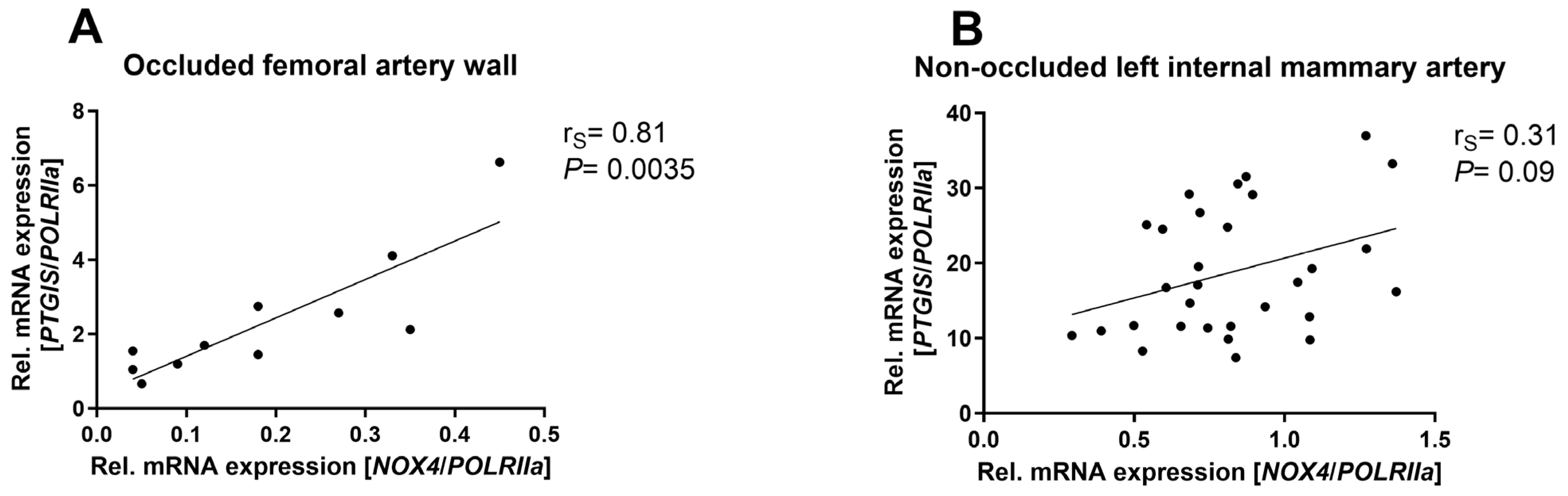
3.1. NOX4 mRNA Expression Highly Correlated with PTGIS Expression in Occluded Arterial Walls of Patients with Peripheral Arterial Disease

We correlated the expression of NOX4 mRNA with prostaglandin I2 synthase (PTGIS) expression in human vessels. From patients with peripheral arterial disease, specimens of the artery wall proximal to the occlusion were obtained. In those arterial walls, where hypoxia is very likely, we observed a tight correlation between NOX4 expression and PTGIS mRNA expression (Figure 1A). The second specimens analyzed were non-occluded left internal mammary arteries from patients undergoing coronary artery bypass graft surgery. Those arteries were not affected by occlusions. Here, no correlation between NOX4 and PTGIS mRNA expression could be found (Figure 1B).

3.2. Hypoxia Elevated NOX4 and PTGIS in Human Endothelial Cells and Murine Vessels

We confirmed a significant increase in NOX4 expression after 8 h of hypoxia in primary human umbilical vein endothelial cells (HUVECs), with sustained elevation at 16 h and 24 h (Figure 2A). This was accompanied by increased H2O2 release following hypoxia, which was abolished in cells with shNOX4 (Figure 2B). Additionally, aortas from wild-type (WT) mice revealed elevated Nox4 mRNA expression after 24 h of hypoxia (Figure 2C). In HUVECs, the mRNA expression of prostaglandin I2 synthase (PTGIS) was significantly upregulated after 24 h of hypoxia (Figure 2D). Correspondingly, concentration of 6-keto-PGF1 alpha, the non-enzymatic hydration product of prostacyclin (PGI2), was increased in the supernatant of HUVECs after 24 h of hypoxia (Figure 2E). Aortas of WT mice indicated a significant upregulation of Ptgis mRNA expression after hypoxia, which was also highly significant in aortas under hypoxia from Nox4−/− mice (Figure 2F). Experiments with Actinomycin D demonstrated similar degradation of NOX4 mRNA under normoxic and hypoxic conditions (Figure 2G). Also, PTGIS mRNA degradation did not differ between normoxic and hypoxic conditions (Figure 2H), which contrasts VEGF mRNA stability, which is known to be increased under hypoxia (Supplementary Figure S1). Therefore, we obtained evidence that the regulation of the mRNA levels of NOX4 and PTGIS in response to hypoxia are controlled at the level of gene transcription rather than by post-transcriptional processes like mRNA stability.

3.3. NOX4 and PTGIS Were Regulated by Different HIFs

Treating human endothelial cells with prolyl-4-hydroxylase inhibitor dimethyloxalylglycine (DMOG) increases endogenous hypoxia-inducible factor (HIF) levels under normoxic conditions [26]. We detected elevated NOX4 (Figure 3A) and PTGIS (Figure 3D) expression in endothelial cells after DMOG application, indicating the HIF dependency of NOX4 and PTGIS upregulation under hypoxia. Inhibiting HIF1a under hypoxia resulted in decreased NOX4 expression (Figure 3B), but not PTGIS expression (Figure 3E). However, inhibiting HIF2a significantly prevented PTGIS upregulation under hypoxia (Figure 3F), while NOX4 was not regulated (Figure 3C).

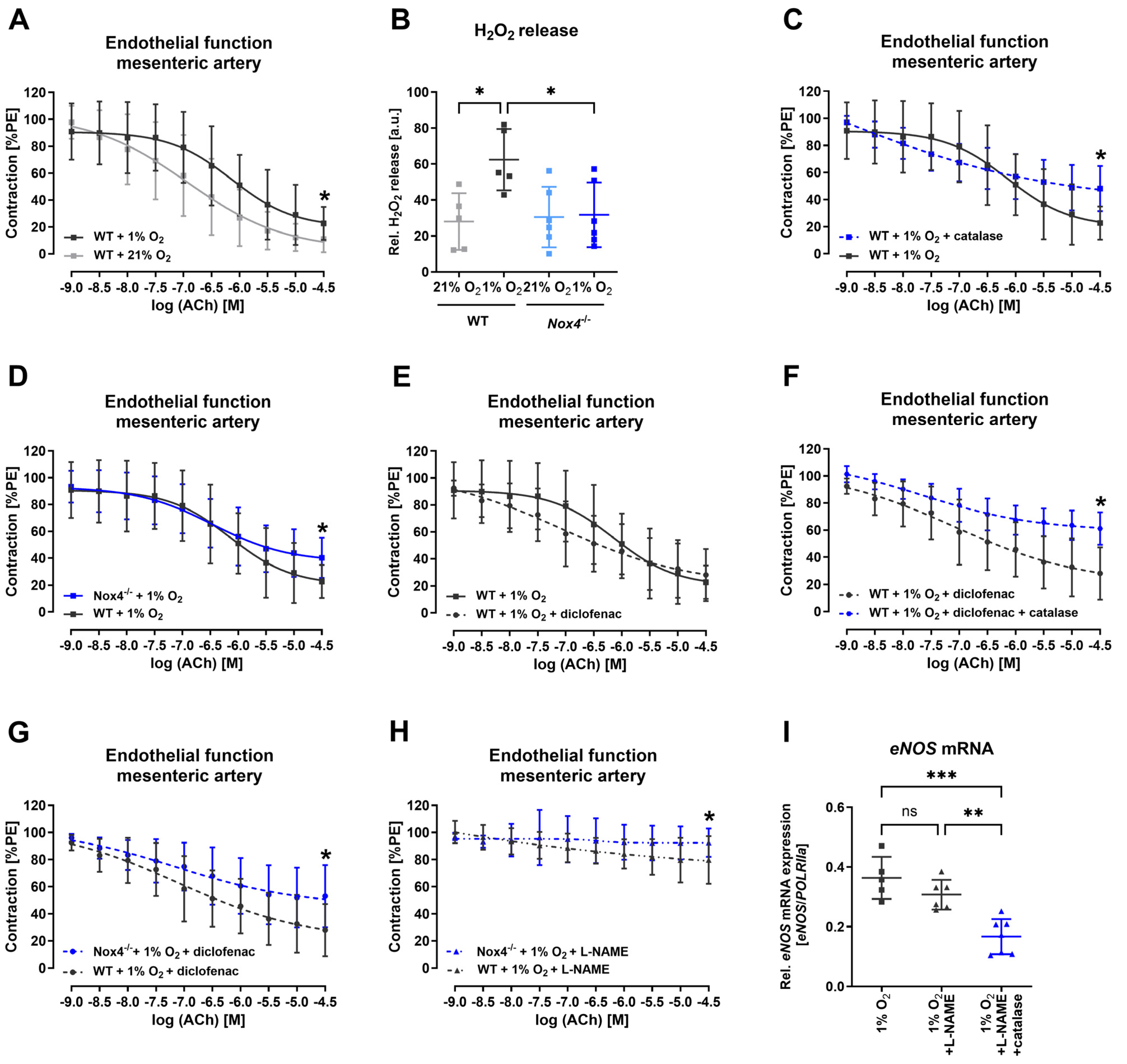
3.4. Nox4 Released Hydrogen Peroxide Maintained Endothelial Function under Hypoxia

To further assess the impact of hypoxia and Nox4 on vascular function, Mulvany myograph experiments were performed. Under hypoxic conditions in the organ chamber, vascular function of mesenteric arteries from WT and Nox4−/− mice was analyzed. Endothelial vasorelaxation was declined in vessels under hypoxic conditions from WT mice (Figure 4A). We also detected an increased hydrogen peroxide release after hypoxia treatment in mesenteric arteries from WT mice, but not Nox4−/− mice (Figure 4B). When we incubated the WT mesenteric arteries under hypoxic conditions with hydrogen peroxide scavenger catalase endothelial function was further altered (Figure 4C). Also, under hypoxic conditions in the organ bath chambers, endothelium-dependent vasorelaxation in the mesenteric artery of Nox4−/− mice was further declined compared with WT mice (Figure 4D). In a next step, mesenteric arteries of WT mice under hypoxia were incubated with diclofenac to inhibit cyclooxygenases to gain insights on the impact of prostaglandins. Maximal vasorelaxation in WT mesenteric arteries was not affected by diclofenac under hypoxia (Figure 4E). However, mesenteric arteries of WT mice incubated with the combination of catalase and diclofenac showed an endothelial dysfunction (Figure 4F). Similarly, mesenteric arteries depleted of Nox4 and incubated with diclofenac under hypoxia revealed an amplified endothelial dysfunction (Figure 4G). Incubating mesenteric arteries with nitric oxide synthase inhibitor L-NAME revealed that, also under hypoxic conditions, endothelial-dependent vasorelaxation was mainly mediated by nitric oxide, even though the impact was significantly increased in the mesenteric arteries of Nox4−/− mice, indicating Nox4 is relevant for endothelium-dependent vasorelaxation (Figure 4H). Additionally, incubating HUVECs with catalase and L-NAME showed the downregulation of eNOS mRNA expression (Figure 4I).

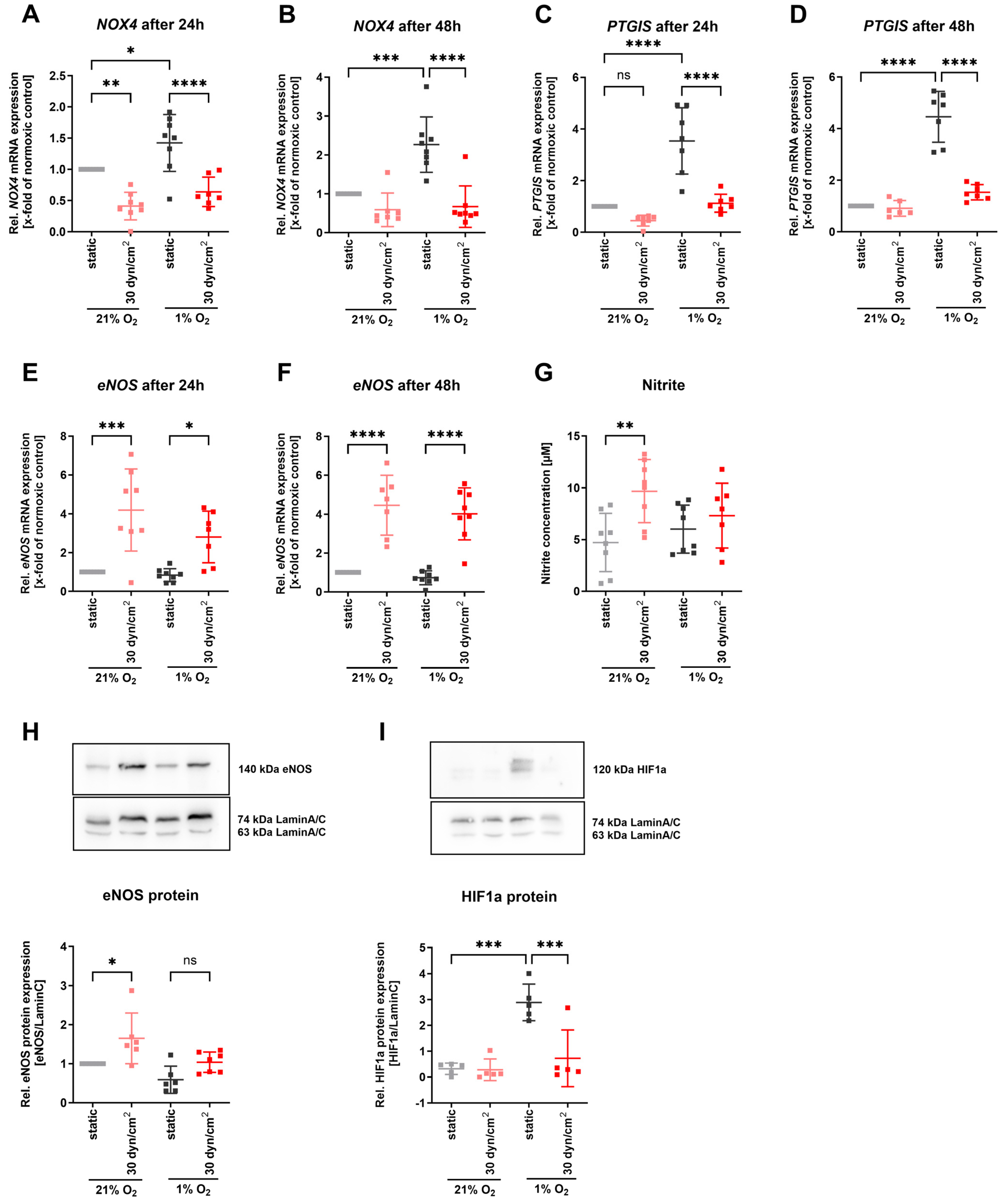
3.5. Restored Laminar Flow Prevented Upregulation of NOX4 and PTGIS under Hypoxia

To study how protective blood flow affects the hypoxic upregulation of NOX4 and PTGIS, we cultured human endothelial cells under hypoxia combined with 30 dyn/cm2 laminar flow to mimic restored blood flow. The elevated expression of NOX4 under hypoxia was significantly diminished when treating endothelial cells additionally under laminar flow conditions for 24 and 48 h (Figure 5A,B). Similarly, PTGIS mRNA expression was significantly decreased under hypoxia when endothelial cells were cultured under protective laminar flow (Figure 5C,D). In contrast, laminar shear stress known to induce eNOS expression still led to upregulation of eNOS mRNA expression for 24 and 48 h when shear stress was applied under hypoxic conditions (Figure 5E,F). However, eNOS protein expression and nitric oxide release only tended to increase due to laminar flow under hypoxia conditions (Figure 5E,G). The other way around, we observed HIF1a protein expression being significantly diminished in those hypoxic endothelial cells where additional laminar flow was applied (Figure 5F).

4. Discussion

In the current study, we present evidence that NOX4 and PGI2 producing the PTGIS enzyme correlate under hypoxia. We observed NOX4 being regulated by HIF1a, while PTGIS was induced by HIF2a. We showed, for the first time, that NOX4 mediated endothelial function under hypoxia, and thereby complemented PGI2 in the vasculature under hypoxia. Restored laminar shear stress restored eNOS expression and blunted the hypoxic response on NOX4 and PTGIS.
We showed for the first time a tight correlation of NOX4 and PGI2 producing PTGIS enzyme expression in human occluded hypoxic vessels. In human endothelial cells, we observed an upregulation of NOX4 expression prior to the upregulation of PTGIS. The NOX4 mRNA expression reached significant levels after 8 h. These data are in agreement with studies showing increased NOX4 expression in bovine and human endothelial cells exposed to hypoxia (1% oxygen) [27,28]. An increased NOX4 expression in response to hypoxia has also been shown in other cell types, including renal cells [6], neuronal cells [7], pulmonary arterial smooth muscle cells [8], pulmonary artery adventitial fibroblasts [9], embryonic stem cells [29], and adipose-derived stem cells [30]. In our experiments, NOX4 expression was only downregulated under hypoxia by the HIF1a inhibitor, while PTGIS expression was affected by HIF2a inhibition. Diebold et al. reported increased NOX4 expression in HIF1a-overexpressing cells [31]. However, other studies also indicated an impact of HIF2a on NOX4 [32]. It has also been reported that NOX4 might act upstream of HIF signaling. For example, in cardiomyocytes, it was shown that the NOX4-dependent upregulation of HIF1a caused increased angiogenesis via VEGF [19]. For PTGIS, in some studies, the regulation is attributed to HIF1a [33,34]; however, recent studies indicate both HIF1a and HIF2a are involved [32,35].
In the cardiovascular system, the role of NOX4 in hypoxia has always been tightly linked to angiogenesis rather than vascular function. Thus, our experimental settings focused on vascular function under hypoxia. We were able to detect a hypoxia-induced endothelial dysfunction in WT mesenteric arteries. However, in mesenteric arteries from Nox4−/− mice, the hypoxia-induced endothelial dysfunction was further altered. Inhibition of COX-1 and COX-2, leading to reduced PGI2, resulted in the most pronounced endothelial dysfunction in Nox4−/− mesenteric arteries under hypoxia. Therefore, our findings implicate an endothelium-dependent vasodilatory effect of Nox4 under hypoxic conditions, accommodating PGI2 [18].
The interplay of NOX4 and PGI2 under hypoxic conditions facilitates vascular adaptation to enhance tissue oxygenation and blood supply. When laminar protective blood flow is restored, the hypoxia-induced signaling is blunted. We noted that a laminar flow of 30 dyn/cm2 led to increased eNOS expression even under hypoxic conditions, resulting in decreased expression of HIFa and its target genes, NOX4 and PTGIS. Others have shown that laminar shear stress also diminished hypoxia-induced apoptosis in HUVECs [36]. In human pulmonary endothelial cells, modification of hypoxia-induced signaling pathways due to laminar blood flow was also demonstrated [37]. Similarly, our results showed that hypoxia-induced NOX4 expression was inhibited by laminar shear stress. These observations might explain the controversial reports on NOX4 being involved in hypoxia-induced vascular diseases. Veith et al. could not detect any effect of Nox4 deletion on pulmonary vasoconstriction and hypertension caused by hypoxia [38]. Other studies showed the upregulation of NOX4 in hypoxic pulmonary artery smooth muscle cells and animal models of hypoxia-induced pulmonary hypertension [39]. Shear stress–hypoxia interactions might account for these different observations.
Additionally, we found, that laminar shear stress-induced eNOS expression was accompanied by a reduced HIF1a expression. It has been reported that long-term exposure to nitric oxide, comparable to our experimental conditions, under flow has reducing effects on HIF1a accumulation and activity [40,41,42]. Mechanistically, it has been suggested that nitric oxide leads to the redistribution of oxygen towards PHDs by inhibiting cytochrome-c oxidase and thus mitochondrial respiration [43]. Cattaneo et al. also demonstrated that treatment of endothelial cells with nitric oxide synthase inhibitor NG-Nitro-L-arginine methyl ester (L-NAME) led to increased HIF1a stability and elevated HIF1a target gene expression [44]. Additionally, H2O2 from NOX4 can directly increase endothelial NO release via the AKT serine/threonine kinase 1-dependent phosphorylation of eNOS [45,46] or via the redox-sensitive Ca2+ channel transient receptor potential cation channel, subfamily M, member 2 (TRPM2) [47], again counteracting the hypoxic response.

5. Conclusions

For the first time, we demonstrated a tight correlation of NOX4 and PTGIS in endothelial cells under hypoxia in human occluded hypoxic vessels. NOX4 was essential to ensure endothelium-dependent vasorelaxation under hypoxia. Thus, NOX4, one of the main sources for hydrogen peroxide, might contribute to enhancing vascular function to restore blood circulation under hypoxia. Therefore, NOX4 inhibitors that are currently explored for the treatment of diseases such as fibrosis and certain cancers need to be verified for cardiovascular side effects.

6. Limitations

In this study, human femoral arteries, internal mammary arteries, as well as murine mesenteric vessels and aortas were investigated. The broad range of examined vessels also represents a limitation, as different factors have varying degrees of significance in the different types of vessels. Additionally, all human samples were obtained either from patients undergoing coronary artery bypass grafting surgery or those with peripheral vascular disease, limiting our ability to draw conclusions about human vessels under healthy physiological conditions.
The use of the Griess assay for nitrite measurement in our experiments presented certain methodological limitations. Specifically, nitrate was not accounted for, which may have led to an underestimation of total nitric oxide production.
Furthermore, our study focused on hydrogen peroxide released by NOX4. Other potential sources of hydrogen peroxide, such as mitochondrial dysfunction and eNOS monomerization, might play a role in our context and need to be addressed in further studies.

Supplementary Materials

The following supporting information can be downloaded. Supplementary Figure S1: VEGF mRNA expression as controls for experiments with DMOG treatment and actinomycin D; Supplementary Figure S2: No correlation of NOX4 and eNOS expression; Supplementary Figure S3: Endothelium-independent vasorelaxation.

Author Contributions

Conceptualization, H.B., H.M. and C.B.; Formal analysis, H.B., J.M., A.H. and C.B.; Funding acquisition, H.B., H.M. and C.B.; Investigation, H.B., J.M., H.H., S.G., P.D.-N. and C.B.; Sources, A.H., S.W. and C.R.; Supervision, C.B.; Visualization, H.B. and J.M.; Writing—original draft preparation, H.B.; Writing—review and editing, H.B., H.M. and C.B. All authors have read and agreed to the published version of the manuscript.

Funding

This work was supported by research grant of the German Cardiac Society (DGK) awarded to H.B., by research grants of the Deutsche Forschungsgemeinschaft (DFG) Projektnummer 470814312 awarded to C.B. and H.B., as well as MO 1695/4-1 to H.M., by the Government of Ghana and the Deutscher Akademischer Austauschdienst (91642764) to P.D.-N. and by the German Centre for Cardiovascular Research (DZHK) (Grant 81X2800207 to H.M.) We are grateful to Ralf P. Brandes and Katrin Schröder for providing us with the Nox4−/− mice.

Institutional Review Board Statement

The study was conducted in accordance with the Declaration of Helsinki. The study was approved by the Ethics Committee of the Technische Universität Dresden (EK 307-12-2007).

Informed Consent Statement

Informed consent was obtained from all patients involved in the study.

Data Availability Statement

The raw data supporting the conclusions of this article will be made available by the authors on request.

Conflicts of Interest

The authors declare no conflicts of interest.

References

  1. Ullah, K.; Wu, R. Hypoxia-Inducible Factor Regulates Endothelial Metabolism in Cardiovascular Disease. Front. Physiol. 2021, 12, 670653. [Google Scholar] [CrossRef] [PubMed]
  2. Nallamshetty, S.; Chan, S.Y.; Loscalzo, J. Hypoxia: A master regulator of microRNA biogenesis and activity. Free Radic. Biol. Med. 2013, 64, 20–30. [Google Scholar] [CrossRef] [PubMed]
  3. Brocato, J.; Chervona, Y.; Costa, M. Molecular responses to hypoxia-inducible factor 1alpha and beyond. Mol. Pharmacol. 2014, 85, 651–657. [Google Scholar] [CrossRef] [PubMed]
  4. Wong, B.W.; Kuchnio, A.; Bruning, U.; Carmeliet, P. Emerging novel functions of the oxygen-sensing prolyl hydroxylase domain enzymes. Trends Biochem. Sci. 2013, 38, 3–11. [Google Scholar] [CrossRef]
  5. Brahimi-Horn, M.C.; Pouyssegur, J. HIF at a glance. J. Cell Sci. 2009, 122, 1055–1057. [Google Scholar] [CrossRef]
  6. Suliman, H.B.; Ali, M.; Piantadosi, C.A. Superoxide dismutase-3 promotes full expression of the EPO response to hypoxia. Blood 2004, 104, 43–50. [Google Scholar] [CrossRef]
  7. Vallet, P.; Charnay, Y.; Steger, K.; Ogier-Denis, E.; Kovari, E.; Herrmann, F.; Michel, J.P.; Szanto, I. Neuronal expression of the NADPH oxidase NOX4, and its regulation in mouse experimental brain ischemia. Neuroscience 2005, 132, 233–238. [Google Scholar] [CrossRef]
  8. Mittal, M.; Roth, M.; König, P.; Hofmann, S.; Dony, E.; Goyal, P.; Selbitz, A.C.; Schermuly, R.T.; Ghofrani, H.A.; Kwapiszewska, G.; et al. Hypoxia-dependent regulation of nonphagocytic NADPH oxidase subunit NOX4 in the pulmonary vasculature. Circ. Res. 2007, 101, 258–267. [Google Scholar] [CrossRef]
  9. Li, S.; Tabar, S.S.; Malec, V.; Eul, B.G.; Klepetko, W.; Weissmann, N.; Grimminger, F.; Seeger, W.; Rose, F.; Hänze, J. NOX4 regulates ROS levels under normoxic and hypoxic conditions, triggers proliferation, and inhibits apoptosis in pulmonary artery adventitial fibroblasts. Antioxid. Redox Signal. 2008, 10, 1687–1698. [Google Scholar] [CrossRef]
  10. Sylvester, A.L.; Zhang, D.X.; Ran, S.; Zinkevich, N.S. Inhibiting NADPH Oxidases to Target Vascular and Other Pathologies: An Update on Recent Experimental and Clinical Studies. Biomolecules 2022, 12, 823. [Google Scholar] [CrossRef]
  11. Touyz, R.M.; Briones, A.M.; Sedeek, M.; Burger, D.; Montezano, A.C. NOX isoforms and reactive oxygen species in vascular health. Mol. Interv. 2011, 11, 27–35. [Google Scholar] [CrossRef] [PubMed]
  12. Kwon, O.S.; Noh, S.G.; Park, S.H.; Andtbacka, R.H.I.; Hyngstrom, J.R.; Richardson, R.S. Ageing and endothelium-mediated vascular dysfunction: The role of the NADPH oxidases. J. Physiol. 2023, 601, 451–467. [Google Scholar] [CrossRef] [PubMed]
  13. Wang, D.; Li, J.; Luo, G.; Zhou, J.; Wang, N.; Wang, S.; Zhao, R.; Cao, X.; Ma, Y.; Liu, G.; et al. Nox4 as a novel therapeutic target for diabetic vascular complications. Redox Biol. 2023, 64, 102781. [Google Scholar] [CrossRef] [PubMed]
  14. Takac, I.; Schröder, K.; Zhang, L.; Lardy, B.; Anilkumar, N.; Lambeth, J.D.; Shah, A.M.; Morel, F.; Brandes, R.P. The E-loop is involved in hydrogen peroxide formation by the NADPH oxidase Nox4. J. Biol. Chem. 2011, 286, 13304–13313. [Google Scholar] [CrossRef] [PubMed]
  15. Peshavariya, H.M.; Liu, G.S.; Chang, C.W.; Jiang, F.; Chan, E.C.; Dusting, G.J. Prostacyclin signaling boosts NADPH oxidase 4 in the endothelium promoting cytoprotection and angiogenesis. Antioxid. Redox Signal. 2014, 20, 2710–2725. [Google Scholar] [CrossRef] [PubMed]
  16. Zeng, C.; Liu, J.; Zheng, X.; Hu, X.; He, Y. Prostaglandin and prostaglandin receptors: Present and future promising therapeutic targets for pulmonary arterial hypertension. Respir. Res. 2023, 24, 263. [Google Scholar] [CrossRef]
  17. Vane, J.; Corin, R.E. Prostacyclin: A vascular mediator. Eur. J. Vasc. Endovasc. Surg. 2003, 26, 571–578. [Google Scholar] [CrossRef]
  18. Busse, R.; Forstermann, U.; Matsuda, H.; Pohl, U. The role of prostaglandins in the endothelium-mediated vasodilatory response to hypoxia. Pflug. Arch. 1984, 401, 77–83. [Google Scholar] [CrossRef]
  19. Zhang, M.; Brewer, A.C.; Schröder, K.; Santos, C.X.; Grieve, D.J.; Wang, M.; Anilkumar, N.; Yu, B.; Dong, X.; Walker, S.J.; et al. NADPH oxidase-4 mediates protection against chronic load-induced stress in mouse hearts by enhancing angiogenesis. Proc. Natl. Acad. Sci. USA 2010, 107, 18121–18126. [Google Scholar] [CrossRef]
  20. Cybularz, M.; Langbein, H.; Zatschler, B.; Brunssen, C.; Deussen, A.; Matschke, K.; Morawietz, H. Endothelial function and gene expression in perivascular adipose tissue from internal mammary arteries of obese patients with coronary artery disease. Atheroscler. Suppl. 2017, 30, 149–158. [Google Scholar] [CrossRef]
  21. Lee, K.; Kang, J.E.; Park, S.K.; Jin, Y.; Chung, K.S.; Kim, H.M.; Lee, K.; Kang, M.R.; Lee, M.K.; Song, K.B.; et al. LW6, a novel HIF-1 inhibitor, promotes proteasomal degradation of HIF-1alpha via upregulation of VHL in a colon cancer cell line. Biochem. Pharmacol. 2010, 80, 982–989. [Google Scholar] [CrossRef]
  22. Zimmer, M.; Ebert, B.L.; Neil, C.; Brenner, K.; Papaioannou, I.; Melas, A.; Tolliday, N.; Lamb, J.; Pantopoulos, K.; Golub, T.; et al. Small-molecule inhibitors of HIF-2a translation link its 5′UTR iron-responsive element to oxygen sensing. Mol. Cell 2008, 32, 838–848. [Google Scholar] [CrossRef] [PubMed]
  23. Korten, S.; Brunssen, C.; Poitz, D.M.; Großklaus, S.; Brux, M.; Schnittler, H.J.; Strasser, R.H.; Bornstein, S.R.; Morawietz, H.; Goettsch, W. Impact of Hey2 and COUP-TFII on genes involved in arteriovenous differentiation in primary human arterial and venous endothelial cells. Basic Res. Cardiol. 2013, 108, 362. [Google Scholar] [CrossRef] [PubMed]
  24. Giebe, S.; Cockcroft, N.; Hewitt, K.; Brux, M.; Hofmann, A.; Morawietz, H.; Brunssen, C. Cigarette smoke extract counteracts atheroprotective effects of high laminar flow on endothelial function. Redox Biol. 2017, 12, 776–786. [Google Scholar] [CrossRef] [PubMed]
  25. Pfaffl, M.W. A new mathematical model for relative quantification in real-time RT-PCR. Nucleic Acids Res. 2001, 29, e45. [Google Scholar] [CrossRef] [PubMed]
  26. Liang, X.; Arullampalam, P.; Yang, Z.; Ming, X.F. Hypoxia Enhances Endothelial Intercellular Adhesion Molecule 1 Protein Level Through Upregulation of Arginase Type II and Mitochondrial Oxidative Stress. Front. Physiol. 2019, 10, 1003. [Google Scholar] [CrossRef]
  27. Craige, S.M.; Chen, K.; Pei, Y.; Li, C.; Huang, X.; Chen, C.; Shibata, R.; Sato, K.; Walsh, K.; Keaney, J.F., Jr. NADPH oxidase 4 promotes endothelial angiogenesis through endothelial nitric oxide synthase activation. Circulation 2011, 124, 731–740. [Google Scholar] [CrossRef]
  28. Wang, J.; Hong, Z.; Zeng, C.; Yu, Q.; Wang, H. NADPH oxidase 4 promotes cardiac microvascular angiogenesis after hypoxia/reoxygenation in vitro. Free Radic. Biol. Med. 2014, 69, 278–288. [Google Scholar] [CrossRef]
  29. Ateghang, B.; Wartenberg, M.; Gassmann, M.; Sauer, H. Regulation of cardiotrophin-1 expression in mouse embryonic stem cells by HIF-1alpha and intracellular reactive oxygen species. J. Cell Sci. 2006, 119, 1043–1052. [Google Scholar] [CrossRef]
  30. DeNies, M.S.; Johnson, J.; Maliphol, A.B.; Bruno, M.; Kim, A.; Rizvi, A.; Rustici, K.; Medler, S. Diet-induced obesity alters skeletal muscle fiber types of male but not female mice. Physiol. Rep. 2014, 2, e00204. [Google Scholar] [CrossRef]
  31. Diebold, I.; Petry, A.; Hess, J.; Gorlach, A. The NADPH oxidase subunit NOX4 is a new target gene of the hypoxia-inducible factor-1. Mol. Biol. Cell 2010, 21, 2087–2096. [Google Scholar] [CrossRef]
  32. Bartoszewski, R.; Moszyńska, A.; Serocki, M.; Cabaj, A.; Polten, A.; Ochocka, R.; Dell’Italia, L.; Bartoszewska, S.; Króliczewski, J.; Dąbrowski, M.; et al. Primary endothelial cell-specific regulation of hypoxia-inducible factor (HIF)-1 and HIF-2 and their target gene expression profiles during hypoxia. FASEB J. Off. Publ. Fed. Am. Soc. Exp. Biol. 2019, 33, 7929–7941. [Google Scholar] [CrossRef]
  33. Camacho, M.; Rodríguez, C.; Guadall, A.; Alcolea, S.; Orriols, M.; Escudero, J.R.; Martínez-González, J.; Vila, L. Hypoxia upregulates PGI-synthase and increases PGI2 release in human vascular cells exposed to inflammatory stimuli. J. Lipid Res. 2011, 52, 720–731. [Google Scholar] [CrossRef]
  34. Manalo, D.J.; Rowan, A.; Lavoie, T.; Natarajan, L.; Kelly, B.D.; Ye, S.Q.; Garcia, J.G.; Semenza, G.L. Transcriptional regulation of vascular endothelial cell responses to hypoxia by HIF-1. Blood 2005, 105, 659–669. [Google Scholar] [CrossRef]
  35. Cabaj, A.; Moszynska, A.; Charzynska, A.; Bartoszewski, R.; Dabrowski, M. Functional and HRE motifs count analysis of induction of selected hypoxia-responsive genes by HIF-1 and HIF-2 in human umbilical endothelial cells. Cell. Signal. 2022, 90, 110209. [Google Scholar] [CrossRef]
  36. Mattiussi, S.; Turrini, P.; Testolin, L.; Martelli, F.; Zaccagnini, G.; Mangoni, A.; Barlucchi, L.M.; Antonini, A.; Illi, B.; Cirielli, C.; et al. p21(Waf1/Cip1/Sdi1) mediates shear stress-dependent antiapoptotic function. Cardiovasc. Res. 2004, 61, 693–704. [Google Scholar] [CrossRef]
  37. Kostyunina, D.S.; Rowan, S.C.; Pakhomov, N.V.; Dillon, E.; Rochfort, K.D.; Cummins, P.M.; O’Rourke, M.J.; McLoughlin, P. Shear Stress Markedly Alters the Proteomic Response to Hypoxia in Human Pulmonary Endothelial Cells. Am. J. Respir. Cell Mol. Biol. 2023, 68, 551–565. [Google Scholar] [CrossRef]
  38. Veith, C.; Kraut, S.; Wilhelm, J.; Sommer, N.; Quanz, K.; Seeger, W.; Brandes, R.P.; Weissmann, N.; Schröder, K. NADPH oxidase 4 is not involved in hypoxia-induced pulmonary hypertension. Pulm. Circ. 2016, 6, 397–400. [Google Scholar] [CrossRef]
  39. Nisbet, R.E.; Bland, J.M.; Kleinhenz, D.J.; Mitchell, P.O.; Walp, E.R.; Sutliff, R.L.; Hart, C.M. Rosiglitazone attenuates chronic hypoxia-induced pulmonary hypertension in a mouse model. Am. J. Respir. Cell Mol. Biol. 2010, 42, 482–490. [Google Scholar] [CrossRef]
  40. Sogawa, K.; Numayama-Tsuruta, K.; Ema, M.; Abe, M.; Abe, H.; Fujii-Kuriyama, Y. Inhibition of hypoxia-inducible factor 1 activity by nitric oxide donors in hypoxia. Proc. Natl. Acad. Sci. USA 1998, 95, 7368–7373. [Google Scholar] [CrossRef]
  41. Wang, F.; Sekine, H.; Kikuchi, Y.; Takasaki, C.; Miura, C.; Heiwa, O.; Shuin, T.; Fujii-Kuriyama, Y.; Sogawa, K. HIF-1alpha-prolyl hydroxylase: Molecular target of nitric oxide in the hypoxic signal transduction pathway. Biochem. Biophys. Res. Commun. 2002, 295, 657–662. [Google Scholar] [CrossRef]
  42. Huang, L.E.; Willmore, W.G.; Gu, J.; Goldberg, M.A.; Bunn, H.F. Inhibition of hypoxia-inducible factor 1 activation by carbon monoxide and nitric oxide. Implications for oxygen sensing and signaling. J. Biol. Chem. 1999, 274, 9038–9044. [Google Scholar] [CrossRef]
  43. Hagen, T.; Taylor, C.T.; Lam, F.; Moncada, S. Redistribution of intracellular oxygen in hypoxia by nitric oxide: Effect on HIF1alpha. Science 2003, 302, 1975–1978. [Google Scholar] [CrossRef]
  44. Cattaneo, M.G.; Cappellini, E.; Benfante, R.; Ragni, M.; Omodeo-Sale, F.; Nisoli, E.; Borgese, N.; Vicentini, L.M. Chronic deficiency of nitric oxide affects hypoxia inducible factor-1alpha (HIF-1alpha) stability and migration in human endothelial cells. PLoS ONE 2011, 6, e29680. [Google Scholar] [CrossRef] [PubMed]
  45. Drummond, G.R.; Cai, H.; Davis, M.E.; Ramasamy, S.; Harrison, D.G. Transcriptional and posttranscriptional regulation of endothelial nitric oxide synthase expression by hydrogen peroxide. Circ. Res. 2000, 86, 347–354. [Google Scholar] [CrossRef]
  46. Dimmeler, S.; Fleming, I.; Fisslthaler, B.; Hermann, C.; Busse, R.; Zeiher, A.M. Activation of nitric oxide synthase in endothelial cells by Akt-dependent phosphorylation. Nature 1999, 399, 601–605. [Google Scholar] [CrossRef]
  47. Alves-Lopes, R.; Lacchini, S.; Neves, K.B.; Harvey, A.; Montezano, A.C.; Touyz, R.M. Vasoprotective effects of NOX4 are mediated via polymerase and transient receptor potential melastatin 2 cation channels in endothelial cells. J. Hypertens. 2023, 41, 1389–1400. [Google Scholar] [CrossRef]
Figure 1. NOX4 highly correlates with PTGIS in human occluded vessels. (A) Correlation of NOX4 mRNA expression and prostaglandin I2 synthase (PTGIS) in femoral artery walls from patients with peripheral arterial disease (n = 11). POLRIIa mRNA expression was used as reference. (B) Correlation of NOX4 mRNA expression and prostaglandin I2 synthase (PTGIS) in human non-occluded left internal mammary arteries (n = 30). POLRIIa mRNA expression was used as reference. Statistics: Spearman’s correlation coefficient (rS) between PTGIS and NOX4 expression. Abbreviations: NOX4, NADPH oxidase 4; PTGIS, prostaglandin I2 synthase; POLRIIa, RNA polymerase II subunit A.
Antioxidants 13 01178 g001
Figure 2. Increased NOX4 and PTGIS expression after hypoxia in human endothelial cells and murine aorta. (A) NOX4 mRNA expression after 8 h, 16 h, and 24 h normoxia (21% oxygen) or hypoxia (1% oxygen) (n = 3). TBP mRNA expression was used as reference. (B) Hydrogen peroxide production in HUVECs subjected to transduction with lentiviral particles containing shNOX4 and exposed to 24 h hypoxia (1% oxygen) (n ≥ 5). Untransduced cells (untr) and cells transduced with scrambled shRNA (shC) were used as controls. (C) Relative Nox4 mRNA expression in aorta from WT and Nox4−/− mice after 24 h hypoxia (1% oxygen) (n = 6). B2m mRNA expression was used as reference. (D) PTGIS mRNA expression after 8 h, 16 h, and 24 h normoxia (21% oxygen) or hypoxia (1% oxygen) (n = 4). TBP mRNA expression was used as reference. (E) 6-keto-PGF1 alpha concentration in supernatant of HUVECs exposed to 24 h hypoxia (1% oxygen) (n ≥ 5). (F) Relative Ptgis mRNA expression in aorta from WT and Nox4−/− mice after 24 h hypoxia (1% oxygen) (n = 6). B2m mRNA expression was used as reference. (G) Relative NOX4 mRNA expression in HUVECs treated subsequently with actinomycin D (ActD) for 8 h under normoxia and hypoxia (1% oxygen) (n = 5). POLRIIa mRNA expression was used as reference. Mean relative NOX4 mRNA expression in HUVECs treated subsequently with actinomycin D (ActD) for 8 h under normoxia and hypoxia (1% oxygen) (n = 5). POLRIIa mRNA expression was used as reference. (H) Relative PTGIS mRNA expression in HUVECs treated subsequently with actinomycin D (ActD) for 8 h under normoxia and hypoxia (1% oxygen) (n = 5). POLRIIa mRNA expression was used as reference. Mean relative PTGIS mRNA expression in HUVECs treated subsequently with actinomycin D (ActD) for 8 h under normoxia and hypoxia (1% oxygen) (n = 5). POLRIIa mRNA expression was used as reference. Statistics: Normal (Gaussian) distribution was tested by Shapiro–Wilk normality tests. All data had Gaussian distributions. Data were then analyzed with t-tests or one-way ANOVAs followed by Tukey’s multiple comparisons tests or two-way ANOVA and Sidak’s multiple comparisons tests, respectively. * p < 0.05; ** p < 0.01; *** p < 0.001; **** p < 0.0001. Abbreviations: ActD, actinomycin D; B2m, beta-2 microglobulin; crtl, control; HUVECs, human umbilical vein endothelial cells; NOX4, NADPH oxidase 4; PTGIS, prostaglandin I2 synthase; POLRIIa, RNA polymerase II subunit A; shRNA, short hairpin RNA; TBP, TATA-box binding protein; untr, untreated; WT, wild-type.
Antioxidants 13 01178 g002
Figure 3. NOX4 and PTGIS were predominantly regulated by different HIFs. (A) Relative NOX4 mRNA expression in HUVECs after 24 h 10 mM DMOG treatment under normoxic conditions (n ≥ 7). TBP mRNA expression was used as reference. (B) Relative NOX4 mRNA expression in HUVECs treated subsequently with HIF1a inhibitor for 16 h under normoxia and hypoxia (1% oxygen) (n = 7). POLRIIa mRNA expression was used as reference. (C) Relative NOX4 mRNA expression in HUVECs treated subsequently with HIF2a inhibitor for 16 h under normoxia and hypoxia (1% oxygen) (n = 5). POLRIIa mRNA expression was used as reference. (D) Relative PTGIS mRNA expression in HUVECs after 24 h 10 mM DMOG treatment under normoxic conditions (n ≥ 7). TBP mRNA expression was used as reference. (E) Relative PTGIS mRNA expression in HUVECs treated subsequently with HIF1a inhibitor for 16 h under normoxia and hypoxia (1% oxygen) (n = 8). POLRIIa mRNA expression was used as reference. (F) Relative PTGIS mRNA expression in HUVECs treated subsequently with HIF2a inhibitor for 16 h under normoxia and hypoxia (1% oxygen) (n = 6). POLRIIa mRNA expression was used as reference. Statistics: Normal (Gaussian) distribution was tested by Shapiro–Wilk normality tests. Non-Gaussian distributed data were analyzed by Kruskal–Wallis and Dunn’s multiple comparison tests (D,E). Gaussian distributed data (A,B,C,F) were analyzed with one-way ANOVA followed by Tukey’s multiple comparisons tests or two-way ANOVA and Sidak’s multiple comparisons tests, respectively. * p < 0.05; ** p < 0.01; **** p < 0.0001. Abbreviations: crtl, control; DMOG, dimethyloxallyl glycine; HIF, hypoxia-inducible factor; HUVECs, human umbilical vein endothelial cells; NOX4, NADPH oxidase 4; PTGIS, prostaglandin I2 synthase; POLRIIa, RNA polymerase II subunit A; TBP, TATA-box binding protein.
Antioxidants 13 01178 g003
Figure 4. H2O2 derived from Nox4 maintained vascular response of mesenteric arteries under hypoxia. (A) Endothelium-dependent vasorelaxation of mesenteric arteries from WT mice under normoxic and hypoxic conditions ex vivo (n ≥ 11). (B) Hydrogen peroxide release in WT and Nox4−/− mice mesenteric arteries ex vivo after hypoxia (n = 6). (C) Endothelium-dependent vasorelaxation of mesenteric arteries from WT mice and catalase treatment under hypoxic conditions ex vivo (n ≥ 6). (D) Endothelium-dependent vasorelaxation of mesenteric arteries from WT mice and Nox4−/− mice under hypoxic conditions ex vivo (n ≥ 11). (E) Endothelium-dependent vasorelaxation of mesenteric arteries from WT mice and diclofenac treatment under hypoxic conditions ex vivo (n ≥ 6). (F) Endothelium-dependent vasorelaxation of mesenteric arteries from WT mice with diclofenac and catalase treatment under hypoxic conditions ex vivo (n ≥ 5). (G) Endothelium-dependent vasorelaxation of mesenteric arteries from WT mice and Nox4−/− mice under hypoxic conditions and diclofenac treatment ex vivo (n ≥ 6). (H) Endothelium-dependent vasorelaxation of mesenteric arteries from WT mice and Nox4−/− mice under hypoxic conditions and L-NAME treatment ex vivo (n ≥ 8). (I) Relative eNOS mRNA expression in HUVEC treated with hypoxia and L-NAME or L-NAME and catalase combination (n ≥ 5). POLRIIa mRNA expression was used as reference. Statistics: Normal (Gaussian) distribution was tested by Shapiro–Wilk normality tests. All data has Gaussian distribution. Data was then analyzed with t-tests or one-way ANOVAs, followed by Tukey’s multiple comparisons tests or two-way ANOVA and Sidak’s multiple comparisons tests, respectively. * p < 0.05; ** p < 0.01; *** p < 0.001. Abbreviations: Ach, acetylcholine; eNOS, endothelial nitric oxide synthase; L-NAME, NG-Nitro-L-arginine methyl ester; NOX4, NADPH oxidase 4; PE, phenylephrine; WT, wild-type.
Antioxidants 13 01178 g004
Figure 5. Laminar flow of 30 dyn/cm2 on human endothelial cells prevented upregulation of NOX4 and PTGIS under hypoxia. (A) Relative NOX4 mRNA expression in endothelial cells under laminar 30 dyn/cm2 flow after 24 h and (B) 48 h normoxic or hypoxic conditions (n = 8). TBP mRNA expression was used as reference. (C) Relative PTGIS mRNA expression in endothelial cells under laminar 30 dyn/cm2 flow after 24 h and (D) 48 h normoxic or hypoxic conditions (n = 8). TBP mRNA expression was used as reference. (E) Relative eNOS mRNA expression in endothelial cells under laminar 30 dyn/cm2 flow after 24 h and (F) 48 h normoxic or hypoxic conditions (n = 8). TBP mRNA expression was used as reference. (G) Nitrite concentration in supernatant of endothelial cells under laminar 30 dyn/cm2 flow after 24 h normoxic or hypoxic conditions (n = 8). (H) Relative eNOS protein expression of endothelial cells under static and laminar 30 dyn/cm2 flow after 24 h normoxic or hypoxic conditions (n = 6). (I) Relative HIF1a protein expression in endothelial cells under static and laminar 30 dyn/cm2 flow after 24 h hypoxic conditions (n = 5). Statistics: Normal (Gaussian) distribution was tested by Shapiro–Wilk normality tests. All data had Gaussian distributions. Data were then analyzed with two-way ANOVAs and Sidak’s multiple comparisons tests, respectively. * p < 0.05; ** p < 0.01; *** p < 0.001; **** p < 0.0001. Abbreviations: eNOS, endothelial nitric oxide synthase; HIF1a, hypoxia-inducible factor 1a; NOX4, NADPH oxidase 4; PTGIS, prostaglandin I2 synthase; TBP, TATA-box binding protein.
Antioxidants 13 01178 g005
Table 1. Patient data.
Parameter
Age, years, median with range, n73.0 (54–81), 11
Sex, male:female, % male9:2, 81%
BMI, kg/m2, median with range, n28.7 (22.9–39.9), 11
Blood glucose, mmol/L, median with range, n5.12 (3.91–12.74), 8
LDL cholesterol, mmol/L, median with range, n1.6 (1.17–3.26), 6
HDL cholesterol, mmol/L, median with range, n1.04 (0.84–1.54), 5
Total cholesterol, mmol/L, median with range, n3.24 (2.35–5.53), 6
Triglycerides, mmol/L, median with range, n1.77 (1.36–2.98), 6
CRP, mg/L, median with range, n7.40 (3.91–12.74), 8
Table 2. Primers used for real-time PCR.
GenePrimersSequence, 5′-3′
B2m murineForwardTCTCACTGACCGGCCTGTATNM_009735.3
ReverseGATTTCAATGTGAGGCGGGTG
eNOS humanForwardGAACCTGTGTGACCCTCACCNM_000603.5; NM_001160109.2; NM_001160110.1; NM_001160111.1
ReverseTGGCTAGCTGGTAACTGTGC
NOX4 humanForwardTAACCTCAACTGCAGCCTTATCNM_001143836.3; NM_001143837.2; NM_001291926.2; NM_001291927.1; NM_001291929.2; NM_001300995.1; NM_016931.5
ReverseCTTTTATCCAACAATCTCCTGGTTCTC
Nox4 murineForwardTGTTGGGCCTAGGATTGTGTTNM_001285833.1; NM_001285835.1; NM_015760.5
ReverseAGGGACCTTCTGTGATCCTCG
POLRIIa humanForwardACCTGCGGTCCACGTTGTGTNM_000937.4
ReverseCCACCATTTCCCCGGGATGCG
PTGIS humanForwardACTGCCTGGGGAGGAGTTATNM_000961.4
ReverseGGGATCTCCACATCTGCGTT
Ptgis murineForwardGTTGGTGGCGGTGACTTGTTNM_001420752.1; NM_008968.5; NM_001420756.1;
ReverseCAGCATCTCTCCCAAACTCCA
TBP humanForwardCGCCGGCTGTTTAACTTCGNM_003194.5
ReverseAGAGCATCTCCAGCACACTC
Table 3. Antibodies used.
Primary AntibodyCompanyDilutionMolecular Weight
HIF1aBD Biosciences1:1000120 kDa
eNOSBD Biosciences1:1000140 kDa
Lamin A/CCell signaling1:200074 kDa/63 kDa
Disclaimer/Publisher’s Note: The statements, opinions and data contained in all publications are solely those of the individual author(s) and contributor(s) and not of MDPI and/or the editor(s). MDPI and/or the editor(s) disclaim responsibility for any injury to people or property resulting from any ideas, methods, instructions or products referred to in the content.

