Article

Scavenging of Alkylperoxyl Radicals by Addition to Ascorbate: An Alternative Mechanism to Electron Transfer

by
Gabriel Robert
1 and
J. Richard Wagner
2,*
1
Department of Biochemistry and Functional Genomics, Faculty of Medicine and Health Sciences, Université de Sherbrooke, 3001, 12e Avenue Nord, Sherbrooke, QC J1H 5N4, Canada
2
Department of Medical Imaging and Radiation Sciences, Faculty of Medicine and Health Sciences, Université de Sherbrooke, 3001, 12e Avenue Nord, Sherbrooke, QC J1H 5N4, Canada
*
Author to whom correspondence should be addressed.
Antioxidants 2024, 13(10), 1194; https://doi.org/10.3390/antiox13101194
Submission received: 6 September 2024 / Revised: 25 September 2024 / Accepted: 26 September 2024 / Published: 1 October 2024
(This article belongs to the Section Aberrant Oxidation of Biomolecules)

Abstract

:
Vitamin C (ascorbate; Asc) is a biologically important antioxidant that scavenges reactive oxygen species such as deleterious alkylperoxyl radicals (ROO), which are generated by radical-mediated oxidation of biomolecules in the presence of oxygen. The radical trapping proprieties of Asc are conventionally attributed to its ability to undergo single-electron transfers with reactive species. According to this mechanism, the reaction between Asc and ROO results in the formation of dehydroascorbate (DHA) and the corresponding hydroperoxides (ROOH). When studying the reactivity of DNA 5-(2′-deoxyuridinyl)methylperoxyl radicals, we discovered a novel pathway of ROO scavenging by Asc. The purpose of this study is to elucidate the underlying mechanism of this reaction with emphasis on the characterization of intermediate and final decomposition products. We show that the trapping of ROO by Asc leads to the formation of an alcohol (ROH) together with an unstable cyclic oxalyl-l-threonate intermediate (cOxa-Thr), which readily undergoes hydrolysis into a series of open-chain oxalyl-l-threonic acid regioisomers. The structure of products was determined by detailed MS and NMR analyses. The above transformation can be explained by initial peroxyl radical addition (PRA) onto the C2=C3 enediol portion of Asc. Following oxidation of the resulting adduct radical, the product subsequently undergoes Baeyer-Villiger rearrangement, which releases ROH and generates the ring expansion product cOxa-Thr. The present investigation provides robust clarifications of the peroxide-mediated oxidation chemistry of Asc and DHA that has largely been obscured in the past by interference with autooxidation reactions and difficulties in analyzing and characterizing oxidation products. Scavenging of ROO by PRA onto Asc may have beneficial consequences since it directly converts ROO into ROH, which prevents the formation of potentially deleterious ROOH, although it induces the breakdown of Asc into fragments of oxalyl-l-threonic acid.

1. Introduction

Biologically relevant antioxidant molecules and enzymes play a major role in the protection of cellular components against the deleterious effects of oxidative stress. They operate on multiple levels to counteract damage by oxidants. Firstly, scavenging of primary free radicals and diffusible reactive species prevents or limits the initial generation of cellular lesions. Additionally, the reaction of small-molecule antioxidants with biomolecule-derived radical species can minimize damage and even, to a certain extent, chemically repair sites of attack by reducing carbon-centered radicals (R) back to the corresponding alkyl groups (RH). However, oxygen competitively reacts with R at close to diffusion-controlled rates [1] to generate oxidizing alkylperoxyl radicals (ROO) that can amplify damage further if not eliminated by radical-trapping antioxidants (RTA). In fact, the ability of ROO to undergo successive reactions with nearby macromolecules has the potential to produce two or more lesions in close proximity. This phenomenon has been typically associated with lipid peroxidation of membranes containing polyunsaturated fatty acids in which ROO undergoes a chain reaction, thereby leading to extensive oxidation of lipids from a single initiating radical [2]. The chain-breaking antioxidant α-tocopherol is essential for the inhibition of cellular lipid peroxidation, which has been shown to occur at a steady state rate of approximately 33 nM/h under basal cell culture conditions [3]. It is increasingly recognized that ROO can amplify radical-mediated damage to macromolecules, apart from the well-established case of polyunsaturated lipids [4,5,6,7]. The formation of peroxyl radical species in DNA and their subsequent reaction with neighboring constituents can lead to the modification of two vicinal nucleotides, referred to as tandem lesions [8].
In a previous investigation [9], we studied the reactivity of the 5-(uracilyl)methyl peroxyl radical (TOO), one of the most abundant peroxyl radicals formed upon exposure of DNA to HO generating systems [10,11,12]. Using a photolabile precursor incorporated in a trinucleotide with the sequence 5′-d(GpTpA)-3′ (G–T–A), it was possible to analyze in detail the fate of independently generated TOO (Figure 1). We showed that TOO undergoes addition to 5′-flanking guanines, leading to the formation of three tandem lesions, which consist of 8-oxo-7,8-dihydroguanine (oxoG) in tandem with either 5-formyluracil (fU) or 5-hydroxymethyluracil (hmU), as well as 2,4-diamino-5-formamido-pyrimid-6-one (fapyG) in tandem with fU. The effect of redox active reagents, e.g., Fe3+ and dithiothreitol (DTT), on the formation of tandem lesions was examined. The low rate constant estimated for the addition of TOO to guanine underlines the importance of small-molecule antioxidants to effectively compete against the formation of deleterious tandem lesions. Other biologically relevant RTA that react with ROO in the aqueous phase include l-ascorbate (Asc) and thiol-containing compounds, such as glutathione (GSH).
The main function of Asc in biology is as an antioxidant and redox cofactor in numerous biochemical reactions, including the biosynthesis of collagen, neurotransmitters, and the demethylation of epigenetic marks within histones and DNA [13,14,15]. The scavenging properties of Asc can be ascribed to its relatively elevated concentration in most cells and high rate constants of reaction with a variety of radicals. As illustrated in Scheme 1A, one-electron oxidation of Asc generates the ascorbyl radical that rapidly deprotonates (pKa < 0) [16,17] into the ascorbyl radical anion (Asc•−). Although the latter readily reacts with other radical species, it is a stable radical delocalized over three carbonyl groups and typically undergoes dismutation into Asc and its oxidized congener, l-dehydroascorbate (DHA). The chemistry of Asc•− has been studied in detail using various one-electron oxidants, notably metal ions [18,19,20,21,22]. Likewise, numerous reports in the literature invoke single electron transfers with concurrent formation of Asc•− as the underlying mechanism in the trapping of a wide range of oxygen radicals by Asc, including: hydroxyl radicals (HO), superoxide radical anions (O2•−), alkoxyl radicals (RO), peroxyl radicals (ROO), and aryloxyl radicals (ArO) [23,24,25,26,27,28,29,30,31,32,33]. Accordingly, the specific reaction between Asc and ROO affords the corresponding hydroperoxides (ROOH) (Scheme 1B).
Asc can also act as a pro-oxidant under certain circumstances. This effect arises from the ability of Asc to recycle Fe2+ or other redox active metal ions (Fe3+ + Asc → Fe2+ + Asc•− + H+), which produces O2•− and H2O2 in the presence of O2 that in turn can lead to the generation of HO as a potent oxidant by the well-known Fenton reaction (Fe2+ + H2O2 → Fe3+ + HO + HO) [23,34,35,36]. Thereby, Asc can catalyze the oxidation of biomolecules in solution. Notably, the pro-oxidant effect of Asc can be observed in laboratory experiments due to the presence of trace amounts of Fe2+ and Cu+. Indeed, a common method to verify the presence of trace metals in aqueous solutions is the “ascorbate test” in which the decomposition of Asc as a function of time depends on the amount of transition metal ions [25]. The above properties of Asc have led to controversial results with in vitro and in vivo studies. For example, Asc was reported to induce the formation of potentially toxic lipid-derived electrophiles via the conversion of ROOH into highly reactive RO [37]. Similarly, numerous studies have reported on the toxicity of Asc in cell culture, particularly with cancer cells [38,39,40,41]. However, findings such as these can be attributed to artifactual oxidation due to the ability of Asc to recycle transition metals as well as react with components of cell-culture media such that high amounts of H2O2 (~100 µM) are generated extracellularly and taken up by cells [42,43]. Although evidence for potential detrimental effects in vivo under normal physiological conditions remains inconclusive in nutritional studies regarding vitamin C supplementation [44,45], Asc may act as a pro-oxidant at supraphysiological levels in plasma (1–2 mM), which is achieved by intravenous administration, a procedure that is under intense investigation as an alternative treatment of cancer [46,47].
In the present study, we investigate the reactivity of TOO in greater detail and report a novel reaction pathway of ROO scavenging by Asc that differs from the conventional single electron transfer mechanism.

2. Materials and Methods

All chemicals were of the highest purity and purchased from Sigma-Aldrich (St. Louis, MO, USA) unless stated otherwise, except thymidine (Chemgenes, Wilmington, MA, USA), 2′-deoxyuridine (Fisher Scientific, Waltham, MA, USA), ascorbic acid (VWR International, Radnor, PA, USA). The chemicals used for analyses by LC-MS/MS, i.e., ammonium formate, formic acid, and water (Fisher Scientific – Optima, Waltham, MA, USA), were LC-MS grade. NMR measurements were performed using a Bruker 400 MHz (Billerica, MA, USA). Purifications of products by HPLC were carried out using an Alliance system (Waters 2695, Milford, MA, USA) connected to a photo diode array detector (Waters 996) and controlled by a Millenium workstation (Waters version 4) using a semipreparative Luna Omega Polar C18, 250 Å, 5 µm, 10 mm × 250 mm column (Phenomenex, Torrance, CA, USA) operating at a flow rate of 3 mL/min with mobile phase systems consisting of 90% acetonitrile for eluent B and an aqueous eluent A. Elution programs and composition of eluent A are described where appropriate. High resolution mass spectrometry (HRMS) was performed by infusion on an ESI-Q-TOF (maXis) instrument (Bruker, Billerica, MA, USA).
LC-MS/MS analyses. Analyses of products were carried out by ultra-high-performance liquid chromatography (LC) online with tandem mass spectrometry (LC-MS/MS; API 3000 with Turbo Ionspray, AB-Sciex, Concord, Canada). The LC system (Nexera X2, Shimadzu, Kyoto, Japan) consisted of dual pumps (Model 30AD), an autosampler (SIL 30AC), a column oven (CTO-10AC), and an UV/Vis detector (SPD-20A) set at 260 and 220 nm. The chromatography was performed on an Acquity BEH C18, 130 Å, 1.7 µm, 2.1 mm × 100 mm column (Waters, Milford, MA, USA) protected by a pre-column of the same material, operating at a flow rate of 0.5 mL/min. The mobile phase programs consisted of a linear gradient of eluent B (90% acetonitrile) in eluent A (composition depending on the method) followed by a 2 min wash cycle with 95% eluent B and 3 min equilibration back to initial conditions. Modified nucleosides were separated using an elution program going from 2.5–12% eluent B over 4.5 min in eluent A composed of 0.1% formic acid + 0.5 mM ammonium formate. The MS/MS transitions used for MRM analyses are included in Table S1. Derivatives of Asc-PA were separated using an elution program going from 6% to 42% eluent B over 6 min in eluent A composed of 5 mM ammonium formate + 0.02% formic acid. The MS/MS transitions used for multiple reaction monitoring (MRM) analyses are included in Table S2.
Synthesis of 5-hydroperoxymethyl-2′-deoxyuridine (hpmdU). The nucleoside hpmdU was prepared on a small scale by menadione photosensitization [48,49]. Briefly, 20 µL of a 50 mM solution of 2-methyl-l,4-naphthoquinone dissolved in acetonitrile was added to a 25 mM aqueous solution of thymidine. The mixture was exposed for 15 min to near-UV light emitted from two medium-pressure 200-W Hg lamps with continuous O2 bubbling. The product was purified twice by HPLC using a gradient of eluent B going from 2% to 20% over 12 min in eluent A composed of 0.1% formic acid. Calibration of the product was created with UV absorption spectrometry using the extinction coefficient of hmdU (λ264, ε = 10.5) [10].
Synthesis of 6-O-(phenylacetyl)-l-ascorbic acid (Asc-PA) and 5-O-(phenylacetyl)-l-ascorbic acid. Asc-PA was synthesized using a previously published procedure with minor modifications [50]. l-Ascorbic acid (1.76 g, 10 mmol) and phenylacetic acid (1.36 g, 10 mmol) were added to 10 mL of 98% H2SO4 and the mixture was stirred vigorously at room temperature for 18 h. The dark solution was poured onto crushed ice and mixed with ethyl acetate. The aqueous layer was then extracted 3 times with ethyl acetate. The combined fractions were concentrated under reduced pressure, and the product was precipitated by addition of hexanes. 6-O-(phenylacetyl)-l-ascorbic acid was recrystallized twice in acetone/hexane giving 50% approximate yield (see characterization; Figures S1–S4, S32 and S33). Small quantities of 5-O-(phenylacetyl)-l-ascorbic acid were obtained by HPLC purification of the dried ethyl acetate fractions using an elution program going from 5% to 65% eluent B within 20 min in eluent A composed of 0.1% formic acid.
Synthesis of l-dehydroascorbate (DHA) and 6-O-phenylacetyl-l-dehydroascorbate (DHA-PA). DHA and DHA-PA were prepared according to the procedure of Washko et al. [51] by mixing 7 µL of Br2 to 1 mL of a 100 mM solution of ascorbic acid or Asc-PA, then flushed with air for 10 min. Stoichiometric yields are assumed based on the complete regeneration of Asc by DTT reduction at pH 7. Solutions of DHA and DHA-PA were always prepared fresh and neutralized with phosphate buffer before use.
Synthesis of 2-O-oxalyl-4-O-(phenylacetyl)-l-threonic acid (Oxa-Thr-PA). The preparation of Oxa-Thr-PA was readily performed by treating Asc-PA (14.7 mg, 50 µmol) with Oxone (95 mg, 155 µmol) in 30% acetonitrile in the presence of 100 mM phosphate buffer at pH 7.4 [52]. Full conversion was complete after 2 h. In the absence of buffer (at lower pH), the reaction proceeded more slowly but led to decreased isomerization. The product was purified by HPLC using an elution program going from 1% to 50% B within 30 min and held at 50% for 5 min, in eluent A composed of 10 mM ammonium formate + 0.05% formic acid.
Synthesis of 4-O-(phenylacetyl)-l-threonic acid (Thr-PA). Thr-PA was prepared using the same procedure as that of Oxa-Thr-PA, except the reaction was performed at pH 10 and heated at 60 °C for 5 h [52].
Synthesis of 1,2-O-cyclo-oxalyl-4-O-(phenylacetyl)-l-threonate (cOxa-Thr-PA). The preparation of cOxa-Thr-PA in the absence of water was achieved by reacting Asc-PA (10 mg, 34 µmol) with 25 mg of mCPBA (≤77%) in freshly distilled acetone for 3 h at room temperature [52]. For MS analyses, the reaction mixture was diluted in dry acetonitrile. To obtain the NMR spectra of the product, the reaction was performed instead in (CD3)2CO (99.9 atom-% D) and analyzed directly. The preparation of cOxa-Thr-PA in aqueous solution was also possible with the method employed for the preparation of Oxa-Thr-PA in the absence of buffer and by limiting the reaction time to 15 min. Using the same HPLC elution program, the product eluted after 30 min.
Synthesis of 5,6-O-isopropilidene-l-ascorbic acid (Asc-iPr). Asc-iPr was synthesized according to the method of Jackson and Jones [53].
Photolysis. UV irradiation was carried out using a 1000 W Hg-Xe arc lamp (Spectra Physics, Oriel Instruments, Stratford, CT, USA) fitted with a water filter and diffracting monochromator (Spectral Energy Corp., Chester, NY, USA) set at 260 nm. The beam of light was focused on a quartz cuvette (1 cm path length). The average number of incident photons was 4.0 × 10¹⁵ photons s−1 as determined by a photometer (Industrial Fiber Optics, Inc., Tempe, AZ, USA). The solutions were bubbled with O2 for 5 min prior to and during photolysis.

3. Results

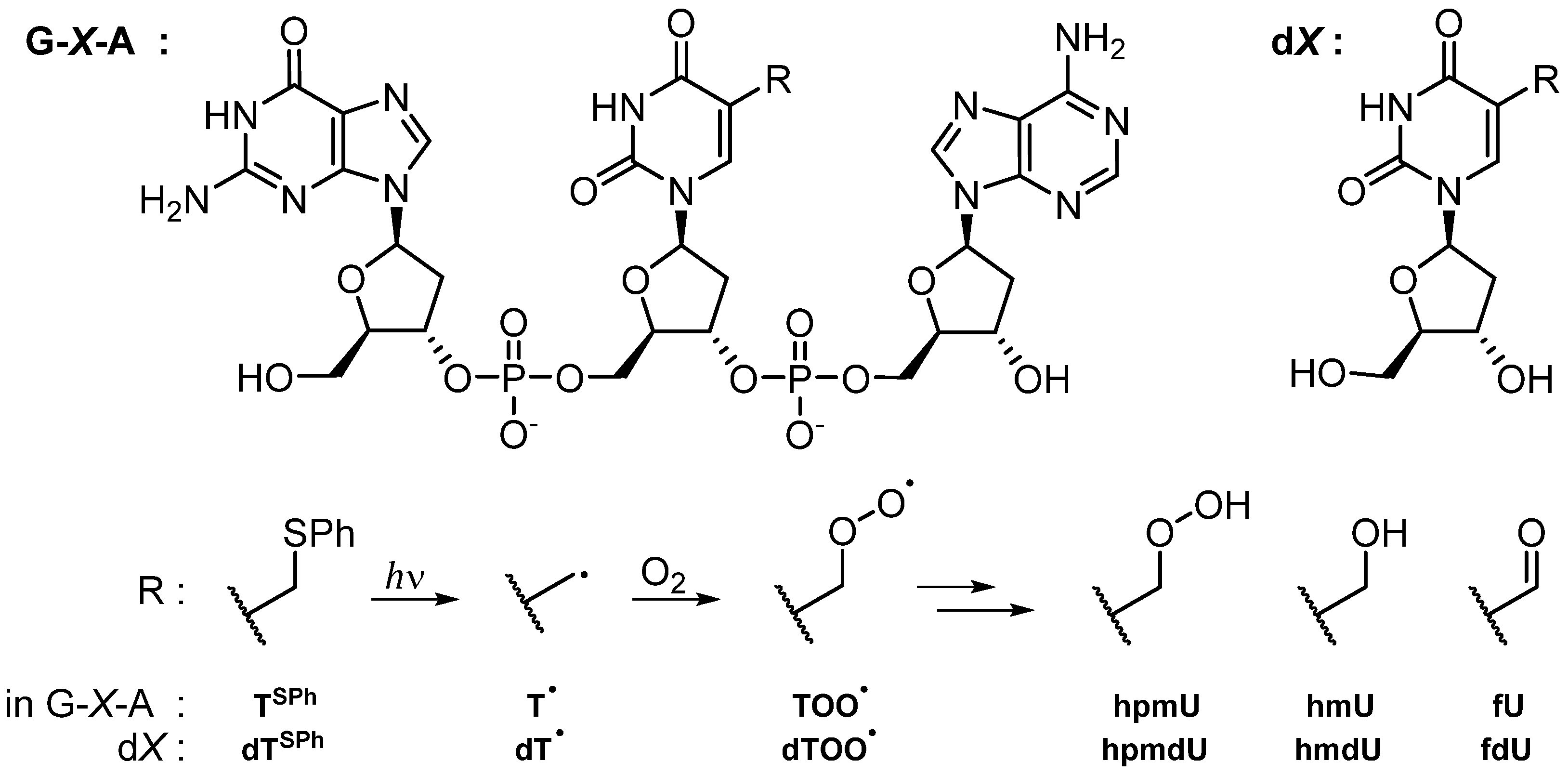
3.1. Fate of 5-(Uracilyl)Methyl Peroxyl Radicals in the Presence of Ascorbate

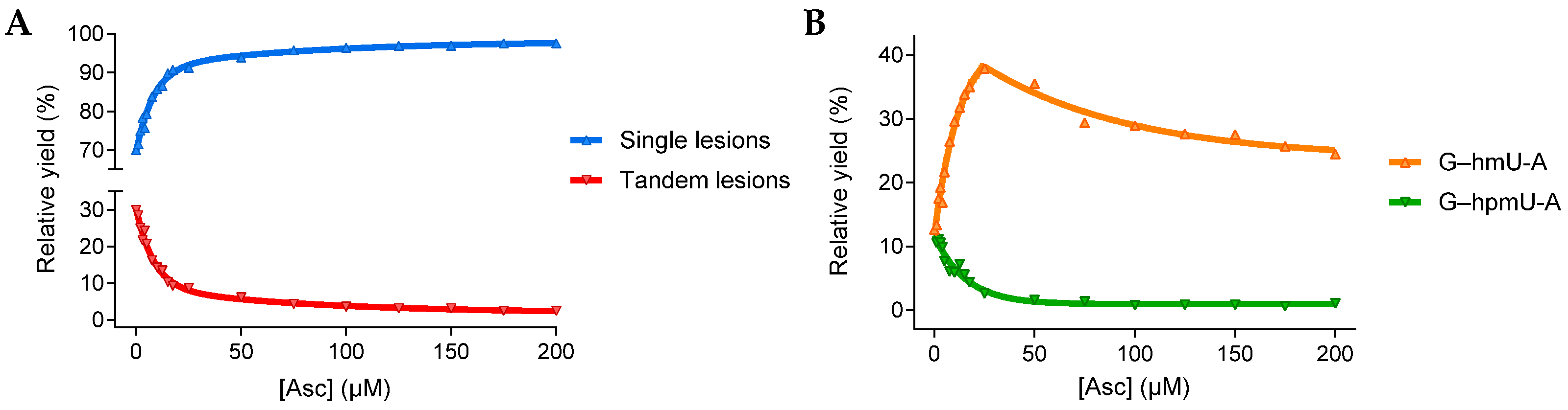
3.1.1. Single and Tandem Lesions Formation in a Trinucleotide Model

The selective photoinduced generation of TOO within G–T–A (Figure 1) and the analysis of associated lesions by LC-MS/MS were performed as described [9]. The phenylsulfide photocleavable system allows one to investigate the specific reactions of TOO with reducing agents that produce single modifications of thymine in competition with the addition to vicinal guanine that leads to tandem base modifications. When TOO was generated in the absence of Asc, single and tandem lesions accounted for approximately 70% and 30% of products, respectively. The addition of Asc efficiently blocked the production of tandem lesions, with less than 25 µM required to decrease their formation to below 10% of the total products (Figure 2A). To our surprise, the formation of the expected ROOH product 5-hydroperoxymethyluracil (hpmU) was minor and decreased to near zero levels from 0 to 25 µM Asc, while the formation of hmU greatly increased (Figure 2B). The kinetics of hmU formation as a function of Asc matched the kinetics of single lesion formation, indicating that the reaction of Asc with TOO leads to hmU rather than hpmU. These results contradicted the classical mechanism of electron transfer from Asc to ROO to produce ROOH. Thus, a series of experiments including mechanistic studies were undertaken to better understand this phenomenon.

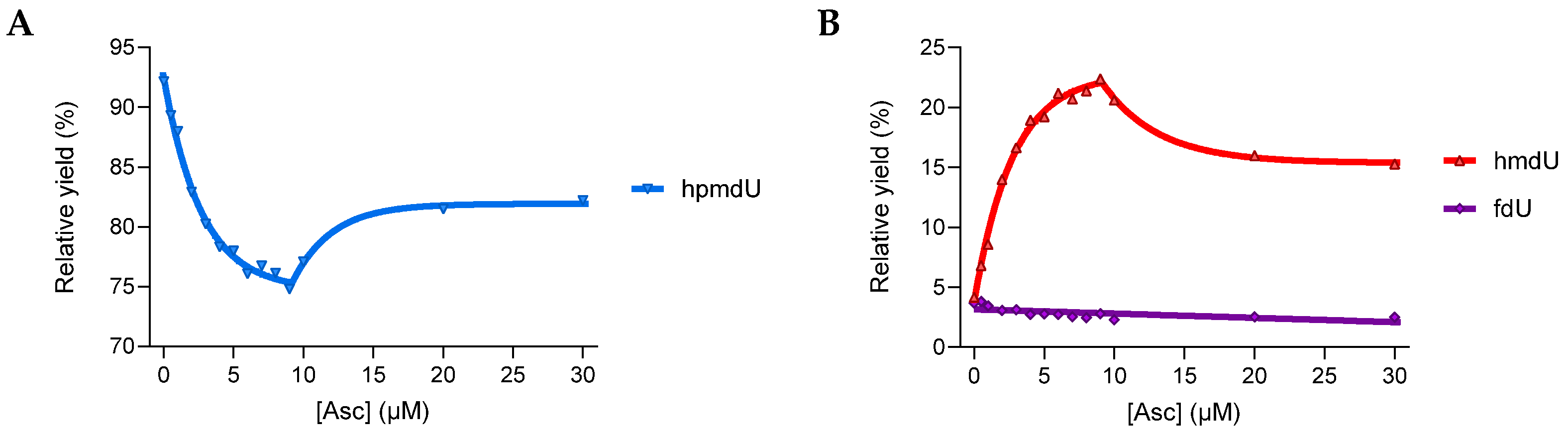
3.1.2. Reactivity in a 2′-Deoxyribonucleoside Model

To simplify the chemistry, the trinucleotide system was changed to a 2-deoxyribonucleoside monomer model (dX in Figure 1). This eliminates reactions with vicinal nucleotides and allows for the accurate quantification of 5-hydroperoxymethyl-2′-deoxyuridine (hpmdU), which is relatively stable as a free nucleoside [49]. The photolabile precursor homologue 5-phenylthiomethyl-2′-deoxuridine (dTSPh) was used to selectively generate 5-(2′-deoxyuridinyl)methylperoxyl radicals (dTOO). Product analysis carried out under control conditions (dTSPh concentration = 125 µM, 2 min photolysis, phosphate-buffered (pH 7.4), O2-saturated solutions) showed that methyl oxidation products of thymidine amount to approximately 5 µM, affording hydroperoxide hpmdU as the predominant product (93%) while the formation of 5-hydroxymethyl-2′-deoxyuridine (hmdU) and 5-formyl-2′-deoxyuridine (fdU) was minor (ca. 4% each). The high yield of hpmdU in control experiments likely arises from the reduction in dTOO by intermediate products derived from thiophenyl photocleavage. In the presence of 10 µM Asc, however, the relative yield of fdU remained unchanged, whereas hmdU increased from 4 to 22% and hpmdU dropped from 93 to 75%, a difference of 18% (Figure 3). Thus, the results indicate that Asc modulates the distribution of products by diverting the conventional pathway giving hpmdU to a novel pathway involving the formation of hmdU. This pathway also accounts for the slight increase in hpmdU observed at higher concentrations of Asc (10–30 µM).

3.2. Mechanistic Studies

Several mechanisms can tentatively explain the unconventional Asc-mediated production of alcohols (ROH) from initial ROO. The following section details our investigation of 3 possible mechanisms that are summarized in reaction pathways 1, 2, and 3.

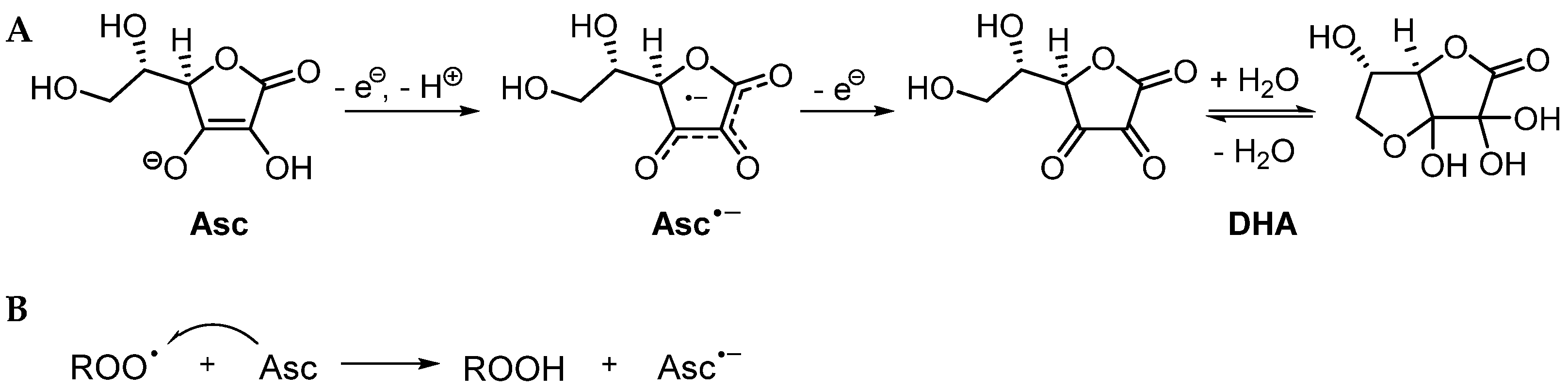
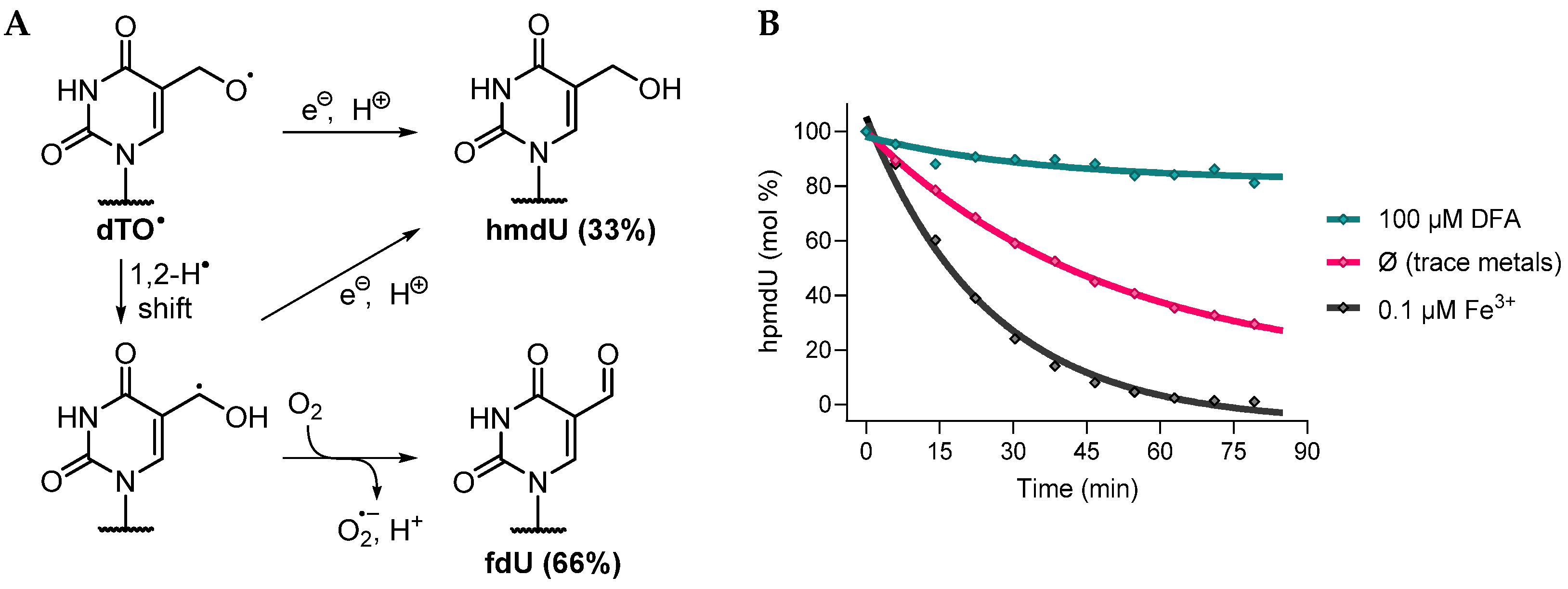
3.2.1. Possible Mechanism #1: Electron Transfers Involving Intermediate Alkoxyl Radical

The radical trapping character of Asc is generally attributed to its low oxidation potential that allows it to undergo single-electron transfers with reactive species. According to this well-established mechanism, a series of single-electron transfers may explain the formation of ROH from ROO (Pathway 1). First, Asc reduces ROO to ROOH (R1). Next, the hydroperoxide reacts with a second electron in a Fenton-type cleavage, generating an intermediate alkoxyl radical (RO) (R2). Lastly, Asc reduces RO to provide ROH (R3). To clarify if the proposed mechanism accurately explains the formation of ROH from ROO, the chemistry of RO was investigated independently.
  • Pathway 1
Antioxidants 13 01194 i001
Antioxidants 13 01194 i002
Antioxidants 13 01194 i003
A suitable procedure to generate RO consists of treating ROOH with one-electron reductants, such as Fe2+, in a so-called Fenton-type reaction. With this method, the specific chemistry of 5-(2′-deoxyuridinyl)methoxyl radicals (dTO) was investigated starting with hpmdU. It should be noted that HPLC purified hpmdU has a relatively long lifetime in aqueous solution (months at 5 °C), and thus, thermal decomposition does not contribute to the formation of products during our experiments [49]. Treatment of 5 µM hpmdU with 20 equivalents of Fe2+ consumed the compound instantaneously and gave a mixture of fdU and hmdU in a 2:1 ratio. The high relative yields of fdU can be explained by the propensity of alkoxyl radicals to undergo fast unimolecular decay in competition with intermolecular proton-coupled electron transfer. Here, the formation of fdU from dTO is rationalized by a 1,2-H-atom shift followed by O2 oxidation of the ensuing ketyl radical (Figure 4A) [54,55,56,57,58,59,60,61]. Alternatively, 2′-deoxyuridine (dU), the expected product of C–C β-scission of dTO, did not form in the reaction mixtures.
Next, the ability of Asc to react with hpmdU was assessed. The reaction of 5 µM hpmdU with 20 equivalents of Asc afforded an identical ratio of products but with much slower kinetics (t1/2 between 17 and 35 min). The same 2:1 carbonyl-to-alcohol ratio obtained in this transformation indicates that the reaction proceeds via a Fenton-type cleavage of the O–O bond of hpmdU. Higher concentrations of Asc (0.1–5 mM) had no effect on product distribution. Our results are consistent with a recent study investigating the chemistry of cholesterol alkoxyl radicals generated in the presence of Asc, which observed high carbonyl-to-alcohol ratios of products [62]. However, the addition of a strong chelator of iron, i.e., deferoxamine (DFA; 100 µM), drastically inhibited the Asc-induced decomposition of hpmdU, while the addition of 0.1 µM Fe3+ greatly accelerated it (Figure 4B). Thus, one can conclude that the decomposition of hpmdU by Asc occurs primarily through catalysis by metal ions present in trace amounts in aqueous solutions. A complete inhibition of hpmdU decomposition in the presence of DFA demonstrates that Asc does not undergo direct electron transfer with the hydroperoxide to generate dTO (R2). Previous claims that report a direct reaction of hydroperoxides with Asc are likely incorrect due to the presence of trace metal ions in buffered solutions [37]. Furthermore, because fdU is a prominent and characteristic product of dTO, and the formation of fdU was not observed in the reaction of dTOO with Asc (Figure 3B), the intermediacy of dTO may be excluded as a possible mechanism. In conclusion, our results show that the mechanistic Pathway 1 does not explain the formation of ROH from ROO in the presence of Asc.

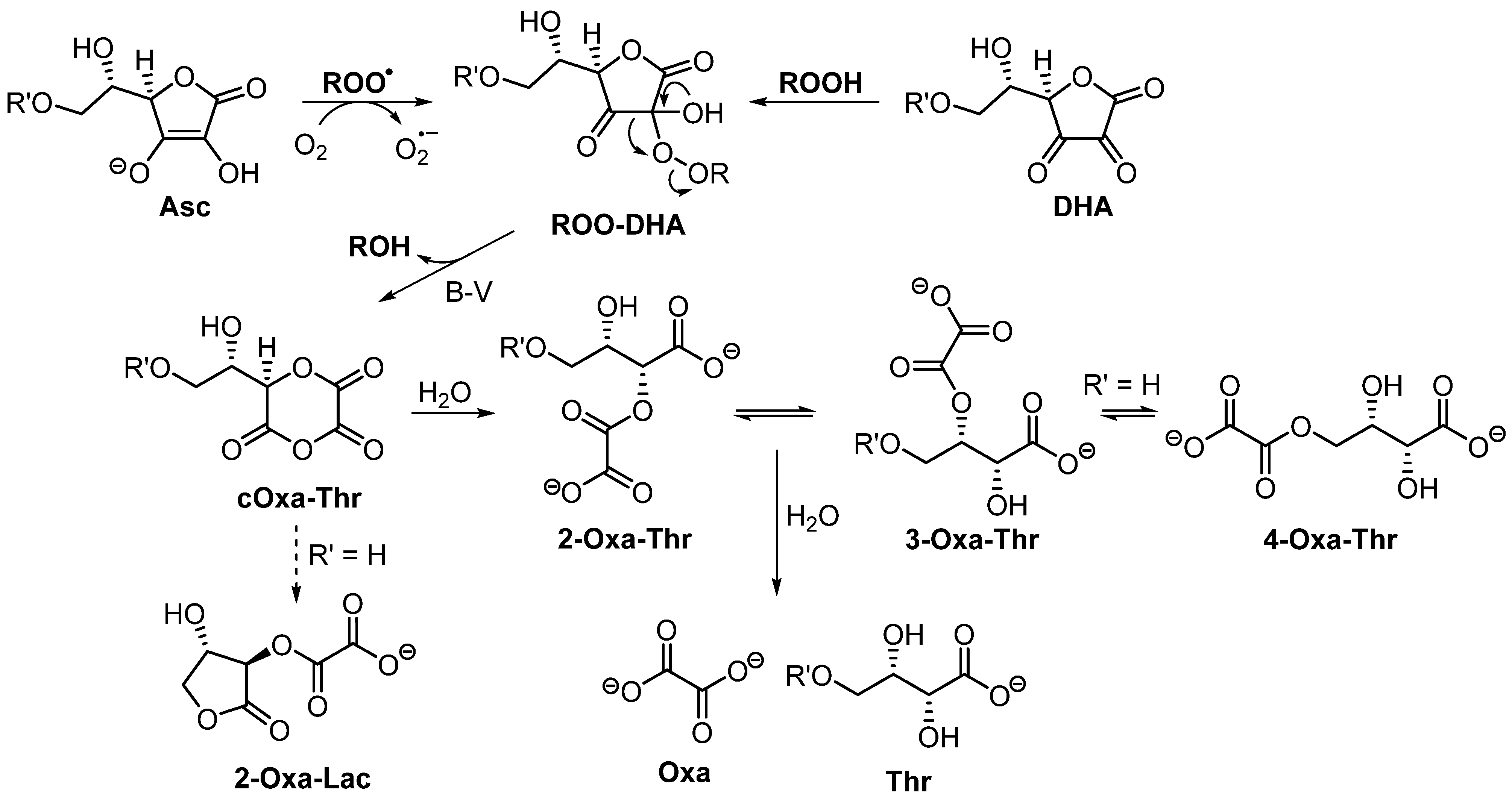
3.2.2. Possible Mechanism #2: Peroxyl Radical Addition

Peroxyl radicals display various uni- and bi-molecular decomposition pathways. The conversion of ROO into ROOH via proton-coupled electron transfer takes place by either a stepwise mechanism (i.e., one-electron reduction followed by protonation) or a concerted mechanism (i.e., formal H-atom transfer (HAT)). The former pathway is conventionally ascribed to reactions of ROO with Asc, while the latter one is involved in the chain propagation step of lipid peroxidation and scavenging reactions by thiols (e.g., GSH) and tocopherol. On the other hand, peroxyl radical addition (PRA) to double bonds represents a major pathway that often competes with HAT [63], as exemplified by the autoxidation of cholesterol [64]. In DNA, a similar competition arises between intramolecular PRA to vicinal guanine and intermolecular HAT with protective thiols [8]. PRA leads to the generation of peroxide adducts, i.e., endoperoxyl radicals, whose fate depends on the chemical structure of the adduct and can be modulated by the presence of redox agents. For instance, in our previous study involving the addition of TOO to 5′-flanking guanine, the formation of three distinct tandem modifications was shown to originate from the subsequent reactions of a common guanine-thymine endoperoxyl radical intermediate [9]. In this case, the guanine-centered radical undergoes either oxidation or reduction due to its redox ambivalence (illustrated in Scheme 5 of Ref. [8]). A similar series of reactions can be proposed for Asc (Pathway 2).
  • Pathway 2
Antioxidants 13 01194 i004
Antioxidants 13 01194 i005
Antioxidants 13 01194 i006
In theory, the addition of ROO to the enediol portion of Asc results in the formation of a ketyl radical anion (R4). Ketyl radicals are a type of species known to rapidly undergo oxidation in aerated conditions to the corresponding carbonyl-containing product via O2 addition followed by HO2 elimination [56,57,58,62]. The fast elimination of HO2 facilitated by the α-hydroxyl group generally prevents subsequent reactions by intermediate peroxyl radicals [65,66,67,68,69,70]. Additionally, the rate constant of O2•− elimination from α-hydroxyalkylperoxyl radical anions (i.e., with deprotonated α-OH groups) is accelerated by multiple orders of magnitude compared to their protonated counterparts [66,71]. Consequently, it is reasonable to propose initial PRA to Asc followed by oxidation of ROO-Asc ketyl radical anions by O2 providing nonradical peroxide adducts (R5).
The proposed ROO–DHA adducts are structurally analogous to so-called Criegee intermediates (gem-hydroxy peroxides) arising from the nucleophilic addition of hydroperoxides/peroxyacids to aldehydes and ketones that can subsequently undergo Baeyer-Villiger rearrangements [52,72]. To determine whether this pathway gives rise to the major alcohol product hmdU (R6), the reaction between hpmdU and DHA was examined independently (Scheme 2). As predicted, treatment of hpmdU with a very large excess of DHA (150 eq.) induced its complete transformation into hmdU (95% yield). The use of lower concentrations of DHA led to decreased rates of conversion, probably because of its fast hydrolysis at neutral pH (t1/2 = 22 min [73]). The implication of a Baeyer-Villiger rearrangement is supported by an acceleration of the reaction rate as a function of pH (Figure 5). At pH > 7, conversion of hpmdU into hmdU was complete in approximately 10 min, whereas acidic conditions retarded the reaction considerably (t1/2 = 75 min at pH 4). Similar to Dakin reactions [74], base catalysis observed here is rationalized by deprotonation of the gem-OH group of a Criegee intermediate, which accelerates migration to the peroxide, the rate-limiting step [75]. From the inflection point of the pH curve (Figure 5B), it is reasonable to assume that the pKa of the gem-OH of dTOO–DHA is approximately 5.4. The above results demonstrate that ROO–DHA adducts undergo rearrangement to provide ROH products, thus supporting reaction R6 of Pathway 2. The formation of hmdU from the reaction of dTOO with Asc under our conditions does not arise from the nonradical addition of hpmdU to DHA because very little amounts of DHA are formed in photolysis mixtures (<10 µM). Furthermore, no conversion was observed when 5 µM hpmdU was treated with 10 µM of DHA for 10 min under similar conditions as those during photolysis. A substantial excess of DHA (>10 eq.) is necessary for the nonradical reaction to proceed. Thus, we conclude that both the reaction between hpmdU and DHA and the one between dTOO and Asc lead to a common intermediate (dTOO–DHA Criegee adduct) and that the formation of hmdU is explained by the subsequent decomposition of this intermediate via a Baeyer–Villiger rearrangement.

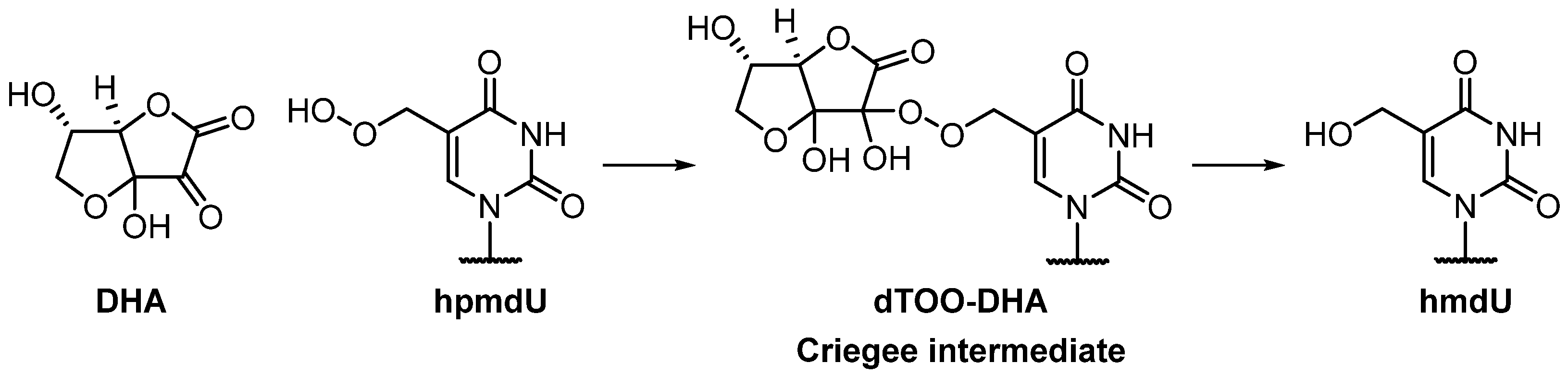
3.2.3. Possible Mechanism #3: Peroxyl Radical Addition Followed by Epoxide Formation

Following PRA, the resulting endoperoxyl radicals can subsequently undergo various types of reactions depending on the redox properties of the radical and the chemical environment. For example, carbon-centered radicals generated by PRA to alkenes engage in two competing pathways: bimolecular addition of O2, affording additional peroxyl radicals, or unimolecular decay via O–O homolytic substitution with concomitant formation of RO and an epoxide [76,77,78]. These reactions are showcased in the oxidation of various lipids [64,79,80]. The latter pathway may be proposed as an alternative explanation for the Asc-induced formation of ROH from initial ROO, as expressed in Pathway 3.
  • Pathway 3
Antioxidants 13 01194 i004
Antioxidants 13 01194 i007
Antioxidants 13 01194 i003
Antioxidants 13 01194 i008
This pathway features PRA to the C2=C3 bond of Asc (R4), followed by O–O homolytic substitution, and the release of RO and epoxidized Asc (R7), which can decompose into ROH (R3) and DHA (R8), respectively. However, as already shown in the previous section, Asc-mediated transformation of dTOO into hmdU does not involve the intermediacy of alkoxyl radicals because dTO leads to the characteristic formation of fdU, and the yield of this product was negligible (Figure 3B). Thus, both Pathways 1 and 3 can be discarded leaving Pathway 2 as the most likely route that converts dTOO into hmdU.

3.3. Product Characterization

The oxidation of Asc was examined using the ester derivative 6-O-(phenylacetyl)-l-ascorbic acid (Asc-PA). The presence of a nonpolar substituent at C6 of Asc greatly improves the analysis of Asc-derived oxidation products with respect to their retention on reversed phase chromatography and detection by UV and MS. A mixture of oxidation products of Asc-PA was observed by LC-MS/MS following the independent generation of dTOO upon photolysis of dTSPh in the presence of 50 µM Asc-PA (Figure 6B). The same products with a similar profile were also observed by treatment of Asc-PA with mild oxidants, including oxone (KHSO5), meta-chloroperoxybenzoic acid (mCPBA), and H2O2 in aqueous solutions at pH 7.4 (Figure 6). Of the products obtained, two are well-known oxidation products of Asc (products at 4.65 and 4.30 min; Figure 6B). The product at 4.65 min was identified as the 6-O-phenylacetate ester of dehydroascorbate (DHA-PA) on the basis of MS analysis (Figures S5–S11, S28 and Table 1) and comparison of the LC and MS properties with authentic compounds obtained by the reaction of Asc-PA and Br2 that quantitatively gives DHA-PA [51]. The identity of another common oxidation product of Asc, i.e., 4-O-(phenylacetyl)-l-threonic acid (Thr-PA; peak at 4.30 min in Figure 6B) was confirmed by MS (Figures S12–S15, S29 and Table 1) and NMR analysis (Figures S34–S37). The phenylacetyl group attached to Asc-PA, Thr-PA, and other products depicted the corresponding proton and carbon atoms of the substituent (Table 2). Based on 1H-13C heteronuclear analysis (HMBC), the carbonyl associated with the phenylacetyl group shows strong cross-peaks with the vicinal CH2 group (Figure S37). The presence and position of the carboxylic acid group of Thr-PA were confirmed by 13C-NMR showing a peak at 174.6 ppm and corresponding 1H-13C cross-peaks with H3 and H4 in the HMBC spectrum (Figures S36 and S37). These analyses are consistent with those reported in the literature on synthetic compounds [81].
In addition to known products, several unidentified peaks of variable stability were observed in the oxidation mixture of Asc-PA, with four of them having a mass of 326 Da (peaks at 2.4, 2.7, 3.3, and 3.6 min; Figure 6B). The least polar product at 5.6 min, which had a mass of 308 Da, was found to be particularly unstable. Following purification by HPLC and addition to a phosphate-buffered solution at pH 7.4, the latter compound rapidly converted into additional products found at 3.3 and 3.6 min (Figure 6C,D). Moreover, upon purification followed by its incubation at pH 7.4 and 37 °C, the major product at 3.3 min was found to give rise to the three other polar products with an equal molecular mass (peaks at 2.4, 2.7, and 3.6 min; Figure 6B) and a small amount of Thr-PA. These results clearly show that the product at 5.6 min is the sole precursor of the four early eluting products. Thus, we conclude that the initial product of oxidation of Asc-PA by dTOO is the product at 5.6 min (308 Da) and that this compound irreversibly undergoes hydrolysis (+18 Da) into the series of four polar isomers described above (326 Da; peaks at 2.4, 2.7, 3.3, and 3.6 min). Furthermore, the mass and corresponding fragment ions indicate that the early eluting products at 2.4, 2.7, 3.3, and 3.6 min are isomers, bearing an intact Thr-PA portion and an oxalate substituent. A very similar product profile was also observed from the reaction of other peroxide-based oxidants with Asc-PA (oxone and mCPBA).
The structures of unknown products were determined by extensive MS and NMR analyses. The product at 5.6 min was identified as the diglycolic anhydride derivative, namely, 1,2-O-cyclo-oxalyl-4-O-(phenylacetyl)-l-threonate (cOxa-Thr-PA). The exact mass (Table 1) and fragmentation of the parent compound, showing fragments at 235 m/z (M–OH–COCO2) and 189 m/z (M–PhCH2CO) in negative mode (Figures S18 and S30), support the proposed structure. To examine the structure by NMR, it was necessary to carry out the oxidation of Asc-PA in situ in the NMR tube using mCPBA as the oxidant in a non-nucleophilic solvent (deuterated acetone). The oxidation was complete under these conditions and afforded cOxa-Thr-PA (70%) together with a product from hydrolysis (30%) (Figures S38–S44). Although the spectra contained signals from excess mCPBA and meta-chlorobenzoic acid, the signals associated with cOxa-Thr-PA were clearly discernable from those of the reagent and byproducts that appeared in a specific aromatic region of the spectra. To simplify comparison between compounds in the remaining text, the numbering of atoms refers to carbon positions in the original Asc backbone. The NMR spectra of cOxa-Thr-PA were similar to those of Asc-PA and Thr-PA (Table 2) except that the protons corresponding to –CH2–CH(OH)–CH– of the Asc side chain (carbons from left to right are numbered C6, C5, and C4) were shifted to the lower field, likely due to deshielding caused by the cyclic oxalate substituent at C4 of the backbone. The carbonyl groups of the cyclic oxalate group at C1, C2, and C3 were assigned based on observable cross-peaks with H4 and H5 from 2D HMBC analysis (Figure S44). In addition, the assignment of C1 and C2 to the oxalate group of cOxa is supported by simulation of the 1H- and 13C-NMR spectra (Figure S52).
The major polar product at 3.3 min was identified as 2-O-oxalyl-4-O-(phenylacetyl)-l-threonic acid (2-Oxa-Thr-PA). This product was observed as a minor component (30%) in the spectra of cOxa-Thr-PA (Figure S38–S44). It was also purified by HPLC and subjected to detailed NMR analyses (Figure S45–S51; Table 2). Based on integration of CH2 protons of the phenylacetyl substituent, 1H-NMR analysis indicates that the purified product at 3.3 min transforms into a mixture of four products with a ratio of 65:20:8:7 (Figures S45 and S46). The relative proportion of each product aligns with the intensity of the four peaks observed previously by LC-MS/MS. The major product representing 65% of the mixture depicted the phenylacetyl substituent, the –CH2–CH(OH)–CH– side chain, as well as additional carbonyl groups associated with an oxalate substituent and a carboxylic acid (Figures S48–S51). In comparison to the major product, the product representing 20% of the mixture showed dramatic changes in the chemical shifts of H4 and H5. In particular, the chemical shift of H4 decreased from 5.23 to 4.56 ppm (−0.8 ppm), while that of H5 increased from 4.49 to 5.59 ppm (+1.1 ppm). In addition, the chemical shift of C4 decreased by −0.8 ppm while that of C5 increased by 1.3 ppm. These changes are consistent with the migration of a largely deshielding oxalate group from O4 to O5. Thus, the other major product in the mixture was identified as a regioisomer 3-O-oxalyl-4-O-(phenylacetyl)-l-threonic acid (3-Oxa-Thr-PA; Figure 6E). In addition to the major products at 3.3 and 3.6 min, two minor products that arise from isomerization of 2-Oxa-Thr-PA and that have the same mass were observed at 2.4 and 2.7 min by LC-MS/MS. To ascertain the structure of these products, we examined the oxidation of Asc bearing the phenylacetate group at C5 rather than C6 (i.e., 5-O-(phenylacetyl)-l-ascorbate rather than 6-O-(phenylacetyl)-l-ascorbate). In this case, the predominant product was identified by MS and HPLC as the same product that elutes at 2.4 min. Thus, we conclude that peaks at 2.4 and 2.7 min arise from migration of the phenylacetate group, i.e., either 1,4-shift for 2-Oxa-Thr-PA or 1,5-shift for 3-Oxa-Thr-PA (Figure 6E). The migration of oxalate and phenylacetate from one OH group to another results in a mixture of four regioisomers. The proportion of Oxa-Thr-PA isomers depends on the nucleophilicity of the OH group, such that substitution was more efficient for the primary alcohol at O6 relative to the secondary alcohol at O5. These findings were corroborated using 5,6-O-isopropylidene-ascorbate (Asc-iPr), in which both O5 and O6 are substituted by the derivatization group. Upon oxidation, the disubstituted derivative produced only one open-chain oxalate derivative, i.e., 2-O-oxalyl-3,4-O-isopropylidene-l-threonic acid (Oxa-Thr-iPr); the product nevertheless was slightly susceptible to hydrolysis into 3,4-O-isopropylidene-l-threonic acid (Thr-iPr). A final structural characterization was achieved by converting the carboxylic acid-containing phenylacetate derivatives into amides of phenylethanamine using EDC-mediated coupling methods (Figures S53–S67). This provided further support for the structures of Oxa-Thr-PA regioisomers, as well as threonate and oxalate.
In conclusion, the cyclic compound cOxa-Thr-PA was unambiguously identified as a novel intermediate in the peroxide-mediated oxidation of Asc-PA, as inferred by detailed MS and NMR analysis. In neutral aqueous solutions, this product rapidly hydrolyzes into the open chain Oxa-Thr-PA, which in turn can undergo acyl group migrations to yield a mixture of four regioisomers, as well as hydrolysis into Thr-PA. The identification of cOxa-Thr-PA validates the proposed mechanism presented in reaction Pathway 2, which features a key step involving a Baeyer–Villiger rearrangement following PRA to Asc.

3.4. Oxidation of Asc-PA by Peroxyl Radicals Derived from Azo Compounds

To further validate the novel reaction pathway, we examined the oxidation of Asc-PA by ROO derived from the thermal decomposition of azo compounds, i.e., 2,2′-azobis(2-amidinopropane) dihydrochloride (ABAP) and 2,2′-azobis(2-methylpropionitrile) (AIBN). Asc-PA (50 µM) was exposed at 37 °C to either ABAP (100 µM) in aqueous solution buffered at pH 7.4 in the presence of DFA (100 µM), or AIBN (100 µM) in acetonitrile (unbuffered). As expected, we observed a time-dependent formation of Oxa-Thr-PA in the aqueous reaction mixture, and cOxa-Thr-PA in the organic reaction mixture, with minute amounts of hydrolyzed products (Figure S68). These results confirm that the PRA reaction to Asc occurs for alkylperoxyl radicals generated by ABAP and AIBN.

4. Discussion

Ascorbate is an effective antioxidant in vivo because it scavenges reactive species with high rate constants of reaction, it usually exists at elevated intracellular concentrations, and it is rapidly recycled upon oxidation. The reactivity of Asc toward radicals is commonly attributed to its ability to donate electrons, like its reaction with oxidizing metal ions. However, pioneering work demonstrated that Asc oxidation by HO does not exclusively afford Asc•− via direct electron transfer but rather produces a mixture of ascorbate radicals, including C2=C3 addition products in considerable proportions [16,82,83,84,85,86]. Likewise, a very detailed kinetic analysis by Cabelli and Bielski showed that the reaction between Asc and O2•− does not properly generate Asc•−; instead, they proposed the formation of a “transient adduct or product” which rapidly engages in redox reactions involving either O2•− or disproportionation [87]. These studies, along with the present one, reflect the unusual reactivity of Asc toward oxygen radicals and demonstrate that, besides electron transfer, addition reactions are involved in radical trapping by Asc. Peroxyl radical addition to double bonds is a well-known phenomenon, especially in the context of hydrocarbon autoxidation. The present study provides direct evidence of PRA to support product analysis and mechanistic studies.
Our results show that a reaction between a peroxyl radical (i.e., dTOO) and Asc leads to the distinctive formation of an alcohol (i.e., hmdU). The intermediacy of an alkoxyl radical in the reaction sequence is ruled out because of the lack of formation of the corresponding carbonyl product (i.e., fdU). The absence of a carbonyl product excludes alternative mechanisms: stepwise electron transfers involving O–O bond cleavage by one-electron reduction (reaction Pathway 1), and PRA to Asc followed by unimolecular decay via radical epoxidation (reaction Pathway 3). To explain the formation of an alcohol, we propose PRA followed by O2-mediated oxidation and Baeyer–Villiger rearrangement of the resulting peroxide adduct (reaction Pathway 2). This mechanism is substantiated by the modulating effect of pH on the rate of rearrangement and unambiguous identification of the ring-expansion product, cOxa-Thr. Furthermore, our results clarify the chemistry of Asc oxidation as well as the reaction of DHA with H2O2, which afford oxalate esters of threonic acid (Oxa-Thr) and threonic acid (Thr) as observed in earlier reports [88,89,90,91]. Previously, Fry and coworkers proposed a cyclic oxalate intermediate to explain the oxidation of Asc and DHA by H2O2 [90,91]. They hypothesized that this intermediate undergoes decomposition into three open-chain oxalyl-threonate derivatives that interconvert via oxalate migration (note that the authors studied unsubstituted Asc with a free hydroxyl group at C6 that can participate in migration). The latter oxidation products of Asc were tentatively identified based uniquely on their electrophoretic and chromatographic properties and comparison to threonate and oxalate derivatives. Although the structure of the cyclic oxalate intermediate proposed by Fry and colleagues was incorrect, the basic structure of the more stable open-chain threonate derivatives compares well with those deduced from our complete analysis.
Here, we show that either reactions of Asc with ROO, or DHA with ROOH and H2O2, lead to a Criegee intermediate that undergoes a Baeyer–Villiger rearrangement to generate cOxa-Thr. This intermediate hydrolyzes into 2-Oxa-Thr, which interconverts via acyl migration into 3-Oxa-Thr and the more stable 4-Oxa-Thr if this position is unsubstituted. Further hydrolysis of the latter oxalate esters leads to the formation of two final decomposition products: threonic acid and oxalic acid. It is reasonable to assume that a similar mechanism takes place not only for 6-O-(phenylacetyl)-ascorbate but also for the nonderivatized Asc in its biologically active form. Thus, we propose a revised mechanism for the oxidation of Asc by peroxyl radicals and other peroxides shown in Scheme 3. Fry and coworkers also proposed the formation of an additional compound arising from H2O2 oxidation, namely l-threonolactone [91]. Based on our revision of the chemistry, we suggest that this product arises from an alternative mechanism involving intramolecular cyclization of unsubstituted C6-OH of cOxa-Thr to produce 2-O-oxalyl-l-threonolactone (2-Oxa-Lac) in competition with hydrolysis that gives Oxa-Thr isomers.
It is unclear whether PRA to Asc occurs at position C2 or C3, since both are probable and would lead to the formation of cOxa-Thr. On one hand, addition at C3 generates a stable radical that is delocalized through C1 and C2, compared to the C2 adduct that simply generates a radical anion. On the other hand, nucleophilic addition of hpmdU to DHA likely takes place at the more electron-deficient C2 position, which gives an intermediate that demonstrably undergoes Baeyer–Villiger rearrangement leading to hmdU. It has been previously shown that HO addition to Asc can occur at both C2 and C3 [85,86]. Thus, the addition of ROO may occur at both sites, although it is likely more selective than HO suggesting that addition to the C2 position is the most probable.
The fate of ROO–Asc PRA adducts may involve alternative pathways that were not evaluated in the present study (depicted in Scheme 4). Firstly, it is possible that ROO–Asc undergo unimolecular decay into Asc•− and peroxide anions, which are the same products obtained via the classical electron transfer mechanism. Secondly, we have shown that O2-mediated oxidation of ROO–Asc (R4 in Pathway 2) ultimately leads to the formation of an alcohol via a key Baeyer–Villiger rearrangement. However, the Criegee intermediate required for this reaction is in equilibrium with the dissociation products, which include ROOH and DHA. Thus, the effectiveness of the formation of ROH is limited by both the rate of ROO addition to Asc and the rate of dissociation of ROO–DHA adducts. Moreover, in parallel with previous propositions related to O2•− and Asc adducts [87], the oxidation ROO–Asc may compete with their reduction. A gradual increase in hmpdU accompanied with a loss of hmdU at high concentrations of Asc (>10 µM) (Figure 2B and Figure 3B) can be explained by the reduction in PRA adducts by Asc. Reduction in ROO–Asc will prevent the Baeyer–Villiger rearrangement necessary for the formation of ROH and divert the pathway to the formation of ROOH, the same product that arises from initial electron transfer.
Because both Asc and Asc•− undergo electron transfer with reactive species according to the classical mechanism (Scheme 1A), the stoichiometry of peroxyl radical trapping theoretically involves two ROO for the oxidation of Asc to DHA. Inferior values have been obtained experimentally, decreasing as low as ~0.1 [29,85,92,93]. Such small values are generally rationalized in terms of Asc depletion by trace metal-catalyzed autoxidation. In view of the present work, the observed lack of stoichiometry can be explained by the ability of Asc to undergo PRA leading to the formation of Oxa-Thr. Thus, two outcomes for the reactions of alkylperoxyl radicals with Asc need to be taken into consideration. The trapping of ROO and their conversion into corresponding ROOH has the potential to be detrimental due to the degradation of hydroperoxides into electrophilic carbonyl compounds and highly reactive radicals such as RO, R, and ROO that can propagate oxidative damage. On the other hand, the conversion of ROO into the corresponding ROH by Asc (as evidenced here) circumvents the generation of subsequent reactive radical species, with the trade-off being the irreversible loss of a molecule of Asc. In biological contexts, the latter reaction likely carries less severe consequences.

5. Conclusions

The ability of Asc to undergo single electron transfers with ROO is a widely accepted reaction that underscores its role as a biological antioxidant. We demonstrate a novel pathway of reaction between ROO and Asc that leads to unconventional products and likely occurs in competition with the electron transfer mechanism. This pathway consists of initial peroxyl radical addition to the enediol portion of Asc, followed by one-electron oxidation of the resulting radical site by O2. The intermediate thereby produced undergoes facile Baeyer–Villiger rearrangement that ultimately leads to the conversion of ROO into the corresponding alcohol product (ROH) and conversion of Asc into the unstable cOxa-Thr. The decomposition of cOxa-Thr affords a series of products that arise from its hydrolysis, including Oxa-Thr regioisomers, together with threonate and oxalate as final decomposition products. Although it was necessary to employ a C6-substiuted derivative of Asc to demonstrate this reaction in the current study, one can expect that similar reactions between Asc and peroxyl radicals take place in cells and tissues. The present findings expand our understanding of the complex chemistry of Asc, which should help investigate its function as a major antioxidant in redox biology.

Supplementary Materials

The following supporting information can be downloaded at: https://www.mdpi.com/article/10.3390/antiox13101194/s1, MS/MS transitions used for MRM analyses, MS, and NMR spectra of phenylacetate derivatives, textual interpretation of NMR spectra, identification of compounds via EDC coupling, oxidation of Asc-PA by ABAP and AIBN.

Author Contributions

Conceptualization, G.R. and J.R.W.; Methodology, G.R. and J.R.W.; Validation, J.R.W.; Formal analysis, G.R.; Investigation, G.R.; Resources, J.R.W.; Data curation, G.R.; Writing—original draft, G.R. and J.R.W.; Writing—review & editing, G.R. and J.R.W.; Supervision, J.R.W.; Project administration, J.R.W.; Funding acquisition, J.R.W. All authors have read and agreed to the published version of the manuscript.

Funding

This research was funded by the Natural Sciences and Engineering Research Council of Canada (NSERC) grant number (RGPIN-2022-03974).

Institutional Review Board Statement

Not applicable.

Informed Consent Statement

Not applicable.

Data Availability Statement

Data is contained within the article and supplementary material.

Acknowledgments

The authors would like to thank Philippe Venne for HRMS analyses and Klaus Klarskov for the helpful discussions and revision of the manuscript.

Conflicts of Interest

The authors declare no conflicts of interest.

References

  1. Neta, P.; Huie, R.E.; Ross, A.B. Rate Constants for Reactions of Peroxyl Radicals in Fluid Solutions. J. Phys. Chem. Ref. Data 1990, 19, 413–513. [Google Scholar] [CrossRef]
  2. Valgimigli, L. Lipid Peroxidation and Antioxidant Protection. Biomolecules 2023, 13, 1291. [Google Scholar] [CrossRef] [PubMed]
  3. Greene, L.E.; Lincoln, R.; Cosa, G. Rate of Lipid Peroxyl Radical Production during Cellular Homeostasis Unraveled via Fluorescence Imaging. J. Am. Chem. Soc. 2017, 139, 15801–15811. [Google Scholar] [CrossRef] [PubMed]
  4. Isildar, M.; Schuchmann, M.N.; Schulte-Frohlinde, D.; von Sonntag, C. Oxygen Uptake in the Radiolysis of Aqueous Solutions of Nucleic Acids and Their Constituents. Int. J. Radiat. Biol. Relat. Stud. Physics, Chem. Med. 1982, 41, 525–533. [Google Scholar] [CrossRef] [PubMed]
  5. Bergeron, F.; Auvré, F.; Radicella, J.P.; Ravanat, J.L. HO Radicals Induce an Unexpected High Proportion of Tandem Base Lesions Refractory to Repair by DNA Glycosylases. Proc. Natl. Acad. Sci. USA 2010, 107, 5528–5533. [Google Scholar] [CrossRef]
  6. Davies, M.J. Protein Oxidation and Peroxidation. Biochem. J. 2016, 473, 805–825. [Google Scholar] [CrossRef]
  7. Gebicki, J.M.; Nauser, T. Initiation and Prevention of Biological Damage by Radiation-Generated Protein Radicals. Int. J. Mol. Sci. 2021, 23, 396. [Google Scholar] [CrossRef]
  8. Robert, G.; Wagner, J.R.; Cadet, J. Oxidatively Generated Tandem DNA Modifications by Pyrimidinyl and 2-Deoxyribosyl Peroxyl Radicals. Free Radic. Biol. Med. 2023, 196, 22–36. [Google Scholar] [CrossRef]
  9. Robert, G.; Wagner, J.R. Tandem Lesions Arising from 5-(Uracilyl)Methyl Peroxyl Radical Addition to Guanine: Product Analysis and Mechanistic Studies. Chem. Res. Toxicol. 2020, 33, 565–575. [Google Scholar] [CrossRef]
  10. Frelon, S.; Douki, T.; Ravanat, J.L.; Pouget, J.P.; Tornabene, C.; Cadet, J. High-Performance Liquid Chromatography - Tandem Mass Spectrometry Measurement of Radiation-Induced Base Damage to Isolated and Cellular DNA. Chem. Res. Toxicol. 2000, 13, 1002–1010. [Google Scholar] [CrossRef]
  11. Samson-Thibault, F.; Madugundu, G.S.; Gao, S.; Cadet, J.; Wagner, J.R. Profiling Cytosine Oxidation in DNA by LC-MS/MS. Chem. Res. Toxicol. 2012, 25, 1902–1911. [Google Scholar] [CrossRef] [PubMed]
  12. Madugundu, G.S.; Cadet, J.; Wagner, J.R. Hydroxyl-Radical-Induced Oxidation of 5-Methylcytosine in Isolated and Cellular DNA. Nucleic Acids Res. 2014, 42, 7450–7460. [Google Scholar] [CrossRef]
  13. Chen, J.; Guo, L.; Zhang, L.; Wu, H.; Yang, J.; Liu, H.; Wang, X.; Hu, X.; Gu, T.; Zhou, Z.; et al. Vitamin C Modulates TET1 Function during Somatic Cell Reprogramming. Nat. Genet. 2013, 45, 1504–1509. [Google Scholar] [CrossRef]
  14. Young, J.I.; Züchner, S.; Wang, G. Regulation of the Epigenome by Vitamin C. Annu. Rev. Nutr. 2015, 35, 545–564. [Google Scholar] [CrossRef] [PubMed]
  15. Fujii, J.; Osaki, T.; Bo, T. Ascorbate Is a Primary Antioxidant in Mammals. Molecules 2022, 27, 6187. [Google Scholar] [CrossRef]
  16. Laroff, G.P.; Fessenden, R.W.; Schuler, R.H. The Electron Spin Resonance Spectra of Radical Intermediates in the Oxidation of Ascorbic Acid and Related Substances. J. Am. Chem. Soc. 1972, 94, 9062–9073. [Google Scholar] [CrossRef]
  17. Davis, H.F.; McManus, H.J.; Fessenden, R.W. An ESR Study of Free-Radical Protonation Equilibria in Strongly Acid Media. J. Phys. Chem. 1986, 90, 6400–6404. [Google Scholar] [CrossRef]
  18. Lagercrantz, C. Free Radicals in the Auto-Oxidation of Ascorbic Acid. Acta Chem. Scand. 1964, 18, 562. [Google Scholar] [CrossRef]
  19. Iyanagi, T.; Yamazaki, I.; Anan, K.F. One-Electron Oxidation-Reduction Properties of Ascorbic Acid. Biochim. Biophys. Acta (BBA)—Bioenerg. 1985, 806, 255–261. [Google Scholar] [CrossRef]
  20. Pelizzetti, E.; Mentasti, E.; Pramauro, E. Outer-Sphere Oxidation of Ascorbic Acid. Inorg. Chem. 1978, 17, 1181–1186. [Google Scholar] [CrossRef]
  21. Williams, N.H.; Yandell, J.K. Outer-Sphere Electron-Transfer Reactions of Ascorbate Anions. Aust. J. Chem. 1982, 35, 1133–1144. [Google Scholar] [CrossRef]
  22. Bielski, B.H.J. Chemistry of Ascorbic Acid Radicals. In Ascorbic Acid: Chemistry, Metabolism, and Uses; Seib, P.A., Tolbert, B.M., Eds.; American Chemical Society: Washington, DC, USA, 1982; pp. 81–100. [Google Scholar]
  23. Buettner, G.R.; Jurkiewicz, B.A. Catalytic Metals, Ascorbate and Free Radicals: Combinations to Avoid. Radiat. Res. 1996, 145, 532–541. [Google Scholar] [CrossRef]
  24. Njus, D.; Kelley, P.M.; Tu, Y.J.; Schlegel, H.B. Ascorbic Acid: The Chemistry Underlying Its Antioxidant Properties. Free Radic. Biol. Med. 2020, 159, 37–43. [Google Scholar] [CrossRef] [PubMed]
  25. Buettner, G.R. The Pecking Order of Free Radicals and Antioxidants: Lipid Peroxidation, α-Tocopherol, and Ascorbate. Arch. Biochem. Biophys. 1993, 300, 535–543. [Google Scholar] [CrossRef]
  26. Nadezhdin, A.D.; Dunford, H.B. The Oxidation of Ascorbic Acid and Hydroquinone by Perhydroxyl Radicals. A Flash Photolysis Study. Can. J. Chem. 1979, 57, 3017–3022. [Google Scholar] [CrossRef]
  27. Packer, J.E.; Willson, R.L.; Bahnemann, D.; Asmus, K.-D. Electron Transfer Reactions of Halogenated Aliphatic Peroxyl Radicals: Measurement of Absolute Rate Constants by Pulse Radiolysis. J. Chem. Soc. Perkin Trans. 2 1980, 296. [Google Scholar] [CrossRef]
  28. Valgimigli, L.; Pratt, D.A. Antioxidants in Chemistry and Biology. In Encyclopedia of Radicals in Chemistry, Biology and Materials; John Wiley & Sons: Hoboken, NJ, USA, 2012; ISBN 9780470971253. [Google Scholar]
  29. Haidasz, E.A.; Van Kessel, A.T.M.; Pratt, D.A. A Continuous Visible Light Spectrophotometric Approach to Accurately Determine the Reactivity of Radical-Trapping Antioxidants. J. Org. Chem. 2016, 81, 737–744. [Google Scholar] [CrossRef]
  30. Domazou, A.S.; Zelenay, V.; Koppenol, W.H.; Gebicki, J.M. Efficient Depletion of Ascorbate by Amino Acid and Protein Radicals under Oxidative Stress. Free Radic. Biol. Med. 2012, 53, 1565–1573. [Google Scholar] [CrossRef]
  31. Domazou, A.S.; Gebicki, J.M.; Nauser, T.; Koppenol, W.H. Repair of Protein Radicals by Antioxidants. Isr. J. Chem. 2014, 54, 254–264. [Google Scholar] [CrossRef]
  32. Nauser, T.; Gebicki, J.M. Physiological Concentrations of Ascorbate Cannot Prevent the Potentially Damaging Reactions of Protein Radicals in Humans. Chem. Res. Toxicol. 2017, 30, 1702–1710. [Google Scholar] [CrossRef]
  33. Packer, J.E.; Slater, T.F.; Wilson, R.L. Direct Observation of a Free Radical Interaction between Vitamin E and Vitamin C. Nature 1979, 278, 737–738. [Google Scholar] [CrossRef] [PubMed]
  34. Buettner, G.R. In the Absence of Catalytic Metals Ascorbate Does Not Autoxidize at PH 7: Ascorbate as a Test for Catalytic Metals. J. Biochem. Biophys. Methods 1988, 16, 27–40. [Google Scholar] [CrossRef]
  35. Boatright, W.L. Oxygen Dependency of One-Electron Reactions Generating Ascorbate Radicals and Hydrogen Peroxide from Ascorbic Acid. Food Chem. 2016, 196, 1361–1367. [Google Scholar] [CrossRef] [PubMed]
  36. Shen, J.; Griffiths, P.T.; Campbell, S.J.; Utinger, B.; Kalberer, M.; Paulson, S.E. Ascorbate Oxidation by Iron, Copper and Reactive Oxygen Species: Review, Model Development, and Derivation of Key Rate Constants. Sci. Rep. 2021, 11, 7417. [Google Scholar] [CrossRef] [PubMed]
  37. Lee, S.H.; Oe, T.; Blair, I.A. Vitamin C-Induced Decomposition of Lipid Hydroperoxides to Endogenous Genotoxins. Science 2001, 292, 2083–2086. [Google Scholar] [CrossRef]
  38. Chen, Q.; Espey, M.G.; Krishna, M.C.; Mitchell, J.B.; Corpe, C.P.; Buettner, G.R.; Shacter, E.; Levine, M. Pharmacologic Ascorbic Acid Concentrations Selectively Kill Cancer Cells: Action as a pro-Drug to Deliver Hydrogen Peroxide to Tissues. Proc. Natl. Acad. Sci. USA 2005, 102, 13604–13609. [Google Scholar] [CrossRef]
  39. Verrax, J.; Calderon, P.B. Pharmacologic Concentrations of Ascorbate Are Achieved by Parenteral Administration and Exhibit Antitumoral Effects. Free Radic. Biol. Med. 2009, 47, 32–40. [Google Scholar] [CrossRef]
  40. Halliwell, B. Artefacts with Ascorbate and Other Redox-Active Compounds in Cell Culture: Epigenetic Modifications, and Cell Killing Due to Hydrogen Peroxide Generation in Cell Culture Media. Free Radic. Res. 2018, 52, 907–909. [Google Scholar] [CrossRef]
  41. Vissers, M.C.M.; Das, A.B. Potential Mechanisms of Action for Vitamin C in Cancer: Reviewing the Evidence. Front. Physiol. 2018, 9, 809. [Google Scholar] [CrossRef]
  42. Clément, M.-V.; Ramalingam, J.; Long, L.H.; Halliwell, B. The In Vitro Cytotoxicity of Ascorbate Depends on the Culture Medium Used to Perform the Assay and Involves Hydrogen Peroxide. Antioxid. Redox Signal. 2001, 3, 157–163. [Google Scholar] [CrossRef]
  43. Halliwell, B.; Clement, M.V.; Ramalingam, J.; Long, L.H. Hydrogen Peroxide. Ubiquitous in Cell Culture and In Vivo? IUBMB Life 2000, 50, 251–257. [Google Scholar] [CrossRef] [PubMed]
  44. Carr, A.; Frei, B. Does Vitamin C Act as a Pro-oxidant under Physiological Conditions? FASEB J. 1999, 13, 1007–1024. [Google Scholar] [CrossRef] [PubMed]
  45. Møller, P.; Loft, S. Interventions with Antioxidants and Nutrients in Relation to Oxidative DNA Damage and Repair. Mutat. Res. Mol. Mech. Mutagen. 2004, 551, 79–89. [Google Scholar] [CrossRef] [PubMed]
  46. Chen, Q.; Espey, M.G.; Sun, A.Y.; Lee, J.-H.; Krishna, M.C.; Shacter, E.; Choyke, P.L.; Pooput, C.; Kirk, K.L.; Buettner, G.R.; et al. Ascorbate in Pharmacologic Concentrations Selectively Generates Ascorbate Radical and Hydrogen Peroxide in Extracellular Fluid in Vivo. Proc. Natl. Acad. Sci. USA 2007, 104, 8749–8754. [Google Scholar] [CrossRef]
  47. Böttger, F.; Vallés-Martí, A.; Cahn, L.; Jimenez, C.R. High-Dose Intravenous Vitamin C, a Promising Multi-Targeting Agent in the Treatment of Cancer. J. Exp. Clin. Cancer Res. 2021, 40, 343. [Google Scholar] [CrossRef]
  48. Wagner, J.R.; Van Lier, J.E.; Decarroz, C.; Berger, M.; Cadet, J. Photodynamic Methods for Oxy Radical-Induced DNA Damage. In Methods in Enzymology. Oxygen Radicals in Biological Systems; Packer, L., Glazer, A.N., Eds.; Academic Press Inc.: London, UK, 1990; Volume 186, pp. 502–511. [Google Scholar]
  49. Wagner, J.R.; van Lier, J.E.; Berger, M.; Cadet, J. Thymidine Hydroperoxides: Structural Assignment, Conformational Features, and Thermal Decomposition in Water. J. Am. Chem. Soc. 1994, 116, 2235–2242. [Google Scholar] [CrossRef]
  50. Cousins, R.C.; Seib, P.A.; Hoseney, R.C.; Deyoe, C.W.; Liang, Y.T.; Lillard, D.W. Synthesis of 6-fatty Acid Esters of L-ascorbic Acid. J. Am. Oil Chem. Soc. 1977, 54, 308–312. [Google Scholar] [CrossRef] [PubMed]
  51. Washko, P.W.; Wang, Y.; Levine, M. Ascorbic Acid Recycling in Human Neutrophils. J. Biol. Chem. 1993, 268, 15531–15535. [Google Scholar] [CrossRef]
  52. Renz, M.; Meunier, B. 100 Years of Baeyer–Villiger Oxidations. Eur. J. Org. Chem. 1999, 1999, 737–750. [Google Scholar] [CrossRef]
  53. Jackson, K.G.A.; Jones, J.K.N. Synthesis of 3-Hexuloses. Can. J. Chem. 1969, 47, 2498–2501. [Google Scholar] [CrossRef]
  54. Gilbert, B.C.; Holmes, R.G.G.; Laue, H.A.H.; Norman, R.O.C. Electron Spin Resonance Studies. Part L. Reactions of Alkoxyl Radicals Generated from Alkyl Hydroperoxides and Titanium(III) Ion in Aqueous Solution. J. Chem. Soc. Perkin Trans. 2 1976, 9, 1047. [Google Scholar] [CrossRef]
  55. Elford, P.E.; Roberts, B.P. EPR Studies of the Formation and Transformation of Isomeric Radicals [C3H5O]˙. Rearrangement of the Allyloxyl Radical in Non-Aqueous Solution Involving a Formal 1,2-Hydrogen-Atom Shift Promoted by Alcohols. J. Chem. Soc. Perkin Trans. 2 1996, 5, 2247–2256. [Google Scholar] [CrossRef]
  56. Ingold, K.U.; Paul, T.; Young, M.J.; Doiron, L. Invention of the First Azo Compound To Serve as a Superoxide Thermal Source under Physiological Conditions: Concept, Synthesis, and Chemical Properties. J. Am. Chem. Soc. 1997, 119, 12364–12365. [Google Scholar] [CrossRef]
  57. Konya, K.G.; Paul, T.; Lin, S.; Lusztyk, J.; Ingold, K.U. Laser Flash Photolysis Studies on the First Superoxide Thermal Source. First Direct Measurements of the Rates of Solvent-Assisted 1,2-Hydrogen Atom Shifts and a Proposed New Mechanism for This Unusual Rearrangement. J. Am. Chem. Soc. 2000, 122, 7518–7527. [Google Scholar] [CrossRef]
  58. Görner, H. Oxygen Uptake and Involvement of Superoxide Radicals upon Photolysis of Ketones in Air-saturated Aqueous Alcohol, Formate, Amine or Ascorbic Acid Solutions. Photochem. Photobiol. 2006, 82, 801–808. [Google Scholar] [CrossRef] [PubMed]
  59. Fernández-Ramos, A.; Zgierski, M.Z. Theoretical Study of the Rate Constants and Kinetic Isotope Effects of the 1,2-Hydrogen-Atom Shift of Methoxyl and Benzyloxyl Radicals Assisted by Water. J. Phys. Chem. A 2002, 106, 10578–10583. [Google Scholar] [CrossRef]
  60. Litwinienko, G.; Beckwith, A.L.J.; Ingold, K.U. The Frequently Overlooked Importance of Solvent in Free Radical Syntheses. Chem. Soc. Rev. 2011, 40, 2157–2163. [Google Scholar] [CrossRef]
  61. Zhang, J.; Liu, D.; Liu, S.; Ge, Y.; Lan, Y.; Chen, Y. Visible-Light-Induced Alkoxyl Radicals Enable α-C(Sp3)-H Bond Allylation. iScience 2020, 23, 100755. [Google Scholar] [CrossRef]
  62. Saraev, D.D.; Wu, Z.; Kim, H.-Y.H.; Porter, N.A.; Pratt, D.A. Intramolecular H-Atom Transfers in Alkoxyl Radical Intermediates Underlie the Apparent Oxidation of Lipid Hydroperoxides by Fe(II). ACS Chem. Biol. 2023, 18, 2073–2081. [Google Scholar] [CrossRef]
  63. Do, Q.; Lee, D.D.; Dinh, A.N.; Seguin, R.P.; Zhang, R.; Xu, L. Development and Application of a Peroxyl Radical Clock Approach for Measuring Both Hydrogen-Atom Transfer and Peroxyl Radical Addition Rate Constants. J. Org. Chem. 2021, 86, 153–168. [Google Scholar] [CrossRef]
  64. Zielinski, Z.A.M.; Pratt, D.A. H-Atom Abstraction vs Addition: Accounting for the Diverse Product Distribution in the Autoxidation of Cholesterol and Its Esters. J. Am. Chem. Soc. 2019, 141, 3037–3051. [Google Scholar] [CrossRef] [PubMed]
  65. Rabani, J.; Klug-Roth, D.; Henglein, A. Pulse Radiolytic Investigations of OHCH₂O₂ Radicals. J. Phys. Chem. 1974, 78, 2089–2093. [Google Scholar] [CrossRef]
  66. Ilan, Y.; Rabani, J.; Henglein, A. Pulse Radiolytic Investigations of Peroxy Radicals Produced from 2-Propanol and Methanol. J. Phys. Chem. 1976, 80, 1558–1562. [Google Scholar] [CrossRef]
  67. Bothe, E.; Behrens, G.; Schulte-Frohlinde, D. Mechanism of the First Order Decay of 2-Hydroxy-Propyl-2-Peroxyl Radicals and of O₂˙¯ Formation in Aqueous Solution. Zeitschrift fur Naturforsch.—Sect. B J. Chem. Sci. 1977, 32, 886–889. [Google Scholar] [CrossRef]
  68. Bothe, E.; Schulte-Frohlinde, D.; von Sonntag, C. Radiation Chemistry of Carbohydrates. Part 16. Kinetics of HO₂˙ Elimination from Peroxyl Radicals Derived from Glucose and Polyhydric Alcohols. J. Chem. Soc. Perkin Trans. 2 1978, 5, 416. [Google Scholar] [CrossRef]
  69. Bothe, E.; Schuchmann, M.N.; Schulte-Frohlinde, D.; Sonntag, C. von HO₂˙ Elimination from α-Hydroxyalkylperoxyl Radicals in Aqueous Solution. Photochem. Photobiol. 1978, 28, 639–643. [Google Scholar] [CrossRef]
  70. Schuchmann, M.N.; Schuchmann, H.; von Sonntag, C. Oxidation of Hydroxymalonic Acid by OH Radicals in the Presence and in the Absence of Molecular Oxygen. A Pulse-Radiolysis and Product Study. J. Phys. Chem. 1995, 99, 9122–9129. [Google Scholar] [CrossRef]
  71. Bothe, E.; Schuchmann, M.N.; Schulte-Frohlinde, D.; Sonntag, C. von Hydroxyl Radical-Induced Oxidation of Ethanol in Oxygenated Aqueous Solutions. A Pulse Radiolysis and Product Study. Zeitschrift für Naturforsch. B 1983, 38, 212–219. [Google Scholar] [CrossRef]
  72. Yaremenko, I.A.; Vil’, V.A.; Demchuk, D.V.; Terent’ev, A.O. Rearrangements of Organic Peroxides and Related Processes. Beilstein J. Org. Chem. 2016, 12, 1647–1748. [Google Scholar] [CrossRef]
  73. Cioffi, N.; Losito, I.; Terzano, R.; Zambonin, C.G. An Electrospray Ionization Ion Trap Mass Spectrometric (ESI-MS-MSn) Study of Dehydroascorbic Acid Hydrolysis at Neutral PH. Analyst 2000, 125, 2244–2248. [Google Scholar] [CrossRef]
  74. Bora, P.; Bora, B.; Bora, U. Recent Developments in Synthesis of Catechols by Dakin Oxidation. New J. Chem. 2021, 45, 17077–17084. [Google Scholar] [CrossRef]
  75. ten Brink, G.-J.; Arends, I.W.C.E.; Sheldon, R.A. The Baeyer−Villiger Reaction: New Developments toward Greener Procedures. Chem. Rev. 2004, 104, 4105–4124. [Google Scholar] [CrossRef] [PubMed]
  76. Mayo, F.R. The Oxidation of Unsaturated Compounds. VIII. The Oxidation of Aliphatic Unsaturated Compounds. J. Am. Chem. Soc. 1958, 80, 2497–2500. [Google Scholar] [CrossRef]
  77. Mayo, F.R.; Miller, A.A.; Russell, G.A. The Oxidation of Unsaturated Compounds. IX. The Effects of Structure on the Rates and Products of Oxidation of Unsaturated Compounds. J. Am. Chem. Soc. 1958, 80, 2500–2507. [Google Scholar] [CrossRef]
  78. Bloodworth, A.J.; Courtneidge, J.L.; Davies, A.G. Rate Constants for the Formation of Oxiranes by γ-Scission in Secondary β-t-Butylperoxyalkyl Radicals. J. Chem. Soc. Perkin Trans. 2 1984, 3, 523–527. [Google Scholar] [CrossRef]
  79. Yin, H.; Xu, L.; Porter, N.A. Free Radical Lipid Peroxidation: Mechanisms and Analysis. Chem. Rev. 2011, 111, 5944–5972. [Google Scholar] [CrossRef]
  80. Kennedy, T.A.; Liebler, D.C. Peroxyl Radical Oxidation of β-Carotene: Formation of β-Carotene Epoxides. Chem. Res. Toxicol. 1991, 4, 290–295. [Google Scholar] [CrossRef]
  81. Wei, C.C.; De Bernardo, S.; Tengi, J.P.; Borgese, J.; Weigele, M. Synthesis of Chiral β-Lactams Using L-Ascorbic Acid. J. Org. Chem. 1985, 50, 3462–3467. [Google Scholar] [CrossRef]
  82. Kirino, Y.; Kwan, T. Free Radicals Formed during the Oxidation of L-Ascorbic Acid or Hydroxytetronic Acid with Hydrogen Peroxide and Titanium (III) Ions. Chem. Pharm. Bull. 1971, 19, 718–721. [Google Scholar] [CrossRef]
  83. Kirino, Y.; Schuler, R.H. Electron Spin Resonance Spectra of Radicals Related to the Intermediates in the Oxidation of Ascorbic Acid. Radical Produced from γ-Methyl-α-Hydroxytetronic Acid. J. Am. Chem. Soc. 1973, 95, 6926–6928. [Google Scholar] [CrossRef]
  84. Schuler, M.A.; Bhatia, K.; Schuler, R.H. Radiation Chemical Studies on Systems Related to Ascorbic Acid. The Radiolysis of Aqueous Solutions of α-Bromotetronic Acid. J. Phys. Chem. 1974, 78, 1063–1074. [Google Scholar] [CrossRef]
  85. Fessenden, R.W.W.; Verma, N.C.C. A Time-Resolved Electron Spin Resonance Study of the Oxidation of Ascorbic Acid by Hydroxyl Radical. Biophys. J. 1978, 24, 93–101. [Google Scholar] [CrossRef]
  86. Cabelli, D.E.; Comstock, D.A.; Bielski, B.H.J. Free Radical Mechanisms for the Oxidation of Substituted Ascorbates. A Pulse Radiolysis Study of L-Ascorbic Acid-2-Sulfate. Radiat. Res. 1983, 95, 530–540. [Google Scholar] [CrossRef]
  87. Cabelli, D.E.; Bielski, B.H.J. Kinetics and Mechanism for the Oxidation of Ascorbic Acid/Ascorbate by HO₂˙/O₂˙¯ Radicals. A Pulse Radiolysis and Stopped-Flow Photolysis Study. J. Phys. Chem. 1983, 87, 1809–1812. [Google Scholar] [CrossRef]
  88. Isbell, H.S.; Frush, H.L. Oxidation of L-Ascorbic Acid by Hydrogen Peroxide: Preparation of L-Threonic Acid. Carbohydr. Res. 1979, 72, 301–304. [Google Scholar] [CrossRef]
  89. Deutsch, J.C. Ascorbic Acid Oxidation by Hydrogen Peroxide. Anal. Biochem. 1998, 255, 1–7. [Google Scholar] [CrossRef]
  90. Green, M.A.; Fry, S.C. Vitamin C Degradation in Plant Cells via Enzymatic Hydrolysis of 4-O-Oxalyl-l-Threonate. Nature 2005, 433, 83–87. [Google Scholar] [CrossRef] [PubMed]
  91. Parsons, H.T.; Fry, S.C. Oxidation of Dehydroascorbic Acid and 2,3-Diketogulonate under Plant Apoplastic Conditions. Phytochemistry 2012, 75, 41–49. [Google Scholar] [CrossRef]
  92. Barclay, L.R.C.; Locke, S.J.; MacNeil, J.M. The Autoxidation of Unsaturated Lipids in Micelles. Synergism of Inhibitors Vitamins C and E. Can. J. Chem. 1983, 61, 1288–1290. [Google Scholar] [CrossRef]
  93. Doba, T.; Burton, G.W.; Ingold, K.U. Antioxidant and Co-Antioxidant Activity of Vitamin C. The Effect of Vitamin C, Either Alone or in the Presence of Vitamin E or a Water-Soluble Vitamin E Analogue, upon the Peroxidation of Aqueous Multilamellar Phospholipid Liposomes. Biochim. Biophys. Acta (BBA)/Lipids Lipid Metab. 1985, 835, 298–303. [Google Scholar] [CrossRef]
Figure 1. Structures of products analyzed in this study. The independent generation of 5-(uracilyl)-methylperoxyl radicals is possible by incorporation of a photolabile phenylsulfide precursor at the allylic position of thymine, either within the trinucleotide G–T–A or thymidine.
Antioxidants 13 01194 g001
Scheme 1. (A) Oxidation of ascorbate (Asc) by electron transfer leads to the ascorbyl radical anion (Asc•−). This intermediate decays further by one-electron oxidation or disproportionation, ultimately producing dehydroascorbate (DHA) that exists in solution as a hydrated cyclized hemiacetal. (B) The conventional mechanism of peroxyl radical trapping by Asc involves electron transfer followed by protonation to afford a hydroperoxide.
Antioxidants 13 01194 sch001
Figure 2. Distribution of lesions obtained by LC-MS/MS arising from TOO within G–T–A as a function of Asc concentration. (A) Total single and total tandem lesions. (B) Formation of ROOH (G–hpmU–A; green curve) and ROH (G–hmU–A; orange curve).
Antioxidants 13 01194 g002
Figure 3. Distribution of products obtained by LC-MS/MS arising from 2′-deoxynucleoside dTOO as a function of Asc concentration. (A) Formation of ROOH (hpmdU; blue curve). (B) Formation of ROH (hmdU; red curve) and aldehyde (fdU; purple curve).
Antioxidants 13 01194 g003
Figure 4. (A) Fate of the 5-(2′-deoxyuridinyl)methoxyl radical under reducing conditions. Reaction of 5 µM hpmdU with either 100 µM Fe2+ or Asc (0.1–5 mM) without DFA yields fdU and hmdU in a 2:1 ratio. (B) Ascorbate-induced decomposition of hpmdU. Phosphate-buffered (pH 7.4) solutions containing 5 µM of hpmdU were treated at 37 °C with 100 µM Asc either in the absence of additive (pink curve), in the presence of 100 µM DFA (green curve), or in the presence of 0.1 µM Fe3+ (black curve).
Antioxidants 13 01194 g004
Scheme 2. Proposed reaction between hpmdU and DHA giving rise to hmdU via the formation of a Criegee intermediate.
Antioxidants 13 01194 sch002
Figure 5. (A) Kinetics of decomposition of hpmdU (5 µM) by DHA (750 µM; 150 eq.) in the pH range 4–10. (B) Sigmoidal representation of the effect of pH on the kinetics of the reaction (pHV50 = 5.4).
Antioxidants 13 01194 g005
Figure 6. Analysis of Asc-PA derivatives by LC-MS/MS. (A) Structure of products identified from peroxide-mediated oxidations. (B) Representative chromatographic separation of products from the oxidation of Asc-PA. (C) Purification and immediate analysis of cOxa-Thr-PA and (D) its hydrolysis products following decomposition in phosphate-buffered solution (pH 7.4). (E) Example of reversible acyl shifts observed in Oxa-Thr-PA involving either the oxalyl group (red) or the phenylacetyl group (blue).
Antioxidants 13 01194 g006
Scheme 3. Proposed mechanism for the oxidation of Asc via PRA, and DHA by either hydroperoxides or H2O2, involving Baeyer–Villiger rearrangement that generates cOxa-Thr. Subsequent hydrolysis of cOxa-Thr produces open-chain Oxa-Thr regioisomers. R’ = H, Asc derivatives; R’ = COCH2Ph, 6-O-phenylacetate derivatives.
Antioxidants 13 01194 sch003
Scheme 4. Reaction pathways of ROO with Asc involving either conventional electron transfer that produces ROOH and Asc•−, or peroxyl radical addition (PRA). Oxidation of the PRA adduct by O2 leads to the formation of either ROH and Oxa-Thr via Baeyer–Villiger rearragement or ROOH and DHA via dissociation. Conversely, reduction in the PRA adduct by another molecule of Asc may lead to release of ROOH by dissociation with concomitant regeneration of initial Asc.
Antioxidants 13 01194 sch004
Table 1. (−)ESI-TOF HRMS analysis of compounds [M−H].
CompdCalcd forCalcdFoundΔ ppm
Asc-PAC14H14O7293.0667293.06752.7
DHA-PAC14H12O5291.0510291.05151.7
cOxa-Thr-PAC14H12O8307.0459307.04580.3
Oxa-Thr-PAC14H14O9325.0565325.05670.6
Thr-PAC12H14O6253.0718253.07200.8
Table 2. Summary of 1H and 13C-NMR analyses.
1H-NMRAsc-PA
(Ace)
cOxa-Thr-PA
(Ace)
Oxa-Thr-PA
(Ace)
Oxa-Thr-PA
(Major/Minor)
(D2O)
Thr-PA
(DMSO)
H44.72 (d)5.66 (d)5.30 (d)5.23 (d)/4.563.56 (dd)
H54.24–4.29 (m)5.37 (ddd)4.52 (dd)4.49 (m)/5.593.81 (m)
H6a4.12–4.21 (m)4.84 (dd)4.24 (dd)4.30 (m)/4.453.88 (m)
H6b4.12–4.21 (m)4.64 (dd)4.31 (dd)4.23 (m)/4.434.03 (dd)
PhCH23.68 (s)3.65 (s)3.69 (s)3.77 (s)3.66 (s)
Ar-H7.19–7.30 (m)7.22–7.34 (m)7.22–7.34 (m)7.28–7.44 (m)7.24–7.35 (m)
13C-NMRAsc-PA
(Ace)
cOxa-Thr-PA
(Ace)
Oxa-Thr-PA
(Ace)
Oxa-Thr-PA
(Major/Minor)
(D2O)
Thr-PA
(DMSO)
C1170.3152.5158.4161.3/161.2---
C2120.0153.2158.6161.7/161.2---
C3151.1169.2168.1170.7/174.0174.6
C476.176.075.374.2/73.470.1
C567.576.069.167.9/69.269.6
C665.864.064.964.3/62.966.3
PhCH241.341.041.340.3/40.540.2
PhCH2CO171.7171.1171.6174.2/174.0171.3
Ar-C127.7, 129.2, 130.2, 135.4127.9, 129.3, 130.3, 134.7127.7, 129.2, 130.2, 134.3127.5, 128.9, 129.4, 133.8126.7, 128.3, 129.4, 134.5
Disclaimer/Publisher’s Note: The statements, opinions and data contained in all publications are solely those of the individual author(s) and contributor(s) and not of MDPI and/or the editor(s). MDPI and/or the editor(s) disclaim responsibility for any injury to people or property resulting from any ideas, methods, instructions or products referred to in the content.

