Review

Neuroinflammation in Age-Related Neurodegenerative Diseases: Role of Mitochondrial Oxidative Stress

1
Department of Cell Death and Proliferation, Institut d’Investigacions Biomèdiques de Barcelona (IIBB), Consejo Superior de Investigaciones Científicas (CSIC), Institut d’Investigacions Biomèdiques August Pi i Sunyer (IDIBAPS), 08036 Barcelona, Spain
2
Centro de Investigación Biomédica en Red de Enfermedades Neurodegenerativas (CIBERNED), Instituto de Salud Carlos III, 28029 Madrid, Spain
3
Departament de Biomedicina, Facultat de Medicina, Universitat de Barcelona, 08036 Barcelona, Spain
4
High Technology Unit, Vall d’Hebron Research Institute, 08035 Barcelona, Spain
5
Centro de Investigación Biomédica en Red en Enfermedades Hepáticas y Digestivas (CIBEREHD), Instituto de Salud Carlos III, 28029 Madrid, Spain
6
Department of Biochemistry and Molecular Biology, Faculty of Pharmacy, Universidad de Sevilla, Instituto de Biomedicina de Sevilla (IBiS)-Hospital Universitario Virgen del Rocío/CSIC, 41013 Sevilla, Spain
*
Authors to whom correspondence should be addressed.
Antioxidants 2024, 13(12), 1440; https://doi.org/10.3390/antiox13121440
Submission received: 11 October 2024 / Revised: 14 November 2024 / Accepted: 18 November 2024 / Published: 22 November 2024
(This article belongs to the Special Issue Mitochondrial Oxidative Stress in Aging and Disease—2nd Edition)

Abstract

:
A shared hallmark of age-related neurodegenerative diseases is the chronic activation of innate immune cells, which actively contributes to the neurodegenerative process. In Alzheimer’s disease, this inflammatory milieu exacerbates both amyloid and tau pathology. A similar abnormal inflammatory response has been reported in Parkinson’s disease, with elevated levels of cytokines and other inflammatory intermediates derived from activated glial cells, which promote the progressive loss of nigral dopaminergic neurons. Understanding the causes that support this aberrant inflammatory response has become a topic of growing interest and research in neurodegeneration, with high translational potential. It has been postulated that the phenotypic shift of immune cells towards a proinflammatory state combined with the presence of immunogenic cell death fuels a vicious cycle in which mitochondrial dysfunction plays a central role. Mitochondria and mitochondria-generated reactive oxygen species are downstream effectors of different inflammatory signaling pathways, including inflammasomes. Dysfunctional mitochondria are also recognized as important producers of damage-associated molecular patterns, which can amplify the immune response. Here, we review the major findings highlighting the role of mitochondria as a checkpoint of neuroinflammation and immunogenic cell deaths in neurodegenerative diseases. The knowledge of these processes may help to find new druggable targets to modulate the inflammatory response.

1. The Immune System in the Brain and Its Role in Neurodegenerative Disorders

The ability of our organism to discriminate between harmful and innocuous molecules is not a piece of luck. Self-defense is mediated by a complex, well-organized immune system that uses both innate and adaptive mechanisms to recognize and scavenge these harmful elements and secrete cytokines in response to damage. The brain was previously considered an “immune privileged” organ isolated from the peripheral immune system. This statement has been revisited, and the existence of intricate neuroimmune interactions with the involvement of resident and peripheral immunological players that guarantee efficient central nervous system (CNS) surveillance is now recognized [1]. Two brain-resident immune populations have been described: parenchymal microglia and CNS-associated macrophages (CAMs) that are located in specific immunological niches in the CNS borders, including the meninges, the choroid plexus, and the perivascular spaces. These immunological compartments also harbor adaptive immune cells that support healthy brain function, mostly indirectly by releasing cytokines and neurotrophic factors, while limiting exposure to peripheral immune challenges [2]. Alternatively, these cells can access the CNS parenchyma in response to damage signals and provide additional support to resident immune cells [3].
Associated with normal aging, there is an increased infiltration of a variety of peripheral immune cells, including CD4+, CD8+, and CD3+ lymphocytes, natural killer (NK) cells, and myeloid cells, that, together with an abnormal immune response of brain-resident cells, may contribute to maintaining a proinflammatory environment, which is a common feature of all neurodegenerative diseases [4]. The sequence of events that promote the mobilization of peripheral immune cells to the brain remains under debate; however, evidence points to age-dependent changes in microglia with the release of chemotactic signals and the blood–brain barrier (BBB)’s integrity loss as crucial factors [5,6]. Aging has been described as the primary risk factor for most neurodegenerative diseases, including Alzheimer’s disease (AD), Parkinson’s disease (PD), Huntington’s disease (HD), and frontotemporal lobar dementia (FTD) [7]. Hallmarks of cellular aging, such as cellular senescence, genomic instability, dysfunctional mitochondria, and defective proteostasis mechanisms (macroautophagy, hereafter referred to as autophagy), are shared by all neurodegenerative diseases [8]. Alteration of these cellular pathways results in the accumulation of specific pathogenic proteins, for instance, extracellular amyloid beta (Aβ) plaques and intraneuronal tau-containing neurofibrillary tangles (NFTs) in AD or misfolded α-synuclein (α-syn) in PD, which are ultimately responsible for the clinical and pathological diversity of disease phenotypes [9]. Neuroinflammation was initially considered a bystander in neurodegeneration; however, there are now growing data highlighting the key role of a dysfunctional immune system in the neurodegenerative process. This is further supported by the discovery of a high number of immune-related genetic mutations identified as risk factors for neurodegeneration [10,11]. In this review, we will outline new findings addressing the aberrant mechanisms and affected inflammatory signaling pathways leading to neuroinflammation. We will mainly focus on reviewing studies that link neuroinflammation to AD and PD, the most prevalent age-related neurodegenerative disorders, and discuss the involvement of mitochondria and mitochondrial oxidative stress.

The Contribution of the Innate–Adaptive Immunity to Neurodegeneration

Aging is known to affect the cross-talk between adaptive and innate immunity in the brain. Disruption of this relationship has consistently been associated with age-related neurodegenerative diseases. In mouse models of AD, an imbalance between type I and type II interferons (IFNs) has been reported in the choroid plexus, resulting in defective immune surveillance [12]. The role of adaptive immunity in neurodegeneration remains poorly understood, exhibiting both beneficial and detrimental functions. Marsh et al. have shown that immune-deficient 5xfAD (Rag-5xfAD) mice lacking IgG-producing B cells display impaired microglial phagocytosis accompanied by increased Aβ deposition [13]. In these mice, defective plaque clearance is restored by the administration of non-specific IgGs, which are recognized by Fcγ receptors on microglia and activate the phagocytic Src/spleen tyrosine kinase (Syk)/phosphatidylinositol 3-kinase signal transduction pathway [13]. However, IgG influx into the brain is a double-edged sword, potentially leading to harmful effects. In the presence of Aβ or α-syn, IgGs can trigger an immune complex-mediated inflammatory response associated with neuronal death by activating microglia through Fcγ receptors [14,15]. Additionally, the genetic loss of B cells, and thus IgGs, in various AD mouse models has been shown to reduce Aβ deposition and the presence of reactive microglia, improving disease symptoms [16]. Recent data from postmortem studies also confirm the presence of monocyte-derived cells in prefrontal cortices affected by PD [17] and in the hippocampal parenchyma during advanced stages of AD [18]. Whether this infiltration is beneficial or detrimental to the neurodegenerative process remains to be fully elucidated. In APPswe/PSEN1dE1 mice, monocyte extravasation into the brain, facilitated by peripheral immune modulation, mitigates AD pathology [19]. In contrast, in a mouse model of PD, monocyte invasion has been described as required for α-syn-induced inflammation and neuronal death [20]. Growing evidence indicates that excessive extravasation of lymphocytes and neutrophils from the bloodstream results in cytotoxicity and perpetuates a proinflammatory state in neurodegenerative disorders [21,22,23]. The close connection between innate and adaptive immunity in neurodegeneration is highlighted in a study by Chen et al. The study shows that in mice with tauopathy but not amyloid, there is a distinct immune response. In this response, T cells actively engage with disease-associated microglia subgroups to promote tau-mediated brain atrophy [24]. Furthermore, in these mice, microglia depletion abolishes T cell infiltration, while depletion of T cells blocks microglia activation and ameliorates neurodegeneration and cognition. Similar dysregulated peripheral immunity has been described in patients with AD, PD, and Lewy body dementia, showing clonally expanded peripheral T cells that migrate to and accumulate in the CNS parenchyma and cerebrospinal fluid (CSF) and correlate with disease progression [25,26,27,28].

The Role of Microglia in the Neurodegenerative Process

Microglia constitute a heterogeneous population with essential roles in brain development and maintenance [29]. Under homeostatic conditions, microglial phagocytosis and the secretion of precise neurotrophic factors and cytokines have been shown to regulate synaptic plasticity and neuronal activity [30,31,32]. These resident macrophages are also considered the sentinels of the brain, constantly scanning the environment to quickly respond to different insults [33]. Upon immune challenge, microglia tend to adopt an amoeboid morphology accompanied by increased phagocytic activity and production of cytokines and reactive oxygen species (ROS) aimed at resolving these threats to homeostasis. Microglia were traditionally classified according to peripheral macrophage terminology as M1 (proinflammatory and neurotoxic type) and M2 (protective type), a dualistic classification now recognized as extremely simplistic. Recent advances in single-cell genomic technologies support the existence of multiple states of microglial activation with different gene expression signatures that are tailored to the stages and severity of a pathological process [34]. Indeed, specific genetic profiles have been associated with neurodegeneration. Initial works in AD mouse models identified triggering receptors expressed on myeloid cells 2 (TREM2)-dependent transcriptional profiles, termed neurodegenerative microglial (MGnD) or disease-associated microglia (DAM) signatures, in sub-clusters of microglia located near Aβ plaques and exposed to neuron or myelin debris [35,36]. These DAM-like signatures have later been shown to coexist with other more neurotoxic profiles associated with tau pathology [37]. Furthermore, their presence has been extended to other neurodegenerative diseases, including amyotrophic lateral sclerosis (ALS) and multiple sclerosis (MS) [38]. Similar microglial heterogeneity has been described in PD [39], with a midbrain-enriched microglia subset that displays transcriptional features of inflammatory and reactive microglia. The key role of microglia in the etiology of age-related neurodegenerative diseases is further substantiated by recent genome-wide association studies (GWASs) that identify genes mainly expressed in microglia in the genetic risk score for AD and related dementias [10,11]. Despite these findings, how the complexity of brain immune network responses is regulated and what promotes the gain of neurotoxic functions of microglia during pathology is largely unknown.
Activation of microglial phagocytosis has been shown to slow AD pathology and cognitive impairment [40,41]. The protective function of microglia benefits from astrocyte-sourced interleukin-3 (IL-3) that stimulates its capacity to cluster and eliminate Aβ and tau aggregates [42]. In contrast, inefficient clearance, aggravated by defective autophagy, has been shown to facilitate the spreading of Aβ and tau seeds [43,44] and promote senile plaque formation [45,46]. Likewise, in mice that express human α-syn, disruption of autophagy in microglia has been shown to favor the accumulation of misfolded α-syn and cause midbrain dopaminergic neuron degeneration [47]. Dysfunctional microglia may also participate in the intercellular propagation and dissemination of protopathic seeds through extracellular vesicle (EV) secretion [48,49,50]. Concordantly, in PS19 mice that express mutant human tau, suppression of tau-containing EVs by pharmacological therapy significantly reduces tau pathology and improves cognitive impairment [51].
Altered microglia phagocytosis in neurodegeneration is part of an aberrant immune response, accompanied by the release of proinflammatory cytokines [52,53,54]. The inflammatory response is initiated when pattern-recognition receptors (PRRs) sense danger signals, which can be categorized into pathogen-associated molecular patterns (PAMPs), immune-responsive molecules from microorganisms, and damage-associated molecular patterns (DAMPs), intracellular molecules released in response to injury [55]. There are membrane-bond PRRs, such as toll-like receptors (TLRs), and cytosol-localized PRRs, such as nucleotide-binding domain and leucine-rich repeat-containing receptors (NLRs), that assemble into inflammasome complexes [56]. Persistent inflammasome activation is thought to play a crucial role in creating a chronic inflammatory environment and eventual neurodegeneration. In addition to Aβ and other misfolded or aggregated proteins related to neurodegenerative diseases, mitochondrial oxidative stress has been shown to trigger inflammasome activation. Recently, Peruzzotti-Jametti et al., using a preclinical model of MS disease and a multi-omic approach, have identified a molecular signature that sustains microglia activation by inducing complex I-mediated ROS generation coupled with increased expression of inflammasome-related proteins [57]. The mechanisms involved in inflammasome assembly and regulation, particularly the role of mitochondria and mitochondrial oxidative stress, are becoming better understood. These new insights will be summarized in the following sections.

2. Mitochondrial Control of the Inflammatory Response

Mitochondria are very dynamic organelles, crucial for cell function and survival [58]. In addition to oxidative phosphorylation (OXPHOS) metabolism and energy generation, mitochondria are involved in multiple cellular processes, including redox signaling, stress response, thermogenesis, and calcium homeostasis. During mitochondrial respiration, the electron transport chain (ETC) makes this organelle the central producer of cellular ROS. Mitochondrial ROS (mtROS), along with other mitochondrial constituents, such as mitochondrial DNA (mtDNA), cardiolipin, and metabolic byproducts released into the cytosol or extracellular environment under cellular stress, have been identified as key regulators of innate immunity [59]. Accordingly, the term “mito-inflammation” has recently been coined to refer to mitochondrial involvement in the inflammatory signaling cascade. Mitochondria are sources of mitochondrial DAMPs but can also act downstream in intracellular signaling pathways triggered by external PAMPs or DAMPs [60]. ROS produced by mitochondria activate redox-sensitive transcription factors, such as nuclear factor kappa B (NF-κB) and activator protein-1 (AP-1), that increase the expression of genes related to inflammation [61]. Mitochondria are also the target of self-generated ROS, especially newly synthesized mtDNA before being packaged into nucleoid structures due to its proximity to the ETC [62]. Oxidized DNA fragments are more prone to being released and triggering a stronger immune response [63]. Cytosolic mtDNA can trigger inflammasome assembly [64,65] and the induction of the cyclic GMP–AMP synthase (cGAS)–stimulator of interferon genes (STING) pathway [64,66].

The mtDNA as a Danger Signal That Activates the cGAS–STING Signaling Pathway

The mtDNA can exit mitochondria under cellular stress through different mechanisms, such as the formation of BCL2 antagonist/killer 1 (BAK1) and BCL2-associated X, apoptosis regulator (BAX) macropores [67], or the coordinated participation of mitochondrial permeability transition pore (PTP) with voltage-dependent anion channel 1 (VDAC1) [64,68]. The mtDNA leakage is also promoted by abnormal packaging of the mtDNA due to transcription factor A, mitochondrial (TFAM), or ATPase family AAA domain containing 3A (ATAD3A) deficiency [69,70,71]. Moreover, under mtDNA replication stress conditions, DNA-containing nucleoids have been shown to exit mitochondria into endosomes that ultimately become leaky [69]. Cytosolic mtDNA is sensed by cGAS, linking the recognition of misplaced self-DNA to the start of a type I IFN immune response [72] (Figure 1).
The induction of the c-GAS–STING pathway is controlled by the ER-resident three prime repair exonuclease 1 (TREX1), which breaks down unwanted cytosolic DNA [73]. Remarkably, oxidized DNA has been found to resist degradation by TREX1, leading to its accumulation in the cytosol and the activation of cGAS [74]. Similarly, a mtDNA-mediated induction of the cGAS-dependent immune response has been observed when mitophagy is disrupted [75].
The cGAS–STING signaling pathway has been postulated as a critical driver of aging-related inflammation, which could be therapeutically targeted to halt the neurodegenerative processes in the aged brain [76,77]. Activation of cGAS–STING signaling has been reported in AD mouse brains and human AD fibroblasts [78]. Furthermore, administration of a STING inhibitor ameliorates pathogenesis in 5xfAD mice [79]. Increased cGAS–STING activity has also been associated with neurodegeneration arising from α-synucleinopathies, including PD [80]. cGAS-initiated signaling contributes to the microglial inflammatory response in the mouse model of PD induced by the mitochondrial toxin 1-methyl- 4-phenyl-1, 2, 3, 6-tetrahydropyridine (MPTP) [81]. Notably, a study by Sliter et al. shows that the combination of mtDNA mutational stress with the absence of mitophagy-related proteins leads to dopaminergic neurodegeneration and motor defects that are rescued by genetic inactivation of STING, thus linking mito-inflammation and the cGAS–STING pathway with the onset of PD-like pathology [75].

3. Inflammasome Signaling in Neuroinflammation

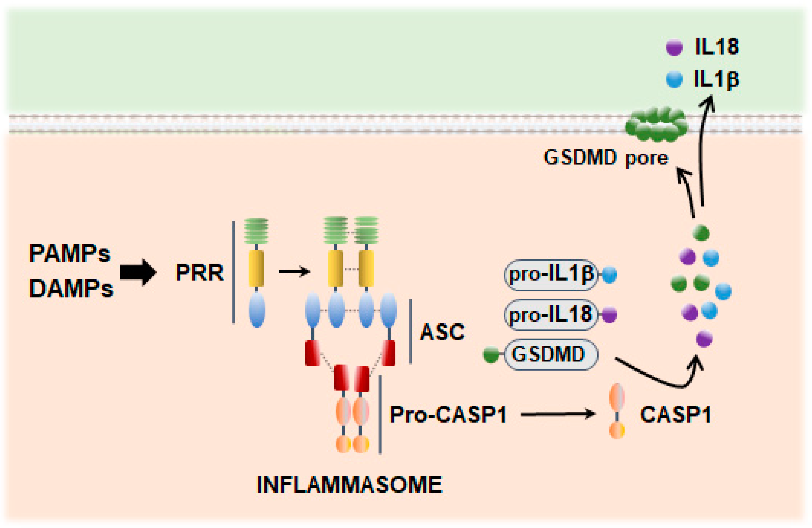
First described by Martinon et al. [82], inflammasomes are multiprotein complexes that act as cytosolic scaffolds to recruit and oligomerize the cysteine protease caspase 1 (CASP1). The function of inflammasomes is best characterized in myeloid lineages where CASP1 catalyzes the maturation of inflammatory cytokines, which are released into the extracellular space and promote both local and systemic immune responses (Figure 2). Alternatively, inflammasome activation can cleave the pore-forming protein gasdermin D (GSDMD), the executor of a lytic mode of cell death termed pyroptosis (Figure 2). Inflammasome proteins have been observed in the CNS, usually in monocytes and microglia responding to acute infection or cell loss, as occurs during neurodegeneration. Although less understood, activated inflammasome signaling has also been reported in neurons linked to pyroptosis [83], providing a mechanism through which neurons may initiate or influence inflammation.
Inflammasomes are constituted by three main components: a sensor consisting of a PRR with a pyrin domain (PYD) and/or a caspase recruitment domain (CARD), the adaptor apoptosis-associated speck-like protein containing a CARD (PYCARD, also known as ASC), and the zymogen of CASP1 (pro-CASP1) (Figure 2). CASP1 consists of an N-terminal CARD, a CARD domain linker, the p20 subunit containing the catalytic sides, an inter-domain linker (IDL), and the p10 subunit involved in dimerization. Mechanistically speaking, it has been proposed that PRR clustering promotes a nucleation-induced polymerization of pro-CASP1 into filaments, facilitating the protease zymogens to undergo proximity-induced autoactivation [84]. The autocatalytic cleavage of pro-CASP1 results in two subunits that form an active p10/p20 tetramer [85], which has been proposed as responsible for the processing of pro-IL-1β and pro-IL-18 [86] and GSDMD (Figure 2). Additionally, CASP1 may also induce NF-κB-mediated pathways [87,88]. Recently, using a biotin-labeled probe that covalently binds to active CASP1, Boucher et al. have questioned the catalytic role of the p20/p10 tetramer under physiological conditions [89]. The study demonstrates that CASP1 clustering on inflammasomes produces active p46 dimers that self-process at the IDL to generate a second active species (p33/p10), which remains associated with the inflammasome. The activity of p33/p10 is blocked by a second self-cleavage event, which removes the CARD domain, releasing a highly unstable dimer (p20/p10) that terminates CASP1 signaling. Notably, the full-length dimeric p46 can effectively process GSDMD but not IL-1β, while p33/p10 can cleave both GSDMD and IL-1β.
Pyroptotic death and cytokine secretion are often simultaneous outcomes of inflammasome activation. However, these events may be uncoupled under certain circumstances [90]. GSDMD sublytic pore formation or GSDMD-independent pathways involving unconventional secretion could result in predominant cytokine uncoupling, whereas regulation of CASP1 cleavage dynamics, which can generate active species with different activities in terms of GSDMD or IL-1β processing, could lead to pyroptosis uncoupling. Recently, in macrophages, the expression of the Toll-1L-1R protein SARM has been identified as a key regulatory player [91]. Nonetheless, the underlying mechanisms of uncoupling remain mostly elusive.

3.1. Inflammasome Scaffold Proteins

The NLR family members were the first sensor proteins identified in inflammasome formation (Figure 3). Recently, other families of inflammasome-associated PRRs have been described. These last include absent in melanoma 2 (AIM2)-like receptors (ALRs) that respond to self and pathogen double-stranded (ds)DNA molecules and pyrin receptors (also known as tripartite motif-containing (TRIM) 20), which assemble in response to perturbations of cytoplasmic homeostasis and cytoskeletal dynamics caused by infection (Figure 3). Nonetheless, the majority of inflammasomes rely on NLR proteins for pathogen sensing and scaffold assembly. All NLR sensors contain a central nucleotide-binding domain (NBD/NACHT) responsible for their self-oligomerization, a leucine-rich repeats (LRRs) domain crucial in the recognition of ligands (PAMPs and DAMPs), and signaling domains (PYR and CARD) that enable the recruitment of pro-CASP1 [92] (Figure 3). Major inflammasome-forming members are NLRP1, NLRP3, and NLRC4. Other atypical members of the family are NLRP2, NLRP6, NLRP7, NLRP12, and NLRC5, which, although less characterized, can also form an inflammasome complex [93]. Pro-CASP1 can directly interact with NLRC4 and mouse (m)NLRP1, through the CARD motifs, while in PYD-containing members, such as NLRP3 and human (h)NLRP1, the cysteine protease is recruited indirectly through homotypic interactions with the bipartite PYD/CARD adaptor ASC [94].
The NLRP3 is the most extensively studied inflammasome. Canonical activation of the NLRP3 inflammasome requires a priming step with the transcriptional upregulation of NLRP3, CASP1, and IL-1β/IL-18 pro-forms [95,96]. The activation of the NLRP3 inflammasome is thought to be tightly associated with the regulator never in mitosis A (NIMA)-related kinase 7 (NEK7) [97]. Additionally, recent research indicates that post-translational modifications of NLRP3, such as acetylation and phosphorylation, are necessary for full activation of the complex [95,98,99]. These priming mechanisms may serve as a checkpoint to prevent accidental activation of NLRP3, which is the sensor that responds to a wide spectrum of infection and stress-associated signals [100]. The exact molecular mechanism that triggers NLRP3 activation in response to such diverse signals is still under investigation. Muñoz-Planillo et al. proposed K+ efflux through purinergic receptor P2X 7 (P2RX7) channels as a universal requirement for NLRP3 activation [101], which is accompanied by the loss of lysosomal integrity [102]. This view has been challenged later by studies showing that NLRP3 activation can be triggered independently of K+ efflux by small-molecule TLR7 agonists that target mitochondrial complex I and produce mtROS [103].

3.2. Dysfunctional Mitochondria and Mitochondrial ROS as Drivers of Inflammasome Activation

NLRP3 inflammasome signaling has been consistently related to mitochondrial dysfunction and oxidative stress (Figure 4). Accordingly, mitophagy induction prevents inflammasome assembly [104], while mtROS serves as a priming signal [61]. On the other hand, the increase in mtROS promotes the binding of NEK7 with NLRP3 [97,103] and facilitates the formation of the complex in mitochondria-associated membranes (MAMs) of dysfunctional mitochondria [105]. This process is suppressed when mitophagy is stimulated or when VDAC2, an isoform of the mitochondrial carrier linked to mtROS regulation, is silenced [105,106,107]. Additionally, it has been described that Ca2+ mobilization from the endoplasmic reticulum (ER) enhances inflammasome activation by inflicting damage to mitochondria [108]. Furthermore, optimal NLRP3 inflammasome signaling has been shown to require the interaction between NLRP3 and thioredoxin-interacting protein (TXNIP), a nuclear protein that relocates to mitochondria during oxidative stress [109,110]. In cerebral ischemia-reperfusion injury, the NLRP3-TXNIP binding and subsequent inflammasome activation are prevented by NFE2-like bZIP transcription factor 2 (NFE2L2, also known as NRF2), a master regulator of the cellular antioxidant system, which upregulates the expression levels of thioredoxin 1 (TRX1), a binding partner of TXNIP [111]. On the other hand, ROS-mediated externalization of cardiolipin to the outer membrane has been reported to promote the binding between NLRP3 and CASP1, which assemble upon ASC recruitment to the mitochondrial surface [112,113]. Externalized cardiolipin can also bind inflammasome-activated GSDMD, a necessary event for pyroptosis to proceed [114]. GSDMD permeabilizes the mitochondrial membranes, facilitating mtROS generation and the release of mitochondrial proteins and DNA. In turn, as indicated previously, the NLRP3 inflammasome can be activated by ox-mtDNA released into the cytosol [64,65] and is downregulated by autophagy and mitochondrial integrity preservation [115]. Zhong et al. described that NLRP3 priming relies on the induction of new mtDNA synthesis, downstream of TLR4 engagement, which ultimately triggers an interferon regulatory factor 1 (IRF1)-dependent expression of the mitochondrial nucleoside cytidine/uridine monophosphate kinase 2 (CMPK2) [116] (Figure 4). The resulting increase in CMPK2 levels regulates mtDNA replication by supplying the required deoxyribonucleotides. Cytosolic mtDNA also enables AIM2 inflammasome assembly [117]. Notably, while the NLRP3 inflammasome shows a preferential response to oxidized DNA [65], AIM2-containing counterparts have been suggested to primarily recognize non-oxidized DNA [116].

3.3. GSDM-Mediated Inflammatory Cell Death: Pyroptosis

As part of the innate immune response, inflammasomes can also trigger a caspase-dependent cell death that prevents pathogen replication and activates phagocytic immune cells. First coined by Cookson and Brennan [118], this type of programmed necrosis called pyroptosis is featured by cell swelling and the appearance of protrusions on the plasma membrane before its rupture. Pyroptosis initiates after CASP1-mediated cleavage of GSDMD. The proteolytic processing of GSDMD generates a 31 kDa N-terminal domain, which aggregates to form 10–15 nm diameter pores in the cell membrane [119]. The pyroptotic pore enables the release of the proinflammatory cytokines matured by the inflammasome [120] and causes the loss of osmotic gradient, which ultimately leads to cell death, with or without cell lysis [121]. Remarkably, recent research has revealed that the pore-forming activity of GSDM is redox-sensitive [122], highlighting the crucial role of ROS in regulating inflammasome-mediated inflammatory response at multiple levels. The mtROS-mediated oxidative modification of specific cysteine residues promotes GSDMD cleavage by CASP1 [123]. Additionally, mitochondrial oxidative stress, induced by the mechanistic target of rapamycin kinase (MTOR) signaling pathway, has been shown to stimulate pore formation downstream of GSDM cleavage [124]. Likewise, it has been described that mtROS controls the palmitoylation of C191, which is crucial for GSDMD oligomerization [125]. On the other hand, cleaved GSDM can also permeabilize the mitochondrial membrane, resulting in increased mtROS and release of mtDNA, which, in turn, can trigger GSDM oligomerization [126,127].
Pyroptosis has been implicated in the inflammatory pathogenesis of multiple neurological diseases. Preclinical and clinical data provide evidence of GSDMD-mediated pyroptosis in age-related neurodegenerative diseases [54,83,128]. On the other hand, recent research using experimental autoimmune encephalomyelitis (EAE) models indicates that pyroptosis in peripheral myeloid cells may contribute to MS pathogenesis by providing essential signals to engage a T cell immune response [129]. Furthermore, GSDMD inhibitors have been shown to alleviate inflammation and disrupt EAE development [130]. The occurrence of GSDMD-mediated pyroptosis has also been documented in glial and brain microvascular endothelial cells across different ischemic brain injury models [131,132]. There is also evidence that pyroptosis-induced inflammation plays a role in secondary damage in CNS injuries, such as traumatic brain injury and spinal cord injury, resulting in prolonged neuroinflammation and functional decline. Studies indicate that inhibiting pyroptosis, particularly by targeting the NLRP3/CASP1 pathway, could provide neuroprotective effects in these conditions [133].

3.4. Inflammasome-Mediated Neuroinflammation in Alzheimer’s and Parkinson’s Disease

Genetic and pharmacologic evidence demonstrates the involvement of the NLR-dependent regulation of inflammation in the progression of different neurodegenerative diseases [134]. In 2008, Halle et al. described the NLRP3 inflammasome activation by fibrillar Aβ in primary microglia cultures [135], which was replicated later using soluble Aβ oligomers [136] and tau seeds [137]. Notably, in cultured cortical neurons, Aβ exposure causes an NLRP1-mediated pyroptosis [83]. Activated microglia can, in turn, release ASC specks that interact with extracellular Aβ, showing cross-seeding and inflammatory boosting activities [45,138]. Similarly, α-syn aggregates in microglia serve as a priming signal via TLR2 ligation and trigger NLRP3 inflammasome assembly associated with ROS production and impaired lysosomal function [139,140]. Conversely, blockage of the NLRP3-mediated signaling pathway restores microglial phagocytosis of the toxic aggregates [137,139].
Markers of inflammasome activation, such as cleaved CASP1 and GSDMD, have been found in brain and CSF samples from AD individuals and transgenic mice [54,128]. Likewise, increased expression of inflammasome-related proteins has been reported in the substantia nigra of patients with PD [52,141]. Inflammasome activation is also observed in the blood cells and correlates with disease progression [142], which has led to their consideration as potential early biomarkers of PD [143].
The involvement of the NLRP3 inflammasome in promoting chronic brain inflammation and neurodegeneration is further supported by preclinical studies, which show that NLRP3 ablation in APPswe/PSEN1dE1 mice induces a microglial shift toward an anti-inflammatory phenotype, associated with lower Aβ deposition and improvement in cognitive decline [54]. A similar reduction in Aβ pathology has been reported in Nlrp1 knock-out mice [83,144]. Along the same lines, pharmacological inhibition of NLRP3 [145] and GSDMD [146] attenuates pathological symptoms in PD mice. Consistently, mice lacking Nlrp3 or Casp1 are resistant to developing PD-like neurodegeneration evoked by neurotoxins that target mitochondria [147,148,149,150]. Moreover, the NLRP3-mediated inflammatory response is blunted when classical PD mitochondrial neurotoxicants are combined with mitochondria-targeted antioxidants [151]. Strong evidence for the role of mitochondria in regulating neuroinflammation is further provided by our recent study showing a differential inflammatory response in microglia and neuronal cells that depends on their mitochondrial antioxidant capacity [152]. In early works, using APPswe/PSEN1dE1 mice overexpressing the sterol regulatory element-binding transcription factor 2 (SREBF2), we demonstrated that elevated brain cholesterol levels accelerate and worsen the pathological features of Alzheimer’s disease, including neuroinflammation [153]. We next have shown that intracellular cholesterol enrichment in microglia triggers a neuroprotective profile mediated by the NLRP3 inflammasome assembly [152]. After the bacterial toxins challenge, cholesterol-enriched microglia display an enhanced release of neurotrophic factors such as nerve growth factor (NGF) and brain-derived neurotrophic factor (BDNF), higher phagocytic capacity, and increased release of encapsulated Il-1β. Moreover, conditioned media from these cells significantly blunt the loss of viability of neuronal cells exposed to Aβ. In contrast, in neuronal cells, increased cholesterol levels compromise the mitochondrial antioxidant defense [152]. In these cells, the exacerbated oxidative stress after Aβ exposure leads to pyroptosis. Pyroptotic neurons, in turn, can inhibit microglia phagocytosis, potentially establishing a proinflammatory vicious cycle.

4. Role of Immunogenic Cell Death as a Contributor to Neurodegeneration

There is a close relationship between inflammation and cell death. When necrotic cells lose their cell membrane integrity, they release DAMPs that can be recognized by different cellular receptors, leading to a sterile inflammatory response. In recent decades, different types of regulated necrosis-like death have been described [154]. Understanding the mechanisms that regulate this proinflammatory cell death in neurodegeneration has become a field of intense study. As mentioned earlier, inflammasome induction can trigger pyroptosis, a process regulated by mitochondrial oxidative stress. In the following sections, we will review recent data showing how dysfunctional mitochondria can control different types of immunogenic cell death relevant to neurodegeneration, such as necroptosis and metal ion-induced cell death.

4.1. Necroptosis

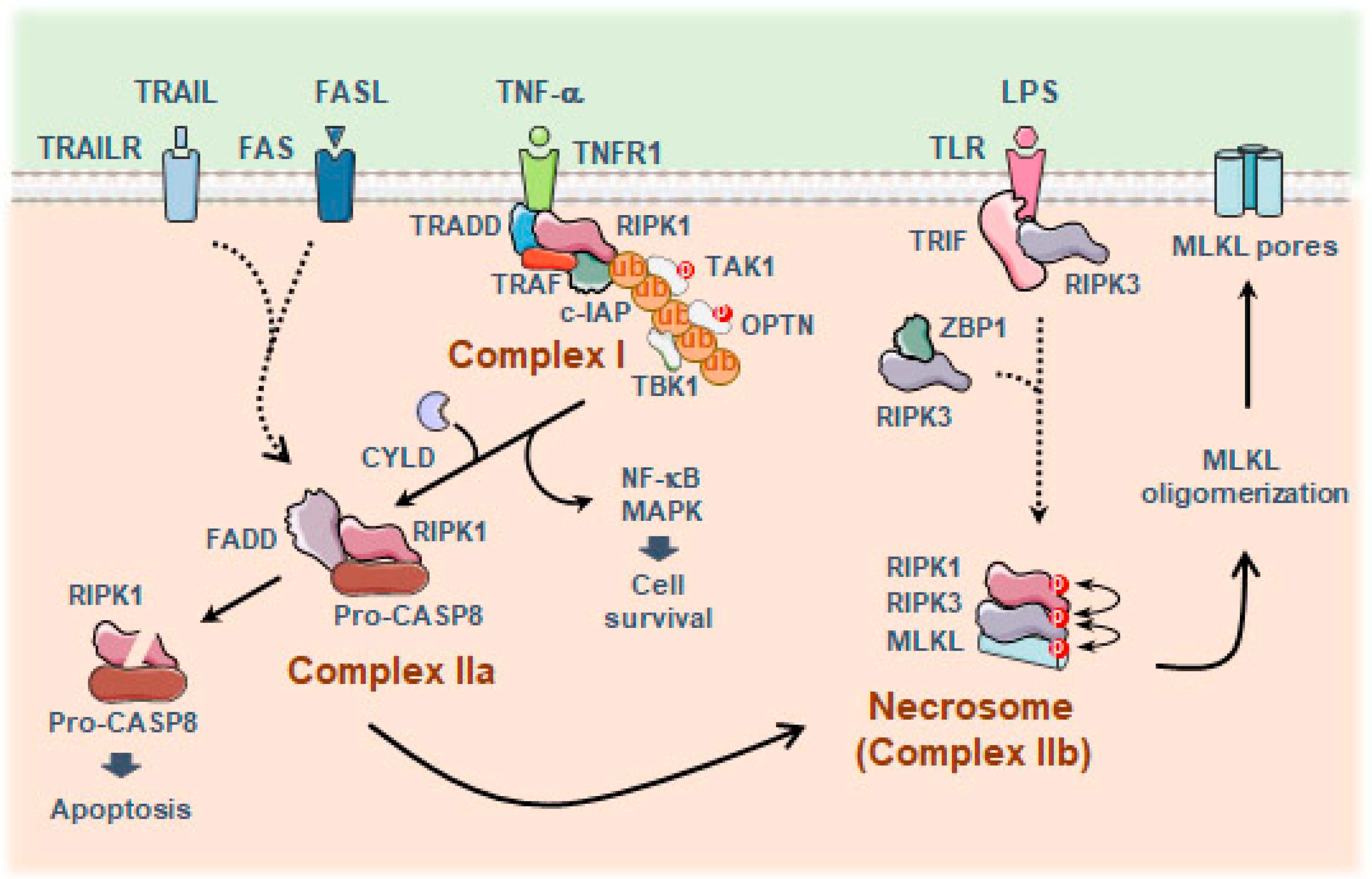
Necroptosis is a form of programmed necrosis mediated by the combined action of the receptor-interacting protein kinase 3 (RIPK3) and the pseudo-kinase mixed lineage kinase domain-like (MLKL) protein, with the participation of RIPK1 when death receptors are engaged [155]. The necroptotic molecular pathway has been most thoroughly studied in the context of the tumor necrosis factor receptor 1 (TNFR1) complex [156], although other members of the death receptor family, such as CD95/Fas and TRAILR, have also been implicated [157] (Figure 5).
Activation of RIPK3 has also been described independently of RIPK1, by binding to TIR-domain-containing adapter-inducing interferon-β (TRIF), a downstream mediator of TLRs [158], or the cytoplasmic viral DNA sensor Z-DNA binding protein 1 (ZBP1), which allows a rapid necrotic response upon viral infection [159] (Figure 5). Remarkably, activated RIPK3 and MLKL in immune cells have been shown to induce an inflammatory response independently of cell death by triggering the assembly of the NLRP3 inflammasome [160,161,162,163]. Subsequently, CASP8 has been indicated to directly cleave GSDMD and cause pyroptosis [164], thus further reinforcing the concept that different modes of inflammatory cell death display a considerable degree of interconnectivity.
Like other forms of programmed cell death, necroptosis is tightly regulated, with transcriptional changes, post-translational modifications, and elimination of necrosome members [165]. The autophagy machinery has been proposed as a molecular platform for necrosome assembly [166]. Additionally, autophagy controls the availability of RIPK1 and RIPK3 [167,168], and robust RIPK- and ROS-dependent necroptosis is observed after autophagy flux is chemically inhibited or when the lysosomal capacity is compromised [167,169,170]. Likewise, the ubiquitin E3 ligase Parkin, whose loss-of-function mutations are linked to autosomal PD, was recently identified as a negative regulator of necroptosis by leading to K33-linked polyubiquitination of RIPK3 and its proteasomal degradation [171]. Additionally, cellular extrusion of phospho-MLKL within extracellular vesicles has been proposed as a mechanism to downregulate MLKL activity and prevent necroptosis [172].
The occurrence of necroptosis has also been linked to mitochondrial function. Despite initial studies showing that mitochondrial depletion by mitophagy does not affect the kinetics of necroptosis [173], further works indicate that necrosome formation requires the production of mtROS [174,175]. Zhang et al. have identified three cysteine residues (C257, C268, and C586) in RIPK1 that can sense mtROS during necrosome execution [176]. These oxidative modifications activate RIPK1 autophosphorylation on S161, enabling RIPK1 to recruit RIPK3 and form a functional complex. Remarkably, at least in TNF-induced necroptosis, mitochondrial oxidative stress is exacerbated by RIPK3-dependent induction of the pyruvate dehydrogenase complex, resulting in enhanced aerobic respiration and high mtROS as a byproduct [177]. MLKL activity is also subjected to redox control by the thiol oxidoreductase thioredoxin-1, which maintains the monomeric MLKL in a reduced inactive state, thereby preventing disulfide bond-dependent polymer formation [178]. On the other hand, recent reports have confirmed the key role of mtROS in triggering necroptosis in experimental acute pancreatitis by a mechanism that requires p53-induced downregulation of the mitochondrial antioxidant enzymes sulfiredoxin and peroxiredoxin III [179]. In contrast, the mitochondrial membrane protein phosphoglycerate mutase family member 5 (PGAM5), whose deficiency impairs PINK1-mediated mitophagy and leads to a PD-like phenotype [180], has been reported to downregulate necroptosis by promoting mitophagy and reducing mtROS [181]. Interestingly, inflammasome activation, normally associated with GASDMD-dependent pyroptotic death, has been shown to shift toward necroptotic cell death in leucine-rich repeat kinase 2 (Lrrk2) G2019S mutant macrophages [182]. The presence of the disease-associated gain-of-function allele Lrrk2G2019S (a common genetic cause of PD) impairs mitochondrial function, resulting in high mtROS, which directs GSDMD to the mitochondria and activates the necroptotic pathway [182].

Necroptosis and Aging-Related Neurodegenerative Diseases

Emerging evidence underscores the significance of necroptosis in neurodegenerative processes [183]. In cell cultures and genetically engineered mouse models that recapitulate AD and PD, treatment with necrostatin-1, an inhibitor of RIPK1, has shown promise in reducing neuronal loss and improving cognitive functions [184,185,186,187]. Similar results have been obtained in subsequent studies inhibiting MLKL activity [188,189]. This aligns with observations of active necrosomes in the substantia nigra pars compacta of PD patients and in postmortem AD brains [187,190,191]. In AD, necrosome components tend to accumulate in granulovacuolar degeneration bodies [192], which are linked to tau-associated lysosomal disturbances [193]. Salvadores et al. described that neuronal necroptosis induction is triggered by Aβ-stimulated microglia [194]. Recently, using a human–mouse chimeric AD model, Balusu et al. found that human neuronal progenitor cells, when differentiated and transplanted into an Aβ-producing mouse, develop tau pathology and undergo necroptosis [195]. Their findings suggest a human-specific response to Aβ plaques, linked to the upregulation of the long non-coding RNA maternally expressed gene 3 (MEG3), which ultimately activates the necroptotic pathway. Hyperphosphorylated tau and Aβ oligomers have both been described to facilitate the formation of the necrosome complex [196,197,198], with the participation of mitochondria and the production of mtROS [199]. Moreover, activation of the necroptotic pathway has been consistently shown in preclinical models of PD, using toxins such as 1-methyl 4-phenylpyridinium (MPP+) and 6-hydroxydopamine (6-OHDA) that interfere with the activity of mitochondrial complex I of the respiratory chain [184,185,190], which further supports a key role of mitochondria in this process and underscores the potential of targeting mitochondrial pathways as a therapeutic strategy to mitigate necroptosis and neuroinflammation in neurodegeneration.

4.2. Metal Ion-Induced Cell Death: Ferroptosis and Cuproptosis

Transition metal ions, including iron (Fe), copper (Cu), zinc (Zn), and manganese (Mn), are involved in many biochemical processes essential for cell survival, and therefore, their levels are tightly regulated. However, under some conditions, excess redox-active essential metals such as Fe and Cu can induce hydroxy radical (·OH) via Fenton or Fenton-like reactions, causing cell damage and inflammation.

4.2.1. Ferroptosis

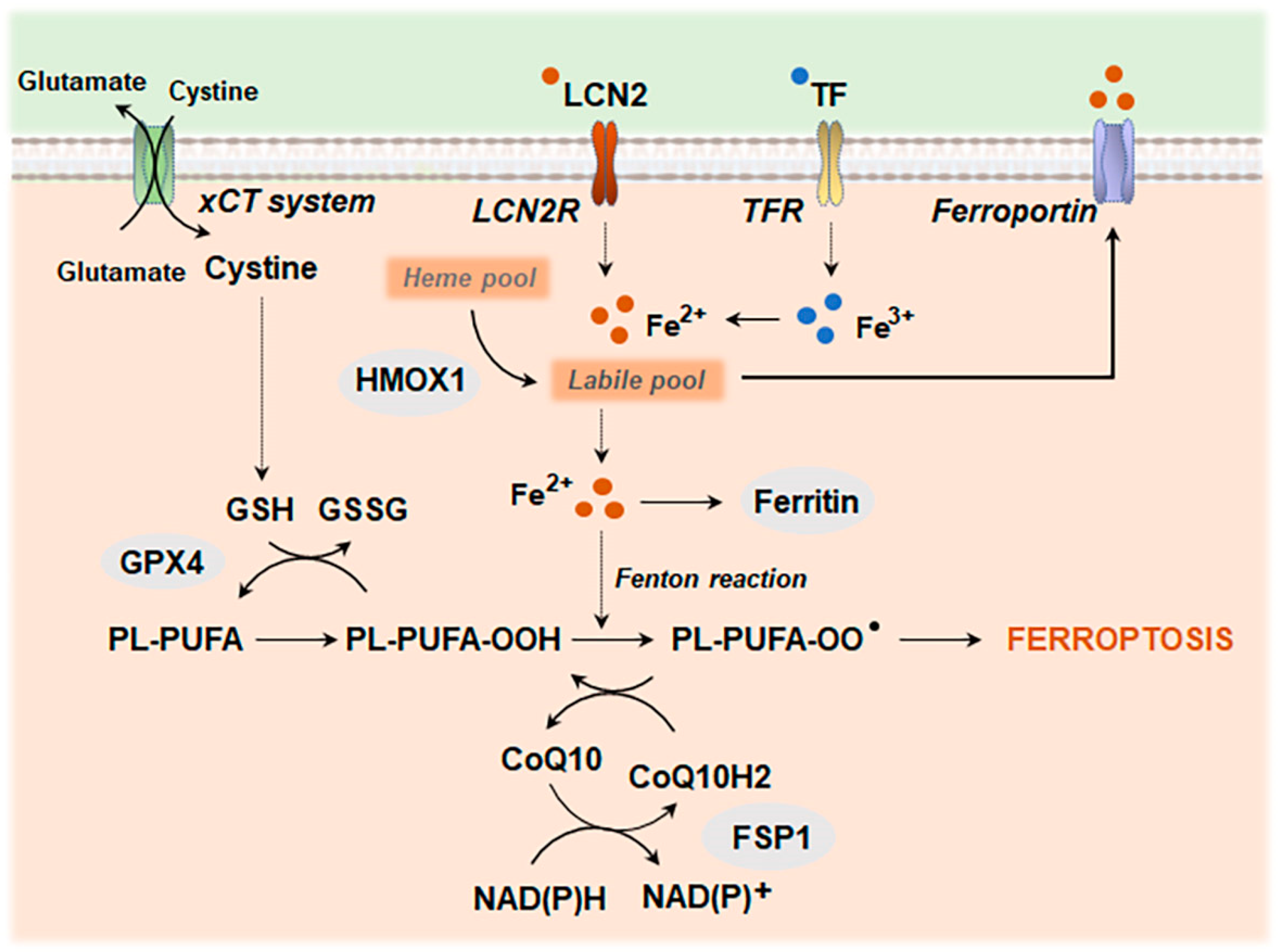
Dysregulation of iron metabolism and redox systems leads to the accumulation of intracellular lipid hydroperoxides, ultimately causing ferroptosis (Figure 6). Lipid peroxidation may kill cells directly by affecting cellular membrane integrity or indirectly through the generation of aldehydes, such as 4-hydroxynonenal (4-HNE) and malondialdehyde (MDA), which form adducts with different biomolecules and can act as a signal to drive cell death [200,201]. Mitochondria play a crucial role in the regulatory network of ferroptosis. Dysfunctional mitochondria-related alterations, including mitochondrial membrane depolarization, mtROS overproduction, and mtDNA release, can favor this type of cell death [202,203]. In particular, recent studies point to mtROS as an essential step in the activation of the ferroptotic pathway when the cystine–glutamate antiporter solute carrier family 7 member 1 (SLC7A11, also known as the xCT system) that supplies cystine for glutathione (GSH) synthesis or the glutathione peroxidase 4 (GPX4) are inhibited [202]. Concordantly, the inhibition of NRF2, which controls the expression levels of an array of antioxidant response element (ARE)-dependent genes, enhances the pathway, while mtROS scavengers prevent ferroptosis [202,203,204].
Ferroptosis has been described in many inflammation-related diseases, closely connected with the activation of NF-κB-driven inflammatory signaling pathways [205,206]. In a retinal degeneration mouse model, increased lipid peroxidation and ferroptosis have also been associated with a cGAS–STING-mediated inflammasome activation due to the autophagy and lysosomal dysfunction induced by iron homeostasis deregulation [207]. Likewise, NLRP3 inflammasome induction has been reported in a rat model of pulmonary hypertension after high mobility group box 1 (HMGB1) was released from ferroptotic cells, a critical DAMP that binds to TLR4 [208]. Both the lipophilic radical scavenger ferrostatin-1 and specific GPX4 activators can downregulate NF-κB signaling and suppress inflammatory conditions [209,210,211]. Furthermore, GPX4 and its ability to reduce lipid peroxides have been shown to downregulate GSDMD-mediated pyroptosis [212,213].
Iron overload is a pathological feature shared by multiple neurodegenerative diseases, including AD and PD [214,215,216]. Although the precise mechanism of iron-driven toxicity is unclear, there is evidence for the activation of the ferroptotic pathway. Increased endogenous levels of α-syn have been reported to sensitize dopamine neurons to ferroptosis [217]. A similar susceptibility to ferroptosis is displayed by cells carrying AD causal mutations of presenilins (PSEN) [218]. Astrocytes from AD patients and APPswe/PSEN1dE1 mice display elevated levels of aldehydes associated with high levels of NADPH oxidase 4 (NOX4) [219]. Upregulation of NOX4 in human astrocyte cultures promotes ferroptosis by affecting mitochondrial metabolism [219]. The role of mitochondria is further supported by different studies showing that strategies aimed at preserving mitochondrial function are effective against neuronal ferroptosis [220,221]. Interestingly, a recent work by Ryan et al. shows that iron overload causes an early shift in the microglial transcriptional state that overlaps with the transcriptomics displayed by microglia from postmortem PD brains [222]. The study further identifies SEC24 homolog B, COPII coat complex component (SEC24B), a protein involved in cargo packaging and intracellular trafficking, as the regulator of microglial ferroptosis and points to microglia as the main cause of ferroptosis-driven neurodegeneration [222]. The induction of ferroptosis as a triggering factor for neurodegeneration is gaining increasing interest and is beginning to be recognized as a bona fide target for future development of treatment and prevention strategies [223].

4.2.2. Cuproptosis

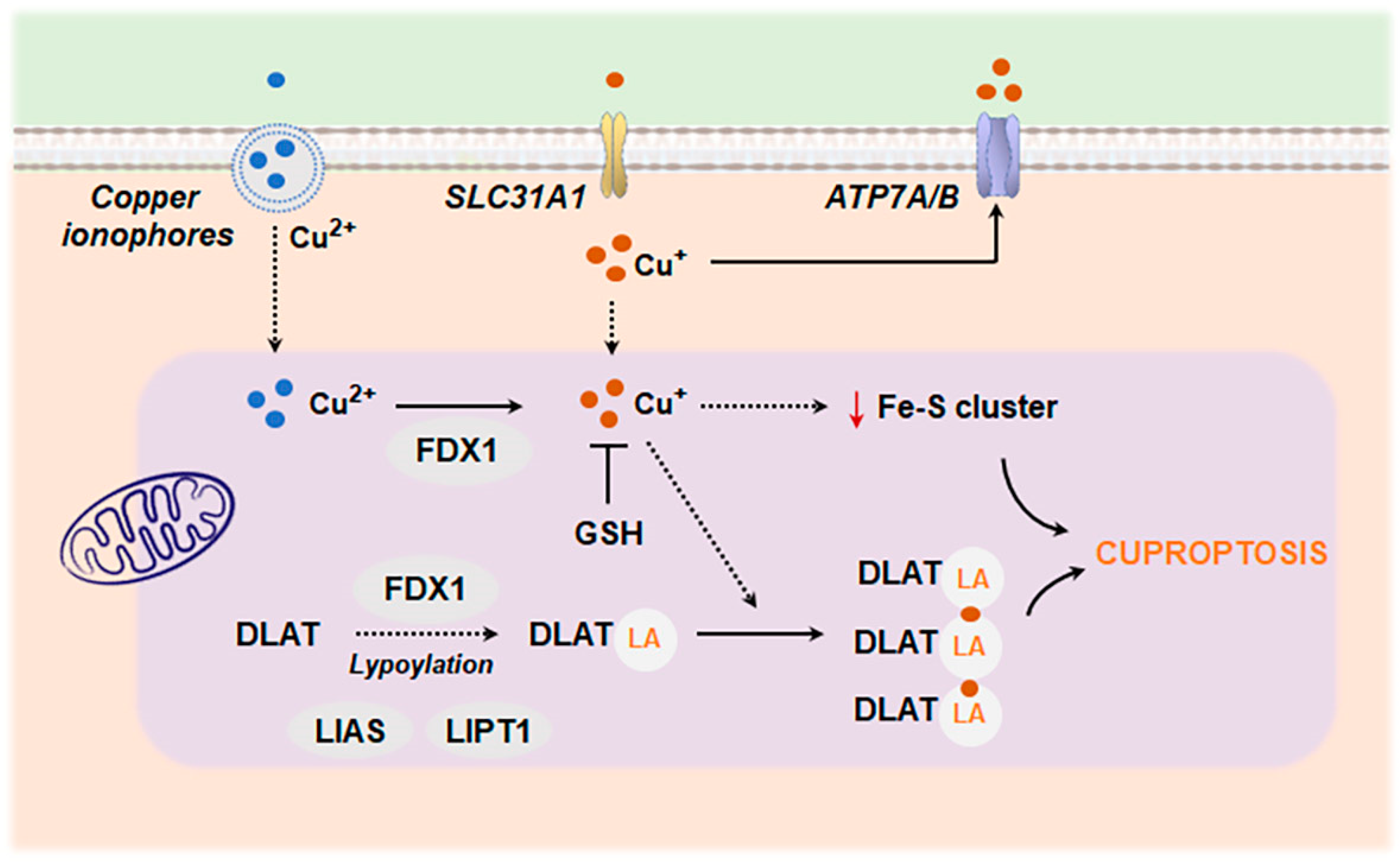
Copper is an important cofactor of several mitochondrial respiration complexes. Therefore, cells heavily dependent on mitochondrial respiration are especially vulnerable to mitochondrial copper accumulation. In physiological conditions, copper homeostasis is tightly regulated by chaperones (ATOX1), storage proteins (CuL), export mechanisms (ATP7A/B), and mitochondrial antioxidant defense or mitophagy [224]. Deregulation of these mechanisms can cause copper accumulation in mitochondria, which ultimately triggers a type of cell death known as cuproptosis. This type of cell death is regulated by ferredoxin 1 (FDX1), which reduces cupric (Cu2+) to cuprous (Cu+) ions. Cu+ binds to mitochondrial lipoylated proteins that participate in the tricarboxylic acid (TCA) cycle, specifically dihydrolipoyl transacetylase (DLAT), an enzyme involved in the formation of the pyruvate dehydrogenase (PDH) complex. The lipoyl moiety enables direct copper binding to these proteins, favoring their aggregation and loss of function. Also, Cu+ disrupts the synthesis of iron–sulfur (Fe-S) clusters, redox-active cofactors in the ETC [225] (Figure 7).
Cuproptosis has been related to cognitive impairment in mice exposed to copper overload [226]. Additionally, using integrated bioinformatics analyses and machine learning algorithms, recent studies have identified dysregulated cuproptosis-related genes (CRGs) linked to specific immune infiltration patterns in the substantia nigra of PD patients, suggesting that CRGs participate in PD progression through regulating immune cell infiltration and activity [227,228,229]. Similarly, two cuproptosis-related molecular clusters were defined in AD [230]. Functional analysis showed that cluster-specific and differentially expressed genes were closely related to various immune responses [230]. Aberrant intracellular copper accumulation may also modulate the microglial inflammatory response. Microglia cells exposed to Cu display an increased presence of inflammatory mediators such as NF-kB, fueled by elevated levels of mtROS [231,232,233]. Furthermore, it has been shown that the redox activity of Cu is necessary to properly activate the NLRP3 inflammasome [233,234]. In AD, mitochondrial oxidative stress elicited by chronic Cu overload exacerbates the impact of Aβ on microglial activation [235,236], promoting a shift toward degenerative gene expression signatures [237].
In recent years, several metal-chelating agents have been proposed as a potential therapeutic approach to restore metal ion balance in the brain and treat neurodegenerative diseases [238]. Prominent among them is a new generation of weaker chelating agents, known as metal protein-attenuating compounds (MPACs), which downregulate abnormal metal ion levels without causing damage to other essential metal-dependent processes. However, further research is still needed to improve their ability to cross the blood–brain barrier (BBB), understand their mode of action, and enhance their specificity, among other aspects.

5. Concluding Remarks and Future Perspectives

Collectively, there is compelling evidence that links an aberrant inflammatory response with the onset and progression of neurodegeneration. Immune system changes are particularly relevant in age-related neurodegenerative diseases where other alterations associated with aging, such as defective proteostasis and mitochondrial dysfunction, converge and may fuel neuroinflammation.
Preclinical studies have demonstrated the benefits of blocking NLRP3 inflammasome signaling. Inflammasome inhibitors reduce misfolded protein aggregates associated with neurodegenerative diseases and alleviate cognitive decline, suggesting that targeting the NLRP3 inflammasome could represent a novel therapeutic approach for neurodegenerative disorders. Various strategies to block this complex are under investigation, ranging from RNA-targeted therapies, nanobodies, and novel small molecule inhibitors [239]. However, a challenge for the field is the ability to translate findings from cellular and animal models of neurodegeneration into effective drug development for clinical use. Our recent studies have emphasized the importance of neuro–glia crosstalk in regulating inflammasome-mediated immune response [152], which should also be taken into consideration. Additionally, it is crucial to note that inflammasomes are necessary for a proper innate immune defense system. Therefore, finding a way to reduce NLRP3 activity without harming the immune system presents a significant challenge.
Beyond NLRP3 inhibitors, new therapeutic strategies focused on microglial modulation or cGAS–STING inhibitors are emerging to treat neurodegenerative diseases [240,241]. Most microglial interventions aim to shift their phenotype from proinflammatory or cytotoxic to more protective profiles, enhancing their phagocytic capacity, modulating their metabolism, or reducing the synthesis and secretion of exosomes and proinflammatory cytokines [240]. Recent results from preclinical and phase I clinical trials using AL002c, a TREM2 agonist antibody, have demonstrated good brain penetrance and tolerability, and its efficacy is currently being tested in early AD [242]. On the other hand, the type I IFN response can be ameliorated by using inhibitors of cGAS, STING, or TBK1 [243]. Although there are currently no clinical trials of cGAS–STING inhibitors in neurological diseases, a recent phase I trial with a cGAS antagonist (VENT-03) is ongoing, with the further aim of testing it in autoimmune diseases like lupus [244]. This new clinical approach, along with its extended use in preclinical studies in neurological disorders [241], positions modulators of the type I IFN pathway as potential therapeutic targets for controlling neuroinflammation.
One plausible therapeutic intervention could involve antioxidant supplementation, given the prominent role of oxidative stress in regulating neuroinflammation at different levels. In this sense, many efforts have been focused on exploring the properties of natural and synthetic antioxidant compounds. However, despite their potential, the clinical use of antioxidants is still limited due to issues such as low bioavailability, easy degradation, and poor solubility in water. To address these problems, nanomedicine has been proposed as an innovative approach to deliver antioxidants to the brain [245]. Nanoparticles can encapsulate the drug, protecting it from degradation and increasing its bioavailability and pharmacokinetic properties. In addition, nanoparticles can be functionalized with specific ligands to target particular organelles, such as mitochondria [246,247,248]. The rapid advancement of nanomedicine offers the possibility of designing tailored nanocarriers for targeted antioxidant delivery to the brain, paving the way for innovative therapeutic solutions.

Funding

This research was funded by MCIN/AEI/10.13039/501100011033, co-funded by NextGenera-tionEU/PRTR, and by “ERDF A way of making Europe”, grants PID2022-143279OB-100 (A.C.), PID2021-123564OB-100 (A.M.), PDC2023-145806-100 (A.M and M.M.), PID2020-115091RB-I00 (R.T.), and RED2022-134786-T (A.C. and R.T.); Instituto de Salud Carlos III (ISCIII) and European Union NextGenerationEU/PRTR, grants PMP22/00100 (R.T. and A.C.) and PI22/00475 (M.M.); Fundació la Marató de TV3 (A.M.); AGAUR, grant 2021-SGR00490 (A.C.); and Junta de Andalucia (Consejería de Economia y Conocimiento) co-funded by FEDER 214-2020 and by the CIBERNED (Convocatoria 2022 Proyectos Colaborativos 2020/21), grants US-1262734 (J.V.) and P20-00843 (J.V.). V.R.-A. has a Juan de la Cierva postdoctoral contract (JDC2022-048952-I) funded by MCIN/AEI/10.13039/501100011033 and by NextGenerationEU/PRTR. C.D. has a contract (2022 INV-1 00065) funded by “Programa Investigo” MCIN/PRTR.

Institutional Review Board Statement

Not applicable.

Informed Consent Statement

Not applicable.

Data Availability Statement

Not applicable.

Acknowledgments

Figures were partly generated using Servier Medical Art, provided by Servier, licensed under a Creative Commons Attribution 3.0 unported license.

Conflicts of Interest

The authors declare no conflicts of interest.

References

  1. Greenhalgh, A.D.; David, S.; Bennett, F.C. Immune Cell Regulation of Glia during CNS Injury and Disease. Nat. Rev. Neurosci. 2020, 21, 139–152. [Google Scholar] [CrossRef] [PubMed]
  2. Prinz, M.; Priller, J. The Role of Peripheral Immune Cells in the CNS in Steady State and Disease. Nat. Neurosci. 2017, 20, 136–144. [Google Scholar] [CrossRef] [PubMed]
  3. Greenhalgh, A.D.; Zarruk, J.G.; Healy, L.M.; Baskar Jesudasan, S.J.; Jhelum, P.; Salmon, C.K.; Formanek, A.; Russo, M.V.; Antel, J.P.; McGavern, D.B.; et al. Peripherally Derived Macrophages Modulate Microglial Function to Reduce Inflammation after CNS Injury. PLoS Biol. 2018, 16, e2005264. [Google Scholar] [CrossRef] [PubMed]
  4. Netzahualcoyotzi, C.; Santillán-Cigales, J.J.; Adalid-Peralta, L.V.; Velasco, I. Infiltration of Immune Cells to the Brain and Its Relation to the Pathogenesis of Alzheimer’s and Parkinson’s Diseases. J. Neurochem. 2024, 168, 2316–2334. [Google Scholar] [CrossRef]
  5. Zhang, X.; Wang, R.; Chen, H.; Jin, C.; Jin, Z.; Lu, J.; Xu, L.; Lu, Y.; Zhang, J.; Shi, L. Aged Microglia Promote Peripheral T Cell Infiltration by Reprogramming the Microenvironment of Neurogenic Niches. Immun. Ageing 2022, 19, 34. [Google Scholar] [CrossRef]
  6. Montagne, A.; Barnes, S.R.; Sweeney, M.D.; Halliday, M.R.; Sagare, A.P.; Zhao, Z.; Toga, A.W.; Jacobs, R.E.; Liu, C.Y.; Amezcua, L.; et al. Blood-Brain Barrier Breakdown in the Aging Human Hippocampus. Neuron 2015, 85, 296–302. [Google Scholar] [CrossRef]
  7. Wyss-Coray, T. Ageing, Neurodegeneration and Brain Rejuvenation. Nature 2016, 539, 180–186. [Google Scholar] [CrossRef]
  8. López-Otín, C.; Blasco, M.A.; Partridge, L.; Serrano, M.; Kroemer, G. The Hallmarks of Aging. Cell 2013, 153, 1194–1217. [Google Scholar] [CrossRef]
  9. Bourdenx, M.; Koulakiotis, N.S.; Sanoudou, D.; Bezard, E.; Dehay, B.; Tsarbopoulos, A. Protein Aggregation and Neurodegeneration in Prototypical Neurodegenerative Diseases: Examples of Amyloidopathies, Tauopathies and Synucleinopathies. Prog. Neurobiol. 2017, 155, 171–193. [Google Scholar] [CrossRef]
  10. Efthymiou, A.G.; Goate, A.M. Late Onset Alzheimer’s Disease Genetics Implicates Microglial Pathways in Disease Risk. Mol. Neurodegener. 2017, 12, 43. [Google Scholar] [CrossRef]
  11. Bellenguez, C.; Küçükali, F.; Jansen, I.E.; Kleineidam, L.; Moreno-Grau, S.; Amin, N.; Naj, A.C.; Campos-Martin, R.; Grenier-Boley, B.; Andrade, V.; et al. New Insights into the Genetic Etiology of Alzheimer’s Disease and Related Dementias. Nat. Genet. 2022, 54, 412–436. [Google Scholar] [CrossRef] [PubMed]
  12. Gorlé, N.; Vandenbroucke, R.E. Interferons: A Molecular Switch between Damage and Repair in Ageing and Alzheimer’s Disease. Mech. Ageing Dev. 2019, 183, 111148. [Google Scholar] [CrossRef] [PubMed]
  13. Marsh, S.E.; Abud, E.M.; Lakatos, A.; Karimzadeh, A.; Yeung, S.T.; Davtyan, H.; Fote, G.M.; Lau, L.; Weinger, J.G.; Lane, T.E.; et al. The Adaptive Immune System Restrains Alzheimer’s Disease Pathogenesis by Modulating Microglial Function. Proc. Natl. Acad. Sci. USA 2016, 113, E1316–E1325. [Google Scholar] [CrossRef] [PubMed]
  14. Cao, S.; Theodore, S.; Standaert, D.G. Fcγ Receptors Are Required for NF-κB Signaling, Microglial Activation and Dopaminergic Neurodegeneration in an AAV-Synuclein Mouse Model of Parkinson’s Disease. Mol. Neurodegener. 2010, 5, 42. [Google Scholar] [CrossRef] [PubMed]
  15. Teeling, J.L.; Carare, R.O.; Glennie, M.J.; Perry, V.H. Intracerebral Immune Complex Formation Induces Inflammation in the Brain That Depends on Fc Receptor Interaction. Acta Neuropathol. 2012, 124, 479–490. [Google Scholar] [CrossRef]
  16. Kim, K.; Wang, X.; Ragonnaud, E.; Bodogai, M.; Illouz, T.; DeLuca, M.; McDevitt, R.A.; Gusev, F.; Okun, E.; Rogaev, E.; et al. Therapeutic B-Cell Depletion Reverses Progression of Alzheimer’s Disease. Nat. Commun. 2021, 12, 2185. [Google Scholar] [CrossRef]
  17. Batchu, S. Prefrontal Cortex Transcriptomic Deconvolution Implicates Monocyte Infiltration in Parkinson’s Disease. Neurodegener. Dis. 2020, 20, 110–112. [Google Scholar] [CrossRef]
  18. Muñoz-Castro, C.; Mejias-Ortega, M.; Sanchez-Mejias, E.; Navarro, V.; Trujillo-Estrada, L.; Jimenez, S.; Garcia-Leon, J.A.; Fernandez-Valenzuela, J.J.; Sanchez-Mico, M.V.; Romero-Molina, C.; et al. Monocyte-Derived Cells Invade Brain Parenchyma and Amyloid Plaques in Human Alzheimer’s Disease Hippocampus. Acta Neuropathol. Commun. 2023, 11, 31. [Google Scholar] [CrossRef]
  19. Koronyo, Y.; Salumbides, B.C.; Sheyn, J.; Pelissier, L.; Li, S.; Ljubimov, V.; Moyseyev, M.; Daley, D.; Fuchs, D.-T.; Pham, M.; et al. Therapeutic Effects of Glatiramer Acetate and Grafted CD115+ Monocytes in a Mouse Model of Alzheimer’s Disease. Brain 2015, 138, 2399–2422. [Google Scholar] [CrossRef]
  20. Harms, A.S.; Thome, A.D.; Yan, Z.; Schonhoff, A.M.; Williams, G.P.; Li, X.; Liu, Y.; Qin, H.; Benveniste, E.N.; Standaert, D.G. Peripheral Monocyte Entry Is Required for Alpha-Synuclein Induced Inflammation and Neurodegeneration in a Model of Parkinson Disease. Exp. Neurol. 2018, 300, 179–187. [Google Scholar] [CrossRef]
  21. Subbarayan, M.S.; Hudson, C.; Moss, L.D.; Nash, K.R.; Bickford, P.C. T Cell Infiltration and Upregulation of MHCII in Microglia Leads to Accelerated Neuronal Loss in an α-Synuclein Rat Model of Parkinson’s Disease. J. Neuroinflammation 2020, 17, 242. [Google Scholar] [CrossRef] [PubMed]
  22. Laurent, C.; Dorothée, G.; Hunot, S.; Martin, E.; Monnet, Y.; Duchamp, M.; Dong, Y.; Légeron, F.-P.; Leboucher, A.; Burnouf, S.; et al. Hippocampal T Cell Infiltration Promotes Neuroinflammation and Cognitive Decline in a Mouse Model of Tauopathy. Brain 2017, 140, 184–200. [Google Scholar] [CrossRef] [PubMed]
  23. Zenaro, E.; Pietronigro, E.; Della Bianca, V.; Piacentino, G.; Marongiu, L.; Budui, S.; Turano, E.; Rossi, B.; Angiari, S.; Dusi, S.; et al. Neutrophils Promote Alzheimer’s Disease-like Pathology and Cognitive Decline via LFA-1 Integrin. Nat. Med. 2015, 21, 880–886. [Google Scholar] [CrossRef] [PubMed]
  24. Chen, X.; Firulyova, M.; Manis, M.; Herz, J.; Smirnov, I.; Aladyeva, E.; Wang, C.; Bao, X.; Finn, M.B.; Hu, H.; et al. Microglia-Mediated T Cell Infiltration Drives Neurodegeneration in Tauopathy. Nature 2023, 615, 668–677. [Google Scholar] [CrossRef]
  25. Lindestam Arlehamn, C.S.; Dhanwani, R.; Pham, J.; Kuan, R.; Frazier, A.; Rezende Dutra, J.; Phillips, E.; Mallal, S.; Roederer, M.; Marder, K.S.; et al. α-Synuclein-Specific T Cell Reactivity Is Associated with Preclinical and Early Parkinson’s Disease. Nat. Commun. 2020, 11, 1875. [Google Scholar] [CrossRef]
  26. Gate, D.; Tapp, E.; Leventhal, O.; Shahid, M.; Nonninger, T.J.; Yang, A.C.; Strempfl, K.; Unger, M.S.; Fehlmann, T.; Oh, H.; et al. CD4+ T Cells Contribute to Neurodegeneration in Lewy Body Dementia. Science 2021, 374, 868–874. [Google Scholar] [CrossRef]
  27. Gate, D.; Saligrama, N.; Leventhal, O.; Yang, A.C.; Unger, M.S.; Middeldorp, J.; Chen, K.; Lehallier, B.; Channappa, D.; De Los Santos, M.B.; et al. Clonally Expanded CD8 T Cells Patrol the Cerebrospinal Fluid in Alzheimer’s Disease. Nature 2020, 577, 399–404. [Google Scholar] [CrossRef]
  28. Piehl, N.; van Olst, L.; Ramakrishnan, A.; Teregulova, V.; Simonton, B.; Zhang, Z.; Tapp, E.; Channappa, D.; Oh, H.; Losada, P.M.; et al. Cerebrospinal Fluid Immune Dysregulation During Healthy Brain Aging and Cognitive Impairment. Cell 2022, 185, 5028–5039.e13. [Google Scholar] [CrossRef]
  29. Tan, Y.-L.; Yuan, Y.; Tian, L. Microglial Regional Heterogeneity and Its Role in the Brain. Mol. Psychiatry 2020, 25, 351–367. [Google Scholar] [CrossRef]
  30. Paolicelli, R.C.; Bolasco, G.; Pagani, F.; Maggi, L.; Scianni, M.; Panzanelli, P.; Giustetto, M.; Ferreira, T.A.; Guiducci, E.; Dumas, L.; et al. Synaptic Pruning by Microglia Is Necessary for Normal Brain Development. Science 2011, 333, 1456–1458. [Google Scholar] [CrossRef]
  31. Parkhurst, C.N.; Yang, G.; Ninan, I.; Savas, J.N.; Yates, J.R.; Lafaille, J.J.; Hempstead, B.L.; Littman, D.R.; Gan, W.-B. Microglia Promote Learning-Dependent Synapse Formation through Brain-Derived Neurotrophic Factor. Cell 2013, 155, 1596–1609. [Google Scholar] [CrossRef] [PubMed]
  32. Sierra, A.; Encinas, J.M.; Deudero, J.J.P.; Chancey, J.H.; Enikolopov, G.; Overstreet-Wadiche, L.S.; Tsirka, S.E.; Maletic-Savatic, M. Microglia Shape Adult Hippocampal Neurogenesis Through Apoptosis-Coupled Phagocytosis. Cell Stem Cell 2010, 7, 483–495. [Google Scholar] [CrossRef] [PubMed]
  33. Nimmerjahn, A.; Kirchhoff, F.; Helmchen, F. Resting Microglial Cells Are Highly Dynamic Surveillants of Brain Parenchyma In Vivo. Science 2005, 308, 1314–1318. [Google Scholar] [CrossRef] [PubMed]
  34. Ransohoff, R.M. A Polarizing Question: Do M1 and M2 Microglia Exist? Nat. Neurosci. 2016, 19, 987–991. [Google Scholar] [CrossRef]
  35. Krasemann, S.; Madore, C.; Cialic, R.; Baufeld, C.; Calcagno, N.; El Fatimy, R.; Beckers, L.; O’Loughlin, E.; Xu, Y.; Fanek, Z.; et al. The TREM2-APOE Pathway Drives the Transcriptional Phenotype of Dysfunctional Microglia in Neurodegenerative Diseases. Immunity 2017, 47, 566–581.e9. [Google Scholar] [CrossRef]
  36. Keren-Shaul, H.; Spinrad, A.; Weiner, A.; Matcovitch-Natan, O.; Dvir-Szternfeld, R.; Ulland, T.K.; David, E.; Baruch, K.; Lara-Astaiso, D.; Toth, B.; et al. A Unique Microglia Type Associated with Restricting Development of Alzheimer’s Disease. Cell 2017, 169, 1276–1290.e17. [Google Scholar] [CrossRef]
  37. Gerrits, E.; Brouwer, N.; Kooistra, S.M.; Woodbury, M.E.; Vermeiren, Y.; Lambourne, M.; Mulder, J.; Kummer, M.; Möller, T.; Biber, K.; et al. Distinct Amyloid-β and Tau-Associated Microglia Profiles in Alzheimer’s Disease. Acta Neuropathol. 2021, 141, 681–696. [Google Scholar] [CrossRef]
  38. Deczkowska, A.; Keren-Shaul, H.; Weiner, A.; Colonna, M.; Schwartz, M.; Amit, I. Disease-Associated Microglia: A Universal Immune Sensor of Neurodegeneration. Cell 2018, 173, 1073–1081. [Google Scholar] [CrossRef]
  39. Uriarte Huarte, O.; Kyriakis, D.; Heurtaux, T.; Pires-Afonso, Y.; Grzyb, K.; Halder, R.; Buttini, M.; Skupin, A.; Mittelbronn, M.; Michelucci, A. Single-Cell Transcriptomics and In Situ Morphological Analyses Reveal Microglia Heterogeneity Across the Nigrostriatal Pathway. Front. Immunol. 2021, 12, 639613. [Google Scholar] [CrossRef]
  40. Singh, N.; Das, B.; Zhou, J.; Hu, X.; Yan, R. Targeted BACE-1 Inhibition in Microglia Enhances Amyloid Clearance and Improved Cognitive Performance. Sci. Adv. 2022, 8, eabo3610. [Google Scholar] [CrossRef]
  41. Heckmann, B.L.; Teubner, B.J.W.; Tummers, B.; Boada-Romero, E.; Harris, L.; Yang, M.; Guy, C.S.; Zakharenko, S.S.; Green, D.R. LC3-Associated Endocytosis Facilitates β-Amyloid Clearance and Mitigates Neurodegeneration in Murine Alzheimer’s Disease. Cell 2019, 178, 536–551.e14. [Google Scholar] [CrossRef] [PubMed]
  42. McAlpine, C.S.; Park, J.; Griciuc, A.; Kim, E.; Choi, S.H.; Iwamoto, Y.; Kiss, M.G.; Christie, K.A.; Vinegoni, C.; Poller, W.C.; et al. Astrocytic Interleukin-3 Programs Microglia and Limits Alzheimer’s Disease. Nature 2021, 595, 701–706. [Google Scholar] [CrossRef] [PubMed]
  43. d’Errico, P.; Ziegler-Waldkirch, S.; Aires, V.; Hoffmann, P.; Mezö, C.; Erny, D.; Monasor, L.S.; Liebscher, S.; Ravi, V.M.; Joseph, K.; et al. Microglia Contribute to the Propagation of Aβ into Unaffected Brain Tissue. Nat. Neurosci. 2022, 25, 20–25. [Google Scholar] [CrossRef] [PubMed]
  44. Xu, Y.; Propson, N.E.; Du, S.; Xiong, W.; Zheng, H. Autophagy Deficiency Modulates Microglial Lipid Homeostasis and Aggravates Tau Pathology and Spreading. Proc. Natl. Acad. Sci. USA 2021, 118, e2023418118. [Google Scholar] [CrossRef]
  45. Venegas, C.; Kumar, S.; Franklin, B.S.; Dierkes, T.; Brinkschulte, R.; Tejera, D.; Vieira-Saecker, A.; Schwartz, S.; Santarelli, F.; Kummer, M.P.; et al. Microglia-Derived ASC Specks Cross-Seed Amyloid-β in Alzheimer’s Disease. Nature 2017, 552, 355–361. [Google Scholar] [CrossRef]
  46. Huang, Y.; Happonen, K.E.; Burrola, P.G.; O’Connor, C.; Hah, N.; Huang, L.; Nimmerjahn, A.; Lemke, G. Microglia Use TAM Receptors to Detect and Engulf Amyloid β Plaques. Nat. Immunol. 2021, 22, 586–594. [Google Scholar] [CrossRef]
  47. Choi, I.; Zhang, Y.; Seegobin, S.P.; Pruvost, M.; Wang, Q.; Purtell, K.; Zhang, B.; Yue, Z. Microglia Clear Neuron-Released α-Synuclein via Selective Autophagy and Prevent Neurodegeneration. Nat. Commun. 2020, 11, 1386. [Google Scholar] [CrossRef]
  48. Joshi, P.; Turola, E.; Ruiz, A.; Bergami, A.; Libera, D.D.; Benussi, L.; Giussani, P.; Magnani, G.; Comi, G.; Legname, G.; et al. Microglia Convert Aggregated Amyloid-β into Neurotoxic Forms through the Shedding of Microvesicles. Cell Death Differ. 2014, 21, 582–593. [Google Scholar] [CrossRef]
  49. Clayton, K.; Delpech, J.C.; Herron, S.; Iwahara, N.; Ericsson, M.; Saito, T.; Saido, T.C.; Ikezu, S.; Ikezu, T. Plaque Associated Microglia Hyper-Secrete Extracellular Vesicles and Accelerate Tau Propagation in a Humanized APP Mouse Model. Mol. Neurodegener. 2021, 16, 18. [Google Scholar] [CrossRef]
  50. Xia, Y.; Zhang, G.; Han, C.; Ma, K.; Guo, X.; Wan, F.; Kou, L.; Yin, S.; Liu, L.; Huang, J.; et al. Microglia as Modulators of Exosomal Alpha-Synuclein Transmission. Cell Death Dis. 2019, 10, 174. [Google Scholar] [CrossRef]
  51. Ruan, Z.; Delpech, J.-C.; Venkatesan Kalavai, S.; Van Enoo, A.A.; Hu, J.; Ikezu, S.; Ikezu, T. P2RX7 Inhibitor Suppresses Exosome Secretion and Disease Phenotype in P301S Tau Transgenic Mice. Mol. Neurodegener. 2020, 15, 47. [Google Scholar] [CrossRef] [PubMed]
  52. Gordon, R.; Albornoz, E.A.; Christie, D.C.; Langley, M.R.; Kumar, V.; Mantovani, S.; Robertson, A.A.B.; Butler, M.S.; Rowe, D.B.; O’Neill, L.A.; et al. Inflammasome Inhibition Prevents α-Synuclein Pathology and Dopaminergic Neurodegeneration in Mice. Sci. Transl. Med. 2018, 10, eaah4066. [Google Scholar] [CrossRef] [PubMed]
  53. Su, Q.; Ng, W.L.; Goh, S.Y.; Gulam, M.Y.; Wang, L.-F.; Tan, E.-K.; Ahn, M.; Chao, Y.-X. Targeting the Inflammasome in Parkinson’s Disease. Front. Aging Neurosci. 2022, 14, 957705. [Google Scholar] [CrossRef] [PubMed]
  54. Heneka, M.T.; Kummer, M.P.; Stutz, A.; Delekate, A.; Schwartz, S.; Vieira-Saecker, A.; Griep, A.; Axt, D.; Remus, A.; Tzeng, T.-C.; et al. NLRP3 Is Activated in Alzheimer’s Disease and Contributes to Pathology in APP/PS1 Mice. Nature 2013, 493, 674–678. [Google Scholar] [CrossRef]
  55. Gong, T.; Liu, L.; Jiang, W.; Zhou, R. DAMP-Sensing Receptors in Sterile Inflammation and Inflammatory Diseases. Nat. Rev. Immunol. 2020, 20, 95–112. [Google Scholar] [CrossRef]
  56. Schroder, K.; Tschopp, J. The Inflammasomes. Cell 2010, 140, 821–832. [Google Scholar] [CrossRef]
  57. Peruzzotti-Jametti, L.; Willis, C.M.; Krzak, G.; Hamel, R.; Pirvan, L.; Ionescu, R.-B.; Reisz, J.A.; Prag, H.A.; Garcia-Segura, M.E.; Wu, V.; et al. Mitochondrial Complex I Activity in Microglia Sustains Neuroinflammation. Nature 2024, 628, 195–203. [Google Scholar] [CrossRef]
  58. Suomalainen, A.; Nunnari, J. Mitochondria at the Crossroads of Health and Disease. Cell 2024, 187, 2601–2627. [Google Scholar] [CrossRef]
  59. Murphy, M.P.; O’Neill, L.A.J. A Break in Mitochondrial Endosymbiosis as a Basis for Inflammatory Diseases. Nature 2024, 626, 271–279. [Google Scholar] [CrossRef]
  60. Patergnani, S.; Bouhamida, E.; Leo, S.; Pinton, P.; Rimessi, A. Mitochondrial Oxidative Stress and “Mito-Inflammation”: Actors in the Diseases. Biomedicines 2021, 9, 216. [Google Scholar] [CrossRef]
  61. Park, J.; Min, J.-S.; Kim, B.; Chae, U.-B.; Yun, J.W.; Choi, M.-S.; Kong, I.-K.; Chang, K.-T.; Lee, D.-S. Mitochondrial ROS Govern the LPS-Induced pro-Inflammatory Response in Microglia Cells by Regulating MAPK and NF-κB Pathways. Neurosci. Lett. 2015, 584, 191–196. [Google Scholar] [CrossRef] [PubMed]
  62. Kang, D.; Kim, S.H.; Hamasaki, N. Mitochondrial Transcription Factor A (TFAM): Roles in Maintenance of mtDNA and Cellular Functions. Mitochondrion 2007, 7, 39–44. [Google Scholar] [CrossRef] [PubMed]
  63. Alam, K.; Moinuddin; Jabeen, S. Immunogenicity of Mitochondrial DNA Modified by Hydroxyl Radical. Cell. Immunol. 2007, 247, 12–17. [Google Scholar] [CrossRef] [PubMed]
  64. Xian, H.; Watari, K.; Sanchez-Lopez, E.; Offenberger, J.; Onyuru, J.; Sampath, H.; Ying, W.; Hoffman, H.M.; Shadel, G.S.; Karin, M. Oxidized DNA Fragments Exit Mitochondria via mPTP- and VDAC-Dependent Channels to Activate NLRP3 Inflammasome and Interferon Signaling. Immunity 2022, 55, 1370–1385.e8. [Google Scholar] [CrossRef]
  65. Shimada, K.; Crother, T.R.; Karlin, J.; Dagvadorj, J.; Chiba, N.; Chen, S.; Ramanujan, V.K.; Wolf, A.J.; Vergnes, L.; Ojcius, D.M.; et al. Oxidized Mitochondrial DNA Activates the NLRP3 Inflammasome during Apoptosis. Immunity 2012, 36, 401–414. [Google Scholar] [CrossRef]
  66. West, A.P.; Khoury-Hanold, W.; Staron, M.; Tal, M.C.; Pineda, C.M.; Lang, S.M.; Bestwick, M.; Duguay, B.A.; Raimundo, N.; MacDuff, D.A.; et al. Mitochondrial DNA Stress Primes the Antiviral Innate Immune Response. Nature 2015, 520, 553–557. [Google Scholar] [CrossRef]
  67. McArthur, K.; Whitehead, L.W.; Heddleston, J.M.; Li, L.; Padman, B.S.; Oorschot, V.; Geoghegan, N.D.; Chappaz, S.; Davidson, S.; San Chin, H.; et al. BAK/BAX Macropores Facilitate Mitochondrial Herniation and mtDNA Efflux during Apoptosis. Science 2018, 359, eaao6047. [Google Scholar] [CrossRef]
  68. Kim, J.; Gupta, R.; Blanco, L.P.; Yang, S.; Shteinfer-Kuzmine, A.; Wang, K.; Zhu, J.; Yoon, H.E.; Wang, X.; Kerkhofs, M.; et al. VDAC Oligomers Form Mitochondrial Pores to Release mtDNA Fragments and Promote Lupus-like Disease. Science 2019, 366, 1531–1536. [Google Scholar] [CrossRef]
  69. Newman, L.E.; Weiser Novak, S.; Rojas, G.R.; Tadepalle, N.; Schiavon, C.R.; Grotjahn, D.A.; Towers, C.G.; Tremblay, M.-È.; Donnelly, M.P.; Ghosh, S.; et al. Mitochondrial DNA Replication Stress Triggers a Pro-Inflammatory Endosomal Pathway of Nucleoid Disposal. Nat. Cell Biol. 2024, 26, 194–206. [Google Scholar] [CrossRef]
  70. Lu, T.; Zhang, Z.; Bi, Z.; Lan, T.; Zeng, H.; Liu, Y.; Mo, F.; Yang, J.; Chen, S.; He, X.; et al. TFAM Deficiency in Dendritic Cells Leads to Mitochondrial Dysfunction and Enhanced Antitumor Immunity through cGAS-STING Pathway. J. Immunother. Cancer 2023, 11, e005430. [Google Scholar] [CrossRef]
  71. Lepelley, A.; Della Mina, E.; Van Nieuwenhove, E.; Waumans, L.; Fraitag, S.; Rice, G.I.; Dhir, A.; Frémond, M.-L.; Rodero, M.P.; Seabra, L.; et al. Enhanced cGAS-STING-Dependent Interferon Signaling Associated with Mutations in ATAD3A. J. Exp. Med. 2021, 218, e20201560. [Google Scholar] [CrossRef] [PubMed]
  72. Decout, A.; Katz, J.D.; Venkatraman, S.; Ablasser, A. The cGAS-STING Pathway as a Therapeutic Target in Inflammatory Diseases. Nat. Rev. Immunol. 2021, 21, 548–569. [Google Scholar] [CrossRef] [PubMed]
  73. Stetson, D.B.; Ko, J.S.; Heidmann, T.; Medzhitov, R. Trex1 Prevents Cell-Intrinsic Initiation of Autoimmunity. Cell 2008, 134, 587–598. [Google Scholar] [CrossRef]
  74. Gehrke, N.; Mertens, C.; Zillinger, T.; Wenzel, J.; Bald, T.; Zahn, S.; Tüting, T.; Hartmann, G.; Barchet, W. Oxidative Damage of DNA Confers Resistance to Cytosolic Nuclease TREX1 Degradation and Potentiates STING-Dependent Immune Sensing. Immunity 2013, 39, 482–495. [Google Scholar] [CrossRef]
  75. Sliter, D.A.; Martinez, J.; Hao, L.; Chen, X.; Sun, N.; Fischer, T.D.; Burman, J.L.; Li, Y.; Zhang, Z.; Narendra, D.P.; et al. Parkin and PINK1 Mitigate STING-Induced Inflammation. Nature 2018, 561, 258–262. [Google Scholar] [CrossRef]
  76. Gulen, M.F.; Samson, N.; Keller, A.; Schwabenland, M.; Liu, C.; Glück, S.; Thacker, V.V.; Favre, L.; Mangeat, B.; Kroese, L.J.; et al. cGAS-STING Drives Ageing-Related Inflammation and Neurodegeneration. Nature 2023, 620, 374–380. [Google Scholar] [CrossRef]
  77. Jiménez-Loygorri, J.I.; Villarejo-Zori, B.; Viedma-Poyatos, Á.; Zapata-Muñoz, J.; Benítez-Fernández, R.; Frutos-Lisón, M.D.; Tomás-Barberán, F.A.; Espín, J.C.; Area-Gómez, E.; Gomez-Duran, A.; et al. Mitophagy Curtails Cytosolic mtDNA-Dependent Activation of cGAS/STING Inflammation during Aging. Nat. Commun. 2024, 15, 830. [Google Scholar] [CrossRef]
  78. Larrick, J.W.; Mendelsohn, A.R. Modulation of cGAS-STING Pathway by Nicotinamide Riboside in Alzheimer’s Disease. Rejuvenation Res. 2021, 24, 397–402. [Google Scholar] [CrossRef]
  79. Xie, X.; Ma, G.; Li, X.; Zhao, J.; Zhao, Z.; Zeng, J. Activation of Innate Immune cGAS-STING Pathway Contributes to Alzheimer’s Pathogenesis in 5×FAD Mice. Nat. Aging 2023, 3, 202–212. [Google Scholar] [CrossRef]
  80. Hinkle, J.T.; Patel, J.; Panicker, N.; Karuppagounder, S.S.; Biswas, D.; Belingon, B.; Chen, R.; Brahmachari, S.; Pletnikova, O.; Troncoso, J.C.; et al. STING Mediates Neurodegeneration and Neuroinflammation in Nigrostriatal α-Synucleinopathy. Proc. Natl. Acad. Sci. USA 2022, 119, e2118819119. [Google Scholar] [CrossRef]
  81. Ma, C.; Liu, Y.; Li, S.; Ma, C.; Huang, J.; Wen, S.; Yang, S.; Wang, B. Microglial cGAS Drives Neuroinflammation in the MPTP Mouse Models of Parkinson’s Disease. CNS Neurosci. Ther. 2023, 29, 2018–2035. [Google Scholar] [CrossRef] [PubMed]
  82. Martinon, F.; Burns, K.; Tschopp, J. The Inflammasome: A Molecular Platform Triggering Activation of Inflammatory Caspases and Processing of proIL-Beta. Mol. Cell 2002, 10, 417–426. [Google Scholar] [CrossRef]
  83. Tan, M.-S.; Tan, L.; Jiang, T.; Zhu, X.-C.; Wang, H.-F.; Jia, C.-D.; Yu, J.-T. Amyloid-β Induces NLRP1-Dependent Neuronal Pyroptosis in Models of Alzheimer’s Disease. Cell Death Dis. 2014, 5, e1382. [Google Scholar] [CrossRef]
  84. Lu, A.; Magupalli, V.G.; Ruan, J.; Yin, Q.; Atianand, M.K.; Vos, M.R.; Schröder, G.F.; Fitzgerald, K.A.; Wu, H.; Egelman, E.H. Unified Polymerization Mechanism for the Assembly of ASC-Dependent Inflammasomes. Cell 2014, 156, 1193–1206. [Google Scholar] [CrossRef]
  85. Walker, N.P.; Talanian, R.V.; Brady, K.D.; Dang, L.C.; Bump, N.J.; Ferenz, C.R.; Franklin, S.; Ghayur, T.; Hackett, M.C.; Hammill, L.D. Crystal Structure of the Cysteine Protease Interleukin-1 Beta-Converting Enzyme: A (P20/P10)2 Homodimer. Cell 1994, 78, 343–352. [Google Scholar] [CrossRef]
  86. Kuida, K.; Lippke, J.A.; Ku, G.; Harding, M.W.; Livingston, D.J.; Su, M.S.; Flavell, R.A. Altered Cytokine Export and Apoptosis in Mice Deficient in Interleukin-1 Beta Converting Enzyme. Science 1995, 267, 2000–2003. [Google Scholar] [CrossRef]
  87. Lamkanfi, M.; Kalai, M.; Saelens, X.; Declercq, W.; Vandenabeele, P. Caspase-1 Activates Nuclear Factor of the Kappa-Enhancer in B Cells Independently of Its Enzymatic Activity. J. Biol. Chem. 2004, 279, 24785–24793. [Google Scholar] [CrossRef]
  88. Miggin, S.M.; Pålsson-McDermott, E.; Dunne, A.; Jefferies, C.; Pinteaux, E.; Banahan, K.; Murphy, C.; Moynagh, P.; Yamamoto, M.; Akira, S.; et al. NF-kappaB Activation by the Toll-IL-1 Receptor Domain Protein MyD88 Adapter-like Is Regulated by Caspase-1. Proc. Natl. Acad. Sci. USA 2007, 104, 3372–3377. [Google Scholar] [CrossRef]
  89. Boucher, D.; Monteleone, M.; Coll, R.C.; Chen, K.W.; Ross, C.M.; Teo, J.L.; Gomez, G.A.; Holley, C.L.; Bierschenk, D.; Stacey, K.J.; et al. Caspase-1 Self-Cleavage Is an Intrinsic Mechanism to Terminate Inflammasome Activity. J. Exp. Med. 2018, 215, 827–840. [Google Scholar] [CrossRef]
  90. Li, Y.; Jiang, Q. Uncoupled Pyroptosis and IL-1β Secretion Downstream of Inflammasome Signaling. Front. Immunol. 2023, 14, 1128358. [Google Scholar] [CrossRef]
  91. Carty, M.; Kearney, J.; Shanahan, K.A.; Hams, E.; Sugisawa, R.; Connolly, D.; Doran, C.G.; Muñoz-Wolf, N.; Gürtler, C.; Fitzgerald, K.A.; et al. Cell Survival and Cytokine Release after Inflammasome Activation Is Regulated by the Toll-IL-1R Protein SARM. Immunity 2019, 50, 1412–1424.e6. [Google Scholar] [CrossRef] [PubMed]
  92. Lu, A.; Wu, H. Structural Mechanisms of Inflammasome Assembly. FEBS J. 2015, 282, 435–444. [Google Scholar] [CrossRef] [PubMed]
  93. Janowski, A.M.; Sutterwala, F.S. Atypical Inflammasomes. Methods Mol. Biol. 2016, 1417, 45–62. [Google Scholar] [CrossRef] [PubMed]
  94. Ciążyńska, M.; Bednarski, I.A.; Wódz, K.; Narbutt, J.; Lesiak, A. NLRP1 and NLRP3 Inflammasomes as a New Approach to Skin Carcinogenesis. Oncol. Lett. 2020, 19, 1649–1656. [Google Scholar] [CrossRef]
  95. McKee, C.M.; Coll, R.C. NLRP3 Inflammasome Priming: A Riddle Wrapped in a Mystery inside an Enigma. J. Leukoc. Biol. 2020, 108, 937–952. [Google Scholar] [CrossRef]
  96. Bauernfeind, F.G.; Horvath, G.; Stutz, A.; Alnemri, E.S.; MacDonald, K.; Speert, D.; Fernandes-Alnemri, T.; Wu, J.; Monks, B.G.; Fitzgerald, K.A.; et al. Cutting Edge: NF-kappaB Activating Pattern Recognition and Cytokine Receptors License NLRP3 Inflammasome Activation by Regulating NLRP3 Expression. J. Immunol. 2009, 183, 787–791. [Google Scholar] [CrossRef]
  97. Shi, H.; Wang, Y.; Li, X.; Zhan, X.; Tang, M.; Fina, M.; Su, L.; Pratt, D.; Bu, C.H.; Hildebrand, S.; et al. NLRP3 Activation and Mitosis Are Mutually Exclusive Events Coordinated by NEK7, a New Inflammasome Component. Nat. Immunol. 2016, 17, 250–258. [Google Scholar] [CrossRef]
  98. Zhang, Y.; Luo, L.; Xu, X.; Wu, J.; Wang, F.; Lu, Y.; Zhang, N.; Ding, Y.; Lu, B.; Zhao, K. Acetylation Is Required for Full Activation of the NLRP3 Inflammasome. Nat. Commun. 2023, 14, 8396. [Google Scholar] [CrossRef]
  99. Niu, T.; De Rosny, C.; Chautard, S.; Rey, A.; Patoli, D.; Groslambert, M.; Cosson, C.; Lagrange, B.; Zhang, Z.; Visvikis, O.; et al. NLRP3 Phosphorylation in Its LRR Domain Critically Regulates Inflammasome Assembly. Nat. Commun. 2021, 12, 5862. [Google Scholar] [CrossRef]
  100. Swanson, K.V.; Deng, M.; Ting, J.P.-Y. The NLRP3 Inflammasome: Molecular Activation and Regulation to Therapeutics. Nat Rev. Immunol. 2019, 19, 477–489. [Google Scholar] [CrossRef]
  101. Muñoz-Planillo, R.; Kuffa, P.; Martínez-Colón, G.; Smith, B.L.; Rajendiran, T.M.; Núñez, G. K+ Efflux Is the Common Trigger of NLRP3 Inflammasome Activation by Bacterial Toxins and Particulate Matter. Immunity 2013, 38, 1142–1153. [Google Scholar] [CrossRef] [PubMed]
  102. Katsnelson, M.A.; Lozada-Soto, K.M.; Russo, H.M.; Miller, B.A.; Dubyak, G.R. NLRP3 Inflammasome Signaling Is Activated by Low-Level Lysosome Disruption but Inhibited by Extensive Lysosome Disruption: Roles for K+ Efflux and Ca2+ Influx. Am. J. Physiol. Cell Physiol. 2016, 311, C83–C100. [Google Scholar] [CrossRef] [PubMed]
  103. Groß, C.J.; Mishra, R.; Schneider, K.S.; Médard, G.; Wettmarshausen, J.; Dittlein, D.C.; Shi, H.; Gorka, O.; Koenig, P.-A.; Fromm, S.; et al. K+ Efflux-Independent NLRP3 Inflammasome Activation by Small Molecules Targeting Mitochondria. Immunity 2016, 45, 761–773. [Google Scholar] [CrossRef] [PubMed]
  104. Kim, M.-J.; Yoon, J.-H.; Ryu, J.-H. Mitophagy: A Balance Regulator of NLRP3 Inflammasome Activation. BMB Rep. 2016, 49, 529–535. [Google Scholar] [CrossRef]
  105. Zhou, R.; Yazdi, A.S.; Menu, P.; Tschopp, J. A Role for Mitochondria in NLRP3 Inflammasome Activation. Nature 2011, 469, 221–225. [Google Scholar] [CrossRef]
  106. Zhong, Z.; Umemura, A.; Sanchez-Lopez, E.; Liang, S.; Shalapour, S.; Wong, J.; He, F.; Boassa, D.; Perkins, G.; Ali, S.R.; et al. NF-κB Restricts Inflammasome Activation via Elimination of Damaged Mitochondria. Cell 2016, 164, 896–910. [Google Scholar] [CrossRef]
  107. Maurya, S.R.; Mahalakshmi, R. VDAC-2: Mitochondrial Outer Membrane Regulator Masquerading as a Channel? FEBS J. 2016, 283, 1831–1836. [Google Scholar] [CrossRef]
  108. Murakami, T.; Ockinger, J.; Yu, J.; Byles, V.; McColl, A.; Hofer, A.M.; Horng, T. Critical Role for Calcium Mobilization in Activation of the NLRP3 Inflammasome. Proc. Natl. Acad. Sci. USA 2012, 109, 11282–11287. [Google Scholar] [CrossRef]
  109. Zhou, R.; Tardivel, A.; Thorens, B.; Choi, I.; Tschopp, J. Thioredoxin-Interacting Protein Links Oxidative Stress to Inflammasome Activation. Nat. Immunol. 2010, 11, 136–140. [Google Scholar] [CrossRef]
  110. Choi, E.-H.; Park, S.-J. TXNIP: A Key Protein in the Cellular Stress Response Pathway and a Potential Therapeutic Target. Exp. Mol. Med. 2023, 55, 1348–1356. [Google Scholar] [CrossRef]
  111. Hou, Y.; Wang, Y.; He, Q.; Li, L.; Xie, H.; Zhao, Y.; Zhao, J. Nrf2 Inhibits NLRP3 Inflammasome Activation Through Regulating Trx1/TXNIP Complex in Cerebral Ischemia Reperfusion Injury. Behav. Brain Res. 2018, 336, 32–39. [Google Scholar] [CrossRef] [PubMed]
  112. Elliott, E.I.; Miller, A.N.; Banoth, B.; Iyer, S.S.; Stotland, A.; Weiss, J.P.; Gottlieb, R.A.; Sutterwala, F.S.; Cassel, S.L. Cutting Edge: Mitochondrial Assembly of the NLRP3 Inflammasome Complex Is Initiated at Priming. J. Immunol. 2018, 200, 3047–3052. [Google Scholar] [CrossRef] [PubMed]
  113. Iyer, S.S.; He, Q.; Janczy, J.R.; Elliott, E.I.; Zhong, Z.; Olivier, A.K.; Sadler, J.J.; Knepper-Adrian, V.; Han, R.; Qiao, L.; et al. Mitochondrial Cardiolipin Is Required for Nlrp3 Inflammasome Activation. Immunity 2013, 39, 311–323. [Google Scholar] [CrossRef] [PubMed]
  114. Miao, R.; Jiang, C.; Chang, W.Y.; Zhang, H.; An, J.; Ho, F.; Chen, P.; Zhang, H.; Junqueira, C.; Amgalan, D.; et al. Gasdermin D Permeabilization of Mitochondrial Inner and Outer Membranes Accelerates and Enhances Pyroptosis. Immunity 2023, 56, 2523–2541.e8. [Google Scholar] [CrossRef]
  115. Nakahira, K.; Haspel, J.A.; Rathinam, V.A.K.; Lee, S.-J.; Dolinay, T.; Lam, H.C.; Englert, J.A.; Rabinovitch, M.; Cernadas, M.; Kim, H.P.; et al. Autophagy Proteins Regulate Innate Immune Responses by Inhibiting the Release of Mitochondrial DNA Mediated by the NALP3 Inflammasome. Nat. Immunol. 2011, 12, 222–230. [Google Scholar] [CrossRef]
  116. Zhong, Z.; Liang, S.; Sanchez-Lopez, E.; He, F.; Shalapour, S.; Lin, X.-J.; Wong, J.; Ding, S.; Seki, E.; Schnabl, B.; et al. New Mitochondrial DNA Synthesis Enables NLRP3 Inflammasome Activation. Nature 2018, 560, 198–203. [Google Scholar] [CrossRef]
  117. Dang, E.V.; McDonald, J.G.; Russell, D.W.; Cyster, J.G. Oxysterol Restraint of Cholesterol Synthesis Prevents AIM2 Inflammasome Activation. Cell 2017, 171, 1057–1071.e11. [Google Scholar] [CrossRef]
  118. Cookson, B.T.; Brennan, M.A. Pro-Inflammatory Programmed Cell Death. Trends Microbiol. 2001, 9, 113–114. [Google Scholar] [CrossRef]
  119. Shi, J.; Gao, W.; Shao, F. Pyroptosis: Gasdermin-Mediated Programmed Necrotic Cell Death. Trends Biochem. Sci. 2017, 42, 245–254. [Google Scholar] [CrossRef]
  120. He, W.; Wan, H.; Hu, L.; Chen, P.; Wang, X.; Huang, Z.; Yang, Z.-H.; Zhong, C.-Q.; Han, J. Gasdermin D Is an Executor of Pyroptosis and Required for Interleukin-1β Secretion. Cell Res. 2015, 25, 1285–1298. [Google Scholar] [CrossRef]
  121. DiPeso, L.; Ji, D.X.; Vance, R.E.; Price, J.V. Cell Death and Cell Lysis Are Separable Events During Pyroptosis. Cell Death Discov. 2017, 3, 17070. [Google Scholar] [CrossRef] [PubMed]
  122. Devant, P.; Boršić, E.; Ngwa, E.M.; Xiao, H.; Chouchani, E.T.; Thiagarajah, J.R.; Hafner-Bratkovič, I.; Evavold, C.L.; Kagan, J.C. Gasdermin D Pore-Forming Activity Is Redox-Sensitive. Cell Rep. 2023, 42, 112008. [Google Scholar] [CrossRef] [PubMed]
  123. Wang, Y.; Shi, P.; Chen, Q.; Huang, Z.; Zou, D.; Zhang, J.; Gao, X.; Lin, Z. Mitochondrial ROS Promote Macrophage Pyroptosis by Inducing GSDMD Oxidation. J. Mol. Cell Biol. 2019, 11, 1069–1082. [Google Scholar] [CrossRef] [PubMed]
  124. Evavold, C.L.; Hafner-Bratkovič, I.; Devant, P.; D’Andrea, J.M.; Ngwa, E.M.; Boršić, E.; Doench, J.G.; LaFleur, M.W.; Sharpe, A.H.; Thiagarajah, J.R.; et al. Control of Gasdermin D Oligomerization and Pyroptosis by the Ragulator-Rag-mTORC1 Pathway. Cell 2021, 184, 4495–4511.e19. [Google Scholar] [CrossRef]
  125. Du, G.; Healy, L.B.; David, L.; Walker, C.; El-Baba, T.J.; Lutomski, C.A.; Goh, B.; Gu, B.; Pi, X.; Devant, P.; et al. ROS-Dependent S-Palmitoylation Activates Cleaved and Intact Gasdermin D. Nature 2024, 630, 437–446. [Google Scholar] [CrossRef]
  126. Rogers, C.; Erkes, D.A.; Nardone, A.; Aplin, A.E.; Fernandes-Alnemri, T.; Alnemri, E.S. Gasdermin Pores Permeabilize Mitochondria to Augment Caspase-3 Activation during Apoptosis and Inflammasome Activation. Nat. Commun. 2019, 10, 1689. [Google Scholar] [CrossRef]
  127. Miao, N.; Wang, Z.; Wang, Q.; Xie, H.; Yang, N.; Wang, Y.; Wang, J.; Kang, H.; Bai, W.; Wang, Y.; et al. Oxidized Mitochondrial DNA Induces Gasdermin D Oligomerization in Systemic Lupus Erythematosus. Nat. Commun. 2023, 14, 872. [Google Scholar] [CrossRef]
  128. Shen, H.; Han, C.; Yang, Y.; Guo, L.; Sheng, Y.; Wang, J.; Li, W.; Zhai, L.; Wang, G.; Guan, Q. Pyroptosis Executive Protein GSDMD as a Biomarker for Diagnosis and Identification of Alzheimer’s Disease. Brain Behav. 2021, 11, e02063. [Google Scholar] [CrossRef]
  129. Li, S.; Wu, Y.; Yang, D.; Wu, C.; Ma, C.; Liu, X.; Moynagh, P.N.; Wang, B.; Hu, G.; Yang, S. Gasdermin D in Peripheral Myeloid Cells Drives Neuroinflammation in Experimental Autoimmune Encephalomyelitis. J. Exp. Med. 2019, 216, 2562–2581. [Google Scholar] [CrossRef]
  130. Humphries, F.; Shmuel-Galia, L.; Ketelut-Carneiro, N.; Li, S.; Wang, B.; Nemmara, V.V.; Wilson, R.; Jiang, Z.; Khalighinejad, F.; Muneeruddin, K.; et al. Succination Inactivates Gasdermin D and Blocks Pyroptosis. Science 2020, 369, 1633–1637. [Google Scholar] [CrossRef]
  131. Zhang, D.; Qian, J.; Zhang, P.; Li, H.; Shen, H.; Li, X.; Chen, G. Gasdermin D Serves as a Key Executioner of Pyroptosis in Experimental Cerebral Ischemia and Reperfusion Model Both In Vivo and In Vitro. J. Neurosci. Res. 2019, 97, 645–660. [Google Scholar] [CrossRef] [PubMed]
  132. Zhang, Y.; Li, X.; Qiao, S.; Yang, D.; Li, Z.; Xu, J.; Li, W.; Su, L.; Liu, W. Occludin Degradation Makes Brain Microvascular Endothelial Cells More Vulnerable to Reperfusion Injury In Vitro. J. Neurochem. 2021, 156, 352–366. [Google Scholar] [CrossRef] [PubMed]
  133. Yang, B.; Zhong, W.; Gu, Y.; Li, Y. Emerging Mechanisms and Targeted Therapy of Pyroptosis in Central Nervous System Trauma. Front. Cell Dev. Biol. 2022, 10, 832114. [Google Scholar] [CrossRef] [PubMed]
  134. Freeman, L.C.; Ting, J.P.-Y. The Pathogenic Role of the Inflammasome in Neurodegenerative Diseases. J. Neurochem. 2016, 136, 29–38. [Google Scholar] [CrossRef]
  135. Halle, A.; Hornung, V.; Petzold, G.C.; Stewart, C.R.; Monks, B.G.; Reinheckel, T.; Fitzgerald, K.A.; Latz, E.; Moore, K.J.; Golenbock, D.T. The NALP3 Inflammasome Is Involved in the Innate Immune Response to Amyloid-Beta. Nat. Immunol. 2008, 9, 857–865. [Google Scholar] [CrossRef]
  136. Lučiūnaitė, A.; McManus, R.M.; Jankunec, M.; Rácz, I.; Dansokho, C.; Dalgėdienė, I.; Schwartz, S.; Brosseron, F.; Heneka, M.T. Soluble Aβ Oligomers and Protofibrils Induce NLRP3 Inflammasome Activation in Microglia. J. Neurochem. 2020, 155, 650–661. [Google Scholar] [CrossRef]
  137. Stancu, I.-C.; Cremers, N.; Vanrusselt, H.; Couturier, J.; Vanoosthuyse, A.; Kessels, S.; Lodder, C.; Brône, B.; Huaux, F.; Octave, J.-N.; et al. Aggregated Tau Activates NLRP3-ASC Inflammasome Exacerbating Exogenously Seeded and Non-Exogenously Seeded Tau Pathology In Vivo. Acta Neuropathol. 2019, 137, 599–617. [Google Scholar] [CrossRef]
  138. Friker, L.L.; Scheiblich, H.; Hochheiser, I.V.; Brinkschulte, R.; Riedel, D.; Latz, E.; Geyer, M.; Heneka, M.T. β-Amyloid Clustering around ASC Fibrils Boosts Its Toxicity in Microglia. Cell Rep. 2020, 30, 3743–3754.e6. [Google Scholar] [CrossRef]
  139. Scheiblich, H.; Bousset, L.; Schwartz, S.; Griep, A.; Latz, E.; Melki, R.; Heneka, M.T. Microglial NLRP3 Inflammasome Activation upon TLR2 and TLR5 Ligation by Distinct α-Synuclein Assemblies. J. Immunol. 2021, 207, 2143–2154. [Google Scholar] [CrossRef]
  140. Codolo, G.; Plotegher, N.; Pozzobon, T.; Brucale, M.; Tessari, I.; Bubacco, L.; de Bernard, M. Triggering of Inflammasome by Aggregated α-Synuclein, an Inflammatory Response in Synucleinopathies. PLoS ONE 2013, 8, e55375. [Google Scholar] [CrossRef]
  141. von Herrmann, K.M.; Salas, L.A.; Martinez, E.M.; Young, A.L.; Howard, J.M.; Feldman, M.S.; Christensen, B.C.; Wilkins, O.M.; Lee, S.L.; Hickey, W.F.; et al. NLRP3 Expression in Mesencephalic Neurons and Characterization of a Rare NLRP3 Polymorphism Associated with Decreased Risk of Parkinson’s Disease. npj Park. Dis. 2018, 4, 24. [Google Scholar] [CrossRef] [PubMed]
  142. Fan, Z.; Pan, Y.-T.; Zhang, Z.-Y.; Yang, H.; Yu, S.-Y.; Zheng, Y.; Ma, J.-H.; Wang, X.-M. Systemic Activation of NLRP3 Inflammasome and Plasma α-Synuclein Levels Are Correlated with Motor Severity and Progression in Parkinson’s Disease. J. Neuroinflammation 2020, 17, 11. [Google Scholar] [CrossRef] [PubMed]
  143. Anderson, F.L.; von Herrmann, K.M.; Andrew, A.S.; Kuras, Y.I.; Young, A.L.; Scherzer, C.R.; Hickey, W.F.; Lee, S.L.; Havrda, M.C. Plasma-Borne Indicators of Inflammasome Activity in Parkinson’s Disease Patients. npj Park. Dis. 2021, 7, 2. [Google Scholar] [CrossRef] [PubMed]
  144. Li, X.; Zhang, H.; Yang, L.; Dong, X.; Han, Y.; Su, Y.; Li, W.; Li, W. Inhibition of NLRP1 Inflammasome Improves Autophagy Dysfunction and Aβ Disposition in APP/PS1 Mice. Behav. Brain Funct. 2023, 19, 7. [Google Scholar] [CrossRef]
  145. Huang, S.; Chen, Z.; Fan, B.; Chen, Y.; Zhou, L.; Jiang, B.; Long, H.; Zhong, W.; Li, X.; Li, Y. A Selective NLRP3 Inflammasome Inhibitor Attenuates Behavioral Deficits and Neuroinflammation in a Mouse Model of Parkinson’s Disease. J. Neuroimmunol. 2021, 354, 577543. [Google Scholar] [CrossRef]
  146. Wang, B.; Ma, Y.; Li, S.; Yao, H.; Gu, M.; Liu, Y.; Xue, Y.; Ding, J.; Ma, C.; Yang, S.; et al. GSDMD in Peripheral Myeloid Cells Regulates Microglial Immune Training and Neuroinflammation in Parkinson’s Disease. Acta Pharm. Sin. B 2023, 13, 2663–2679. [Google Scholar] [CrossRef]
  147. Yan, Y.; Jiang, W.; Liu, L.; Wang, X.; Ding, C.; Tian, Z.; Zhou, R. Dopamine Controls Systemic Inflammation through Inhibition of NLRP3 Inflammasome. Cell 2015, 160, 62–73. [Google Scholar] [CrossRef]
  148. Qiao, C.; Zhang, L.-X.; Sun, X.-Y.; Ding, J.-H.; Lu, M.; Hu, G. Caspase-1 Deficiency Alleviates Dopaminergic Neuronal Death via Inhibiting Caspase-7/AIF Pathway in MPTP/p Mouse Model of Parkinson’s Disease. Mol. Neurobiol. 2017, 54, 4292–4302. [Google Scholar] [CrossRef]
  149. Martinez, E.M.; Young, A.L.; Patankar, Y.R.; Berwin, B.L.; Wang, L.; von Herrmann, K.M.; Weier, J.M.; Havrda, M.C. Editor’s Highlight: Nlrp3 Is Required for Inflammatory Changes and Nigral Cell Loss Resulting from Chronic Intragastric Rotenone Exposure in Mice. Toxicol. Sci. 2017, 159, 64–75. [Google Scholar] [CrossRef]
  150. Mao, Z.; Liu, C.; Ji, S.; Yang, Q.; Ye, H.; Han, H.; Xue, Z. The NLRP3 Inflammasome Is Involved in the Pathogenesis of Parkinson’s Disease in Rats. Neurochem. Res. 2017, 42, 1104–1115. [Google Scholar] [CrossRef]
  151. Sarkar, S.; Malovic, E.; Harishchandra, D.S.; Ghaisas, S.; Panicker, N.; Charli, A.; Palanisamy, B.N.; Rokad, D.; Jin, H.; Anantharam, V.; et al. Mitochondrial Impairment in Microglia Amplifies NLRP3 Inflammasome Proinflammatory Signaling in Cell Culture and Animal Models of Parkinson’s Disease. npj Park. Dis. 2017, 3, 30. [Google Scholar] [CrossRef] [PubMed]
  152. de Dios, C.; Abadin, X.; Roca-Agujetas, V.; Jimenez-Martinez, M.; Morales, A.; Trullas, R.; Mari, M.; Colell, A. Inflammasome Activation under High Cholesterol Load Triggers a Protective Microglial Phenotype While Promoting Neuronal Pyroptosis. Transl. Neurodegener. 2023, 12, 10. [Google Scholar] [CrossRef] [PubMed]
  153. Barbero-Camps, E.; Fernández, A.; Martínez, L.; Fernández-Checa, J.C.; Colell, A. APP/PS1 Mice Overexpressing SREBP-2 Exhibit Combined Aβ Accumulation and Tau Pathology Underlying Alzheimer’s Disease. Hum. Mol. Genet. 2013, 22, 3460–3476. [Google Scholar] [CrossRef]
  154. Galluzzi, L.; Vitale, I.; Aaronson, S.A.; Abrams, J.M.; Adam, D.; Agostinis, P.; Alnemri, E.S.; Altucci, L.; Amelio, I.; Andrews, D.W.; et al. Molecular Mechanisms of Cell Death: Recommendations of the Nomenclature Committee on Cell Death 2018. Cell Death Differ. 2018, 25, 486–541. [Google Scholar] [CrossRef]
  155. Weinlich, R.; Oberst, A.; Beere, H.M.; Green, D.R. Necroptosis in Development, Inflammation and Disease. Nat. Rev. Mol. Cell Biol. 2017, 18, 127–136. [Google Scholar] [CrossRef]
  156. Petrie, E.J.; Czabotar, P.E.; Murphy, J.M. The Structural Basis of Necroptotic Cell Death Signaling. Trends Biochem. Sci. 2019, 44, 53–63. [Google Scholar] [CrossRef]
  157. Lee, E.-W.; Seo, J.; Jeong, M.; Lee, S.; Song, J. The Roles of FADD in Extrinsic Apoptosis and Necroptosis. BMB Rep. 2012, 45, 496–508. [Google Scholar] [CrossRef]
  158. Kaiser, W.J.; Sridharan, H.; Huang, C.; Mandal, P.; Upton, J.W.; Gough, P.J.; Sehon, C.A.; Marquis, R.W.; Bertin, J.; Mocarski, E.S. Toll-like Receptor 3-Mediated Necrosis via TRIF, RIP3, and MLKL. J. Biol. Chem. 2013, 288, 31268–31279. [Google Scholar] [CrossRef]
  159. Chen, X.-Y.; Dai, Y.-H.; Wan, X.-X.; Hu, X.-M.; Zhao, W.-J.; Ban, X.-X.; Wan, H.; Huang, K.; Zhang, Q.; Xiong, K. ZBP1-Mediated Necroptosis: Mechanisms and Therapeutic Implications. Molecules 2022, 28, 52. [Google Scholar] [CrossRef]
  160. Kang, T.-B.; Yang, S.-H.; Toth, B.; Kovalenko, A.; Wallach, D. Caspase-8 Blocks Kinase RIPK3-Mediated Activation of the NLRP3 Inflammasome. Immunity 2013, 38, 27–40. [Google Scholar] [CrossRef]
  161. Duong, B.H.; Onizawa, M.; Oses-Prieto, J.A.; Advincula, R.; Burlingame, A.; Malynn, B.A.; Ma, A. A20 Restricts Ubiquitination of Pro-Interleukin-1β Protein Complexes and Suppresses NLRP3 Inflammasome Activity. Immunity 2015, 42, 55–67. [Google Scholar] [CrossRef] [PubMed]
  162. Conos, S.A.; Chen, K.W.; De Nardo, D.; Hara, H.; Whitehead, L.; Núñez, G.; Masters, S.L.; Murphy, J.M.; Schroder, K.; Vaux, D.L.; et al. Active MLKL Triggers the NLRP3 Inflammasome in a Cell-Intrinsic Manner. Proc. Natl. Acad. Sci. USA 2017, 114, E961–E969. [Google Scholar] [CrossRef] [PubMed]
  163. Huang, Z.; Zhou, T.; Sun, X.; Zheng, Y.; Cheng, B.; Li, M.; Liu, X.; He, C. Necroptosis in Microglia Contributes to Neuroinflammation and Retinal Degeneration Through TLR4 Activation. Cell Death Differ. 2018, 25, 180–189. [Google Scholar] [CrossRef]
  164. Orning, P.; Weng, D.; Starheim, K.; Ratner, D.; Best, Z.; Lee, B.; Brooks, A.; Xia, S.; Wu, H.; Kelliher, M.A.; et al. Pathogen Blockade of TAK1 Triggers Caspase-8-Dependent Cleavage of Gasdermin D and Cell Death. Science 2018, 362, 1064–1069. [Google Scholar] [CrossRef]
  165. Meng, Y.; Sandow, J.J.; Czabotar, P.E.; Murphy, J.M. The Regulation of Necroptosis by Post-Translational Modifications. Cell Death Differ. 2021, 28, 861–883. [Google Scholar] [CrossRef]
  166. Goodall, M.L.; Fitzwalter, B.E.; Zahedi, S.; Wu, M.; Rodriguez, D.; Mulcahy-Levy, J.M.; Green, D.R.; Morgan, M.; Cramer, S.D.; Thorburn, A. The Autophagy Machinery Controls Cell Death Switching Between Apoptosis and Necroptosis. Dev. Cell 2016, 37, 337–349. [Google Scholar] [CrossRef]
  167. Bray, K.; Mathew, R.; Lau, A.; Kamphorst, J.J.; Fan, J.; Chen, J.; Chen, H.-Y.; Ghavami, A.; Stein, M.; DiPaola, R.S.; et al. Autophagy Suppresses RIP Kinase-Dependent Necrosis Enabling Survival to mTOR Inhibition. PLoS ONE 2012, 7, e41831. [Google Scholar] [CrossRef]
  168. Seo, J.; Lee, E.-W.; Sung, H.; Seong, D.; Dondelinger, Y.; Shin, J.; Jeong, M.; Lee, H.-K.; Kim, J.-H.; Han, S.Y.; et al. CHIP Controls Necroptosis through Ubiquitylation- and Lysosome-Dependent Degradation of RIPK3. Nat. Cell Biol. 2016, 18, 291–302. [Google Scholar] [CrossRef]
  169. Liu, S.; Li, Y.; Choi, H.M.C.; Sarkar, C.; Koh, E.Y.; Wu, J.; Lipinski, M.M. Lysosomal Damage after Spinal Cord Injury Causes Accumulation of RIPK1 and RIPK3 Proteins and Potentiation of Necroptosis. Cell Death Dis. 2018, 9, 476. [Google Scholar] [CrossRef]
  170. Oshima, R.; Hasegawa, T.; Tamai, K.; Sugeno, N.; Yoshida, S.; Kobayashi, J.; Kikuchi, A.; Baba, T.; Futatsugi, A.; Sato, I.; et al. ESCRT-0 Dysfunction Compromises Autophagic Degradation of Protein Aggregates and Facilitates ER Stress-Mediated Neurodegeneration via Apoptotic and Necroptotic Pathways. Sci. Rep. 2016, 6, 24997. [Google Scholar] [CrossRef]
  171. Lee, S.B.; Kim, J.J.; Han, S.-A.; Fan, Y.; Guo, L.-S.; Aziz, K.; Nowsheen, S.; Kim, S.S.; Park, S.-Y.; Luo, Q.; et al. The AMPK-Parkin Axis Negatively Regulates Necroptosis and Tumorigenesis by Inhibiting the Necrosome. Nat. Cell Biol. 2019, 21, 940–951. [Google Scholar] [CrossRef] [PubMed]
  172. Gong, Y.-N.; Guy, C.; Olauson, H.; Becker, J.U.; Yang, M.; Fitzgerald, P.; Linkermann, A.; Green, D.R. ESCRT-III Acts Downstream of MLKL to Regulate Necroptotic Cell Death and Its Consequences. Cell 2017, 169, 286–300.e16. [Google Scholar] [CrossRef] [PubMed]
  173. Tait, S.W.G.; Oberst, A.; Quarato, G.; Milasta, S.; Haller, M.; Wang, R.; Karvela, M.; Ichim, G.; Yatim, N.; Albert, M.L.; et al. Widespread Mitochondrial Depletion via Mitophagy Does Not Compromise Necroptosis. Cell Rep. 2013, 5, 878–885. [Google Scholar] [CrossRef] [PubMed]
  174. Basit, F.; van Oppen, L.M.; Schöckel, L.; Bossenbroek, H.M.; van Emst-de Vries, S.E.; Hermeling, J.C.; Grefte, S.; Kopitz, C.; Heroult, M.; Hgm Willems, P.; et al. Mitochondrial Complex I Inhibition Triggers a Mitophagy-Dependent ROS Increase Leading to Necroptosis and Ferroptosis in Melanoma Cells. Cell Death Dis. 2017, 8, e2716. [Google Scholar] [CrossRef] [PubMed]
  175. Lee, J.; Lee, S.; Min, S.; Kang, S.W. RIP3-Dependent Accumulation of Mitochondrial Superoxide Anions in TNF-α-Induced Necroptosis. Mol. Cells 2022, 45, 193–201. [Google Scholar] [CrossRef]
  176. Zhang, Y.; Su, S.S.; Zhao, S.; Yang, Z.; Zhong, C.-Q.; Chen, X.; Cai, Q.; Yang, Z.-H.; Huang, D.; Wu, R.; et al. RIP1 Autophosphorylation Is Promoted by Mitochondrial ROS and Is Essential for RIP3 Recruitment into Necrosome. Nat. Commun. 2017, 8, 14329. [Google Scholar] [CrossRef]
  177. Yang, Z.; Wang, Y.; Zhang, Y.; He, X.; Zhong, C.-Q.; Ni, H.; Chen, X.; Liang, Y.; Wu, J.; Zhao, S.; et al. RIP3 Targets Pyruvate Dehydrogenase Complex to Increase Aerobic Respiration in TNF-Induced Necroptosis. Nat. Cell Biol. 2018, 20, 186–197. [Google Scholar] [CrossRef]
  178. Reynoso, E.; Liu, H.; Li, L.; Yuan, A.L.; Chen, S.; Wang, Z. Thioredoxin-1 Actively Maintains the Pseudokinase MLKL in a Reduced State to Suppress Disulfide Bond-Dependent MLKL Polymer Formation and Necroptosis. J. Biol. Chem. 2017, 292, 17514–17524. [Google Scholar] [CrossRef]
  179. Rius-Pérez, S.; Pérez, S.; Toledano, M.B.; Sastre, J. P53 Drives Necroptosis via Downregulation of Sulfiredoxin and Peroxiredoxin 3. Redox Biol. 2022, 56, 102423. [Google Scholar] [CrossRef]
  180. Lu, W.; Karuppagounder, S.S.; Springer, D.A.; Allen, M.D.; Zheng, L.; Chao, B.; Zhang, Y.; Dawson, V.L.; Dawson, T.M.; Lenardo, M. Genetic Deficiency of the Mitochondrial Protein PGAM5 Causes a Parkinson’s-like Movement Disorder. Nat. Commun. 2014, 5, 4930. [Google Scholar] [CrossRef]
  181. Lu, W.; Sun, J.; Yoon, J.S.; Zhang, Y.; Zheng, L.; Murphy, E.; Mattson, M.P.; Lenardo, M.J. Mitochondrial Protein PGAM5 Regulates Mitophagic Protection Against Cell Necroptosis. PLoS ONE 2016, 11, e0147792. [Google Scholar] [CrossRef] [PubMed]
  182. Weindel, C.G.; Martinez, E.L.; Zhao, X.; Mabry, C.J.; Bell, S.L.; Vail, K.J.; Coleman, A.K.; VanPortfliet, J.J.; Zhao, B.; Wagner, A.R.; et al. Mitochondrial ROS Promotes Susceptibility to Infection via Gasdermin D-Mediated Necroptosis. Cell 2022, 185, 3214–3231.e23. [Google Scholar] [CrossRef] [PubMed]
  183. Zhang, S.; Tang, M.-B.; Luo, H.-Y.; Shi, C.-H.; Xu, Y.-M. Necroptosis in Neurodegenerative Diseases: A Potential Therapeutic Target. Cell Death Dis. 2017, 8, e2905. [Google Scholar] [CrossRef] [PubMed]
  184. Wu, J.-R.; Wang, J.; Zhou, S.-K.; Yang, L.; Yin, J.; Cao, J.-P.; Cheng, Y.-B. Necrostatin-1 Protection of Dopaminergic Neurons. Neural Regen. Res. 2015, 10, 1120–1124. [Google Scholar] [CrossRef] [PubMed]
  185. Iannielli, A.; Bido, S.; Folladori, L.; Segnali, A.; Cancellieri, C.; Maresca, A.; Massimino, L.; Rubio, A.; Morabito, G.; Caporali, L.; et al. Pharmacological Inhibition of Necroptosis Protects from Dopaminergic Neuronal Cell Death in Parkinson’s Disease Models. Cell Rep. 2018, 22, 2066–2079. [Google Scholar] [CrossRef]
  186. Yang, S.-H.; Lee, D.K.; Shin, J.; Lee, S.; Baek, S.; Kim, J.; Jung, H.; Hah, J.-M.; Kim, Y. Nec-1 Alleviates Cognitive Impairment with Reduction of Aβ and Tau Abnormalities in APP/PS1 Mice. EMBO Mol. Med. 2017, 9, 61–77. [Google Scholar] [CrossRef]
  187. Caccamo, A.; Branca, C.; Piras, I.S.; Ferreira, E.; Huentelman, M.J.; Liang, W.S.; Readhead, B.; Dudley, J.T.; Spangenberg, E.E.; Green, K.N.; et al. Necroptosis Activation in Alzheimer’s Disease. Nat. Neurosci. 2017, 20, 1236–1246. [Google Scholar] [CrossRef]
  188. Geng, L.; Gao, W.; Saiyin, H.; Li, Y.; Zeng, Y.; Zhang, Z.; Li, X.; Liu, Z.; Gao, Q.; An, P.; et al. MLKL Deficiency Alleviates Neuroinflammation and Motor Deficits in the α-Synuclein Transgenic Mouse Model of Parkinson’s Disease. Mol. Neurodegener. 2023, 18, 94. [Google Scholar] [CrossRef]
  189. Leem, Y.-H.; Kim, D.-Y.; Park, J.-E.; Kim, H.-S. Necrosulfonamide Exerts Neuroprotective Effect by Inhibiting Necroptosis, Neuroinflammation, and α-Synuclein Oligomerization in a Subacute MPTP Mouse Model of Parkinson’s Disease. Sci. Rep. 2023, 13, 8783. [Google Scholar] [CrossRef]
  190. Oñate, M.; Catenaccio, A.; Salvadores, N.; Saquel, C.; Martinez, A.; Moreno-Gonzalez, I.; Gamez, N.; Soto, P.; Soto, C.; Hetz, C.; et al. The Necroptosis Machinery Mediates Axonal Degeneration in a Model of Parkinson Disease. Cell Death Differ. 2020, 27, 1169–1185. [Google Scholar] [CrossRef]
  191. Jayaraman, A.; Htike, T.T.; James, R.; Picon, C.; Reynolds, R. TNF-Mediated Neuroinflammation Is Linked to Neuronal Necroptosis in Alzheimer’s Disease Hippocampus. Acta Neuropathol. Commun. 2021, 9, 159. [Google Scholar] [CrossRef] [PubMed]
  192. Koper, M.J.; Van Schoor, E.; Ospitalieri, S.; Vandenberghe, R.; Vandenbulcke, M.; von Arnim, C.A.F.; Tousseyn, T.; Balusu, S.; De Strooper, B.; Thal, D.R. Necrosome Complex Detected in Granulovacuolar Degeneration Is Associated with Neuronal Loss in Alzheimer’s Disease. Acta Neuropathol. 2020, 139, 463–484. [Google Scholar] [CrossRef] [PubMed]
  193. Wiersma, V.I.; van Ziel, A.M.; Vazquez-Sanchez, S.; Nölle, A.; Berenjeno-Correa, E.; Bonaterra-Pastra, A.; Clavaguera, F.; Tolnay, M.; Musters, R.J.P.; van Weering, J.R.T.; et al. Granulovacuolar Degeneration Bodies Are Neuron-Selective Lysosomal Structures Induced by Intracellular Tau Pathology. Acta Neuropathol. 2019, 138, 943–970. [Google Scholar] [CrossRef] [PubMed]
  194. Salvadores, N.; Moreno-Gonzalez, I.; Gamez, N.; Quiroz, G.; Vegas-Gomez, L.; Escandón, M.; Jimenez, S.; Vitorica, J.; Gutierrez, A.; Soto, C.; et al. Aβ Oligomers Trigger Necroptosis-Mediated Neurodegeneration via Microglia Activation in Alzheimer’s Disease. Acta Neuropathol. Commun. 2022, 10, 31. [Google Scholar] [CrossRef]
  195. Balusu, S.; Horré, K.; Thrupp, N.; Craessaerts, K.; Snellinx, A.; Serneels, L.; T’Syen, D.; Chrysidou, I.; Arranz, A.M.; Sierksma, A.; et al. MEG3 Activates Necroptosis in Human Neuron Xenografts Modeling Alzheimer’s Disease. Science 2023, 381, 1176–1182. [Google Scholar] [CrossRef]
  196. Chan, H.-H.; Leong, C.-O.; Lim, C.-L.; Koh, R.-Y. Roles of Receptor-Interacting Protein Kinase 1 in SH-SY5Y Cells with Beta Amyloid-Induced Neurotoxicity. J. Cell Mol. Med. 2022, 26, 1434–1444. [Google Scholar] [CrossRef]
  197. Dong, Y.; Yu, H.; Li, X.; Bian, K.; Zheng, Y.; Dai, M.; Feng, X.; Sun, Y.; He, Y.; Yu, B.; et al. Hyperphosphorylated Tau Mediates Neuronal Death by Inducing Necroptosis and Inflammation in Alzheimer’s Disease. J. Neuroinflammation 2022, 19, 205. [Google Scholar] [CrossRef]
  198. Yu, H.; Morihara, R.; Ota-Elliott, R.; Bian, Z.; Bian, Y.; Hu, X.; Sun, H.; Fukui, Y.; Abe, K.; Ishiura, H.; et al. Injection of Exogenous Amyloid-β Oligomers Aggravated Cognitive Deficits, and Activated Necroptosis, in APP23 Transgenic Mice. Brain Res. 2023, 1821, 148565. [Google Scholar] [CrossRef]
  199. Tu, J.-L.; Chen, W.-P.; Cheng, Z.-J.; Zhang, G.; Luo, Q.-H.; Li, M.; Liu, X. EGb761 Ameliorates Cell Necroptosis by Attenuating RIP1-Mediated Mitochondrial Dysfunction and ROS Production in Both In Vivo and In Vitro Models of Alzheimer’s Disease. Brain Res. 2020, 1736, 146730. [Google Scholar] [CrossRef]
  200. Dixon, S.J.; Lemberg, K.M.; Lamprecht, M.R.; Skouta, R.; Zaitsev, E.M.; Gleason, C.E.; Patel, D.N.; Bauer, A.J.; Cantley, A.M.; Yang, W.S.; et al. Ferroptosis: An Iron-Dependent Form of Nonapoptotic Cell Death. Cell 2012, 149, 1060–1072. [Google Scholar] [CrossRef]
  201. Hirschhorn, T.; Stockwell, B.R. The Development of the Concept of Ferroptosis. Free Radic. Biol. Med. 2019, 133, 130–143. [Google Scholar] [CrossRef] [PubMed]
  202. Oh, S.-J.; Ikeda, M.; Ide, T.; Hur, K.Y.; Lee, M.-S. Mitochondrial Event as an Ultimate Step in Ferroptosis. Cell Death Discov. 2022, 8, 414. [Google Scholar] [CrossRef] [PubMed]
  203. Lyamzaev, K.G.; Panteleeva, A.A.; Simonyan, R.A.; Avetisyan, A.V.; Chernyak, B.V. Mitochondrial Lipid Peroxidation Is Responsible for Ferroptosis. Cells 2023, 12, 611. [Google Scholar] [CrossRef] [PubMed]
  204. Merkel, M.; Goebel, B.; Boll, M.; Adhikari, A.; Maurer, V.; Steinhilber, D.; Culmsee, C. Mitochondrial Reactive Oxygen Species Formation Determines ACSL4/LPCAT2-Mediated Ferroptosis. Antioxidants 2023, 12, 1590. [Google Scholar] [CrossRef] [PubMed]
  205. Tsurusaki, S.; Tsuchiya, Y.; Koumura, T.; Nakasone, M.; Sakamoto, T.; Matsuoka, M.; Imai, H.; Yuet-Yin Kok, C.; Okochi, H.; Nakano, H.; et al. Hepatic Ferroptosis Plays an Important Role as the Trigger for Initiating Inflammation in Nonalcoholic Steatohepatitis. Cell Death Dis. 2019, 10, 449. [Google Scholar] [CrossRef]
  206. Li, W.; Feng, G.; Gauthier, J.M.; Lokshina, I.; Higashikubo, R.; Evans, S.; Liu, X.; Hassan, A.; Tanaka, S.; Cicka, M.; et al. Ferroptotic Cell Death and TLR4/Trif Signaling Initiate Neutrophil Recruitment after Heart Transplantation. J. Clin. Investig. 2019, 129, 2293–2304. [Google Scholar] [CrossRef]
  207. Gupta, U.; Ghosh, S.; Wallace, C.T.; Shang, P.; Xin, Y.; Nair, A.P.; Yazdankhah, M.; Strizhakova, A.; Ross, M.A.; Liu, H.; et al. Increased LCN2 (Lipocalin 2) in the RPE Decreases Autophagy and Activates Inflammasome-Ferroptosis Processes in a Mouse Model of Dry AMD. Autophagy 2023, 19, 92–111. [Google Scholar] [CrossRef]
  208. Xie, S.-S.; Deng, Y.; Guo, S.-L.; Li, J.-Q.; Zhou, Y.-C.; Liao, J.; Wu, D.-D.; Lan, W.-F. Endothelial Cell Ferroptosis Mediates Monocrotaline-Induced Pulmonary Hypertension in Rats by Modulating NLRP3 Inflammasome Activation. Sci. Rep. 2022, 12, 3056. [Google Scholar] [CrossRef]
  209. Li, C.; Deng, X.; Xie, X.; Liu, Y.; Friedmann Angeli, J.P.; Lai, L. Activation of Glutathione Peroxidase 4 as a Novel Anti-Inflammatory Strategy. Front. Pharmacol. 2018, 9, 1120. [Google Scholar] [CrossRef]
  210. Yang, W.S.; SriRamaratnam, R.; Welsch, M.E.; Shimada, K.; Skouta, R.; Viswanathan, V.S.; Cheah, J.H.; Clemons, P.A.; Shamji, A.F.; Clish, C.B.; et al. Regulation of Ferroptotic Cancer Cell Death by GPX4. Cell 2014, 156, 317–331. [Google Scholar] [CrossRef]
  211. Xiao, Z.; Kong, B.; Fang, J.; Qin, T.; Dai, C.; Shuai, W.; Huang, H. Ferrostatin-1 Alleviates Lipopolysaccharide-Induced Cardiac Dysfunction. Bioengineered 2021, 12, 9367–9376. [Google Scholar] [CrossRef] [PubMed]
  212. Kang, R.; Zeng, L.; Zhu, S.; Xie, Y.; Liu, J.; Wen, Q.; Cao, L.; Xie, M.; Ran, Q.; Kroemer, G.; et al. Lipid Peroxidation Drives Gasdermin D-Mediated Pyroptosis in Lethal Polymicrobial Sepsis. Cell Host Microbe 2018, 24, 97–108.e4. [Google Scholar] [CrossRef] [PubMed]
  213. Fan, R.; Sui, J.; Dong, X.; Jing, B.; Gao, Z. Wedelolactone Alleviates Acute Pancreatitis and Associated Lung Injury via GPX4 Mediated Suppression of Pyroptosis and Ferroptosis. Free Radic. Biol. Med. 2021, 173, 29–40. [Google Scholar] [CrossRef]
  214. Fu, Y.; He, Y.; Phan, K.; Bhatia, S.; Pickford, R.; Wu, P.; Dzamko, N.; Halliday, G.M.; Kim, W.S. Increased Unsaturated Lipids Underlie Lipid Peroxidation in Synucleinopathy Brain. Acta Neuropathol. Commun. 2022, 10, 165. [Google Scholar] [CrossRef]
  215. Emre, C.; Do, K.V.; Jun, B.; Hjorth, E.; Alcalde, S.G.; Kautzmann, M.-A.I.; Gordon, W.C.; Nilsson, P.; Bazan, N.G.; Schultzberg, M. Age-Related Changes in Brain Phospholipids and Bioactive Lipids in the APP Knock-in Mouse Model of Alzheimer’s Disease. Acta Neuropathol. Commun. 2021, 9, 116. [Google Scholar] [CrossRef]
  216. Ayton, S.; Wang, Y.; Diouf, I.; Schneider, J.A.; Brockman, J.; Morris, M.C.; Bush, A.I. Brain Iron Is Associated with Accelerated Cognitive Decline in People with Alzheimer Pathology. Mol. Psychiatry 2020, 25, 2932–2941. [Google Scholar] [CrossRef]
  217. Mahoney-Sanchez, L.; Bouchaoui, H.; Boussaad, I.; Jonneaux, A.; Timmerman, K.; Berdeaux, O.; Ayton, S.; Krüger, R.; Duce, J.A.; Devos, D.; et al. Alpha Synuclein Determines Ferroptosis Sensitivity in Dopaminergic Neurons via Modulation of Ether-Phospholipid Membrane Composition. Cell Rep. 2022, 40, 111231. [Google Scholar] [CrossRef]
  218. Greenough, M.A.; Lane, D.J.R.; Balez, R.; Anastacio, H.T.D.; Zeng, Z.; Ganio, K.; McDevitt, C.A.; Acevedo, K.; Belaidi, A.A.; Koistinaho, J.; et al. Selective Ferroptosis Vulnerability Due to Familial Alzheimer’s Disease Presenilin Mutations. Cell Death Differ. 2022, 29, 2123–2136. [Google Scholar] [CrossRef]
  219. Park, M.W.; Cha, H.W.; Kim, J.; Kim, J.H.; Yang, H.; Yoon, S.; Boonpraman, N.; Yi, S.S.; Yoo, I.D.; Moon, J.-S. NOX4 Promotes Ferroptosis of Astrocytes by Oxidative Stress-Induced Lipid Peroxidation via the Impairment of Mitochondrial Metabolism in Alzheimer’s Diseases. Redox Biol. 2021, 41, 101947. [Google Scholar] [CrossRef]
  220. Marmolejo-Garza, A.; Krabbendam, I.E.; Luu, M.D.A.; Brouwer, F.; Trombetta-Lima, M.; Unal, O.; O’Connor, S.J.; Majerníková, N.; Elzinga, C.R.S.; Mammucari, C.; et al. Negative Modulation of Mitochondrial Calcium Uniporter Complex Protects Neurons against Ferroptosis. Cell Death Dis. 2023, 14, 772. [Google Scholar] [CrossRef]
  221. Chen, T.; Majerníková, N.; Marmolejo-Garza, A.; Trombetta-Lima, M.; Sabogal-Guáqueta, A.M.; Zhang, Y.; Ten Kate, R.; Zuidema, M.; Mulder, P.P.M.F.A.; den Dunnen, W.; et al. Mitochondrial Transplantation Rescues Neuronal Cells from Ferroptosis. Free Radic. Biol. Med. 2023, 208, 62–72. [Google Scholar] [CrossRef] [PubMed]
  222. Ryan, S.K.; Zelic, M.; Han, Y.; Teeple, E.; Chen, L.; Sadeghi, M.; Shankara, S.; Guo, L.; Li, C.; Pontarelli, F.; et al. Microglia Ferroptosis Is Regulated by SEC24B and Contributes to Neurodegeneration. Nat. Neurosci. 2023, 26, 12–26. [Google Scholar] [CrossRef] [PubMed]
  223. Ryan, S.K.; Ugalde, C.L.; Rolland, A.-S.; Skidmore, J.; Devos, D.; Hammond, T.R. Therapeutic Inhibition of Ferroptosis in Neurodegenerative Disease. Trends Pharmacol. Sci. 2023, 44, 674–688. [Google Scholar] [CrossRef]
  224. Ruiz, L.M.; Libedinsky, A.; Elorza, A.A. Role of Copper on Mitochondrial Function and Metabolism. Front. Mol. Biosci. 2021, 8, 711227. [Google Scholar] [CrossRef]
  225. Tsvetkov, P.; Coy, S.; Petrova, B.; Dreishpoon, M.; Verma, A.; Abdusamad, M.; Rossen, J.; Joesch-Cohen, L.; Humeidi, R.; Spangler, R.D.; et al. Copper Induces Cell Death by Targeting Lipoylated TCA Cycle Proteins. Science 2022, 375, 1254–1261. [Google Scholar] [CrossRef]
  226. Zhang, Y.; Zhou, Q.; Lu, L.; Su, Y.; Shi, W.; Zhang, H.; Liu, R.; Pu, Y.; Yin, L. Copper Induces Cognitive Impairment in Mice via Modulation of Cuproptosis and CREB Signaling. Nutrients 2023, 15, 972. [Google Scholar] [CrossRef]
  227. Zhao, S.; Zhang, L.; Ji, W.; Shi, Y.; Lai, G.; Chi, H.; Huang, W.; Cheng, C. Machine Learning-Based Characterization of Cuprotosis-Related Biomarkers and Immune Infiltration in Parkinson’s Disease. Front. Genet. 2022, 13, 1010361. [Google Scholar] [CrossRef]
  228. Wu, J.; Qin, C.; Cai, Y.; Zhou, J.; Xu, D.; Lei, Y.; Fang, G.; Chai, S.; Xiong, N. Machine Learning Screening for Parkinson’s Disease-Related Cuproptosis-Related Typing Development and Validation and Exploration of Personalized Drugs for Cuproptosis Genes. Ann. Transl. Med. 2023, 11, 11. [Google Scholar] [CrossRef]
  229. Zhang, M.; Meng, W.; Liu, C.; Wang, H.; Li, R.; Wang, Q.; Gao, Y.; Zhou, S.; Du, T.; Yuan, T.; et al. Identification of Cuproptosis Clusters and Integrative Analyses in Parkinson’s Disease. Brain Sci. 2023, 13, 1015. [Google Scholar] [CrossRef]
  230. Lai, Y.; Lin, C.; Lin, X.; Wu, L.; Zhao, Y.; Lin, F. Identification and Immunological Characterization of Cuproptosis-Related Molecular Clusters in Alzheimer’s Disease. Front. Aging Neurosci. 2022, 14, 932676. [Google Scholar] [CrossRef]
  231. Deng, H.; Zhu, S.; Yang, H.; Cui, H.; Guo, H.; Deng, J.; Ren, Z.; Geng, Y.; Ouyang, P.; Xu, Z.; et al. The Dysregulation of Inflammatory Pathways Triggered by Copper Exposure. Biol. Trace Elem Res. 2023, 201, 539–548. [Google Scholar] [CrossRef] [PubMed]
  232. Hu, Z.; Yu, F.; Gong, P.; Qiu, Y.; Zhou, W.; Cui, Y.; Li, J.; Chen, H. Subneurotoxic Copper(II)-Induced NF-κB-Dependent Microglial Activation Is Associated with Mitochondrial ROS. Toxicol. Appl. Pharmacol. 2014, 276, 95–103. [Google Scholar] [CrossRef]
  233. Zhou, Q.; Zhang, Y.; Lu, L.; Zhang, H.; Zhao, C.; Pu, Y.; Yin, L. Copper Induces Microglia-Mediated Neuroinflammation through ROS/NF-κB Pathway and Mitophagy Disorder. Food Chem. Toxicol. 2022, 168, 113369. [Google Scholar] [CrossRef] [PubMed]
  234. Deigendesch, N.; Zychlinsky, A.; Meissner, F. Copper Regulates the Canonical NLRP3 Inflammasome. J. Immunol. 2018, 200, 1607–1617. [Google Scholar] [CrossRef] [PubMed]
  235. Kitazawa, M.; Hsu, H.-W.; Medeiros, R. Copper Exposure Perturbs Brain Inflammatory Responses and Impairs Clearance of Amyloid-Beta. Toxicol. Sci. 2016, 152, 194–204. [Google Scholar] [CrossRef] [PubMed]
  236. Yu, F.; Gong, P.; Hu, Z.; Qiu, Y.; Cui, Y.; Gao, X.; Chen, H.; Li, J. Cu(II) Enhances the Effect of Alzheimer’s Amyloid-β Peptide on Microglial Activation. J. Neuroinflammation 2015, 12, 122. [Google Scholar] [CrossRef]
  237. Lim, S.L.; Rodriguez-Ortiz, C.J.; Hsu, H.-W.; Wu, J.; Zumkehr, J.; Kilian, J.; Vidal, J.; Ayata, P.; Kitazawa, M. Chronic Copper Exposure Directs Microglia towards Degenerative Expression Signatures in Wild-Type and J20 Mouse Model of Alzheimer’s Disease. J. Trace Elem. Med. Biol. 2020, 62, 126578. [Google Scholar] [CrossRef]
  238. Sales, T.A.; Prandi, I.G.; de Castro, A.A.; Leal, D.H.S.; da Cunha, E.F.F.; Kuca, K.; Ramalho, T.C. Recent Developments in Metal-Based Drugs and Chelating Agents for Neurodegenerative Diseases Treatments. Int. J. Mol. Sci. 2019, 20, 1829. [Google Scholar] [CrossRef]
  239. Zahid, A.; Li, B.; Kombe, A.J.K.; Jin, T.; Tao, J. Pharmacological Inhibitors of the NLRP3 Inflammasome. Front. Immunol. 2019, 10, 2538. [Google Scholar] [CrossRef]
  240. Gao, C.; Jiang, J.; Tan, Y.; Chen, S. Microglia in Neurodegenerative Diseases: Mechanism and Potential Therapeutic Targets. Signal Transduct. Target. Ther. 2023, 8, 359. [Google Scholar] [CrossRef]
  241. Huang, Y.; Liu, B.; Sinha, S.C.; Amin, S.; Gan, L. Mechanism and Therapeutic Potential of Targeting cGAS-STING Signaling in Neurological Disorders. Mol. Neurodegener. 2023, 18, 79. [Google Scholar] [CrossRef] [PubMed]
  242. Long, H.; Simmons, A.; Mayorga, A.; Burgess, B.; Nguyen, T.; Budda, B.; Rychkova, A.; Rhinn, H.; Tassi, I.; Ward, M.; et al. Preclinical and First-in-Human Evaluation of AL002, a Novel TREM2 Agonistic Antibody for Alzheimer’s Disease. Alzheimers Res. Ther. 2024, 16, 235. [Google Scholar] [CrossRef] [PubMed]
  243. Yu, X.; Cai, L.; Yao, J.; Li, C.; Wang, X. Agonists and Inhibitors of the cGAS-STING Pathway. Molecules 2024, 29, 3121. [Google Scholar] [CrossRef] [PubMed]
  244. Mullard, A. Biotechs Step on cGAS for Autoimmune Diseases. Nat. Rev. Drug Discov. 2023, 22, 939–941. [Google Scholar] [CrossRef]
  245. Ashok, A.; Andrabi, S.S.; Mansoor, S.; Kuang, Y.; Kwon, B.K.; Labhasetwar, V. Antioxidant Therapy in Oxidative Stress-Induced Neurodegenerative Diseases: Role of Nanoparticle-Based Drug Delivery Systems in Clinical Translation. Antioxidants 2022, 11, 408. [Google Scholar] [CrossRef]
  246. Kwon, H.J.; Cha, M.-Y.; Kim, D.; Kim, D.K.; Soh, M.; Shin, K.; Hyeon, T.; Mook-Jung, I. Mitochondria-Targeting Ceria Nanoparticles as Antioxidants for Alzheimer’s Disease. ACS Nano 2016, 10, 2860–2870. [Google Scholar] [CrossRef]
  247. Swarnakar, N.K.; Jain, A.K.; Singh, R.P.; Godugu, C.; Das, M.; Jain, S. Oral Bioavailability, Therapeutic Efficacy and Reactive Oxygen Species Scavenging Properties of Coenzyme Q10-Loaded Polymeric Nanoparticles. Biomaterials 2011, 32, 6860–6874. [Google Scholar] [CrossRef]
  248. Zhang, L.; Zhao, P.; Yue, C.; Jin, Z.; Liu, Q.; Du, X.; He, Q. Sustained Release of Bioactive Hydrogen by Pd Hydride Nanoparticles Overcomes Alzheimer’s Disease. Biomaterials 2019, 197, 393–404. [Google Scholar] [CrossRef]
Figure 1. Activation of the cGAS–STING signaling pathway by cytosolic mtDNA. Mitochondrial oxidative stress favors the release of mtDNA. In the cytosol, mtDNA is recognized by cGAS, which then assembles into dimers and catalyzes the synthesis of cGAMP from ATP and GTP. cGAMP initiates the signaling cascade by binding to the ER-associated adaptor protein STING, which translocates to the Golgi apparatus, where it recruits and activates TBK1. TBK1 phosphorylates IRF3, which, in turn, targets the nucleus and transcribes interferon-stimulated genes. TBK1 can also relieve the inhibition of NF-κB, triggering the transcription of gene-encoding proinflammatory cytokines [72]. Abbreviations: ER, endoplasmic reticulum; cGAMP, cyclic 2′,3′-cyclic guanosine monophosphate; cGAS, cyclic GMP–AMP synthase; IRF3, interferon regulatory factor 3; NF-κB, nuclear factor kappa B; STING, stimulator of interferon genes; TBK1, TANK-binding kinase.
Antioxidants 13 01440 g001
Figure 2. Inflammasome induction. Inflammasome assembly can be triggered by a repertoire of PPRs that detect and bind microbial ligands (PAMPs) and endogenous molecules exposed under stress conditions (DAMPs). The activated sensor then oligomerizes with the adapter protein ASC through PYD interactions and recruits the CASP1 zymogen, which undergoes auto-proteolysis to generate catalytically active CASP1. Mature CASP1 then cleaves and activates the inflammatory cytokines pro-IL-1β and pro-IL-18 and the pore-forming protein GSDMD. Abbreviations: ASC, associated speck-like protein containing a CARD; CASP1, caspase 1; DAMPs, damage-associated molecular patterns; GSDMD, gasdermin D; PAMPs, pathogen-associated molecular patterns; PPRs, pattern recognition receptors; PYD, pyrin domain.
Antioxidants 13 01440 g002
Figure 3. Inflammasome components and their domain architecture. Members of the NLR receptor family contain NBD and LRR motifs. NLR family members may be further subdivided into NLRP (with a PYD motif) and NLRC (with a CARD motif). The AIM2 receptor is composed of a PYD and a dsDNA-binding Hin-200/HIN domain. Finally, the inflammasome scaffold protein pyrin consists of PYD, BBOX, and CC domains that are followed by a carboxy-terminal B30.2 domain in the human but not its murine ortholog. NLRP3, human (h)NLRP1, AIM2, and pyrin, containing a PYD, bind to the PYD of ASC, allowing the ASC to activate CASP1 by interacting with the CARD of pro-CASP1. Card-containing sensors such as mouse (m)NLRP1 and NLRC4 activate CASP1 by directly binding the CARD of pro-CASP1 without ASC or binding the paired ASC scaffold. Abbreviations: AIM2, absent in melanoma 2; ASC, associated speck-like protein containing a CARD; BBOX, B-Box-type zinc finger; CASP1, caspase 1; CARD, caspase recruitment domain; CC, coiled coil; HIN/Hin-200, hematopoietic interferon-inducible nuclear protein with a 200 amino acid repeat; NBD, nucleotide-binding domain; NLR, nucleotide-binding domain and leucine-rich repeat; LRR, leucine-rich repeat; PYD, pyrin domain.
Antioxidants 13 01440 g003
Figure 4. NLRP3 inflammasome induction by mtROS. The mtROS generated in complexes I and III of the electron transport chain confer the priming signal necessary for the NLRP3 inflammasome activation. The cytosolic spread of mtROS activates redox-sensitive transcription factors, such as NF-κB, which stimulate the expression of inflammasome-related genes. Additionally, excessive production of mtROS causes oxidative modifications of mitochondrial proteins, mtDNA, and membrane lipids such as cardiolipin, which favor inflammasome assembly. ox-mtDNA release requires a TLR-dependent priming signal that triggers mtDNA replication through IRF1-mediated induction of CMPK2, an enzyme responsible for dNTPs supply. Dysfunctional mitochondria also engage the NRF2-ARE signaling pathway and mitophagy, which inhibit inflammasome assembly. Abbreviations: ARE, antioxidant response element; CMPK2, cytidine/uridine monophosphate kinase 2; IRF1, interferon regulatory factor 1; NF-κB, nuclear factor kappa B; NRF2, nuclear factor-erythroid 2 (NFE2) p45-related factor 2; dNTPs, deoxynucleotide triphosphates; TLR, toll-like receptor.
Antioxidants 13 01440 g004
Figure 5. TNFR1-mediated survival and cell death pathways. Cell death signaling engaged by TNFα initiates with the adaptor TRADD protein that binds TNFR1 by its death domain and, in turn, serves as a signal to recruit RIPK1. Then, RIPK1 is polyubiquitinated by the combined action of TRAFs with c-IAPs and triggers the translocation of different ubiquitin-associated proteins and kinases, including OPTN, TAK1, and the serine/threonine-protein kinase TBK1. This signaling cascade promotes the stabilization of RIPK1 in complex I and the activation of pro-survival NF-κB and MAPK pathways. Conversely, inhibition of c-IAPs permits the removal of polyubiquitin chains of RIPK1 by the ubiquitin carboxyl-terminal hydrolase CYLD. RIPK1 is released from complex I and binds FADD and pro-CASP8 in complex IIa. TRAIL/TNFSF10 and FASL-mediated signaling can also participate in the assembly of complex IIa. Then, activated CASP8 can cleave the kinase domain of RIPK1, thereby triggering apoptosis; however, when CASP8 is deficient, RIPK1 remains active, and the signaling pathway is switched from apoptosis to necroptosis. Under necroptotic permissive conditions, RIPK1 interacts with RIPK3 through their RHIMs motifs, leading to the formation of a signaling platform, named necrosome (complex IIb). Upon necrosome assembly, RIPK3 forms homodimers, which leads to activating cis-autophosphorylation of T231/S232 in mouse and S227 in human RIPK3. Then, the kinase domain of RIPK3 binds to the C-terminal domain of MLKL and phosphorylates S345 in mice and T357/S358 in human MLKL, allowing the assembly of MLKL into oligomers. Ultimately, these MLKL polymers form pores in the plasma membrane that drive lytic cell death [156]. Abbreviations: CASP8, caspase 8; c-IAPs, cellular inhibitors of apoptosis; CYLD, CYLD lysine 63 deubiquitinase; FADD, Fas-associated death domain; FASL, Fas ligand; MAPK, mitogen-activated protein kinases; MLKL, mixed lineage kinase domain-like; NF-κB, nuclear factor kappa B; OPTN, optineurin; RIPK, receptor-interacting protein kinase; TAK1, transforming growth factor-β-activated kinase 1; TBK1, TANK-binding kinase; TRADD, TNFR1-associated death domain protein; TRAF, TNFR-associated factor; TRAIL/TNFSF10, TNF superfamily member 10; RHIM, RIP homotypic interaction motifs.
Antioxidants 13 01440 g005
Figure 6. Core mechanisms of ferroptosis. TF and LCN2 transport extracellular iron into the cell through their respective receptors. Meanwhile, ferroportin is responsible for exporting intracellular iron to maintain iron balance. Inside the cells, HMOX1 breaks down heme to release free Fe2+, while ferritin, the iron storage protein, regulates intracellular iron levels to prevent iron overload. Ferrous iron (Fe2+) converts peroxides into free radicals through the Fenton reaction. This process leads to excessive lipid peroxidation and ultimately triggers ferroptosis. Ferroptosis can be prevented by two main antioxidant systems. One involves GPx4, which reduces lipid peroxides in a GSH-dependent reaction. The other is mediated by FSP1, which regenerates ubiquinone (CoQ10) to act as a trap for lipid peroxyl radicals. The cystine–glutamate antiporter SLC7A11 (xCT system) mediates the uptake of cystine, which is used for GSH synthesis. Abbreviations: CoQ10, coenzyme Q10; FSP1, ferroptosis suppressor protein 1; GPX4, glutathione peroxidase 4; HMOX1, heme oxygenase 1; LCN2, lipocalin 2; (PL-PUFA) phospholipids PUFA; SLC7A11, solute carrier family 7 member 1; TF, transferrin.
Antioxidants 13 01440 g006
Figure 7. Molecular mechanisms of Cuproptosis. Extracellular copper can be transported within the cell via a copper ionophore (elesclomol, disulfiram, etc.). FDX1 reduces Cu2+ to Cu+, facilitating the release of copper ions in the mitochondrial matrix. FDX1 also binds to LIAS to promote lipoyl moiety generation. Then, LIPT1 transfers the LA to target proteins, such as DLAT. Lipoylated mitochondrial proteins (specifically DLAT) exhibit a high affinity for Cu+ binding, which causes their aggregation and loss of function. Moreover, excess of Cu+ reduces Fe-S cluster biosynthesis, destabilizes Fe-S cluster-containing proteins and contributes to overload Cu-mediated mitochondrial dysfunction and cell death. Copper importer SLC31A1 and exporters ATP7A/B can regulate the degree of cuproptosis sensitivity by controlling the intracellular concentration of copper ions. Also, GSH can serve as an endogenous copper chelator and protect against cuproptosis. Abbreviations: ATP7A/B, ATPase copper transporting alpha and beta; DLAT, dihydrolipoyl transacetylase; FDX1, ferredoxin 1; GSH, glutathione; LA, lipoic acid; LIAS, lipoic acid synthase; LIPT1, lipoyltransferase 1; SLC31A1, solute carrier family 31 member 1.
Antioxidants 13 01440 g007
Disclaimer/Publisher’s Note: The statements, opinions and data contained in all publications are solely those of the individual author(s) and contributor(s) and not of MDPI and/or the editor(s). MDPI and/or the editor(s) disclaim responsibility for any injury to people or property resulting from any ideas, methods, instructions or products referred to in the content.