Review

Nanotechnological Approaches to Enhance the Potential of α-Lipoic Acid for Application in the Clinic

by
Chiara Bellini
1,†,
Fabrizio Mancin
2,
Emanuele Papini
1 and
Regina Tavano
1,*
1
Department of Biomedical Sciences, University of Padova, Via U. Bassi 58/b, 35121 Padova, Italy
2
Department of Chemical Sciences, University of Padova, Via F. Marzolo 1, 35121 Padova, Italy
*
Author to whom correspondence should be addressed.
Current address: Hevesy György PhD School of Chemistry, Eötvös Loránd University, Pázmány Péter Sétány 1/A, 1117 Budapest, Hungary.
Antioxidants 2024, 13(6), 706; https://doi.org/10.3390/antiox13060706
Submission received: 9 May 2024 / Revised: 30 May 2024 / Accepted: 4 June 2024 / Published: 9 June 2024
(This article belongs to the Special Issue Applications of Antioxidant Nanoparticles, 2nd Edition)

Abstract

:
α-lipoic acid is a naturally occurring compound with potent antioxidant properties that helps protect cells and tissues from oxidative stress. Its incorporation into nanoplatforms can affect factors like bioavailability, stability, reactivity, and targeted delivery. Nanoformulations of α-lipoic acid can significantly enhance its solubility and absorption, making it more bioavailable. While α-lipoic acid can be prone to degradation in its free form, encapsulation within nanoparticles ensures its stability over time, and its release in a controlled and sustained manner to the targeted tissues and cells. In addition, α-lipoic acid can be combined with other compounds, such as other antioxidants, drugs, or nanomaterials, to create synergistic effects that enhance their overall therapeutic benefits or hinder their potential cytotoxicity. This review outlines the advantages and drawbacks associated with the use of α-lipoic acid, as well as various nanotechnological approaches employed to enhance its therapeutic effectiveness, whether alone or in combination with other bioactive agents. Furthermore, it describes the engineering of α-lipoic acid to produce poly(α-lipoic acid) nanoparticles, which hold promise as an effective drug delivery system.

1. Introduction

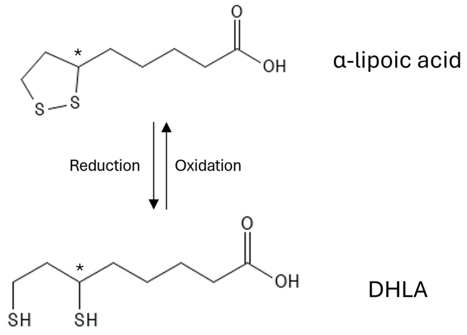
1.1. Chemical Structure of α-Lipoic Acid

α-lipoic acid, also known as thioctic acid, is a naturally occurring compound with potent antioxidant properties.
Structurally, it comprises three components: a five-atom dithiolate ring, an alkyl chain, and a terminal carboxylic acid (see Figure 1). The chiral centre at position C6 results in two enantiomers, R(+)-α-lipoic acid, the naturally available form, and S(-)-α-lipoic acid, which can be synthetically obtained [1].
R(+)-α-lipoic acid is generally recognized to exhibit superior activity compared to S(-)-α-lipoic acid [2,3,4,5,6], even as α-lipoic acid is usually marketed as a racemic mixture containing both enantiomers. To date, the extent to which each enantiomer contributes to α-lipoic acid pharmacological and toxicological effects is not fully understood. While it was proposed that S(-)-α-lipoic acid does not exhibit significant biological side effects [1,2,3,4,5,6,7], some studies indicated that it could potentially hinder the activity of the R(-)-enantiomer [8,9,10].

1.2. Antioxidant Activity of α-Lipoic Acid

The sulfur atoms within the α-lipoic acid ring play a crucial role in exerting antioxidant activity. The oxidised form (α-lipoic acid, ALA) and its reduced derivative (dihydrolipoic acid, DHLA) function as a potent redox couple, participating in different aspects of the antioxidant defence system (see Figure 1).
α-lipoic acid exerts its antioxidant activity through multiple mechanisms, including radical scavenging, the regeneration of other antioxidants, metal chelation, and signalling regulation.
ALA and DHLA can neutralise a variety of reactive oxygen species (ROS) and reactive nitrogen species (RNS), thereby reducing oxidative stress and damage. Different mechanisms have been proposed for the free-radical scavenging activity of α-lipoic acid [11]. ALA was suggested to work through a proton-loss electron transfer mechanism, while DHLA exhibits its activity by single electron transfer followed by proton transfer, or hydrogen atom transfer. In the latter, the ideal spacing between the sulfhydryl and carboxylate groups, determined by the optimal length of the alkyl chain, facilitates the efficient trapping of radicals and the transfer of hydrogen atoms [12].
Generally, antioxidants are categorised based on their solubility characteristics. Water-soluble antioxidants like vitamin C are effective in aqueous environments, while hydrophobic antioxidants like vitamin E prefer lipid-rich environments. Due to its amphiphilic nature, α-lipoic acid represents a unique universal antioxidant, exhibiting activity in aqueous and lipid-based environments, thus protecting a wide range of cellular structures and biomolecules.
The capacity of α-lipoic acid to form complexes with redox-active metals presents a dual benefit. On the one hand, it acts as a chelator for heavy metals like mercury, iron, cadmium, lead, and copper, mitigating their toxicity [13,14,15]. On the other hand, its ability to bind metals, especially gold, has been used to create innovative nanostructures for targeted drug delivery, imaging, and theranostic applications [16].
One distinctive feature of α-lipoic acid is its ability to regenerate the reduced form of either non-enzymatic antioxidants, such as vitamins C and E, glutathione and cysteine, or enzymatic antioxidants, like the coenzyme Q10 [17]. By recycling other antioxidants, α-lipoic acid actively contributes to maintaining the antioxidant network and the cellular redox equilibrium.
Moreover, α-lipoic acid acts as a modulator of proteins involved in cell signalling and transcription through the thiol/disulfide exchange mechanism [18,19,20]. As a result, it has been proposed to influence different aspects, including cell growth and death regulation, inflammatory responses, and glucose and lipid metabolism.

1.3. α-Lipoic Acid as a Therapeutic Agent

The extensive range of protective mechanisms exhibited by α-lipoic acid has attracted interest regarding its potential use as a therapeutic agent. Specifically, it has gained particular attention in clinical conditions involving oxidative stress and inflammation, preserving various cell types and organs from oxidative damage.
Due to its role in contributing to cellular energy homeostasis, α-lipoic acid has proven beneficial against insulin resistance, metabolic syndrome, type 2 diabetes, and diabetes-related complications, such as diabetic neuropathy and nephropathy [21,22,23,24,25,26]. Notably, α-lipoic acid demonstrates nephroprotective effects and holds promise in liver disease prevention due to its rapid hepatic metabolism [27,28,29,30,31,32,33]. Several studies have described the cardiovascular benefits of α-lipoic acid administration [34,35,36,37,38]. Moreover, its ability to cross the blood–brain barrier has shown potential in protecting against neurodegenerative diseases, such as multiple sclerosis and Alzheimer’s and Parkinson’s diseases [39,40,41]. Also, α-lipoic acid has been suggested to have immunomodulatory activity on both innate and adaptive immune cells, down-regulating the proinflammatory markers and reducing the inflammatory response [11,42]. The topical application of α-lipoic acid has proved effective in skin-related conditions, such as ageing and wound healing, in which its antimicrobial properties may ensure a safe repair of skin damage [11,43,44,45,46,47,48]. The use of α-lipoic acid was also investigated in cancer research and it was described as having antiproliferative and antimetastatic activity in different cancer models [49,50,51,52,53].
The unique structure of α-lipoic acid not only makes it a versatile antioxidant but also a promising scaffold for derivatization, providing opportunities for the creation of novel therapeutic agents. From a chemical perspective, the dually modifiable structure of α-lipoic acid allows the functionalization of the dithiolane ring and the carboxylic acid. This feature enables the properties and activity of either the ligands or α-lipoic acid itself to be customised.

1.4. Limitations and Drawbacks in the Therapeutic Use of α-Lipoic Acid

While showing diverse beneficial effects, α-lipoic acid and its therapeutic usage exhibit some drawbacks.
α-lipoic acid is mainly administered orally in solid or liquid form. However, the oral administration of α-lipoic acid results in a limited pharmacokinetic profile, characterised by a short half-life, reaching the maximum plasma concentration in approximately 30 min, and reduced bioavailability (about 30%) [54,55,56,57,58]. This drawback becomes especially significant when administering α-lipoic acid as a racemic mixture. In fact, stereochemistry has been proved to notably affect the absorption, distribution, degradation, and elimination of lipoic acid enantiomers. R-(+)-α-lipoic acid has higher bioavailability than the S-(+) enantiomer, potentially because of the different mechanisms of absorption in the gastrointestinal tract [5,8,54,55,56,57,59,60]. The suboptimal pharmacokinetics of α-lipoic acid have been attributed to its reduced solubility in water and acidic environments and consequent limited gastric stability, and its high first-pass hepatic metabolism [8]. As a result, it needs appropriate formulation to optimise its therapeutic efficacy when used as a supplement or medication [59,61,62,63].
While the prolonged use of α-lipoic acid has been demonstrated to be well-tolerated among healthy individuals [64,65] and it is generally considered not toxic [66,67], registered adverse effects have been correlated with the use of high α-lipoic acid doses [68,69,70], such as gastrointestinal side effects such as stomach discomfort, nausea, or diarrhoea [71]. Moreover, α-lipoic acid at a high dosage negatively affected the liver enzyme activity and induced liver damage in mice and monkeys [66,72]. Also, its supplementation may lower blood sugar levels in genetically predisposed individuals, which can be problematic for diabetic individuals [73]. Finally, α-lipoic acid may interact with certain medications, including chemotherapy drugs, thyroid medications, and drugs that lower blood sugar levels.
Some in vitro and in vivo studies have indicated the pro-oxidant potential of α-lipoic acid, whether in the oxidised form or DHLA [73,74,75]. The result of this effect with either beneficial or detrimental outcomes depends on the specific physiological circumstances, although the pro-oxidant activity of α-lipoic acid has been associated with cardiovascular, renal, and liver damages [73,74,75].
From the chemical point of view, α-lipoic acid exhibits high sensitivity to various environmental factors. It gradually polymerizes, increasing the temperature, and forming an elastic compound [76,77]. Moreover, it is photosensitive, and exposure to UV light can impair the hindered dithiolane ring, leading to undesired polymerization [78]. α-lipoic acid is poorly soluble in water, but soluble in organic solvents. However, the drying process has been suggested to impact its stability. Specifically, Brown and Edwards [79] reported that solvents with easily extractable hydrogens or chlorine atoms minimise α-lipoic acid polymerization in solution. Thus, solvent evaporation at high temperatures and under reduced pressure could reduce the stabiliser effect of hydrogen/chlorine atoms.
Limitations and benefits of the use of α-lipoic acid are summarised in Figure 2.

1.5. Nanomedicine as a Strategy to Improve α-Lipoic Acid

Nanomedicine, the “application of nanotechnology for treatment, diagnosis, monitoring, and control of biological systems” [80,81,82], has raised great expectations [83]. Therapeutic and diagnostic nanoagents (nanotheranostics) may improve the pharmaceutical activity while reducing side effects [84], thanks to multiple functionalisations. Moreover, coating with polymers, like polyethylene glycol, reduces the binding of plasma opsonins, making nanoparticles invisible to macrophages’ clearance [85,86,87].
Nanoparticles with antioxidant properties have recently emerged as possible therapeutic agents for controlling neurodegenerative diseases [88] and cardiovascular [89] and pulmonary diseases [90], and in wound healing [91] and the treatment of cancer [92].
This review aims to outline the application of α-lipoic acid to the nanomedicine field and to explore the advantages associated with its inclusion in nanoplatforms. Indeed, this work describes the strategies employed to increase the shelf life of α-lipoic acid through encapsulation in different nanocarriers, as well as studies utilising mixtures of α-lipoic acid and nanoparticles to enhance the biocompatibility or therapeutic activity of the nanoparticles. Moreover, new frontiers of nanocomposites obtained with poly(α-lipoic acid) will be discussed.

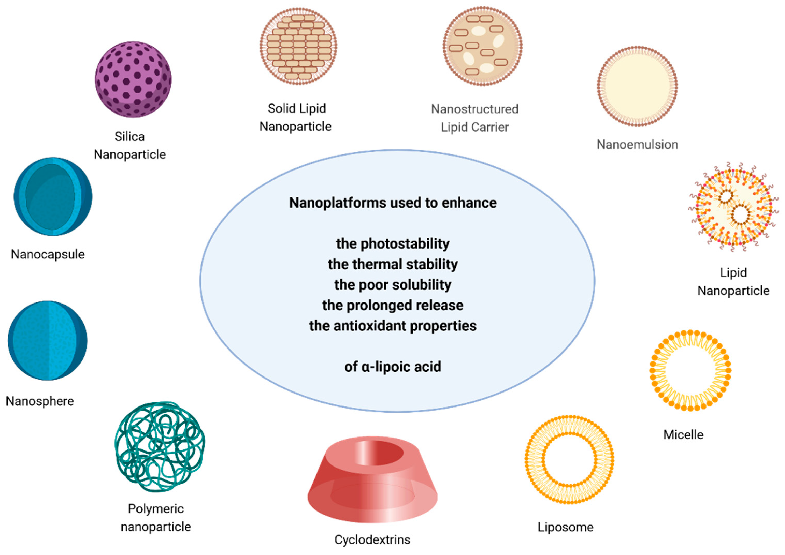
2. Enhancement of α-Lipoic Acid Properties and Activity When Encapsulated in Nanoparticles

As described in the introduction, the primary challenges associated with the use of α-lipoic acid are its short half-life, limited bioavailability, and poor solubility. This section describes several strategies employed to address these drawbacks and improve α-lipoic acid properties by encapsulating the free molecule into different nanotechnological platforms [93]. The delivery systems established are visualised in Figure 3, while Table 1, at the end of this section, summarises the nanotechnological strategies described throughout the text.

2.1. Silica Nanoparticles

Silica nanoparticles represent suitable candidates to overcome several challenges faced by bioactive compounds, including hydrolysis, solubility concerns, and susceptibility to light and heat degradation [92].
To enhance the α-lipoic acid photo- and thermal stability, Dolinina et al. synthesised novel mesoporous silica-based nanocomposites using the widely applied sol–gel method [93]. Two different final products were proposed for α-lipoic acid thermal- and photodegradation, namely polymeric α-lipoic acid and DHLA, respectively. α-lipoic acid was stabilised by the interaction with the silica matrices through H-bonds and electrostatic and hydrophobic forces. As a result, its thermal degradation decreased when compared to the free molecule. As regards UV-induced degradation, the photodegradation rates were comparable to the free α-lipoic acid. The authors suggested that the significant amount of water contained in the nanocomposites, being a suitable hydrogen atom source, favoured the formation of DHLA. Also, the stability of the nanocomposites and the release of α-lipoic acid was investigated, mimicking the pH of digestive fluids. The silica-based composites displayed a release behaviour close to the zero-order kinetic, sustained a plasma concentration of α-lipoic acid comparable to clinically available oral formulations, and preserved its antioxidant activity at all pH values for up to 24 h [94].

2.2. Lipid-Based Nanoparticles (Lipid Carriers, Solid Lipid Nanoparticles, Nanoemulsions, Liposomes, Micelles)

Encapsulation into lipid nanoparticles can overcome the poor solubility of compounds in aqueous media, and their susceptibility to degradation when exposed to oxygen in water. Moreover, lipid nanoparticles have been shown to sustain the release of different drugs, increasing the therapeutic effect and bioavailability and enhancing their biocompatibility.
Platforms belonging to this category include nanostructured lipid carriers, solid lipid nanoparticles, nanoemulsions, liposomes, lipid nanocapsules, and micelles. They are composed of lipids comprising fatty acids, fatty alcohols, glycerides, and waxes, along with stabilising agents like surfactants or polymeric coatings [95].
While solid lipid nanoparticles are formed from solid lipids stabilised by surfactants, nanostructured lipid carriers include both solid and liquid lipids in their matrix. They represent the next generation lipid nanoparticles, designed to overcome the limitations of conventional colloidal carriers, like emulsions, liposomes, and polymeric nanoparticles [96,97].
Indeed, when α-lipoic acid was formulated into nanostructured lipid carriers, solid lipid nanoparticles, and nanoemulsions, the former two displayed a prolonged release of α-lipoic acid compared to nanoemulsions. They also proved high entrapment efficiency and stability over time, and exhibited antioxidant activity comparable to pure α-lipoic acid [98].
Conjugation with stearylamine was used to stabilise α-lipoic acid and synthesise solid lipid nanoparticles, and was then investigated as a delivery system for the chemotherapeutic tamoxifen. The formulation was proved to stabilise α-lipoic acid, which exerted its antioxidant activity, restoring the antioxidant protein network. Moreover, it efficiently enhanced the bioavailability of tamoxifen [99]. Also, solid lipid nanoparticles, co-delivering docetaxel and α-lipoic acid, were demonstrated to have enhanced efficacy against murine mammary carcinoma cells and human breast adenocarcinoma cells. The nanoformulation containing the chemotherapeutic agent and the antioxidant molecule showed increasing cytotoxic effects and significantly higher uptake efficiency compared to free drugs or single drug-loaded solid lipid nanoparticles [100].
Wang et al. synthesised nanostructured lipid carriers using the biodegradable components phosphatidylcholine, stearic acid, oleic acid, and glyceryl monostearate, with the aim of incorporating α-lipoic acid and improving its stability and solubility. The authors found that the obtained lipid carriers were not cytotoxic in vitro, retained α-lipoic acid, and released it within 72 h [101].
Phosphatidylcholine was also employed in the formulation of both nanoemulsions and liposomes containing α-lipoic acid. While both nanosystems efficiently entrapped α-lipoic acid, nanoemulsions exhibited a slower release of α-lipoic acid compared to phosphatidylcholine liposomes. The authors proposed that the slower release from nanoemulsions could be attributed to their multiple-layer structure, whereas liposomes, being primarily single-layered particles, facilitated a faster release. Thus, nanoemulsions might represent a more suitable formulation for α-lipoic acid, prolonging the drug’s action [102].
Different oil-in-water nanoemulsions were also investigated for the co-delivery of α-lipoic acid and vitamin B. Specifically, the influence of using different oils (castor oil and sunflower oil) and agitation methods on the properties of the obtained emulsions was evaluated, including metrics of colloidal stability, encapsulation efficiency and drug release. The formulation prepared by the widely applied solvent displacement method and using castor oil under magnetic stirring resulted in the best colloidal properties, highest thermal and pH stability, and higher encapsulation and release rates [103]. In another study, pumpkin oil was used to solubilize α-lipoic acid and develop a self-nanoemulsifying drug delivery system, stabilised by Tween 80 polyethylene glycol 200. The formulation demonstrates higher protective effects against gastric ulcer models compared to the free α-lipoic acid [104].
Phophatidylcholine-based liposomes were prepared to improve the stability and absorption of α-lipoic acid. The formulation proved to be stable, enhanced the oral absorption and bioavailability of α-lipoic acid, and improved hepatoprotection in hepatic injury model rats [105]. Other authors have synthesised new liposomes, consisting of phosphatidylcholine and curcumin, in which α-lipoic acid has been encapsulated; using these liposomes, they demonstrated a high encapsulation efficiency of lipoic acid and its slow release, which countered cisplatin-induced toxicity in HEI-OC1 cells, a cell line utilised as a model for auditory cells [106].
Polymers can be used to stabilise different platforms, including emulsions, liposomes, and micelles. Huang et al. developed an advance system for preparing a multilayer emulsion. A primary oil-in-water emulsion, stabilised by Tween 20 and phosphatidylcholine, was used to encapsulate α-lipoic acid and linseed oil. Subsequently, a chitosan coating was applied to improve the thermostability of the oil droplets and to address the limited capacity of emulsions to counteract oxidative stress, which could potentially affect the stability of both encapsulated molecules. The system demonstrated a higher thermal stability than the primary emulsion and limited lipolysis of linseed oil in vivo, and inhibited the degradation of α-lipoic acid [107]. The use of a chitosan coating also proved efficient in stabilising liposomes co-encapsulating α-lipoic acid and Coenzyme Q10 with the purpose of improving the solubility and stability of both molecules for transdermal application. It was found that chitosan interacted with α-lipoic acid and Coenzyme Q10 via H-bonds and ionic forces, promoting the permeation and accumulation of both molecules in rabbit skin. Moreover, the system demonstrated higher scavenging activity compared to free α-lipoic acid and Coenzyme Q10 [108]. α-lipoic acid was also incorporated into micelles composed of a copolymer (methoxypoly(ethylene glycol) and poly(caprolactone)). This incorporation led to an enhanced loading capacity of the micelles for IR780, a near-infrared dye for imaging and photothermal therapy, while also preserving their loading capacity for docetaxel, a chemotherapeutic agent [109]. Pluronic nanoparticles were prepared by the oil-in-water emulsion method, encapsulating α-lipoic acid into a soybean oil core and using Pluronic F127 to form the hydrophilic shell. When administered in the mouse middle ear cavity, the system was demonstrated to preserve hearing ability after ototoxicity induction, and could be delivered in a more effective way with respect to free α-lipoic acid [110].

2.3. Nanocapsules and Nanospheres

Nanocapsules are composed of a polymeric shell surrounding an oily core, in which the drug may either dissolve within the core or adhere to the polymeric wall. In contrast, nanospheres lack an oily component and instead constitute a polymeric matrix, in which the drug can be uniformly distributed or adsorbed.
α-lipoic acid was encapsulated in lipid-core nanocapsules with triglycerides of capric and caprylic acid, sorbitan monostearate, and poly (ε-caprolactone) as main components, and stabilised by polysorbate 80. The encapsulation counteracted α-lipoic acid’s undesired pro-oxidant activity, and contributed to the wound healing process, although to a lesser extent than lipoic acid alone [43]. Also, Xia et al. encapsulated α-lipoic acid into lipid nanocapsules containing triglycerides of capric and caprylic acids and phosphatidylcholine as a surfactant. The capsules proved to be stable and have high encapsulation efficiency and drug loading. Moreover, antioxidant activity remained intact within the nanocapsules, and sustained release of α-lipoic acid was achieved [111]. Interestingly, a dual stabilising effect of α-lipoic acid and the antioxidant resveratrol was observed when co-encapsulated into lipid-core nanocapsules. The co-encapsulation was shown to enhance the stability, photostability, and antioxidant properties of both compounds. When α-lipoic acid was encapsulated alone, it exhibited rapid release, consistent with previous findings [112]; however, when co-encapsulated with resveratrol, the release rates of both compounds decreased [113].
Nishiura et al. exploited the self-assembly properties of α-lipoic acid, along with the non-ionic surfactant polyoxyethylene (20) stearylether, to create a nanosphere matrix encapsulating α-lipoic acid. The encapsulation improved the stability and bioavailability of α-lipoic acid by placing its hydrophobic moiety inside the nanocapsule, thereby protecting the unstable disulfide bond from degradation [77]. The same method was used to encapsulate α-lipoic acid and test its antitumor effects in a mouse model of Ehrlich solid tumour. Both free and encapsulated α-lipoic acid significantly reduced oxidative stress, but encapsulated α-lipoic acid remained stable and in circulation for longer periods of time [114]. Also, EL-Gebaly et al.’s approach was used to investigate the beneficial effects of encapsulated α-lipoic acid against 99mTc-MIBI-induced injury, showing significant protective effects and suggesting its efficacy as a radioprotector [115]. Kubota and collaborators used a similar approach by mixing α-lipoic acid with the polyoxyethylene (20) stearylether to improve its skin permeation. The results confirmed that nanoencapsulation is a suitable method for the efficient topical administration of α-lipoic acid [116].

2.4. Polymeric Nanoparticles

Natural polymers have frequently been used to develop biodegradable and biocompatible delivery systems.
Gogoi et al. co-loaded α-lipoic acid and curcumin into chitosan nanoparticles to improve the limited solubility and bioavailability of both drugs. They investigated the in vitro activity of the nanosystem in a breast cancer cell model, highlighting the synergistic activity of curcumin and α-lipoic acid in inhibiting tumour cell growth and inducing tumour cell death [117]. Also, the bioavailability and stability of α-lipoic acid formulated into chitosan nanoparticles and solid lipid nanoparticles was compared [118]. Both systems encapsulated α-lipoic acid effectively, demonstrated high stability, and counteracted the oxidative stress and neurotoxicity induced by aluminium chloride in rats. However, as expected, the effect was superior when α-lipoic acid was loaded into solid lipid nanoparticles compared to chitosan nanoparticles.
To improve its skin delivery, α-lipoic acid was loaded into liquid crystalline nanoparticles. The surface of the platform was modified by lipopolymers composed of Polyethylene glycol (PEG) and 1,2-dioleoyl-sn-glycero-3-phosphoethanolamine, and functionalized with the epidermal growth-factor-receptor-targeting D4 peptide, and the HIV-1-derived TAT cell-penetrating peptide. The system demonstrated a high drug encapsulation rate and released the α-lipoic acid into the deepest layers of the epidermis, where it counteracted the oxidative stress and inflammation after exposure to UVB light [119].
The use of α-lipoic acid is also interesting in suppressing appetite. Traditional formulations (salt or micronized crystals) had poor absorption and a short plasma half-life, not sufficient to suppress appetite. Park and collaborators introduce a novel α-lipoic acid nanoparticulate formulation developed through nano-comminution, employing different polymeric stabilisers (hydroxypropyl cellulose, Pluronic® F127, and polyvinylpyrrolidone). Interestingly, they found that nanoparticle powder formulations, when compared to nanosuspension formulations, were endowed with an improved efficacy in reducing food intake [120].

2.5. Cyclodextrins

Cyclodextrins (CDs) are cyclic oligosaccharides derived from the enzymatic degradation of starch which typically comprise six (α-), seven (β-), or eight (γ-) glucose units linked through α-1,4 glycosidic bonds. They have a toroidal-like shape characterised by a hydrophilic outer surface and a more hydrophobic central cavity. This distinctive arrangement enables them to interact with molecules of appropriate size and geometry, and form complexes with the entire molecule or part of it. They are frequently used as excipients in pharmaceutical formulations to increase the aqueous solubility of poorly soluble active substances, improving the stability and bioavailability of drugs [121,122,123].
α-lipoic acid has been proven to be complexed by α-, β-, and γ-CDs [124,125], and in particular the R-(+)-α-lipoic acid complex with γ-CD exhibited biocompatibility and stability towards heat, humidity, and low pH [76]. γ-CD significantly improved the bioavailability of R-(+)-α-lipoic acid in rats by enhancing its intestinal absorption [126]. As a result, the R-(+)-α-lipoic acid/γ-CD complex exhibited a 2.2 times higher plasma concentration compared to free α-lipoic acid, after oral administration. This evidence was confirmed by a recent clinical trial in healthy volunteers, where the bioavailability of the R-(+)-α-lipoic acid/γ-CD complex demonstrated results 2.5 higher than the non-complexed R-(+)-α-lipoic acid [127].
Table 1. Nanotechnological strategies used to improve α-lipoic acid characteristics and efficacy.
NanoplatformGoalReference
Silica nanoparticlesStabilise[93,94]
Lipid-based nanoparticlesProlong the release [98,100,101,102,103,106,108,110]
Stabilise[99,103,107,110]
Improve solubility[104]
Improve bioavailability[105]
Improve antioxidant activity[108]
Nanocapsules and nanospheresCounteract pro-oxidant activity[43]
Preserve antioxidant activity [111]
Improve the stability [114]
Prolong the release [111,113,114]
Polymeric nanoparticleImprove the solubility and bioavailability[118,119,120]
Improve the release [120]
CyclodextrinsImprove the stability and bioavailability[76,126,127]

3. Exploring the Role of α-Lipoic Acid in Therapeutic Nanoplatforms

α-lipoic acid has been studied in numerous works in combination with several types of nanoparticles. The formulations have been shown to enhance either the effects of nanoparticles or α-lipoic acid antioxidant properties, both in cellular systems and in mouse models of different pathologies.

3.1. Renewing Tissues: Nanoparticles and α-Lipoic Acid in Regenerative Medicine

The application of nanotechnology to non-healing wounds aims to facilitate healing and tissue repair and avoid complications like infections.
The potential benefits of therapies based on a mixture of nanoparticles and antioxidant agents on tissue regeneration have been demonstrated by Leu et al. The topical application of a mixture containing gold nanoparticles, the antioxidant epigallocatechin gallate, and α-lipoic acid significantly enhanced the proliferation and migration of skin cells and accelerated wound healing in mice [128]. The same combination was also investigated in the treatment of diabetic ulcers, reducing the expression of the receptor for advanced glycation end-products, enhancing angiogenesis, and exerting anti-inflammatory effects [129].
Moreover, wound healing was improved by the treatment with α-lipoic acid conjugated with hexagonal boron nitride and boron carbide nanoparticles, exhibiting regenerative and antioxidant properties. α-lipoic acid has also been demonstrated to improve the antimicrobial properties of boron nitride and boron carbide nanoparticles against Staphylococcus aureus and Escherichia coli strains [130].
Finally, α-lipoic acid-capped silver nanoparticles introduced into alginate-based aerogels demonstrated improved biological properties, including higher antioxidant capacity and anti-inflammatory activity, compared to a commercial dressing made of alginate containing colloidal silver [131].

3.2. Exploring the Use of α-Lipoic Acid-Containing Nanoparticles in Tumour Fighting

In cancer nanomedicine, α-lipoic acid has found application in three primary aspects: (1) the exploitation of its intrinsic antitumor activity when nanoformulated; (2) the development of tumour environment-responsive nanoparticles, taking advantage of the characteristics of its disulfide bond; (3) its action as a linker to enhance the binding strength of tumour-targeting ligands on the surface of nanoparticles.
As reviewed elsewhere, α-lipoic acid has different powerful effects against cancer cells, such as inducing apoptosis and suppressing proliferation, and cancer stemness. Moreover, it shows potential in mitigating chemotherapy-induced side-effects and overcoming chemoresistance [49,132].
α-lipoic acid can be nanoformulated and investigated for its potential effects against tumour cells. For example, Usama et al. proposed the synergistic activity of α-lipoic acid with simvastatin, a statin with direct cytotoxic activity against different types of tumour cells. The synthesised α-lipoic acid-simvastatin nanoparticles demonstrated a significant increase in the cellular uptake and cytotoxicity of the statin in breast carcinoma cells [133].
The disulfide bond of α-lipoic acid can be exploited to prepare stimuli-responsive nanoparticles, specifically systems that respond to the reducing environment found in tumour cells. In fact, it is well known that the concentration of glutathione in its reduced form (GSH) is significantly higher in cancer cells, and it can trigger the reduction of the α-lipoic acid dithiolane ring, facilitating the release of the chemotherapeutics.
For example, α-lipoic acid was esterified to phosphatidylcholine to produce stimuli-responsive cross-linked liposomes for the intracellular delivery of doxorubicin. The system demonstrated a high serum stability and reduced-responsive drug release, resulting in an in vitro anticancer activity comparable to the free doxorubicin. Moreover, the liposomal formulation improved the pharmacokinetic profile of doxorubicin, which was maintained in the blood circulation for a longer time [134]. Also, α-lipoic acid was conjugated to a tocopheryl moiety to produce stimuli-responsive nanovesicles designed for the delivery of doxorubicin. The nanovesicles efficiently encapsulated doxorubicin, and released it in a glutathione-responsive manner. Moreover, the system proved to be biocompatible and to have an anticancer activity superior to doxorubicin alone in drug-resistant cancer cells [135]. Also, α-lipoic acid was conjugated to xylan to produce redox-sensitive nanoparticles aimed to deliver the anticancer drug niclosamide. The platform exhibited good stability and biocompatibility as well as higher anticancer activity compared to the free drug [136].
Additionally, dual targeting delivery systems have been developed, combining the stimuli-responsive behaviour of α-lipoic acid and other tumour-specific targets or tumour-specific environmental conditions (see Table 2). In this category, several works exploit the concept of charge-reversible nanoplatforms, capable of adjusting their surface charge according to the surrounding environment [137]. They exhibit either a negative or a neutral charge under physiological pH conditions to prevent adsorption to serum proteins that can induce opsonization, complement activation, and rapid clearance. To achieve this feature, specific functional groups, such as the imidazole moiety and maleic anhydride [138,139,140,141], or coatings like polyethylene glycol, have been introduced [138,141,142]. Consequently, charge-reversible nanosystems exhibit enhanced plasma stability and can accumulate in tumour tissues. Once it has reached the tumour microenvironment, the acidic pH triggers the charge switch and the positively charged surface facilitates the cellular internalisation of the delivery platform.
In other works, tumour-specific ligands were introduced to improve the targeting of the delivery systems, such as binders of the tumour-overexpressed receptors [138,143,144,145,146,147,148].
After internalisation, drug release was achieved by the activity of high glutathione intracellular concentration on α-lipoic acid.
Table 2. α-lipoic acid-based dual responsive drug delivery systems in cancer treatment.
Tumour TargetMoleculeNanoplatformGoalReference
Acidic pH and Overexpressed ASGPR *Dimethylmaleic acid—PEG and lactobionic acidPolymeric
nanoparticle
Delivery of Doxorubicin[138]
Overexpressed ASGPR and folate receptorPullulan and folic acidPolymeric
nanoparticle
Delivery of Paclitaxel[148]
Overexpressed folate receptorFolic acidSilica hybrid
Magnetic nanoparticles
Delivery of Doxorubicin[143]
Acidic pH HistidinePolymeric nanoparticleDelivery of Doxorubicin[139]
Dimethylmaleic anhydridePolymeric nanoparticleDelivery of Doxorubicin[140]
Histidine—PEGLiposomesDelivery of VEGF ***
siRNA and Etoposide
[141]
PEGAlbumin-based nanocarrierDelivery of Doxorubicin[142]
CD44 receptor overexpression Hyaluronic acidPolymeric nanoparticleDelivery 17α-Methyltestosterone[134]
Hyaluronic acidPolymeric nanoparticleDelivery of Doxorubicin[135]
Tumour esterase overexpressionChlorambucilPolymeric nanoparticleDelivery of Doxorubicin and Chlorambucil[136]
αvβ3 receptor overexpressioncRGD peptide **MicelleDelivery of Doxorubicin[137]
* asialoglycoprotein receptor; ** cyclic arginyl-glycyl-aspartic acid peptide; *** vascular endothelial growth factor.
Thiols are commonly used as ligands for attaching drugs to the surface of nanoparticles, such as gold nanoparticles. Using linkers with multiple thiols, such as α-lipoic acid, increases the binding strength between the nanoparticles’ surface and the ligands. Also, α-lipoic acid’s carboxylic group can be directly functionalised with chemotherapeutics, spacers, and tumour-specific targets. The following works, mainly focused on the functionalisation of gold nanoparticles, describe the use of α-lipoic acid for these purposes. Ghorbani et al. used α-lipoic acid as a linker to attach curcumin to the surface of Fe3O4, resulting in a novel, particularly potent medicinal compound with increased cytotoxicity compared to curcumin alone [149]. Also, promising antimetastatic activity was shown by linking a tumour-associated carbohydrate antigen to gold nanoparticles through an analogue of α-lipoic acid, the iso-α-lipoic acid. Moreover, the advantage of having an achiral linker as iso-α-lipoic acid lies in the simplified design, synthesis, and characterisation of the nanosystem [150]. α-lipoic acid was used to link folic acid and doxorubicin to the surface of gold nanoparticles to obtain a targeted delivery system for the chemotherapeutic drug. The approach resulted in enhanced cell penetration and the prolonged release of doxorubicin [151]. α-lipoic acid was used in the development of gold tumour-targeting nanoparticles capable of responding to the acidic intracellular environment and aggregating to amplify nanoparticle retention within tumour cells. Specifically, in the work of Cheng, α-lipoic acid was used to link a biotin-end-derived PEG moiety to the gold surface. As a result, the nanoplatform exhibited stealth properties and active targeting ability [152]. A similar approach was used to conjugate doxorubicin and anti-PD-L1 antibody on the gold nanoparticle surface, through an α-lipoic acid-PEG-N-hydroxysuccinimide linker. The thiol groups of α-lipoic acid interacted with the gold surface, while it formed an amide bond with the N-hydroxysuccinimide moiety binding the cancer-targeting ligands [153].
Wang and collaborators modified star poly(lactic-co-glycolic acid) ends with α-lipoic acid to obtain a new nanoparticle with enhanced loading capacity, reduced drug leakage, and efficient uptake in melanoma cells [154,155].

3.3. Exploring Miscellaneous Applications of Nanoparticles and α-Lipoic Acid

The combination of α-lipoic with nanomaterials, including metal and polymeric nanoparticles, was also investigated in other therapeutic areas.
Indeed, Tudose et al. covalently immobilised α-lipoic acid onto the surface of silver nanoparticles–decorated silica nanoparticles, and they evaluated the antioxidant, cytotoxic, and antimicrobial activities of these new nanoparticles. They found that the functionalisation of silver nanoparticles–decorated silica nanoparticles with α-lipoic acid enhanced the specificity of the interaction with mammalian cell lines and antioxidant activity, while reducing cytotoxicity, suggesting its potential for modulating the cell cycle to achieve desired therapeutic effects [156].
The mixture of α-lipoic acid with gold nanoparticles showed a superior neuroprotective effect compared to each compound alone in vivo, with the inhibition of brain damage induced by radiation in rats [157]. The combination of α-lipoic acid with PEGylated gold nanoparticles proved to counteract oxidative stress in osteoporosis development. α-lipoic acid-gold nanoparticles were biocompatible, and effectively removed reactive oxygen species and promoted osteoblast proliferation [158].
The impact of α-lipoic acid in combination with caffeine-loaded chitosan nanoparticles was evaluated against obesity and its hepatic and renal complications in rats. The treatment was shown to mitigate weight gain and improve hepatic and renal function, reducing biochemical markers and histopathological alterations [159]. The same nanosystem was also tested for its ability to mitigate cardiovascular complications induced by obesity in rats. Treatment with α-lipoic acid and caffeine-loaded chitosan nanoparticles alleviated cardiac complications and the cardioprotective effect of the combined treatment, was more evident than that of the two agents used individually [160]. A nanosphere matrix made of zein (a water-insoluble plant protein derived from maize) and α-lipoic acid was employed to encapsulate Vardenafil, a medication used for the treatment of erectile dysfunction [161]. α-lipoic acid has indeed previously been shown to significantly improve sexual function and quality of life in both animals and human investigations [162]. In the work of Ahmed [161], α-lipoic acid was stabilised by the interaction with the zein polymer; moreover, the bioavailability, delivery, and effects of Vardenafil were improved thanks to the nanoformulation.

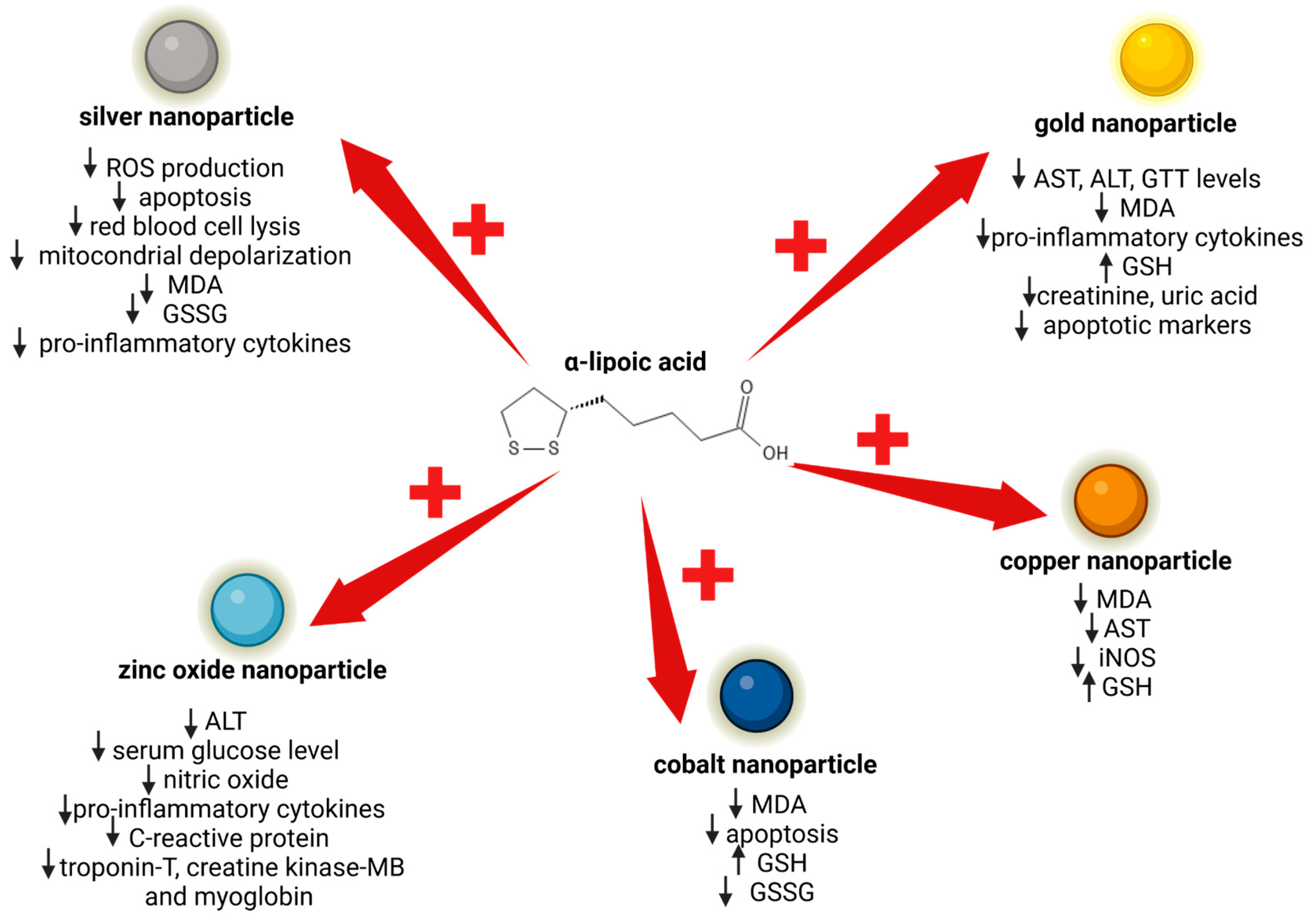
4. Protective Role of α-Lipoic Acid against Nanoparticle Cytotoxicity

Different studies have shown that α-lipoic acid can inhibit the intrinsic cytotoxicity of various types of nanoparticles. Two strategies have been used by researchers: on one hand, directly conjugating α-lipoic acid to the surface of the nanoparticle, and on the other hand, mixing α-lipoic acid with the nanoparticles before application. Using α-lipoic acid in conjunction with various types of nanoparticles has been demonstrated to limit many of the cytotoxic effects induced by the nanoparticles themselves (see Figure 4).

4.1. α-Lipoic Acid and Silver Nanoparticle Cytotoxicity

As is already known, silver nanoparticles possess potent antimicrobial activity, but they can exert cytotoxic effects [163,164]. Hajtuch et al., comparing uncoated silver nanoparticles with α-lipoic acid silver nanoparticles, found that silver nanoparticles coated with α-lipoic acid maintained their antimicrobial properties, while improving their biosafety. In fact, α-lipoic acid silver nanoparticles were found to be biocompatible with Huvec cells, red blood cells, and platelets [165]. Also, Cotton and collaborators mitigated silver nanoparticles’ toxic effects by capping them with α-lipoic acid. The protective effect of α-lipoic acid was investigated on human gingival fibroblasts in comparison to ionic silver and clinical antiseptics (chlorhexidine and silver diamine fluoride). The authors found that α-lipoic acid-capped silver nanoparticles exhibited notably lower toxicity than the clinically utilised controls, while keeping their antimicrobial properties intact [166]. In another study, the damage induced on rat testes by silver nanoparticles was attenuated by mixing them with α-lipoic acid [167]. The authors demonstrated that the co-treatment mitigated the biochemical, oxidative, and apoptotic changes induced by silver nanoparticles. These kinds of nanoparticles, when administered, can damage the blood–brain barrier function and induce neurotoxicity; the co-treatment with α-lipoic acid was demonstrated to be favourable in ameliorating the neurotoxic side effects of silver nanoparticles by reducing the induced apoptosis, inflammation, and oxidative stress [168]. The co-treatment with α-lipoic acid and silver nanoparticles was also used to mitigate adverse effects of these nanoparticles in the treatment of pancreatic ductal adenocarcinoma. α-lipoic acid, in fact, reduced the production of reactive oxygen species (ROS) induced by the silver nanoparticles themselves, protecting non-malignant cells from oxidative stress [169].

4.2. α-Lipoic Acid and Gold Nanoparticle Renal and Hepatic Toxicity

Exposure to gold nanoparticles can induce hepatic oxidative damage and toxicities; it has been shown that the co-administration of α-lipoic acid with gold nanoparticles has protective effects, suppressing oxidative stress and inflammation [170]. Gold nanoparticles can also have nephrotoxic effects: α-lipoic acid has been demonstrated to reduce nanoparticle kidney damage, suggesting that combining natural antioxidants with gold nanoparticles may offer a potential strategy to prevent nanoparticle-induced toxicity [170,171,172].

4.3. α-Lipoic Acid and the Inhibition of Toxicity Induced by Other Types of Nanoparticles

α-lipoic acid exhibited hepatoprotective functions against zinc oxide nanoparticle toxicity, decreasing liver damage and the expression of metabolic disorder markers, which were up-regulated by the treatment with zinc oxide nanoparticles [173]. The co-administration of α-lipoic acid also significantly mitigated the cardiotoxic effects of zinc oxide nanoparticles by reducing serum cardiac injury markers, pro-inflammatory biomarkers, nitric oxide, and vascular endothelial growth factor levels, as well as cardiac calcium concentration and oxidative DNA damage, indicating its potential as s protective agent against ZnO-NP-induced cardiac tissue injury [174]. Moreover, α-lipoic acid displayed a therapeutic role after the exposure to lead and zinc oxide nanoparticles, attenuating the biochemical alterations in neurological, immunological, and male reproductive organs of rats, suggesting its potential as a protective agent against their adverse effects [175]. Cobalt nanoparticles can be generated from cobalt-containing implants and they can induce ferroptosis-like cell death through, among other things, increasing intracellular reactive oxygen species. Cobalt nanoparticle adverse effects have been mitigated by the co-administration of α-lipoic acid, which effectively counteracts the adverse effects induced by CoNPs, thus stabilising the cell’s antioxidant capacity and preventing ferroptosis-like cell death [176]. α-lipoic acid was also shown to be a promising hepatoprotective agent against copper-nanoparticle-induced oxidative damage; its administration mitigated hepatic damage, maintaining an antioxidant status [177].

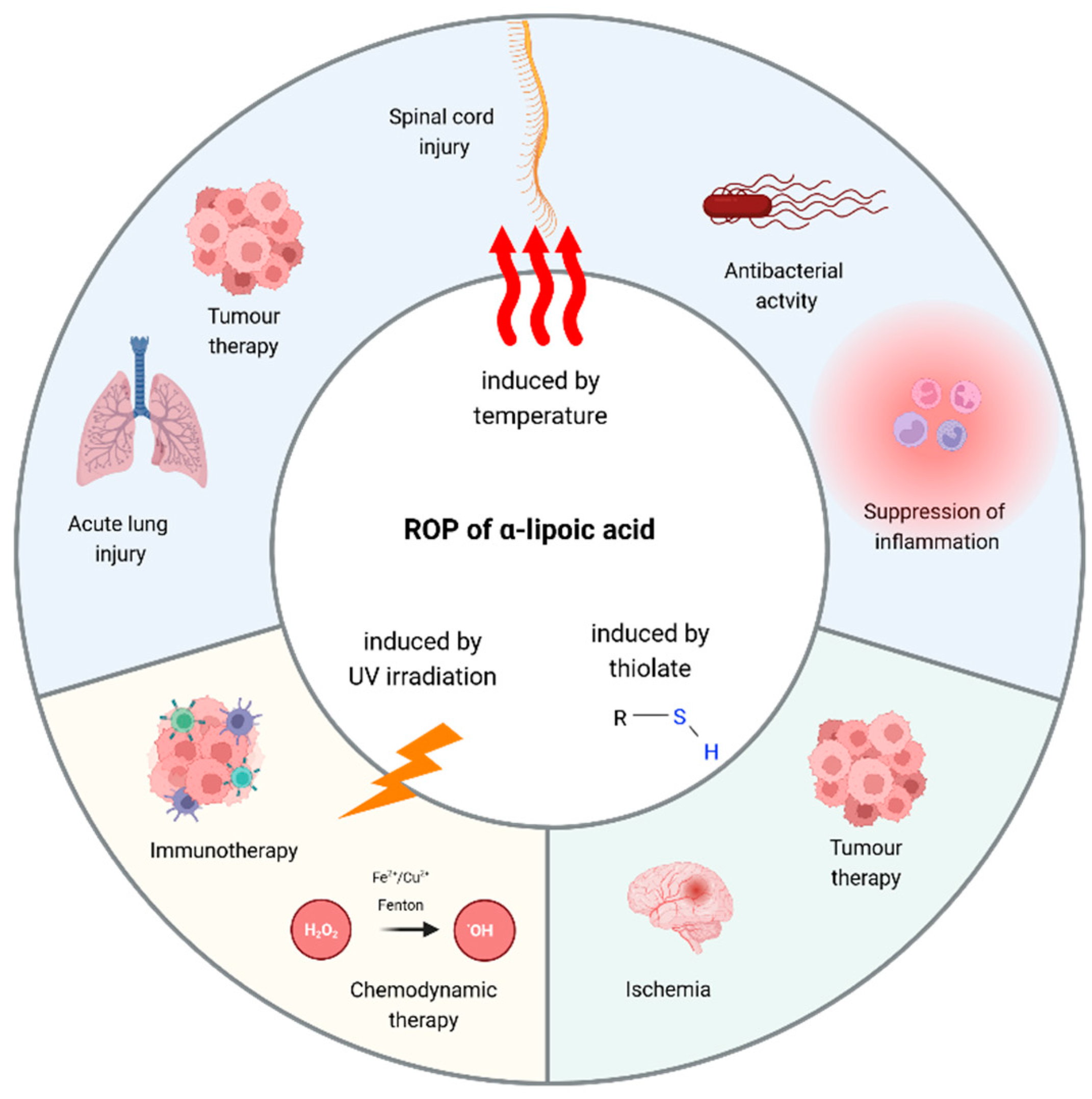
5. Poly(α-Lipoic Acid)-Based Polymeric Nanoparticles: Insights and Perspectives

The 1,2-dithiolane ring contained in α-lipoic acid can undergo ring-opening polymerization (ROP) to produce poly(disulfide)s [178]. ROP [179] is the approach most used to propagate α-lipoic acid polymerization and obtain its cross-linking. ROP can be achieved by using different methods: elevated temperatures, photo-initiated radical polymerization, and thiolate compounds (see Figure 5).
All these approaches have been used to produce nanoparticles based on the polymerization of α-lipoic acid.
It is interesting to note how heat and light are often listed as factors that destabilise α-lipoic acid (see Introduction), but if used properly, they are good alternatives for achieving polymerization. On the other hand, ROP initiated using a thiolate monomer represents a promising approach due to the mild conditions it requires and the better control of the polymerization process [180]. The advantages and limitations of each strategy are summarised in Table 3.

5.1. Poly(α-Lipoic Acid) Nanoparticles Obtained by Thermal Polymerization

Poly(α-lipoic acid) nanoparticles, synthesised via thermal polymerization, were employed alongside various drugs to augment their anticancer efficacy. The general method applied in the studies outlined in this section involves polymerizing α-lipoic acid at a temperature above its melting point, then utilising the resulting poly(α-lipoic acid) to create the desired nanoparticles.
For instance, Yang et al. synthesised a PEGylated poly(α-lipoic acid) copolymer and assembled it into nanoparticles in aqueous solution. The nanoparticles showed the ability to load doxorubicin with high efficiency, and the doxorubicin-loaded nanoparticles were efficiently internalised in 4T1 cancer cells, where doxorubicin was released, displaying its antiproliferation ability. Moreover, these nanoparticles exhibited improved antitumoral efficacy in a mouse model, while causing fewer toxic effects on healthy organs [182]. The same nanoparticles were co-loaded with paclitaxel and doxorubicin and both were studied in a cellular and an animal model for osteosarcoma. The authors found that the dual-drug-loaded nanoparticles were effectively taken up by osteosarcoma cells, releasing drugs intracellularly in response to an acidic pH and the reductive environment of cancer cells. Moreover, the co-loaded nanoparticles exhibited improved biodistribution into the tumour in mice, resulting in a more effective inhibition of tumour growth compared to free drugs [183]. A similar strategy was used to develop pH-responsive nanovesicles from a PEGylated poly(α-lipoic acid) copolymer to deliver the chemotherapeutics doxorubicin and gefitinib for the treatment of ovarian cancer. The drugs were efficiently encapsulated and released intracellularly in a pH-responsive manner, inducing the apoptosis of cancer cells [184]. Liu and collaborators conjugated poly(α-lipoic) nanoparticles to combretastatin A4, a potent vascular disrupting agent, resulting in a specific accumulation of combretastatin A4 in tumour cells and in a prolonged apoptosis of cancer cells because of its long retention time [185]. Also, they used the same approach with the cytotoxic drug honokiol and another vascular disrupting agent, 5,6-dimethylxanthenone-4-acetic acid, obtaining a nanoparticle with high antitumor potency against a murine model of breast tumour [186].
Poly(α-lipoic acid) nanoparticles obtained thanks to elevated temperatures were also tested for their potential ability to treat spinal cord injury. The antibiotic minocycline was encapsulated in polymeric nanoparticles, and loaded with methylprednisolone, a drug used to inhibit inflammation after traumatic spinal cord injury. The combined nanoparticle was biocompatible and exhibited a potent anti-inflammatory activity, able to inhibit proinflammatory cytokine release in a microglia cell line treated with LPS [187].
The encapsulation of Venetoclax, a Bcl2 inhibitor, in polymeric nanoparticles has been demonstrated to enhance its effects when administered freely. It inhibits the production of pro-inflammatory cytokines in a murine model of lung injury by inducing the apoptosis of infiltrated neutrophils [188]. Also, the antioxidant properties of the natural compound quercetin were enhanced through encapsulation in polymeric nanoparticles. This encapsulation favoured its chemical stability and slow release, and the antioxidants properties of encapsulated quercetin have been shown to be improved with respect to its crystalline form in a rat model of acute liver injury [189].
Rhein (an anthraquinone with anti-inflammatory activity) and geraniol (a bioactive compound) were loaded onto poly(α-lipoic acid) acid nanoparticles, showing an enhanced antimicrobial activity against Salmonella infection, compared with free drugs. Additionally, co-loaded nanoparticles were also able to suppress the production of pro-inflammatory cytokines and to maintain the gut microbiota homeostasis [190]. With the aim of tackling Salmonella infection, characterised by the production of high levels of H2S, PEGylated α-lipoic acid was thermally polymerized and polymeric nanoparticles were loaded with the antibiotic compound ciprofloxacin. The nanoplatform, besides demonstrating excellent stability in vitro and biocompatibility toward mammalian cells, selectively delivered the antibiotic to the inflammation site, thus improving its efficacy [191].

5.2. Poly(α-Lipoic Acid) Nanoparticles Obtained by Thiolate-Initiated Polymerization

Matile and colleagues examined the reactivity of cyclic disulfides in disulfide-exchange polymerization. They concluded that α-lipoic acid’s disulfide bond length and the torsion angle of the thiolane ring exhibited the optimal reactivity to achieve optimal and controlled polymerization [192]. In most of the studies detailed in this paragraph, the process begins with the formation of nanoparticles containing α-lipoic acid, followed by the induction of polymerization using a thiolate initiator to yield poly(α-lipoic acid) nanoparticles.
Cross-linked α-lipoic acid nanocapsules using 1,4-dithiothreitol (DTT) thiolate have been employed to encapsulate two precursors of combretastatin (a compound displaying strong inhibitory activity on tumour cell growth) and a copper catalyst. Encapsulation ensured that the prodrug was not prematurely activated. Within the tumour tissue, the high concentration of GSH induces the breakdown of poly(α-lipoic acid) nanoparticles, releasing prodrugs and catalysts and inducing the death of only tumour cells [193]. The polymerization of α-lipoic acid was used to project nanoparticles based on a conjugate of lipoic acid and hyaluronic acid; these cross-linked nanoparticles were used to encapsulate and then deliver doxorubicin specifically to breast cancer and myeloma and leukaemia cells [194]. DTT was also used to induce the polymerization of α-lipoic acid contained in a dimeric surfactant-like amphiphilic molecule: this molecule can form nanoaggregates, due to the hydrophobic head of α-lipoic acid and the hydrophilic tail of gemini surfactant. Nanoaggregates were then stabilised thanks to α-lipoic acid polymerization and loaded with doxorubicin: the nanonetwork was selectively cytotoxic against cancer cells with respect to normal cells [195].
Mancin and collaborators showed that α-lipoic acid polymerization could be realised through a single-step synthesis using 1-octanethiol as initiator, producing cross-linked poly(α-lipoic acid) nanoparticles loaded with dye and protected by PEG directly from small molecule precursors [196]. Interestingly, they conducted a comparison on the long-term stability of nanoparticles synthesised with a thiolate initiator versus those formed without any initiators. The results highlighted that nanoparticles produced through thiol-initiated ROP resulted in the formation of more rigid and stable nanoparticles. Also, α-lipoic acid preserved its antioxidant activity within the nanoformulation, demonstrating in vitro efficacy in mitigating damages caused by post-ischemic reperfusion [197]. The potential of these nanoparticles as cardiomyocyte-targeted drug was validated in rats, revealing their specific accumulation in the heart without inducing any toxicity in cardiomyocytes. Furthermore, the nanoparticles were tested in vitro as a delivery system for nucleic acids in cardiomyocytes, demonstrating their ability to enter this specific cell type and release their cargo in the perinuclear area [198].
Conversely to the previous works described in this section, Gu et al. first induced the polymerization of α-lipoic acid using cysteine hydrochloride, and then prepared poly(α-lipoic acid) nanoparticles through ultrasonic emulsification. The newly obtained nanoplatform was employed for the delivery of docetaxel, demonstrating specific drug targeting in cancer cells and preventing unspecific drug release [199].

5.3. Poly(α-Lipoic Acid) Nanoparticles Obtained by UV light

In the case of irradiation-induced polymerization, as well, the majority of studies first involve the synthesis of nanoparticles containing α-lipoic acid, followed by its polymerization.
A new pH-responsive nanodrug based on a form of camptothecin (an inhibitor of topoisomerase) was loaded into cross-linked R(+)-α-lipoic acid nanoparticles, enhancing the anticancer activity of camptothecin, stabilising the drug, and achieving a higher drug concentration in the HT29 tumour cells [200].
UV cross-linked R(+)-α-lipoic acid nanoparticles were used to anchor quaternary ammonium salts provided with long alkyl chains, which have the ability to destabilise cell membranes. Their link to α-lipoic acid nanoparticles ensured that their activity occurs only within the cell, following the dissolution of the nanocontainer due to the cytosolic redox environment. As a result, they exerted a selective cytotoxic activity only to the tumour environment [201].
Two α-lipoic acid monomers were modified, one with a disulfide Zn-coordinated dipicolylamine analogue, known to boost protein transfection efficiency, and the other with a guanidinium side chain, mimicking the structure of cell-penetrating peptides. The modified monomers were then polymerized by irradiation, and the new compound showed enhanced delivery efficacy for nucleic acids compared to commercial transfection reagents, demonstrating potential for both gene therapy and immunotherapy applications [202].
Chemodynamic therapy is a kind of therapy based on inducing high concentrations of hydroxyl radical (one of the most toxic radicals) from hydrogen peroxide through the Fenton or Fenton-like reaction which can be catalysed by iron-containing compounds. The high concentration of hydroxyl ions in turn induces lipid peroxidation and consequent cell death through ferroptosis. This type of therapy is highly specific but requires high concentrations of iron, which is toxic to non-tumour cells [203]. With the aim of specifically delivering iron into tumour cells, an iron-doped cross-linked α-lipoic acid nano-aggregate was developed. Inside the tumour cells the new nanocompound was able to self-supply H2O2 and accelerate Fe3+/Fe2+ conversion, thus increasing ROS levels [204]. For the same purpose, UV cross-linked α-lipoic acid nanoparticles were utilised to anchor copper, which can catalyse the Fenton reaction even at neutral or slightly acidic pH levels. By conjugating copper to α-lipoic acid nanoparticles, the authors achieved a nanoplatform capable of consuming less copper, facilitated by the ability of dihydrolipoic acid to reduce Cu2+ to Cu+, thus maintaining the production of hydroxyl radicals for a longer period compared to iron [205].
In another study, PSMA (prostate-specific membrane antigen) was cross-linked to poly α-lipoic acid nanoparticles, with a trans-cyclooctene functionalized lysine linker also added. In this way, the authors obtained a nanoplatform specific for prostate cancer cells, with pro-apoptotic activity given by the α-lipoic acid’s ability to inhibit Bcl2 and equipped with pre-targeting for tetrazine (which can selectively conjugate with trans-cyclooctene), thus enabling radiotherapy, suggesting a promising therapeutic approach for metastatic castration-resistant prostate cancer [206].
Poly(α-lipoic acid) nanoparticles were also employed to load indole-3-methanol, a natural compound capable of activating Phosphatase and tensin homolog (PTEN). PTEN is a well-known tumour suppressor, whose expression is found to be three times lower in triple-negative breast cancer cells. The designed nanoplatform delivered indole-3-methanol to tumour cells, where its PTEN-activating ability was enhanced by the co-presence of α-lipoic acid, leading to the specific apoptosis of tumour cells [207].
Interestingly enough, the polymerization of α-lipoic acid induced by irradiation has also been used to stabilise and finely control the size of gold nanoparticles. In this case, Cely-Pinto and co-authors first induced the polymerization of α-lipoic acid through irradiation; then, they mixed poly(α-lipoic acid) with tetralone (a photoinitiator for gold reduction) and HAuCl4, and irradiated the solution again; this way, they obtained stabilised gold nanoparticles with a diameter of 20–40 nm [208].

5.4. Harnessing Poly(α-Lipoic Acid) in Hydrogel Engineering

Hydrogels are three-dimensional networks of hydrophilic polymers capable of retaining large amounts of water within their structure. These materials exhibit a unique combination of properties, including high water content, softness, and flexibility, resembling natural tissues [209,210]. Hydrogels can be synthesised from a variety of polymers, including α-lipoic acid.
For example, both a coenzyme salt polymer (poly(sodium α-lipoate)) and a poly(α-lipoic acid) were incorporated in an elastomer adhesive patch. The patch had hydrogen bonding cross-links between poly(α-lipoic acid) and poly(sodium α-lipoate), which prevented poly(α-lipoic acid) depolymerization. This design enabled the sustainable delivery of bioactive α-lipoic acid and provided durable adhesion to oral mucosal wounds due to the adhesive action of poly(sodium α-lipoate). In mouse and mini-pig models of oral ulcers, the adhesive patch showed promising therapeutic effects. In fact, it accelerated ulcer healing by regulating the inflammatory environment of damaged tissue, maintaining oral microbiota stability, and promoting faster re-epithelialization and angiogenesis, showing promising therapeutic properties for treating oral ulcers [211]. Qi et al. projected an elastic patch made of methacrylated gelatin in combination with a poly(α-lipoic acid). Also, in this case, the crosslinking between poly(α-lipoic acid) and the methacrylated gelatin prevented poly(α-lipoic acid) depolymerization and slowed down its dissociation in water, enabling durable adhesion to oral periodontal tissue and the continuous release of bioactive small molecules in periodontitis wounds [212]. Hydrogels of poly(α-lipoic acid-co-sodium lipoate) were obtained by inducing the polymerization of the α-lipoic acid by heating in the presence of sodium bicarbonate. These green hydrogels maintained the ROS scavenging ability and they have been demonstrated to reduce inflammation in a rat spinal injury model, promoting the functional recovery of the spinal cord [213]. Hydrogels comprising poly(α-lipoic acid) and poly(ethylene glycol) were utilised as drug delivery vehicles to encapsulate doxorubicin, which was shown to be selectively released within the tumour microenvironment due to the presence of disulfide bonds in the hydrogel and the reducing conditions found in the tumour [214]. Additionally, a multifunctional supramolecular polymer hydrogel was engineered by merging gold nanostars with insulin through the ring-opening polymerization of α-lipoic acid. This hydrogel demonstrated the capacity to expedite the healing process of diabetic wounds infected by S. aureus, showcasing remarkable antibacterial efficacy under near-infrared irradiation (attributed to gold nanostars) and redox balance regulation (attributed to α-lipoic acid) [215].
Pluronic F127 has gained attention in the development of wound healing hydrogel due to the possibility of easily adjusting the phase transition temperature. In fact, F127 undergoes a sol–gel thermo-reversible transition within the range of room temperature to physiological temperatures. However, F127 hydrogel rapidly dissolves in physiological conditions, thereby limiting their efficacy. The functionalization of the copolymer was investigated as a strategy to target this drawback.
For instance, the thiol/disulfide exchange reaction was employed to produce an injectable self-healing cross-linked hydrogel. α-lipoic acid-functionalized PEG reacted with thiol-modified F127 to form a dynamic thermoresponsive hydrogel with a rapid sol–gel transition ability at body temperature [216]. Also, α-lipoic acid was linked to Pluronic F127, resulting in a co-polymer that self-assembled in micelles. Subsequently, the hydrogel formation was obtained by the photo-induced cross-linking of α-lipoic acid. The hydrogel exhibited biocompatibility, optimal mechanical properties, and temperature-dependent swelling properties, and provided the release of bovine serum albumin in a reducing environment [217]. A similar approach was used by Wang et al., who conjugated α-lipoic acid to Pluronic F127 to create an injectable tissue-adhesive hydrogel. The exposure of the hydrophobic dithiolane ring of α-lipoic acid facilitated the formation of bigger micelles compared to F127 alone, promoting the hydrophobic aggregation. Moreover, α-lipoic acid was used to enhance the biocompatibility by preventing the potential toxicity of the aldehyde groups of F127. The micelles were combined with Ce3+/tannic acid/ulinastatin nanoparticles with LPS- and ROS-scavenging activity to obtain a wound-healing-promoting hydrogel. The nanohydrogel demonstrated excellent injectability and tissue adherence at physiological temperatures. Moreover, upon UV irradiation, the initially semisolid hydrogel solidified, providing tight tissue adherence. The application of the nanohydrogel on wounds suppressed inflammation and facilitated the healing process [218]. Eventually, an injectable hydrogel with near-infrared, antioxidant, and antiinflammatory properties was developed, incorporating α-lipoic acid-modified palladium nanoparticles into a sodium alginate hydrogel crosslinked with calcium ions [219].

6. Conclusions and Future Perspectives

α-lipoic acid has been extensively studied as a dietary supplement, with a current high impact and growing market. Moreover, it has been investigated in different therapeutic contexts, showing encouraging outcomes in both preclinical and clinical studies.
However, α-lipoic acid use has faced challenges, such as sensitivity to external factors, instability, and poor bioavailability, which affect its pharmacokinetic and pharmacological profile. Nevertheless, certain fundamental aspects are still debated and poorly understood, such as the impact of each enantiomer in the therapeutic activity and the paradoxical pro-oxidant activity despite its being a well-established antioxidant. Thus, additional investigation should be undertaken to elucidate these issues.
Nanotechnology has emerged as a promising strategy to target these limitations and offers the possibility of an efficient formulation of α-lipoic acid. Furthermore, α-lipoic acid represents a promising scaffold for derivatisation and is used in the design of delivery nanoplatforms for different drugs and therapeutic areas. This review highlights numerous ways in which research on α-lipoic acid and nanotechnology has sought to address some of the challenges encountered when working with this compound. Although nanotechnological platforms incorporating α-lipoic acid have shown promise, many studies have focused solely on investigating the effects of nanoformulated α-lipoic acid at low concentrations or short exposure times, not considering the side effects observed with prolonged exposure to α-lipoic acid alone. Furthermore, the potential long-term side effects of the materials used to encapsulate α-lipoic acid (such as surfactants and copolymers) have often not been considered, and few studies provide data on the pharmacokinetic and pharmacodynamic properties of the new nanoplatforms. Further data also need to be obtained regarding the chemical degradation of the new nanoplatforms once in circulation and how they are eliminated from the body. Finally, many studies have been conducted using cellular models, while few studies utilise disease models in small animals, highlighting the need for further studies, including clinical trials testing nanoformulations of α-lipoic acid, to validate their effectiveness as formulations and delivery systems for α-lipoic acid.

Author Contributions

All authors contributed to the writing of this article. All authors have read and agreed to the published version of the manuscript.

Funding

This research was funded by the EU—NRRP Next Generation EU—Mission 4, Component C2, Investment 1.4, project CN00000041 “National Center for Gene Therapy and Drugs based on RNA Technology”.

Acknowledgments

We thank Giulio Fracasso for the critical reading of the manuscript.

Conflicts of Interest

The authors declare no conflicts of interest.

References

  1. Wang, J.-Q.; Ling, X.; Wang, H.-J.; Chen, F.-E. α-Lipoic Acid Chemistry: The Past 70 Years. RSC Adv. 2023, 13, 36346–36363. [Google Scholar] [CrossRef] [PubMed]
  2. Liu, Q.; Li, W.; Huang, S.; Zhao, L.; Zhang, J.; Ji, C.; Ma, Q. R- Is Superior to S-Form of α-Lipoic Acid in Anti-Inflammatory and Antioxidant Effects in Laying Hens. Antioxidants 2022, 11, 1530. [Google Scholar] [CrossRef] [PubMed]
  3. Lechner, S.; Steimbach, R.R.; Wang, L.; Deline, M.L.; Chang, Y.-C.; Fromme, T.; Klingenspor, M.; Matthias, P.; Miller, A.K.; Médard, G.; et al. Chemoproteomic Target Deconvolution Reveals Histone Deacetylases as Targets of (R)-Lipoic Acid. Nat. Commun. 2023, 14, 3548. [Google Scholar] [CrossRef] [PubMed]
  4. Pacini, A.; Tomassoni, D.; Trallori, E.; Micheli, L.; Amenta, F.; Ghelardini, C.; Di Cesare Mannelli, L.; Traini, E. Comparative Assessment of the Activity of Racemic and Dextrorotatory Forms of Thioctic (Alpha-Lipoic) Acid in Low Back Pain: Preclinical Results and Clinical Evidences From an Open Randomized Trial. Front. Pharmacol. 2021, 12, 607572. [Google Scholar] [CrossRef] [PubMed]
  5. Uchida, R.; Okamoto, H.; Ikuta, N.; Terao, K.; Hirota, T. Investigation of Enantioselective Membrane Permeability of α-Lipoic Acid in Caco-2 and MDCKII Cell. Int. J. Mol. Sci. 2016, 17, 155. [Google Scholar] [CrossRef] [PubMed]
  6. Smith, J.R.; Thiagaraj, H.V.; Seaver, B.; Parker, K.K. Differential Activity of Lipoic Acid Enantiomers in Cell Culture. J. Herb. Pharmacother. 2005, 5, 43–54. [Google Scholar] [CrossRef] [PubMed]
  7. Shay, K.P.; Moreau, R.F.; Smith, E.J.; Smith, A.R.; Hagen, T.M. Alpha-Lipoic Acid as a Dietary Supplement: Molecular Mechanisms and Therapeutic Potential. Biochim. Biophys. Acta (BBA) Gen. Subj. 2009, 1790, 1149–1160. [Google Scholar] [CrossRef] [PubMed]
  8. Salehi, B.; Berkay Yılmaz, Y.; Antika, G.; Boyunegmez Tumer, T.; Fawzi Mahomoodally, M.; Lobine, D.; Akram, M.; Riaz, M.; Capanoglu, E.; Sharopov, F.; et al. Insights on the Use of α-Lipoic Acid for Therapeutic Purposes. Biomolecules 2019, 9, 356. [Google Scholar] [CrossRef]
  9. Amenta, F.; Buccioni, M.; Ben, D.D.; Lambertucci, C.; Navia, A.M.; Ngouadjeu Ngnintedem, M.A.; Ricciutelli, M.; Spinaci, A.; Volpini, R.; Marucci, G. Ex-Vivo Absorption Study of Lysine R-Lipoate Salt, a New Pharmaceutical Form of R-ALA. Eur. J. Pharm. Sci. 2018, 118, 200–207. [Google Scholar] [CrossRef]
  10. Lucarini, E.; Trallori, E.; Tomassoni, D.; Amenta, F.; Ghelardini, C.; Pacini, A.; Di Cesare Mannelli, L. Toxicological Profile of the Pain-Relieving Antioxidant Compound Thioctic Acid in Its Racemic and Enantiomeric Forms. Antioxidants 2020, 9, 749. [Google Scholar] [CrossRef]
  11. Tripathi, A.K.; Ray, A.K.; Mishra, S.K.; Bishen, S.M.; Mishra, H.; Khurana, A. Molecular and Therapeutic Insights of Alpha-Lipoic Acid as a Potential Molecule for Disease Prevention. Rev. Bras. Farmacogn. 2023, 33, 272–287. [Google Scholar] [CrossRef] [PubMed]
  12. Castañeda-Arriaga, R.; Alvarez-Idaboy, J.R. Lipoic Acid and Dihydrolipoic Acid. A Comprehensive Theoretical Study of Their Antioxidant Activity Supported by Available Experimental Kinetic Data. J. Chem. Inf. Model. 2014, 54, 1642–1652. [Google Scholar] [CrossRef] [PubMed]
  13. Bjørklund, G.; Aaseth, J.; Crisponi, G.; Rahman, M.; Chirumbolo, S. Insights on Alpha Lipoic and Dihydrolipoic Acids as Promising Scavengers of Oxidative Stress and Possible Chelators in Mercury Toxicology. J. Inorg. Biochem. 2019, 195, 111–119. [Google Scholar] [CrossRef]
  14. Gurer, H.; Ozgunes, H.; Oztezcan, S.; Ercal, N. Antioxidant Role of α-Lipoic Acid in Lead Toxicity. Free Radic. Biol. Med. 1999, 27, 75–81. [Google Scholar] [CrossRef]
  15. Ou, P.; Tritschler, H.J.; Wolff, S.P. Thioctic (Lipoic) Acid: A Therapeutic Metal-Chelating Antioxidant? Biochem. Pharmacol. 1995, 50, 123–126. [Google Scholar] [CrossRef] [PubMed]
  16. Koufaki, M.; Detsi, A.; Kiziridi, C. Multifunctional Lipoic Acid Conjugates. CMC 2009, 16, 4728–4742. [Google Scholar] [CrossRef] [PubMed]
  17. Gorąca, A.; Huk-Kolega, H.; Piechota, A.; Kleniewska, P.; Ciejka, E.; Skibska, B. Lipoic Acid—Biological Activity and Therapeutic Potential. Pharmacol. Rep. 2011, 63, 849–858. [Google Scholar] [CrossRef]
  18. Packer, L.; Cadenas, E. Lipoic Acid: Energy Metabolism and Redox Regulation of Transcription and Cell Signaling. J. Clin. Biochem. Nutr. 2010, 48, 26–32. [Google Scholar] [CrossRef]
  19. Smith, A.R.; Shenvi, S.V.; Widlansky, M.; Suh, J.H.; Hagen, T.M. Lipoic Acid as a Potential Therapy for Chronic Diseases Associated with Oxidative Stress. Curr. Med. Chem. 2004, 11, 1135–1146. [Google Scholar] [CrossRef]
  20. Tibullo, D.; Li Volti, G.; Giallongo, C.; Grasso, S.; Tomassoni, D.; Anfuso, C.D.; Lupo, G.; Amenta, F.; Avola, R.; Bramanti, V. Biochemical and Clinical Relevance of Alpha Lipoic Acid: Antioxidant and Anti-Inflammatory Activity, Molecular Pathways and Therapeutic Potential. Inflamm. Res. 2017, 66, 947–959. [Google Scholar] [CrossRef]
  21. Namazi, N.; Larijani, B.; Azadbakht, L. Alpha-Lipoic Acid Supplement in Obesity Treatment: A Systematic Review and Meta-Analysis of Clinical Trials. Clin. Nutr. 2018, 37, 419–428. [Google Scholar] [CrossRef] [PubMed]
  22. Bobe, G.; Michels, A.J.; Zhang, W.-J.; Purnell, J.Q.; Woffendin, C.; Pereira, C.; Vita, J.A.; Thomas, N.O.; Traber, M.G.; Frei, B.; et al. A Randomized Controlled Trial of Long-Term (R)-α-Lipoic Acid Supplementation Promotes Weight Loss in Overweight or Obese Adults without Altering Baseline Elevated Plasma Triglyceride Concentrations. J. Nutr. 2020, 150, 2336–2345. [Google Scholar] [CrossRef] [PubMed]
  23. Golbidi, S.; Badran, M.; Laher, I. Diabetes and Alpha Lipoic Acid. Front. Pharmacol. 2011, 2, 69. [Google Scholar] [CrossRef]
  24. Ziegler, D. Pathogenetic Treatments for Diabetic Peripheral Neuropathy. Diabetes Res. Clin. Pract. 2023, 206, 110764. [Google Scholar] [CrossRef] [PubMed]
  25. Derosa, G.; D’Angelo, A.; Romano, D.; Maffioli, P. A Clinical Trial about a Food Supplement Containing α-Lipoic Acid on Oxidative Stress Markers in Type 2 Diabetic Patients. Int. J. Mol. Sci. 2016, 17, 1802. [Google Scholar] [CrossRef] [PubMed]
  26. Capece, U.; Moffa, S.; Improta, I.; Di Giuseppe, G.; Nista, E.C.; Cefalo, C.M.A.; Cinti, F.; Pontecorvi, A.; Gasbarrini, A.; Giaccari, A.; et al. Alpha-Lipoic Acid and Glucose Metabolism: A Comprehensive Update on Biochemical and Therapeutic Features. Nutrients 2022, 15, 18. [Google Scholar] [CrossRef] [PubMed]
  27. Kamt, S.F.; Liu, J.; Yan, L.-J. Renal-Protective Roles of Lipoic Acid in Kidney Disease. Nutrients 2023, 15, 1732. [Google Scholar] [CrossRef]
  28. Basu, P.P.; Shah, N.J.; Aloysius, M.M.; Brown, R.S., Jr. Effect of Vitamin E and Alpha Lipoic Acid in Nonalcoholic Fatty Liver Disease: A Randomized, Placebo-Controlled, Open-Label, Prospective Clinical Trial (VAIN Trial). OJGas 2014, 4, 199–207. [Google Scholar] [CrossRef]
  29. Kaya-Dagistanli, F.; Tanriverdi, G.; Altinok, A.; Ozyazgan, S.; Ozturk, M. The Effects of Alpha Lipoic Acid on Liver Cells Damages and Apoptosis Induced by Polyunsaturated Fatty Acids. Food Chem. Toxicol. 2013, 53, 84–93. [Google Scholar] [CrossRef]
  30. Longhitano, L.; Distefano, A.; Musso, N.; Bonacci, P.; Orlando, L.; Giallongo, S.; Tibullo, D.; Denaro, S.; Lazzarino, G.; Ferrigno, J.; et al. (+)-Lipoic Acid Reduces Mitochondrial Unfolded Protein Response and Attenuates Oxidative Stress and Aging in an in Vitro Model of Non-Alcoholic Fatty Liver Disease. J. Transl. Med. 2024, 22, 82. [Google Scholar] [CrossRef]
  31. Uskoković, A.; Dinić, S.; Jovanović, J.A.; Poznanović, G.; Vidaković, M.; Mihailović, M. Liver Diseases: Epigenetic Mechanisms, Oxidative Stress and Use of Alpha-Lipoic Acid. In Handbook of Nutrition, Diet, and Epigenetics; Patel, V., Preedy, V., Eds.; Springer International Publishing: Cham, Switzerland, 2018; pp. 1–21. ISBN 978-3-319-31143-2. [Google Scholar]
  32. Bustamante, J. α-Lipoic Acid in Liver Metabolism and Disease. Free Radic. Biol. Med. 1998, 24, 1023–1039. [Google Scholar] [CrossRef] [PubMed]
  33. El-Maadawy, W.; Hammam, O.; Seif el-Din, S.; El-Lakkany, N. α-Lipoic Acid Modulates Liver Fibrosis: A Cross Talk between TGF-Β1, Autophagy, and Apoptosis. Hum. Exp. Toxicol. 2020, 39, 440–450. [Google Scholar] [CrossRef] [PubMed]
  34. Hu, Q.-F.; Sun, A.-J. Cardioprotective Effect of Alpha-Lipoic Acid and Its Mechanisms. Cardiol. Plus 2020, 5, 109–117. [Google Scholar] [CrossRef]
  35. Wollin, S.D.; Jones, P.J.H. α-Lipoic Acid and Cardiovascular Disease. J. Nutr. 2003, 133, 3327–3330. [Google Scholar] [CrossRef] [PubMed]
  36. Skibska, B.; Goraca, A. The Protective Effect of Lipoic Acid on Selected Cardiovascular Diseases Caused by Age-Related Oxidative Stress. Oxidative Med. Cell. Longev. 2015, 2015, 313021. [Google Scholar] [CrossRef] [PubMed]
  37. Hajizadeh-Sharafabad, F.; Sharifi Zahabi, E. Role of Alpha-Lipoic Acid in Vascular Function: A Systematic Review of Human Intervention Studies. Crit. Rev. Food Sci. Nutr. 2022, 62, 2928–2941. [Google Scholar] [CrossRef] [PubMed]
  38. Rochette, L.; Ghibu, S. Mechanics Insights of Alpha-Lipoic Acid against Cardiovascular Diseases during COVID-19 Infection. Int. J. Mol. Sci. 2021, 22, 7979. [Google Scholar] [CrossRef] [PubMed]
  39. Khan, H.; Singh, T.G.; Dahiya, R.S.; Abdel-Daim, M.M. α-Lipoic Acid, an Organosulfur Biomolecule a Novel Therapeutic Agent for Neurodegenerative Disorders: An Mechanistic Perspective. Neurochem. Res. 2022, 47, 1853–1864. [Google Scholar] [CrossRef] [PubMed]
  40. Kaur, D.; Behl, T.; Sehgal, A.; Singh, S.; Sharma, N.; Chigurupati, S.; Alhowail, A.; Abdeen, A.; Ibrahim, S.F.; Vargas-De-La-Cruz, C.; et al. Decrypting the Potential Role of α-Lipoic Acid in Alzheimer’s Disease. Life Sci. 2021, 284, 119899. [Google Scholar] [CrossRef]
  41. Xie, H.; Yang, X.; Cao, Y.; Long, X.; Shang, H.; Jia, Z. Role of Lipoic Acid in Multiple Sclerosis. CNS Neurosci. Ther. 2022, 28, 319–331. [Google Scholar] [CrossRef]
  42. Liu, W.; Shi, L.; Li, S. The Immunomodulatory Effect of Alpha-Lipoic Acid in Autoimmune Diseases. BioMed Res. Int. 2019, 2019, 1–11. [Google Scholar] [CrossRef] [PubMed]
  43. Külkamp-Guerreiro, I.C.; Souza, M.N.; Bianchin, M.D.; Isoppo, M.; Freitas, J.S.; Alves, J.A.; Piovezan, A.P.; Pohlmann, A.R.; Guterres, S.S. Evaluation of Lipoic Acid Topical Application on Rats Skin Wound Healing. Acta Cir. Bras. 2013, 28, 708–715. [Google Scholar] [CrossRef] [PubMed]
  44. Lateef, H.; Aslam, M.N.; Stevens, M.J.; Varani, J. Pretreatment of Diabetic Rats with Lipoic Acid Improves Healing of Subsequently-Induced Abrasion Wounds. Arch. Dermatol. Res. 2005, 297, 75–83. [Google Scholar] [CrossRef] [PubMed]
  45. Yang, Y.; Xiao, Y.; Jiang, Y.; Luo, J.; Yuan, J.; Yan, J.; Tong, Q. Alpha-Lipoic Acid Promotes Intestinal Epithelial Injury Repair by Regulating MAPK Signaling Pathways. Mediat. Inflamm. 2022, 2022, 1894379. [Google Scholar] [CrossRef] [PubMed]
  46. Fernandes, A.; Rodrigues, P.M.; Pintado, M.; Tavaria, F.K. A Systematic Review of Natural Products for Skin Applications: Targeting Inflammation, Wound Healing, and Photo-Aging. Phytomedicine 2023, 115, 154824. [Google Scholar] [CrossRef] [PubMed]
  47. De Bengy, A.-F.; Decorps, J.; Martin, L.S.; Pagnon, A.; Chevalier, F.P.; Sigaudo-Roussel, D.; Fromy, B. Alpha-Lipoic Acid Supplementation Restores Early Age-Related Sensory and Endothelial Dysfunction in the Skin. Biomedicines 2022, 10, 2887. [Google Scholar] [CrossRef] [PubMed]
  48. Beitner, H. Randomized, Placebo-Controlled, Double Blind Study on the Clinical Efficacy of a Cream Containing 5%alpha-Lipoic Acid Related to Photoageing of Facial Skin. Br. J. Dermatol. 2003, 149, 841–849. [Google Scholar] [CrossRef] [PubMed]
  49. Farhat, D.; Lincet, H. Lipoic Acid a Multi-Level Molecular Inhibitor of Tumorigenesis. Biochim. Biophys. Acta (BBA) Rev. Cancer 2020, 1873, 188317. [Google Scholar] [CrossRef] [PubMed]
  50. Abdullah, K.M.; Sharma, G.; Takkar, S.; Kaushal, J.B.; Pothuraju, R.; Chakravarti, B.; Batra, S.K.; Siddiqui, J.A. α-Lipoic Acid Modulates Prostate Cancer Cell Growth and Bone Cell Differentiation. Sci. Rep. 2024, 14, 4404. [Google Scholar] [CrossRef]
  51. Chakravarti, B.; Rajput, S.; Raza, S.; Rajak, S.; Tewari, A.; Gupta, P.; Upadhyay, A.; Chattopadhyay, N.; Sinha, R.A. Lipoic Acid Blocks Autophagic Flux and Impairs Cellular Bioenergetics in Breast Cancer and Reduces Stemness. Biochim. Biophys. Acta (BBA) Mol. Basis Dis. 2022, 1868, 166455. [Google Scholar] [CrossRef]
  52. Peng, P.; Zhang, X.; Qi, T.; Cheng, H.; Kong, Q.; Liu, L.; Cao, X.; Ding, Z. Alpha-lipoic Acid Inhibits Lung Cancer Growth via mTOR-mediated Autophagy Inhibition. FEBS Open Bio 2020, 10, 607–618. [Google Scholar] [CrossRef] [PubMed]
  53. Bossio, S.; Perri, A.; Gallo, R.; De Bartolo, A.; Rago, V.; La Russa, D.; Di Dio, M.; La Vignera, S.; Calogero, A.E.; Vitale, G.; et al. Alpha-Lipoic Acid Reduces Cell Growth, Inhibits Autophagy, and Counteracts Prostate Cancer Cell Migration and Invasion: Evidence from In Vitro Studies. Int. J. Mol. Sci. 2023, 24, 17111. [Google Scholar] [CrossRef] [PubMed]
  54. Hermann, R.; Mungo, J.; Cnota, P.J.; Ziegler, D. Enantiomer-Selective Pharmacokinetics, Oral Bioavailability, and Sex Effects of Various Alpha-Lipoic Acid Dosage Forms. CPAA 2014, 6, 195–204. [Google Scholar] [CrossRef] [PubMed]
  55. Hermann, R.; Niebch, G.; Borbe, H.O.; Fieger-Büschges, H.; Ruus, P.; Nowak, H.; Riethmüller-Winzen, H.; Peukert, M.; Blume, H. Enantioselective Pharmacokinetics and Bioavailability of Different Racemic α-Lipoic Acid Formulations in Healthy Volunteers. Eur. J. Pharm. Sci. 1996, 4, 167–174. [Google Scholar] [CrossRef]
  56. Gleiter, C.H.; Schug, B.S.; Hermann, R.; Elze, M.; Blume, H.H.; Gundert-Remy, U. Influence of Food Intake on the Bioavailability of Thioctic Acid Enantiomers. Eur. J. Clin. Pharmacol. 1996, 50, 513–514. [Google Scholar] [CrossRef] [PubMed]
  57. Breithaupt-Grögler, K.; Niebch, G.; Schneider, E.; Erb, K.; Hermann, R.; Blume, H.H.; Schug, B.S.; Belz, G.G. Dose-Proportionality of Oral Thioctic Acid—Coincidence of Assessments via Pooled Plasma and Individual Data. Eur. J. Pharm. Sci. 1999, 8, 57–65. [Google Scholar] [CrossRef] [PubMed]
  58. Teichert, J.; Hermann, R.; Ruus, P.; Preiss, R. Plasma Kinetics, Metabolism, and Urinary Excretion of Alpha-Lipoic Acid Following Oral Administration in Healthy Volunteers. J. Clin. Pharmacol. 2003, 43, 1257–1267. [Google Scholar] [CrossRef] [PubMed]
  59. Brufani, M.; Figliola, R. (R)-α-Lipoic Acid Oral Liquid Formulation: Pharmacokinetic Parameters and Therapeutic Efficacy. Acta Biomed. 2014, 85, 108–115. [Google Scholar]
  60. Keith, D.J.; Butler, J.A.; Bemer, B.; Dixon, B.; Johnson, S.; Garrard, M.; Sudakin, D.L.; Christensen, J.M.; Pereira, C.; Hagen, T.M. Age and Gender Dependent Bioavailability of R- and R,S-α-Lipoic Acid: A Pilot Study. Pharmacol. Res. 2012, 66, 199–206. [Google Scholar] [CrossRef]
  61. Lalić-Popović, M.N.; Vuković, M.M.; Jovičić-Bata, J.N.; Čanji-Panić, J.M.; Todorović, N.B. Comparison of Formulation Characteristics of Drugs and Dietary Supplements Containing Alpha-Lipoic Acid Relevant to Therapeutic Efficacy. Eur. Rev. Med. Pharmacol. Sci. 2023, 27, 3159–3170. [Google Scholar] [CrossRef]
  62. Pop, A.; Crișan, S.; Bârcă, M.; Ciobanu, A.-M.; Varlas, V.; Pop, C.; Pali, M.-A.; Cauni, D.; Ozon, E.; Udeanu, D.; et al. Evaluation of Dissolution Profiles of a Newly Developed Solid Oral Immediate-Release Formula Containing Alpha-Lipoic Acid. Processes 2021, 9, 176. [Google Scholar] [CrossRef]
  63. Di Martino, P. Human Bioavailability and Pharmacokinetic Profile of Different Formulations Delivering Alpha Lipoic Acid. J. Clin. Cell. Immunol. 2012, 1, 1–6. [Google Scholar] [CrossRef]
  64. Derosa, G.; D’Angelo, A.; Preti, P.; Maffioli, P. Safety and Efficacy of Alpha Lipoic Acid During 4 Years of Observation: A Retrospective, Clinical Trial in Healthy Subjects in Primary Prevention. DDDT 2020, 14, 5367–5374. [Google Scholar] [CrossRef] [PubMed]
  65. Parente, E.; Colannino, G.; Picconi, O.; Monastra, G. Safety of Oral Alpha-Lipoic Acid Treatment in Pregnant Women: A Retrospective Observational Study. Eur. Rev. Med. Pharmacol. Sci. 2017, 21, 4219–4227. [Google Scholar] [PubMed]
  66. Cremer, D.R.; Rabeler, R.; Roberts, A.; Lynch, B. Safety Evaluation of α-Lipoic Acid (ALA). Regul. Toxicol. Pharmacol. 2006, 46, 29–41. [Google Scholar] [CrossRef] [PubMed]
  67. Fogacci, F.; Rizzo, M.; Krogager, C.; Kennedy, C.; Georges, C.M.G.; Knežević, T.; Liberopoulos, E.; Vallée, A.; Pérez-Martínez, P.; Wenstedt, E.F.E.; et al. Safety Evaluation of α-Lipoic Acid Supplementation: A Systematic Review and Meta-Analysis of Randomized Placebo-Controlled Clinical Studies. Antioxidants 2020, 9, 1011. [Google Scholar] [CrossRef] [PubMed]
  68. Moretti, R.; Angeletti, C.; Minora, S. Multiple Organ Failure and Shock Following Acute Alpha Lipoic Acid (ALA) Intoxication. Clin. Toxicol. 2019, 57, 749–751. [Google Scholar] [CrossRef] [PubMed]
  69. Halabi, Z.; El Helou, C.; Al Balushi, H.; Gittinger, M.; Steck, A.R.; Kaakour, A.; Abu-Alfa, A.; El Zahran, T. Alpha Lipoic Acid Toxicity: The First Reported Mortality in an Adult Patient After Multiorgan Failure. J. Emerg. Med. 2023, 64, 190–194. [Google Scholar] [CrossRef] [PubMed]
  70. Gatti, M.; Ippoliti, I.; Poluzzi, E.; Antonazzo, I.C.; Moro, P.A.; Moretti, U.; Menniti-Ippolito, F.; Mazzanti, G.; De Ponti, F.; Raschi, E. Assessment of Adverse Reactions to α-Lipoic Acid Containing Dietary Supplements through Spontaneous Reporting Systems. Clin. Nutr. 2021, 40, 1176–1185. [Google Scholar] [CrossRef]
  71. Theodosis-Nobelos, P.; Papagiouvannis, G.; Tziona, P.; Rekka, E.A. Lipoic Acid. Kinetics and Pluripotent Biological Properties and Derivatives. Mol. Biol. Rep. 2021, 48, 6539–6550. [Google Scholar] [CrossRef]
  72. Vigil, M.; Berkson, B.M.; Garcia, A.P. Adverse Effects of High Doses of Intravenous Alpha Lipoic Acid on Liver Mitochondria. Glob. Adv. Health Med. 2014, 3, 25–27. [Google Scholar] [CrossRef] [PubMed]
  73. Usacheva, A.M.; Chernikov, A.V.; Karmanova, E.E.; Bruskov, V.I. Pharmacological Aspects of the Use of Lipoic Acid (Review). Pharm. Chem. J. 2022, 55, 1138–1146. [Google Scholar] [CrossRef]
  74. Çakatay, U. Pro-Oxidant Actions of α-Lipoic Acid and Dihydrolipoic Acid. Med. Hypotheses 2006, 66, 110–117. [Google Scholar] [CrossRef] [PubMed]
  75. Bhatti, F.; Mankhey, R.W.; Asico, L.; Quinn, M.T.; Welch, W.J.; Maric, C. Mechanisms of Antioxidant and Pro-Oxidant Effects of α-Lipoic Acid in the Diabetic and Nondiabetic Kidney. Kidney Int. 2005, 67, 1371–1380. [Google Scholar] [CrossRef]
  76. Ikuta, N.; Sugiyama, H.; Shimosegawa, H.; Nakane, R.; Ishida, Y.; Uekaji, Y.; Nakata, D.; Pallauf, K.; Rimbach, G.; Terao, K.; et al. Analysis of the Enhanced Stability of R(+)-Alpha Lipoic Acid by the Complex Formation with Cyclodextrins. Int. J. Mol. Sci. 2013, 14, 3639–3655. [Google Scholar] [CrossRef] [PubMed]
  77. Nishiura, H. A Novel Nano-Capsule of α-Lipoic Acid as a Template of Core-Shell Structure Constructed by Self-Assembly. J. Nanomed. Nanotechnol. 2012, 4, 2. [Google Scholar] [CrossRef]
  78. Wada, N.; Wakami, H.; Konishi, T.; Matsugo, S. The Degradation and Regeneration of α-Lipoic Acid under the Irradiation of UV Light in the Existence of Homocysteine. J. Clin. Biochem. Nutr. 2009, 44, 218–222. [Google Scholar] [CrossRef] [PubMed]
  79. Brown, P.R.; Edwards, J.O. Effect of Solvent on the Photolysis of .Alpha.-Lipoic Acid. J. Org. Chem. 1969, 34, 3131–3135. [Google Scholar] [CrossRef] [PubMed]
  80. Moghimi, S.M.; Hunter, A.C.; Murray, J.C. Nanomedicine: Current Status and Future Prospects. FASEB J. 2005, 19, 311–330. [Google Scholar] [CrossRef]
  81. Haleem, A.; Javaid, M.; Singh, R.P.; Rab, S.; Suman, R. Applications of Nanotechnology in Medical Field: A Brief Review. Glob. Health J. 2023, 7, 70–77. [Google Scholar] [CrossRef]
  82. Malik, S.; Muhammad, K.; Waheed, Y. Emerging Applications of Nanotechnology in Healthcare and Medicine. Molecules 2023, 28, 6624. [Google Scholar] [CrossRef] [PubMed]
  83. Ng, P.Q.; Ling, L.S.C.; Chellian, J.; Madheswaran, T.; Panneerselvam, J.; Kunnath, A.P.; Gupta, G.; Satija, S.; Mehta, M.; Hansbro, P.M.; et al. Applications of Nanocarriers as Drug Delivery Vehicles for Active Phytoconstituents. CPD 2020, 26, 4580–4590. [Google Scholar] [CrossRef] [PubMed]
  84. Gupta, D.; Roy, P.; Sharma, R.; Kasana, R.; Rathore, P.; Gupta, T.K. Recent Nanotheranostic Approaches in Cancer Research. Clin. Exp. Med. 2024, 24, 8. [Google Scholar] [CrossRef] [PubMed]
  85. Shafaei, N.; Khorshidi, S.; Karkhaneh, A. The Immune-Stealth Polymeric Coating on Drug Delivery Nanocarriers: In Vitro Engineering and in Vivo Fate. J. Biomater. Appl. 2023, 38, 159–178. [Google Scholar] [CrossRef] [PubMed]
  86. Aljabali, A.A.; Obeid, M.A.; Bashatwah, R.M.; Serrano-Aroca, Á.; Mishra, V.; Mishra, Y.; El-Tanani, M.; Hromić-Jahjefendić, A.; Kapoor, D.N.; Goyal, R.; et al. Nanomaterials and Their Impact on the Immune System. Int. J. Mol. Sci. 2023, 24, 2008. [Google Scholar] [CrossRef] [PubMed]
  87. Papini, E.; Tavano, R.; Mancin, F. Opsonins and Dysopsonins of Nanoparticles: Facts, Concepts, and Methodological Guidelines. Front. Immunol. 2020, 11, 567365. [Google Scholar] [CrossRef] [PubMed]
  88. Fernandes, C.; Oliveira, C.; Benfeito, S.; Soares, P.; Garrido, J.; Borges, F. Nanotechnology and Antioxidant Therapy: An Emerging Approach for Neurodegenerative Diseases. CMC 2014, 21, 4311–4327. [Google Scholar] [CrossRef] [PubMed]
  89. Cartaya, A.; Maiocchi, S.; Bahnson, E.M. Nanotherapies for Treatment of Cardiovascular Disease: A Case for Antioxidant Targeted Delivery. Curr. Pathobiol. Rep. 2019, 7, 47–60. [Google Scholar] [CrossRef] [PubMed]
  90. Xu, Y.; Liu, H.; Song, L. Novel Drug Delivery Systems Targeting Oxidative Stress in Chronic Obstructive Pulmonary Disease: A Review. J. Nanobiotechnol. 2020, 18, 145. [Google Scholar] [CrossRef]
  91. Zhu, X.; Yuan, W.; Li, Z.; Lin, Y.; Li, W.; Ji, L.; Wang, D.; Zhang, H.; Wang, Y. Progress of Research on Antioxidants and Carriers for Skin Wound Repair. Processes 2023, 11, 2069. [Google Scholar] [CrossRef]
  92. Omran, B.; Baek, K.-H. Nanoantioxidants: Pioneer Types, Advantages, Limitations, and Future Insights. Molecules 2021, 26, 7031. [Google Scholar] [CrossRef] [PubMed]
  93. Dolinina, E.S.; Akimsheva, E.Y.; Parfenyuk, E.V. Development of Novel Silica-Based Formulation of α-Lipoic Acid: Evaluation of Photo and Thermal Stability of the Encapsulated Drug. Pharmaceutics 2020, 12, 228. [Google Scholar] [CrossRef] [PubMed]
  94. Dolinina, E.S.; Parfenyuk, E.V. Development of Novel Oral Formulations of Disulfide Antioxidants Based on Porous Silica for Controlled Release of the Drugs. Materials 2021, 14, 963. [Google Scholar] [CrossRef] [PubMed]
  95. Tenchov, R.; Bird, R.; Curtze, A.E.; Zhou, Q. Lipid Nanoparticles─From Liposomes to mRNA Vaccine Delivery, a Landscape of Research Diversity and Advancement. ACS Nano 2021, 15, 16982–17015. [Google Scholar] [CrossRef] [PubMed]
  96. Naseri, N.; Valizadeh, H.; Zakeri-Milani, P. Solid Lipid Nanoparticles and Nanostructured Lipid Carriers: Structure, Preparation and Application. Adv. Pharm. Bull. 2015, 5, 305–313. [Google Scholar] [CrossRef] [PubMed]
  97. Viegas, C.; Patrício, A.B.; Prata, J.M.; Nadhman, A.; Chintamaneni, P.K.; Fonte, P. Solid Lipid Nanoparticles vs. Nanostructured Lipid Carriers: A Comparative Review. Pharmaceutics 2023, 15, 1593. [Google Scholar] [CrossRef] [PubMed]
  98. Ruktanonchai, U.; Bejrapha, P.; Sakulkhu, U.; Opanasopit, P.; Bunyapraphatsara, N.; Junyaprasert, V.; Puttipipatkhachorn, S. Physicochemical Characteristics, Cytotoxicity, and Antioxidant Activity of Three Lipid Nanoparticulate Formulations of Alpha-Lipoic Acid. AAPS PharmSciTech 2009, 10, 227. [Google Scholar] [CrossRef] [PubMed]
  99. Dhaundiyal, A.; Jena, S.K.; Samal, S.K.; Sonvane, B.; Chand, M.; Sangamwar, A.T. Alpha-Lipoic Acid–Stearylamine Conjugate-Based Solid Lipid Nanoparticles for Tamoxifen Delivery: Formulation, Optimization, in-Vivo Pharmacokinetic and Hepatotoxicity Study. J. Pharm. Pharmacol. 2016, 68, 1535–1550. [Google Scholar] [CrossRef] [PubMed]
  100. Kothari, I.R.; Mazumdar, S.; Sharma, S.; Italiya, K.; Mittal, A.; Chitkara, D. Docetaxel and Alpha-Lipoic Acid Co-Loaded Nanoparticles for Cancer Therapy. Ther. Deliv. 2019, 10, 227–240. [Google Scholar] [CrossRef]
  101. Wang, J.; Xia, Q. Alpha-Lipoic Acid-Loaded Nanostructured Lipid Carrier: Sustained Release and Biocompatibility to HaCaT Cells in Vitro. Drug Deliv. 2014, 21, 328–341. [Google Scholar] [CrossRef]
  102. Shchelkonogov, V.A.; Alyaseva, S.O.; Lotosh, N.Y.; Baranova, O.A.; Chekanov, A.V.; Solov’eva, E.Y.; Kamyshinskii, R.A.; Vasilov, R.G.; Shastina, N.S.; Korepanova, E.A.; et al. Lipoic Acid Nanoforms Based on Phosphatidylcholine: Production and Characteristics. Eur. Biophys. J. 2020, 49, 95–103. [Google Scholar] [CrossRef] [PubMed]
  103. Çoban, Ö.; Yıldırım, S.; Bakır, T. Alpha-Lipoic Acid and Cyanocobalamin Co-Loaded Nanoemulsions: Development, Characterization, and Evaluation of Stability. J. Pharm. Innov. 2022, 17, 510–520. [Google Scholar] [CrossRef]
  104. Badr-Eldin, S.M.; Fahmy, U.A.; Aldawsari, H.M.; Ahmed, O.A.A.; Alhakamy, N.A.; Okbazghi, S.Z.; El-Moselhy, M.A.; Alghaith, A.F.; Anter, A.; Matouk, A.I.; et al. Optimized Self-Nanoemulsifying Delivery System Based on Plant-Derived Oil Augments Alpha-Lipoic Acid Protective Effects Against Experimentally Induced Gastric Lesions. Dose-Response 2021, 19, 155932582110012. [Google Scholar] [CrossRef] [PubMed]
  105. Halder, S.; Mibe, Y.; Rikimura, S.; Kuromi, K.; Sato, H.; Onoue, S. Strategic Application of Liposomal System to R-α-Lipoic Acid for the Improvement of Nutraceutical Properties. Drug Dev. Ind. Pharm. 2022, 48, 239–246. [Google Scholar] [CrossRef] [PubMed]
  106. Curcio, M.; Cirillo, G.; Amato, R.; Guidotti, L.; Amantea, D.; De Luca, M.; Nicoletta, F.P.; Iemma, F.; Garcia-Gil, M. Encapsulation of Alpha-Lipoic Acid in Functional Hybrid Liposomes: Promising Tool for the Reduction of Cisplatin-Induced Ototoxicity. Pharmaceuticals 2022, 15, 394. [Google Scholar] [CrossRef] [PubMed]
  107. Huang, J.; Wang, Q.; Li, T.; Xia, N.; Xia, Q. Multilayer Emulsions as a Strategy for Linseed Oil and A-lipoic Acid Micro-encapsulation: Study on Preparation and In Vitro Characterization. J. Sci. Food Agric. 2018, 98, 3513–3523. [Google Scholar] [CrossRef]
  108. Zhao, G.D.; Sun, R.; Ni, S.L.; Xia, Q. Development and Characterisation of a Novel Chitosan-Coated Antioxidant Liposome Containing Both Coenzyme Q10 and Alpha-Lipoic Acid. J. Microencapsul. 2015, 32, 157–165. [Google Scholar] [CrossRef]
  109. Li, W.; Peng, J.; Yang, Q.; Chen, L.; Zhang, L.; Chen, X.; Qian, Z. α-Lipoic Acid Stabilized DTX/IR780 Micelles for Photoacoustic/Fluorescence Imaging Guided Photothermal Therapy/Chemotherapy of Breast Cancer. Biomater. Sci. 2018, 6, 1201–1216. [Google Scholar] [CrossRef]
  110. Jung, S.Y.; Yoo, J.; Yang, K.-J.; Jang, S.; Yi, G.; Kim, D.-K.; Koo, H. Intratympanic Administration of Alpha-Lipoic Acid-Loaded Pluronic F-127 Nanoparticles Ameliorates Acute Hearing Loss. Nanomed. Nanotechnol. Biol. Med. 2021, 32, 102329. [Google Scholar] [CrossRef]
  111. Xia, N.; Liu, T.; Wang, Q.; Xia, Q.; Bian, X. In Vitro Evaluation of α-Lipoic Acid-Loaded Lipid Nanocapsules for Topical Delivery. J. Microencapsul. 2017, 34, 571–581. [Google Scholar] [CrossRef]
  112. Külkamp, I.C.; Rabelo, B.D.; Berlitz, S.J.; Isoppo, M.; Bianchin, M.D.; Schaffazick, S.R.; Pohlmann, A.R.; Guterres, S.S. Nanoencapsulation Improves the <I>In Vitro</I> Antioxidant Activity of Lipoic Acid. J. Biomed. Nanotechnol. 2011, 7, 598–607. [Google Scholar] [CrossRef] [PubMed]
  113. Davies, S.; Contri, R.V.; Guterres, S.S.; Pohlmann, A.R.; Guerreiro, I.C.K. Simultaneous Nanoencapsulation of Lipoic Acid and Resveratrol with Improved Antioxidant Properties for the Skin. Colloids Surf. B Biointerfaces 2020, 192, 111023. [Google Scholar] [CrossRef] [PubMed]
  114. Rageh, M.M.; El-Gebaly, R.H. Antioxidant Activities of α-Lipoic Acid Free and Nano-Capsule Inhibit the Growth of Ehrlich Carcinoma. Mol. Biol. Rep. 2019, 46, 3141–3148. [Google Scholar] [CrossRef] [PubMed]
  115. EL-Gebaly, R.H.; Rageh, M.M.; Maamoun, I.K. Radio-Protective Potential of Lipoic Acid Free and Nano-Capsule against 99mTc-MIBI Induced Injury in Cardio Vascular Tissue. XST 2019, 27, 83–96. [Google Scholar] [CrossRef] [PubMed]
  116. Kubota, Y.; Musashi, M.; Nagasawa, T.; Shimura, N.; Igarashi, R.; Yamaguchi, Y. Novel Nanocapsule of A-lipoic Acid Reveals Pigmentation Improvement: α-Lipoic Acid Stimulates the Proliferation and Differentiation of Keratinocyte in Murine Skin by Topical Application. Exp. Dermatol. 2019, 28, 55–63. [Google Scholar] [CrossRef] [PubMed]
  117. Gogoi, P.; Dutta, A.; Ramteke, A.; Maji, T.K. Preparation, Characterization and Cytotoxic Applications of Curcumin-(±) α-lipoic Acid Coloaded Phosphorylated Chitosan Nanoparticles in MDA MB 231 Breast Cancer Cell Line. Polym. Adv. Technol. 2020, 31, 2827–2841. [Google Scholar] [CrossRef]
  118. Metwaly, H.H.; Fathy, S.A.; Abdel Moneim, M.M.; Emam, M.A.; Soliman, A.F.; El-Naggar, M.E.; Omara, E.A.; El-Bana, M.A. Chitosan and Solid Lipid Nanoparticles Enhance the Efficiency of Alpha-Lipoic Acid against Experimental Neurotoxicity. Toxicol. Mech. Methods 2022, 32, 268–279. [Google Scholar] [CrossRef] [PubMed]
  119. Campos, P.M.; Praça, F.G.; Mussi, S.V.; Figueiredo, S.A.; Fantini, M.C.D.A.; Fonseca, M.J.V.; Torchilin, V.P.; Bentley, M.V.L.B. Liquid Crystalline Nanodispersion Functionalized with Cell-Penetrating Peptides Improves Skin Penetration and Anti-Inflammatory Effect of Lipoic Acid after in Vivo Skin Exposure to UVB Radiation. Drug Deliv. Transl. Res. 2020, 10, 1810–1828. [Google Scholar] [CrossRef]
  120. Park, C.H.; Lee, K.-U.; Park, J.-Y.; Koh, E.-H.; Kim, H.-S.; Lee, J. Lipoic Acid Nanoparticles: Effect of Polymeric Stabilizer on Appetite Suppression. Pharmazie 2010, 65, 580–584. [Google Scholar]
  121. Del Valle, E.M.M. Cyclodextrins and Their Uses: A Review. Process Biochem. 2004, 39, 1033–1046. [Google Scholar] [CrossRef]
  122. Muankaew, C.; Loftsson, T. Cyclodextrin-Based Formulations: A Non-Invasive Platform for Targeted Drug Delivery. Basic Clin. Pharmacol. Toxicol. 2018, 122, 46–55. [Google Scholar] [CrossRef] [PubMed]
  123. Uekama, K.; Hirayama, F.; Arima, H. Recent Aspect of Cyclodextrin-Based Drug Delivery System. J. Incl. Phenom. Macrocycl. Chem. 2006, 56, 3–8. [Google Scholar] [CrossRef]
  124. Ikuta, N.; Tanaka, A.; Otsubo, A.; Ogawa, N.; Yamamoto, H.; Mizukami, T.; Arai, S.; Okuno, M.; Terao, K.; Matsugo, S. Spectroscopic Studies of R(+)-α-Lipoic Acid—Cyclodextrin Complexes. Int. J. Mol. Sci. 2014, 15, 20469–20485. [Google Scholar] [CrossRef] [PubMed]
  125. Ikuta, N.; Terao, K.; Matsugo, S. Stabilized R -α-Lipoic Acid by Encapsulation Using Cyclodextrins. In Impact of Nanoscience in the Food Industry; Elsevier: Amsterdam, The Netherlands, 2018; pp. 351–366. ISBN 978-0-12-811441-4. [Google Scholar]
  126. Uchida, R.; Iwamoto, K.; Nagayama, S.; Miyajima, A.; Okamoto, H.; Ikuta, N.; Fukumi, H.; Terao, K.; Hirota, T. Effect of γ-Cyclodextrin Inclusion Complex on the Absorption of R-α-Lipoic Acid in Rats. Int. J. Mol. Sci. 2015, 16, 10105–10120. [Google Scholar] [CrossRef] [PubMed]
  127. Ikuta, N.; Okamoto, H.; Furune, T.; Uekaji, Y.; Terao, K.; Uchida, R.; Iwamoto, K.; Miyajima, A.; Hirota, T.; Sakamoto, N. Bioavailability of an R-α-Lipoic Acid/γ-Cyclodextrin Complex in Healthy Volunteers. Int. J. Mol. Sci. 2016, 17, 949. [Google Scholar] [CrossRef]
  128. Leu, J.-G.; Chen, S.-A.; Chen, H.-M.; Wu, W.-M.; Hung, C.-F.; Yao, Y.-D.; Tu, C.-S.; Liang, Y.-J. The Effects of Gold Nanoparticles in Wound Healing with Antioxidant Epigallocatechin Gallate and α-Lipoic Acid. Nanomed. Nanotechnol. Biol. Med. 2012, 8, 767–775. [Google Scholar] [CrossRef]
  129. Chen, S.-A.; Chen, H.-M.; Yao, Y.-D.; Hung, C.-F.; Tu, C.-S.; Liang, Y.-J. Topical Treatment with Anti-Oxidants and Au Nanoparticles Promote Healing of Diabetic Wound through Receptor for Advance Glycation End-Products. Eur. J. Pharm. Sci. 2012, 47, 875–883. [Google Scholar] [CrossRef] [PubMed]
  130. Türkez, H.; Yıldırım, Ö.Ç.; Öner, S.; Kadı, A.; Mete, A.; Arslan, M.E.; Şahin, İ.O.; Yapça, Ö.E.; Mardinoğlu, A. Lipoic Acid Conjugated Boron Hybrids Enhance Wound Healing and Antimicrobial Processes. Pharmaceutics 2022, 15, 149. [Google Scholar] [CrossRef]
  131. Martínez-García, K.D.; Zertuche-Arias, T.; Bernáldez-Sarabia, J.; Iñiguez, E.; Kretzchmar, T.; Camacho-Villegas, T.A.; Lugo-Fabres, P.H.; Licea Navarro, A.F.; Bravo-Madrigal, J.; Castro-Ceseña, A.B. Radical Scavenging, Hemocompatibility, and Antibacterial Activity against MDR Acinetobacter Baumannii in Alginate-Based Aerogels Containing Lipoic Acid-Capped Silver Nanoparticles. ACS Omega 2024, 9, 2350–2361. [Google Scholar] [CrossRef]
  132. Attia, M.; Essa, E.A.; Zaki, R.M.; Elkordy, A.A. An Overview of the Antioxidant Effects of Ascorbic Acid and Alpha Lipoic Acid (in Liposomal Forms) as Adjuvant in Cancer Treatment. Antioxidants 2020, 9, 359. [Google Scholar] [CrossRef]
  133. Fahmy, U.A.; Aljaeid, B.M. Combined Strategy for Suppressing Breast Carcinoma MCF-7 Cell Lines by Loading Simvastatin on Alpha Lipoic Acid Nanoparticles. Expert Opin. Drug Deliv. 2016, 13, 1653–1660. [Google Scholar] [CrossRef]
  134. Ling, L.; Ismail, M.; Du, Y.; Yao, C.; Li, X. Lipoic Acid-Derived Cross-Linked Liposomes for Reduction-Responsive Delivery of Anticancer Drug. Int. J. Pharm. 2019, 560, 246–260. [Google Scholar] [CrossRef]
  135. Maiti, B.; Kumar, K.; Moitra, P.; Kondaiah, P.; Bhattacharya, S. Reduction Responsive Nanovesicles Derived from Novel α-Tocopheryl–Lipoic Acid Conjugates for Efficacious Drug Delivery to Sensitive and Drug Resistant Cancer Cells. Bioconjug. Chem. 2018, 29, 255–266. [Google Scholar] [CrossRef]
  136. Sauraj; Kumar, A.; Kumar, B.; Kulshreshtha, A.; Negi, Y.S. Redox-Sensitive Nanoparticles Based on Xylan-Lipoic Acid Conjugate for Tumor Targeted Drug Delivery of Niclosamide in Cancer Therapy. Carbohydr. Res. 2021, 499, 108222. [Google Scholar] [CrossRef] [PubMed]
  137. Veider, F.; Sanchez Armengol, E.; Bernkop-Schnürch, A. Charge-Reversible Nanoparticles: Advanced Delivery Systems for Therapy and Diagnosis. Small 2024, 20, 2304713. [Google Scholar] [CrossRef]
  138. Chen, W.; Yang, S.; Li, F.; Qu, C.; Liu, Y.; Wang, Y.; Wang, D.; Zhang, X. Programmed pH/Reduction-Responsive Nanoparticles for Efficient Delivery of Antitumor Agents in Vivo. Acta Biomater. 2018, 81, 219–230. [Google Scholar] [CrossRef] [PubMed]
  139. Li, F.; Chen, W.; You, B.; Liu, Y.; Yang, S.; Yuan, Z.; Zhu, W.; Li, J.; Qu, C.; Zhou, Y.; et al. Enhanced Cellular Internalization and On-Demand Intracellular Release of Doxorubicin by Stepwise pH-/Reduction-Responsive Nanoparticles. ACS Appl. Mater. Interfaces 2016, 8, 32146–32158. [Google Scholar] [CrossRef] [PubMed]
  140. Yang, S.; Wang, Y.; Ren, Z.; Chen, M.; Chen, W.; Zhang, X. Stepwise pH/Reduction-Responsive Polymeric Conjugates for Enhanced Drug Delivery to Tumor. Mater. Sci. Eng. C 2018, 82, 234–243. [Google Scholar] [CrossRef]
  141. Li, F.; Wang, Y.; Chen, W.; Wang, D.; Zhou, Y.; You, B.; Liu, Y.; Qu, C.; Yang, S.; Chen, M.; et al. Co-Delivery of VEGF siRNA and Etoposide for Enhanced Anti-Angiogenesis and Anti-Proliferation Effect via Multi-Functional Nanoparticles for Orthotopic Non-Small Cell Lung Cancer Treatment. Theranostics 2019, 9, 5886–5898. [Google Scholar] [CrossRef]
  142. Toosi Moghadam, F.; Mamashli, F.; Khoobi, M.; Ghasemi, A.; Pirhaghi, M.; Delavari, B.; Mahmoudi Aznaveh, H.; Nikkhah, M.; Saboury, A.A. A Dual Responsive Robust Human Serum Albumin-based Nanocarrier for Doxorubicin. Biotechnol. App. Biochem. 2022, 69, 2496–2506. [Google Scholar] [CrossRef]
  143. Birlik Demirel, G.; Aygul, E.; Dag, A.; Atasoy, S.; Cimen, Z.; Cetin, B. Folic Acid-Conjugated pH and Redox-Sensitive Ellipsoidal Hybrid Magnetic Nanoparticles for Dual-Triggered Drug Release. ACS Appl. Bio Mater. 2020, 3, 4949–4961. [Google Scholar] [CrossRef] [PubMed]
  144. Rezaei, S.; Kashanian, S.; Bahrami, Y.; Cruz, L.J.; Motiei, M. Redox-Sensitive and Hyaluronic Acid-Functionalized Nanoparticles for Improving Breast Cancer Treatment by Cytoplasmic 17α-Methyltestosterone Delivery. Molecules 2020, 25, 1181. [Google Scholar] [CrossRef] [PubMed]
  145. Zhong, Y.; Meng, F.; Deng, C.; Mao, X.; Zhong, Z. Targeted Inhibition of Human Hematological Cancers in Vivo by Doxorubicin Encapsulated in Smart Lipoic Acid-Crosslinked Hyaluronic Acid Nanoparticles. Drug Deliv. 2017, 24, 1482–1490. [Google Scholar] [CrossRef] [PubMed]
  146. Poursani, E.; Cirillo, G.; Curcio, M.; Vittorio, O.; De Luca, M.; Leggio, A.; Nicoletta, F.P.; Iemma, F. Dual-Responsive Chondroitin Sulfate Self-Assembling Nanoparticles for Combination Therapy in Metastatic Cancer Cells. Int. J. Pharm. X 2024, 7, 100235. [Google Scholar] [CrossRef] [PubMed]
  147. Chen, T.; Qiu, M.; Zhang, J.; Sun, H.; Deng, C.; Zhong, Z. Integrated Multifunctional Micelles Co-Self-Assembled from Polypeptides Conjugated with Natural Ferulic Acid and Lipoic Acid for Doxorubicin Delivery. ChemPhysChem 2018, 19, 2070–2077. [Google Scholar] [CrossRef] [PubMed]
  148. Huang, L.; Chaurasiya, B.; Wu, D.; Wang, H.; Du, Y.; Tu, J.; Webster, T.J.; Sun, C. Versatile Redox-Sensitive Pullulan Nanoparticles for Enhanced Liver Targeting and Efficient Cancer Therapy. Nanomed. Nanotechnol. Biol. Med. 2018, 14, 1005–1017. [Google Scholar] [CrossRef] [PubMed]
  149. Ghorbani, M.; Bigdeli, B.; Jalili-baleh, L.; Baharifar, H.; Akrami, M.; Dehghani, S.; Goliaei, B.; Amani, A.; Lotfabadi, A.; Rashedi, H.; et al. Curcumin-Lipoic Acid Conjugate as a Promising Anticancer Agent on the Surface of Gold-iron Oxide Nanocomposites: A pH-Sensitive Targeted Drug Delivery System for Brain Cancer Theranostics. Eur. J. Pharm. Sci. 2018, 114, 175–188. [Google Scholar] [CrossRef] [PubMed]
  150. Mondal, A.; Das, S.; Ali, S.M.; Kolay, S.; Sengupta, A.; Molla, M.R. Bioderived Lipoic Acid-Based Dynamic Covalent Nanonetworks of Poly(Disulfide)s: Enhanced Encapsulation Stability and Cancer Cell-Selective Delivery of Drugs. Bioconjug. Chem. 2023, 34, 489–500. [Google Scholar] [CrossRef] [PubMed]
  151. Dzwonek, M.; Załubiniak, D.; Piątek, P.; Cichowicz, G.; Męczynska-Wielgosz, S.; Stępkowski, T.; Kruszewski, M.; Więckowska, A.; Bilewicz, R. Towards Potent but Less Toxic Nanopharmaceuticals—Lipoic Acid Bioconjugates of Ultrasmall Gold Nanoparticles with an Anticancer Drug and Addressing Unit. RSC Adv. 2018, 8, 14947–14957. [Google Scholar] [CrossRef]
  152. Cheng, M.; Zhang, Y.; Zhang, X.; Wang, W.; Yuan, Z. One-Pot Synthesis of Acid-Induced in Situ Aggregating Theranostic Gold Nanoparticles with Enhanced Retention in Tumor Cells. Biomater. Sci. 2019, 7, 2009–2022. [Google Scholar] [CrossRef]
  153. Emami, F.; Banstola, A.; Vatanara, A.; Lee, S.; Kim, J.O.; Jeong, J.-H.; Yook, S. Doxorubicin and Anti-PD-L1 Antibody Conjugated Gold Nanoparticles for Colorectal Cancer Photochemotherapy. Mol. Pharm. 2019, 16, 1184–1199. [Google Scholar] [CrossRef] [PubMed]
  154. Wang, X.; Cheng, R.; Cheng, L.; Zhong, Z. Lipoyl Ester Terminated Star PLGA as a Simple and Smart Material for Controlled Drug Delivery Application. Biomacromolecules 2018, 19, 1368–1373. [Google Scholar] [CrossRef] [PubMed]
  155. Wang, X.; Qiu, M.; Deng, C.; Cheng, R.; Zhong, Z. Targeted and Reduction-Sensitive Cross-Linked PLGA Nanotherapeutics for Safer and Enhanced Chemotherapy of Malignant Melanoma. ACS Biomater. Sci. Eng. 2020, 6, 2621–2629. [Google Scholar] [CrossRef]
  156. Tudose, M.; Culita, D.C.; Musuc, A.M.; Somacescu, S.; Ghica, C.; Chifiriuc, M.C.; Bleotu, C. Lipoic Acid Functionalized SiO2@Ag Nanoparticles. Synthesis, Characterization and Evaluation of Biological Activity. Mater. Sci. Eng. C 2017, 79, 499–506. [Google Scholar] [CrossRef]
  157. Abdelkader, N.F.; El-Batal, A.I.; Amin, Y.M.; Hawas, A.M.; Hassan, S.H.M.; Eid, N.I. Neuroprotective Effect of Gold Nanoparticles and Alpha-Lipoic Acid Mixture against Radiation-Induced Brain Damage in Rats. Int. J. Mol. Sci. 2022, 23, 9640. [Google Scholar] [CrossRef]
  158. Xi, Y.; Pan, W.; Liu, Y.; Liu, J.; Xu, G.; Su, Y.; Chen, D.; Ye, X. α-Lipoic Acid Loaded Hollow Gold Nanoparticles Designed for Osteoporosis Treatment: Preparation, Characterization and in Vitro Evaluation. Artif. Cells Nanomed. Biotechnol. 2023, 51, 131–138. [Google Scholar] [CrossRef]
  159. Sawie, H.G.; Khadrawy, Y.A.; El-Gizawy, M.M.; Mourad, H.H.; Omara, E.A.; Hosny, E.N. Effect of Alpha-Lipoic Acid and Caffeine-Loaded Chitosan Nanoparticles on Obesity and Its Complications in Liver and Kidney in Rats. Naunyn-Schmiedeberg’s Arch. Pharmacol. 2023, 396, 3017–3031. [Google Scholar] [CrossRef]
  160. Hosny, E.N.; Sawie, H.G.; Abou-Seif, H.S.; Khadrawy, Y.A. Effect of Caffeine-Chitosan Nanoparticles and α-Lipoic Acid on the Cardiovascular Changes Induced in Rat Model of Obesity. Int. Immunopharmacol. 2024, 129, 111627. [Google Scholar] [CrossRef] [PubMed]
  161. Ahmed, O.A.A. Development and Single Dose Clinical Pharmacokinetics Investigation of Novel Zein Assisted- Alpha Lipoic Acid Nanoencapsulation of Vardenafil. Sci. Rep. 2018, 8, 15802. [Google Scholar] [CrossRef]
  162. Cai, J.; Chen, J.; Zeng, Q.; Liu, J.; Zhang, Y.; Cheng, H.; Yao, S.; Chen, Q. Assessment of the Efficacy of α-Lipoic Acid in Treatment of Diabetes Mellitus Patients with Erectile Dysfunction: A Protocol for Systematic Review and Meta-Analysis. Medicine 2020, 99, e22161. [Google Scholar] [CrossRef]
  163. Bruna, T.; Maldonado-Bravo, F.; Jara, P.; Caro, N. Silver Nanoparticles and Their Antibacterial Applications. Int. J. Mol. Sci. 2021, 22, 7202. [Google Scholar] [CrossRef] [PubMed]
  164. Ferdous, Z.; Nemmar, A. Health Impact of Silver Nanoparticles: A Review of the Biodistribution and Toxicity Following Various Routes of Exposure. Int. J. Mol. Sci. 2020, 21, 2375. [Google Scholar] [CrossRef] [PubMed]
  165. Hajtuch, J.; Santos-Martinez, M.J.; Wojcik, M.; Tomczyk, E.; Jaskiewicz, M.; Kamysz, W.; Narajczyk, M.; Inkielewicz-Stepniak, I. Lipoic Acid-Coated Silver Nanoparticles: Biosafety Potential on the Vascular Microenvironment and Antibacterial Properties. Front. Pharmacol. 2022, 12, 733743. [Google Scholar] [CrossRef] [PubMed]
  166. Cotton, G.C.; Gee, C.; Jude, A.; Duncan, W.J.; Abdelmoneim, D.; Coates, D.E. Efficacy and Safety of Alpha Lipoic Acid-Capped Silver Nanoparticles for Oral Applications. RSC Adv. 2019, 9, 6973–6985. [Google Scholar] [CrossRef] [PubMed]
  167. Tohamy, H.G.; Lebda, M.A.; Sadek, K.M.; Elfeky, M.S.; El-Sayed, Y.S.; Samak, D.H.; Hamed, H.S.; Abouzed, T.K. Biochemical, Molecular and Cytological Impacts of Alpha-Lipoic Acid and Ginkgo Biloba in Ameliorating Testicular Dysfunctions Induced by Silver Nanoparticles in Rats. Environ. Sci. Pollut. Res. 2022, 29, 38198–38211. [Google Scholar] [CrossRef] [PubMed]
  168. Lebda, M.A.; Sadek, K.M.; Tohamy, H.G.; Abouzed, T.K.; Shukry, M.; Umezawa, M.; El-Sayed, Y.S. Potential Role of α-Lipoic Acid and Ginkgo Biloba against Silver Nanoparticles-Induced Neuronal Apoptosis and Blood-Brain Barrier Impairments in Rats. Life Sci. 2018, 212, 251–260. [Google Scholar] [CrossRef] [PubMed]
  169. An, X.; Liu, L.; Schaefer, M.; Yan, B.; Scholz, C.; Hillmer, S.; Wang, K.; Luo, Y.; Ji, H.; Gladkich, J.; et al. Alpha-Lipoic Acid Prevents Side Effects of Therapeutic Nanosilver without Compromising Cytotoxicity in Experimental Pancreatic Cancer. Cancers 2021, 13, 4770. [Google Scholar] [CrossRef] [PubMed]
  170. Alshammari, G.M.; Al-Ayed, M.S.; Abdelhalim, M.A.; Al-Harbi, L.N.; Yahya, M.A. Effects of Antioxidant Combinations on the Renal Toxicity Induced Rats by Gold Nanoparticles. Molecules 2023, 28, 1879. [Google Scholar] [CrossRef] [PubMed]
  171. Abdelhalim, M.A.K.; Qaid, H.A.Y.; Al-Mohy, Y.H.; Ghannam, M.M. The Protective Roles of Vitamin E and α-Lipoic Acid Against Nephrotoxicity, Lipid Peroxidation, and Inflammatory Damage Induced by Gold Nanoparticles. IJN 2020, 15, 729–734. [Google Scholar] [CrossRef]
  172. Abdelhalim, M.; Moussa, S.; Qaid, H.; Al-Ayed, M. Potential Effects of Different Natural Antioxidants on Inflammatory Damage and Oxidative-Mediated Hepatotoxicity Induced by Gold Nanoparticles. IJN 2018, 13, 7931–7938. [Google Scholar] [CrossRef]
  173. Al-Rasheed, N.M.; Al-Rasheed, N.M.; Abdel Baky, N.A.; Faddah, L.M.; Fatani, A.J.; Hasan, I.H.; Mohamad, R.A. Prophylactic Role of α-Lipoic Acid and Vitamin E against Zinc Oxide Nanoparticles Induced Metabolic and Immune Disorders in Rat’s Liver. Eur. Rev. Med. Pharmacol. Sci. 2014, 18, 1813–1828. [Google Scholar]
  174. Baky, N.A.; Faddah, L.; Al-Rasheed, N.; Al-Rasheed, N.; Fatani, A. Induction of Inflammation, DNA Damage and Apoptosis in Rat Heart after Oral Exposure to Zinc Oxide Nanoparticles and the Cardioprotective Role of α-lipoic Acid and Vitamin E. Drug Res. 2013, 63, 228–236. [Google Scholar] [CrossRef]
  175. Deore, M.S.; S, K.; Naqvi, S.; Kumar, A.; Flora, S.J.S. Alpha-Lipoic Acid Protects Co-Exposure to Lead and Zinc Oxide Nanoparticles Induced Neuro, Immuno and Male Reproductive Toxicity in Rats. Front. Pharmacol. 2021, 12, 626238. [Google Scholar] [CrossRef]
  176. Liu, Y.; Zhu, W.; Ni, D.; Zhou, Z.; Gu, J.; Zhang, W.; Sun, H.; Liu, F. Alpha Lipoic Acid Antagonizes Cytotoxicity of Cobalt Nanoparticles by Inhibiting Ferroptosis-like Cell Death. J. Nanobiotechnol. 2020, 18, 141. [Google Scholar] [CrossRef]
  177. Khalaf, A.; Zaki, A.; Galal, M.; Ogaly, H.; Ibrahim, M.; Hassan, A. The Potential Protective Effect of α-Lipoic Acid against Nanocopper Particle–Induced Hepatotoxicity in Male Rats. Hum. Exp. Toxicol. 2017, 36, 881–891. [Google Scholar] [CrossRef] [PubMed]
  178. Levkovskyi, I.O.; Mochizuki, S.; Zheng, A.; Zhang, X.; Zhang, F. Lipoic Acid-Based Poly(Disulfide)s: Synthesis and Biomedical Applications. Nano Trans. Med. 2023, 2, 100006. [Google Scholar] [CrossRef]
  179. Nuyken, O.; Pask, S. Ring-Opening Polymerization—An Introductory Review. Polymers 2013, 5, 361–403. [Google Scholar] [CrossRef]
  180. Bang, E.-K.; Lista, M.; Sforazzini, G.; Sakai, N.; Matile, S. Poly(Disulfide)s. Chem. Sci. 2012, 3, 1752. [Google Scholar] [CrossRef]
  181. Yu, Q.; Fang, Z.; Luan, S.; Wang, L.; Shi, H. Biological Applications of Lipoic Acid-Based Polymers: An Old Material with New Promise. J. Mater. Chem. B 2024, 12, 4574–4583. [Google Scholar] [CrossRef]
  182. Yang, H.; Shen, W.; Liu, W.; Chen, L.; Zhang, P.; Xiao, C.; Chen, X. PEGylated Poly(α-Lipoic Acid) Loaded with Doxorubicin as a pH and Reduction Dual Responsive Nanomedicine for Breast Cancer Therapy. Biomacromolecules 2018, 19, 4492–4503. [Google Scholar] [CrossRef]
  183. Li, Y.; Hou, H.; Zhang, P.; Zhang, Z. Co-Delivery of Doxorubicin and Paclitaxel by Reduction/pH Dual Responsive Nanocarriers for Osteosarcoma Therapy. Drug Deliv. 2020, 27, 1044–1053. [Google Scholar] [CrossRef]
  184. Zhang, H.; Yang, Y.; Chen, Y.; Zhang, X.; Chen, X. A Convergent Fabrication of Programmed pH/Reduction-Responsive Nanoparticles for Efficient Dual Anticancer Drugs Delivery for Ovarian Cancer Treatment. J. Exp. Nanosci. 2023, 18, 2193400. [Google Scholar] [CrossRef]
  185. Liu, Z.; Shen, N.; Tang, Z.; Zhang, D.; Ma, L.; Yang, C.; Chen, X. An Eximious and Affordable GSH Stimulus-Responsive Poly(α-Lipoic Acid) Nanocarrier Bonding Combretastatin A4 for Tumor Therapy. Biomater. Sci. 2019, 7, 2803–2811. [Google Scholar] [CrossRef]
  186. Liu, Z.; Tang, Z.; Zhang, D.; Wu, J.; Si, X.; Shen, N.; Chen, X. A Novel GSH Responsive Poly(Alpha-Lipoic Acid) Nanocarrier Bonding with the Honokiol-DMXAA Conjugate for Combination Therapy. Sci. China Mater. 2020, 63, 307–315. [Google Scholar] [CrossRef]
  187. Lin, F.; Liu, Y.; Luo, W.; Liu, S.; Wang, Y.; Gu, R.; Liu, W.; Xiao, C. Minocycline-Loaded Poly(α-Lipoic Acid)–Methylprednisolone Prodrug Nanoparticles for the Combined Anti-Inflammatory Treatment of Spinal Cord Injury. IJN 2022, 17, 91–104. [Google Scholar] [CrossRef]
  188. Su, R.; Wang, H.; Xiao, C.; Tao, Y.; Li, M.; Chen, Z. Venetoclax Nanomedicine Alleviates Acute Lung Injury via Increasing Neutrophil Apoptosis. Biomater. Sci. 2021, 9, 4746–4754. [Google Scholar] [CrossRef]
  189. Banik, S.; Yamada, K.; Sato, H.; Onoue, S. Development of Poly(Lipoic Acid) Nanoparticles with Improved Oral Bioavailability and Hepatoprotective Effects of Quercetin. Mol. Pharm. 2022, 19, 1468–1476. [Google Scholar] [CrossRef]
  190. Han, L.; Zang, T.; Tan, L.; Liang, D.; Long, T.; Liu, X.; Shen, X.; Ren, H.; Li, Z.; Lu, Z.; et al. Self-Assembly of H2S-Responsive Nanoprodrugs Based on Natural Rhein and Geraniol for Targeted Therapy against Salmonella Typhimurium. J. Nanobiotechnol. 2023, 21, 483. [Google Scholar] [CrossRef]
  191. Han, L.; Liu, X.-W.; Zang, T.; Ren, H.; Liang, D.-S.; Bai, S.-C.; Li, C.; Liao, X.-P.; Liu, Y.-H.; Zhang, C.; et al. H2S Responsive PEGylated Poly (Lipoic Acid) with Ciprofloxacin for Targeted Therapy of Salmonella. J. Control. Release 2022, 351, 896–906. [Google Scholar] [CrossRef]
  192. Carmine, A.; Domoto, Y.; Sakai, N.; Matile, S. Comparison of Lipoic and Asparagusic Acid for Surface-Initiated Disulfide-Exchange Polymerization. Chem. Eur. J. 2013, 19, 11558–11563. [Google Scholar] [CrossRef]
  193. Wang, L.; Jing, P.; Tan, J.; Liao, C.; Chen, Y.; Yu, Y.; Zhang, S. “One-Stitch” Bioorthogonal Prodrug Activation Based on Cross-Linked Lipoic Acid Nanocapsules. Biomaterials 2021, 273, 120823. [Google Scholar] [CrossRef] [PubMed]
  194. Zhong, Y.; Zhang, J.; Cheng, R.; Deng, C.; Meng, F.; Xie, F.; Zhong, Z. Reversibly Crosslinked Hyaluronic Acid Nanoparticles for Active Targeting and Intelligent Delivery of Doxorubicin to Drug Resistant CD44+ Human Breast Tumor Xenografts. J. Control. Release 2015, 205, 144–154. [Google Scholar] [CrossRef] [PubMed]
  195. Mondal, U.K.; Barchi, J.J. Isolipoic Acid-Linked Gold Nanoparticles Bearing the Thomsen Friedenreich Tumor-Associated Carbohydrate Antigen: Stability and in Vitro Studies. Front. Chem. 2022, 10, 1002146. [Google Scholar] [CrossRef] [PubMed]
  196. Trzciński, J.W.; Morillas-Becerril, L.; Scarpa, S.; Tannorella, M.; Muraca, F.; Rastrelli, F.; Castellani, C.; Fedrigo, M.; Angelini, A.; Tavano, R.; et al. Poly(Lipoic Acid)-Based Nanoparticles as Self-Organized, Biocompatible, and Corona-Free Nanovectors. Biomacromolecules 2021, 22, 467–480. [Google Scholar] [CrossRef] [PubMed]
  197. Bellini, C.; Antonucci, S.; Morillas-Becerril, L.; Scarpa, S.; Tavano, R.; Mancin, F.; Di Lisa, F.; Papini, E. Nanoparticles Based on Cross-Linked Poly(Lipoic Acid) Protect Macrophages and Cardiomyocytes from Oxidative Stress and Ischemia Reperfusion Injury. Antioxidants 2022, 11, 907. [Google Scholar] [CrossRef] [PubMed]
  198. Castellani, C.; Radu, C.M.; Morillas-Becerril, L.; Barison, I.; Menato, F.; Do Nascimento, T.M.; Fedrigo, M.; Giarraputo, A.; Virzì, G.M.; Simioni, P.; et al. Poly(Lipoic Acid)-Based Nanoparticles as a New Therapeutic Tool for Delivering Active Molecules. Nanomed. Nanotechnol. Biol. Med. 2022, 45, 102593. [Google Scholar] [CrossRef] [PubMed]
  199. Gu, F.; Hu, C.; Tai, Z.; Yao, C.; Tian, J.; Zhang, L.; Xia, Q.; Gong, C.; Gao, Y.; Gao, S. Tumour Microenvironment-Responsive Lipoic Acid Nanoparticles for Targeted Delivery of Docetaxel to Lung Cancer. Sci. Rep. 2016, 6, 36281. [Google Scholar] [CrossRef] [PubMed]
  200. Yang, F.; Chen, Y.; Zhang, J.; Liao, C.; Zhang, S. Cross-Linked (R)-(+)-Lipoic Acid Nanoparticles with Prodrug Loading for Synergistic Cancer Therapy. J. Mater. Chem. B 2021, 9, 1583–1591. [Google Scholar] [CrossRef] [PubMed]
  201. Zhou, X.; Dai, X.; Tan, J.; Wu, X.; Zhou, X.; Liao, C.; Zhang, S. Quaternary Ammonium Salts Anchored on Cross-Linked (R)-(+)-Lipoic Acid Nanoparticles for Drug-Resistant Tumor Therapy. ACS Appl. Mater. Interfaces 2021, 13, 56850–56857. [Google Scholar] [CrossRef]
  202. Hei, M.-W.; Zhan, Y.-R.; Chen, P.; Zhao, R.-M.; Tian, X.-L.; Yu, X.-Q.; Zhang, J. Lipoic Acid-Based Poly(Disulfide)s as Versatile Biomolecule Delivery Vectors and the Application in Tumor Immunotherapy. Mol. Pharm. 2023, 20, 3210–3222. [Google Scholar] [CrossRef]
  203. Jia, C.; Guo, Y.; Wu, F. Chemodynamic Therapy via Fenton and Fenton-Like Nanomaterials: Strategies and Recent Advances. Small 2022, 18, 2103868. [Google Scholar] [CrossRef] [PubMed]
  204. Tan, J.; Zhou, X.; Zhang, S. Iron-Doped Cross-Linked Lipoic Acid Nano-Aggregates for Ferroptosis-Mediated Cancer Treatment. Acta Biomater. 2023, 159, 289–299. [Google Scholar] [CrossRef] [PubMed]
  205. Cui, R.; Li, B.; Liao, C.; Zhang, S. Copper-Mediated Chemodynamic Therapy with Ultra-Low Copper Consumption by Doping Cupric Ion on Cross-Linked (R)-(+)-Lipoic Acid Nanoparticles. Regen. Biomater. 2023, 10, rbad021. [Google Scholar] [CrossRef] [PubMed]
  206. Dai, L.; Zhang, X.; Zhou, S.; Li, J.; Pan, L.; Liao, C.; Wang, Z.; Chen, Y.; Shen, G.; Li, L.; et al. Pretargeted Radiotherapy and Synergistic Treatment of Metastatic, Castration-Resistant Prostate Cancer Using Cross-Linked, PSMA-Targeted Lipoic Acid Nanoparticles. J. Mater. Chem. B 2024, 12, 2324–2333. [Google Scholar] [CrossRef] [PubMed]
  207. Xiao, X.; Cui, R.; Liao, C.; Zhang, S. Cross-Linked Lipoic Acid Nanoparticles with Indole-3-Methanol Loading for the PTEN-Mediated TNBC Treatment. J. Mater. Sci. Technol. 2024, 181, 198–208. [Google Scholar] [CrossRef]
  208. Cely-Pinto, M.; Wang, B.; Scaiano, J.C. Understanding α-Lipoic Acid Photochemistry Helps to Control the Synthesis of Plasmonic Gold Nanostructures. Photochem. Photobiol. Sci. 2023, 22, 1299–1307. [Google Scholar] [CrossRef] [PubMed]
  209. Pourshahrestani, S.; Zeimaran, E.; Kadri, N.A.; Mutlu, N.; Boccaccini, A.R. Polymeric Hydrogel Systems as Emerging Biomaterial Platforms to Enable Hemostasis and Wound Healing. Adv. Healthc. Mater. 2020, 9, 2000905. [Google Scholar] [CrossRef] [PubMed]
  210. Zhao, L.; Zhou, Y.; Zhang, J.; Liang, H.; Chen, X.; Tan, H. Natural Polymer-Based Hydrogels: From Polymer to Biomedical Applications. Pharmaceutics 2023, 15, 2514. [Google Scholar] [CrossRef] [PubMed]
  211. Cui, C.; Mei, L.; Wang, D.; Jia, P.; Zhou, Q.; Liu, W. A Self-Stabilized and Water-Responsive Deliverable Coenzyme-Based Polymer Binary Elastomer Adhesive Patch for Treating Oral Ulcer. Nat. Commun. 2023, 14, 7707. [Google Scholar] [CrossRef]
  212. Qi, Y.; Xu, C.; Zhang, Z.; Zhang, Q.; Xu, Z.; Zhao, X.; Zhao, Y.; Cui, C.; Liu, W. Wet Environment-Induced Adhesion and Softening of Coenzyme-Based Polymer Elastic Patch for Treating Periodontitis. Bioact. Mater. 2024, 35, 259–273. [Google Scholar] [CrossRef]
  213. Du, J.; Wang, F.; Li, J.; Yang, Y.; Guo, D.; Zhang, Y.; Yang, A.; He, X.; Cheng, Y. Green Polymer Hydrogels from a Natural Monomer with Inherent Antioxidative Capability for Efficient Wound Healing and Spinal Cord Injury Treatment. Biomater. Sci. 2023, 11, 3683–3694. [Google Scholar] [CrossRef] [PubMed]
  214. Liu, H.; Deng, Z.; Li, T.; Bu, J.; Wang, D.; Wang, J.; Liu, M.; Li, J.; Yang, Y.; Zhong, S. Fabrication, GSH-Responsive Drug Release, and Anticancer Properties of Thioctic Acid-Based Intelligent Hydrogels. Colloids Surf. B Biointerfaces 2022, 217, 112703. [Google Scholar] [CrossRef] [PubMed]
  215. Wang, L.; Fan, G.; Zhu, L.; Zhang, Y.; Wang, X.; Qin, J.; Lu, K.; Hu, J.; Ma, J. A Lipoic Acid Supramolecular Polymer-Based Hydrogel with Self-Regulating ROS, Reduced Blood Sugar, and Antibacterial Ability for Improved Diabetic Wound Healing. J. Sci. Adv. Mater. Devices 2024, 9, 100677. [Google Scholar] [CrossRef]
  216. Yu, H.; Wang, Y.; Yang, H.; Peng, K.; Zhang, X. Injectable Self-Healing Hydrogels Formed via Thiol/Disulfide Exchange of Thiol Functionalized F127 and Dithiolane Modified PEG. J. Mater. Chem. B 2017, 5, 4121–4127. [Google Scholar] [CrossRef] [PubMed]
  217. Song, L.; Zhang, B.; Gao, G.; Xiao, C.; Li, G. Single Component Pluronic F127-Lipoic Acid Hydrogels with Self-Healing and Multi-Responsive Properties. Eur. Polym. J. 2019, 115, 346–355. [Google Scholar] [CrossRef]
  218. Wang, T.; Fang, H.; Yalikun, S.; Li, J.; Pan, Y.; Zhang, K.; Yin, J.; Cui, H. Pluronic F127-Lipoic Acid Adhesive Nanohydrogel Combining with Ce 3+ /Tannic Acid/Ulinastatin Nanoparticles for Promoting Wound Healing. Biomacromolecules 2024, 25, 924–940. [Google Scholar] [CrossRef]
  219. Luo, Y.; Zhou, X.; Liu, C.; Lu, R.; Jia, M.; Li, P.; Zhang, S. Scavenging ROS and Inflammation Produced during Treatment to Enhance the Wound Repair Efficacy of Photothermal Injectable Hydrogel. Biomater. Adv. 2022, 141, 213096. [Google Scholar] [CrossRef]
Figure 1. Structure of α-lipoic acid and its reduced form dihydrolipoic acid (DHLA). The asterisk (*) indicates the chiral center of α-lipoic acid and DHLA.
Antioxidants 13 00706 g001
Figure 2. Beneficial aspects and limitations of α-lipoic acid.
Antioxidants 13 00706 g002
Figure 3. The different nanoplatforms used to encapsulate α-lipoic acid. Created with BioRender.com (accessed on 3 June 2024).
Antioxidants 13 00706 g003
Figure 4. The impact of α-lipoic acid on mitigating the cytotoxic effects induced by various metal nanoparticles. ALT: alanine aminotransferase; AST: aspartate transaminase; MDA: malondialdehyde; GSSG: glutathione disulfide; GSH: glutathione; iNOS: inducible nitric oxide synthase. Created with BioRender.com (accessed on 3 June 2024).
Antioxidants 13 00706 g004
Figure 5. ROP of α-lipoic acid can be obtained thanks to three different strategies; by temperature, by irradiation, and by thiolate initiation. All these protocols were used to prepare poly(α-lipoic acid) nanoparticles, which were then tested for their abilities to fight various pathologies or pathological environments, and to enhance different types of therapies. Created with BioRender.com (accessed on 3 June 2024).
Antioxidants 13 00706 g005
Table 3. Advantages and limitations of the most common methods used to synthesise poly(α-lipoic acid) nanoparticles [181].
MethodMechanismAdvantagesLimitations
Thermal polymerizationFree radical polymerization
  • Solvent-free bulk conditions
  • Rapid above the melting point
  • Potential degradation
  • Uncontrollable (often results in a mixture of poly(α-lipoic acid) and cyclic monomers)
Thiolate-initiated polymerizationThiol–disulfide exchange
  • Mild conditions
  • Controllable (homogeneity)
  • Rapid
  • Stability of the thiolate initiator
  • Control of temperature and light exposure
Photo-initiated polymerizationFree radical polymerization
  • Mild conditions
  • Potential degradation
  • Uncontrollable (often results in a mixture of poly(α-lipoic acid) and cyclic monomers)
Disclaimer/Publisher’s Note: The statements, opinions and data contained in all publications are solely those of the individual author(s) and contributor(s) and not of MDPI and/or the editor(s). MDPI and/or the editor(s) disclaim responsibility for any injury to people or property resulting from any ideas, methods, instructions or products referred to in the content.

