Article

The SOD1 Inhibitor, LCS-1, Oxidizes H2S to Reactive Sulfur Species, Directly and Indirectly, Through Conversion of SOD1 to an Oxidase

by
Kenneth R. Olson
1,2,*,
Tsuyoshi Takata
1,
Kasey J. Clear
3,
Yan Gao
1,
Zhilin Ma
1,2,
Ella Pfaff
1,2,
Karthik Mouli
4,
Thomas A. Kent
4,
Prentiss Jones, Jr.
5,
Jon Fukuto
6,
Gang Wu
7 and
Karl D. Straub
8,9
1
Department of Physiology, Indiana University School of Medicine South Bend, South Bend, IN 46617, USA
2
Department of Biological Sciences, University of Notre Dame, Notre Dame, IN 46556, USA
3
Department of Chemistry and Biochemistry, Indiana University South Bend, South Bend, IN 46615, USA
4
Institute of Biosciences and Technology, Texas A&M Health Sciences Center-Houston Campus, Houston, TX 77030, USA
5
Toxicology Department, Western Michigan University Homer Stryker M.D. School of Medicine, Kalamazoo, MI 49007, USA
6
Department of Chemistry, Sonoma State University, Rohnert Park, CA 94928, USA
7
Department of Internal Medicine, McGovern Medical School, University of Texas, Houston, TX 77030, USA
8
Central Arkansas Veteran’s Healthcare System, Little Rock, AR 72205, USA
9
Departments of Medicine and Biochemistry, University of Arkansas for Medical Sciences, Little Rock, AR 72202, USA
*
Author to whom correspondence should be addressed.
Antioxidants 2024, 13(8), 991; https://doi.org/10.3390/antiox13080991 (registering DOI)
Submission received: 1 July 2024 / Revised: 30 July 2024 / Accepted: 8 August 2024 / Published: 15 August 2024
(This article belongs to the Section Antioxidant Enzyme Systems)

Abstract

:
LCS-1, a putative selective inhibitor of SOD1, is a substituted pyridazinone with rudimentary similarity to quinones and naphthoquinones. As quinones catalytically oxidize H2S to biologically active reactive sulfur species (RSS), we hypothesized LCS-1 might have similar attributes. Here, we examine LCS-1 reactions with H2S and SOD1 using thiol-specific fluorophores, liquid chromatography–mass spectrometry, electron paramagnetic resonance (EPR), UV–vis spectrometry, and oxygen consumption. We show that LCS-1 catalytically oxidizes H2S in buffer solutions to form RSS, namely per- and polyhydrosulfides (H2Sn, n = 2–6). These reactions consume oxygen and produce hydrogen peroxide, but they do not have an EPR signature, nor do they affect the UV–vis spectrum. Surprisingly, LCS-1 synergizes with SOD1, but not SOD2, to oxidize H2S to H2S3-6. LCS-1 forms monothiol adducts with H2S, glutathione (GSH), and cysteine (Cys), but not with oxidized glutathione or cystine; both thiol adducts inhibit LCS-1-SOD1 synergism. We propose that LCS-1 forms an adduct with SOD1 that disrupts the intramolecular Cys57-Cys146 disulfide bond and transforms SOD1 from a dismutase to an oxidase. This would increase cellular ROS and polysulfides, the latter potentially affecting cellular signaling and/or cytoprotection.

1. Introduction

The small-molecule lung cancer screen-1 (LCS-1) is a 4,5-dichloro-3(2H)-pyridazinone compound originally identified from a screen of 189,290 small molecules based on its ability to retard growth of select human lung adenocarcinoma cell lines with EGFR and KRAS mutations [1]. It was subsequently shown to be a potent and specific inhibitor of superoxide dismutase 1 (SOD1 [2]). Since then, targeting SOD1 with LCS-1 has proven to be an effective strategy against multiple myeloma cells [3], glioma tissue [4], colorectal cancer cells [5,6], and breast cancer cells [7]. Conversely, LCS-1 decreased the efficacy of the antioxidant flavonoid, dihydromyricetin, in reactive oxygen species (ROS)-sensitive hepatoblastoma cells [8].
The effects of LCS-1 are generally accepted to be mediated through an increase in ROS resulting from SOD1 inhibition. However, Steverding and Barcelos observed that LCS-1 was also effective against the protozoan parasite Trypanosoma brucei, even though T. brucei lacks SOD1 [9]. Although they did not pursue alternative mechanisms, their study suggests LCS-1 may have effects beyond SOD1 inhibition.
We have shown that SOD1 oxidizes hydrogen sulfide (H2S) to hydrogen per- and polysulfides (H2Sn, where n = 2–5), collectively referred to as reactive sulfur species (RSS), and it has recently been proposed that SOD1 is a major pathway for H2S metabolism in cells [10]. We have also shown that it is difficult to analytically distinguish between the most biologically relevant ROS, i.e., hydrogen peroxide (H2O2) and superoxide (O2), and the RSS, as well as hydrogen persulfide (H2S2) and supersulfide S2 [11]. Furthermore, ROS and RSS signal via common pathways; H2O2 sulfenylates and H2S2 persulfidates redox-sensitive cysteines on many regulatory proteins; and ROS and RSS initiate identical responses. However, H2S2 tends to be more efficacious than H2O2 [12,13].
Given the fact that SOD1 oxidizes H2S to H2S2 and that LCS-1 is a potent SOD1 inhibitor, we initially hypothesized that LCS-1 would increase cellular H2S and decrease H2S2. However, our initial studies in cell-free systems suggested that LCS-1 did not inhibit SOD1 oxidation of H2S, but it augmented it. Furthermore, LCS-1 appeared to directly oxidize H2S in the absence of SOD1. Although LCS-1 is not a quinone, it has some structural resemblance to 2,3-dichloro-1,4-naphthoquinone (DCNQ; Figure 1), and we have shown that DCNQ and a variety of other naphthoquinones (NQs) catalytically oxidize H2S to H2Sn [14,15]. However, to the best of our knowledge, no redox cycling activity has been reported for LCS-1 or other molecules of the 4,5-dichloro-3(2H)-pyridazinone class of compounds. In the present work, we examine the catalytic properties of H2S metabolism by LCS-1 guided by our experience with H2S and NQs.

2. Methods

2.1. H2S and Polysulfide Measurements

AzMC (7-azido-4-methylcoumarin, 365/450 nm Ex/Em) and SSP4 (3’,6’-Di(O-thiosalicyl)fluorescein, 482/515 nm Ex/Em) fluorophores were used to detect H2S and per/polysulfides (H2Sn, n = 2–7). These fluorophores are specific for H2S and H2Sn, respectively, and insensitive to other sulfur compounds, ROS, and reactive nitrogen species [16,17,18]. Fluorophores and reactants were mixed in 96-well plates, and fluorescence was measured with a SpectraMax M5e plate reader (Molecular Devices, Sunnyvale, CA, USA). Plates were covered with tape to minimize H2S volatilization. As both AzMC and SSP4 are irreversible, they provide a cumulative record of H2S and polysulfide production.

2.2. Liquid Chromatography Mass Spectrometry (LC-MS/MS)

LC-MS/MS analysis was conducted using a Waters Micromass Quattro Premier Triple Quadropole Mass Spectrometer coupled to the Waters Alliance 2795 LC system. Chromatographic separation was accomplished using a YMC-Triart C18 column with dimensions of 50 × 2.1 mm inner diameter with a mobile phase consisting of A (0.1% formic acid) and B (0.1% formic acid in methanol) with a flow rate of 0.2 mL/min at 40 °C. Ten μL were injected with a linear gradient (5–90% B, 0–4 min, and 90% B, 4–7 min). The mass spectrometer was operated using electrospray ionization in positive ion mode with the capillary voltage set to 3500 V and drying gas set to 10.0 L/min at 350 °C. Moreover, β-(4-hydroxyphenyl)ethyl iodoacetamide (HPE-IAM, [19]) polysulfide adducts were detected as the [M + H]+ ion using their exact masses of ± 0.002 m/z: S1 (389.153), S2 (421.125), S3 (453.097), S4 (485.069), S5 (517.041), S6 (549.013), S7 (580.985), HSO3 (260.059), HS2O3 (292.031), HS4O (323.985), HS5O (355.957).
In a typical experiment, a solution containing 100 μM Na2S, 1 μM SOD, or 10 μM LCS-1 was incubated at room temperature for 2 h. Following this incubation period, HPE-IAM was introduced to achieve a final concentration of 5 mM. The mixture underwent an additional incubation at 37° for 20 min. The resulting reaction mixtures were analyzed using LC-MS/MS with selected ion recording (SIR).
Polysulfides and sulfoxides can be derived directly from LCS-1-SOD1 oxidation of H2S or indirectly by SOD1 dismutation of superoxide to H2O2, which then oxidizes H2S. To examine these possibilities, 100 μM of H2S was incubated for 2h with 1 μM SOD1 and 10 μM LCS-1 without or with 1 μM catalase and subjected to LC-MS/MS analysis.
For cell experiments, HEK293T cells (kindly provided by Dr. X. Lu, University of Notre Dame, purchased from American Type Culture Collection, Manassas, VA, USA) were cultured for 48 h in 21% or 5% O2 in Dubelco’s medium (Life Technologies Corp. Grand Island, NY, USA) without or with 0.3 μM and 1 μM LCS-1. The medium was removed, the cells were washed 2 times with PBS, and 5 mM HPE-IAM/70% methanol/30 mM acetate buffer (pH 6.5) (0.4 mL/dish) was added. The cells were collected, sonicated (power output 4, 10 s), and incubated at 37° for 20 min. They were then centrifuged (10,000× g, 10 min), and the supernatants were diluted by 0.1% FA to 1/2 and analyzed by LC-MS/MS.

2.3. Colloidal Sulfur (S8)

Progressive catenation of polysulfides to cyclic S8 forms an insoluble, turbid colloid over 10–30 min that can be detected by comparing optical density at 263 nm (OD263), where light is also absorbed by soluble sulfur compounds to OD at 650 nm (OD650), which is only affected by turbidity. To determine if a detectable level of S8 was formed in our experiments, 300 μM H2S was incubated with 10 μM LCS-1 without or with 1 μM SOD in air-saturated buffer, pH 7.4, 22 °C, and OD monitored for 30 min.

2.4. Oxygen Consumption

Oxygen tension was monitored in a stirred 1 mL water-jacketed chamber at room temperature with a FireStingO2 oxygen-sensing system (Pyroscience Sensor Technology, Aachen, Germany) and a non-oxygen-consuming, 3 mm diameter OXROB10 fiberoptic probe. The probe was calibrated with room air (21% O2) or nitrogen gas (0% O2). Compounds of interest were added to PBS buffer containing 50 μM of desferrioxamine at timed intervals, and percent oxygen (100% equals room air) was measured every 0.1–0.3 s for 60 min. Oxygen concentration (in μM) was estimated by multiplying the percent oxygen by the solubility coefficient for oxygen in 300 mOsm saline at 20 °C (2.65 μmol⋅L−1⋅% O2−1, i.e., for air-saturated buffer, 2.65 × 100 = 265 μmol oxygen). Oxygen consumption was calculated from the difference between the oxygen concentration immediately after adding the compounds of interest and the oxygen concentration at the response nadir.

2.5. H2O2 Detection with Amplex Red

LCS-1, 50 mM in DMSO, was diluted into PBS, pH 7.4 to 25 uM. Amplex Red and horseradish peroxidase were then added with final concentrations of 9.4 uM and 0.02 μM, respectively. Then, one equivalent (25 μM) of H2S as Na2S was added, and the oxidation of Amplex Red was followed by the change in A570. Catalase (0.028 μM) or SOD (0.1 μM) was included in parallel experiments.

2.6. SOD1 Assay

Two methods were used to measure the effects of LCS-1 on SOD1. In the first, SOD1 activity was measured using the method of Ewing and Janero [20], where superoxide is produced by the reaction of phenazine methosulfate (PMS) with NADH. This method is independent of other enzymes that could potentially react with H2S or polysulfides. Moreover, 200 μL of reaction buffer (0.1 mM EDTA, 62 μM nitro blue tetrazolium, and 100 μM NADH in 50 mM phosphate buffer (PBS)) were placed in 96-well plates without or with various concentrations of SOD. Furthermore, 25 μL of 150 μM PMS with 0.1 mM EDTA in 50 mM PBS were added, and absorbance at 560 nm was measured after 5 min on the plate reader. The effect of LCS-1 or the copper chelator ATN-244 (2-hydroxy-N,N,N-trimethylethanaminium tetrathiomolybdate) [2] was examined by incubating 1 μM SOD in reaction buffer with various concentrations of LCS-1 and/or ATN-244 for 30 min prior to addition of PMS.
For comparison, a method similar to that used by Somwar et al. [2] to measure SOD was also examined. This method uses the tetrazolium dye, WST1, for the detection of superoxide radicals generated from the reaction catalyzed by xanthine in the presence of xanthine, hypoxanthine, and diatomic oxygen. SOD1 inhibits the reaction of the tetrazolium indicator with superoxide radicals and attenuates the increase in optical absorbance. Briefly, bovine erythrocyte-derived SOD1 at a final well concentration of 0.2 U/mL was incubated with varying concentrations of LCS-1 in DMSO for 20 min at 25 °C while shaken at 500 rpm. The DMSO concentration of each well was kept constant at 0.417% by volume. Following incubation, 20 µL of the SOD1-LCS-1 solution were added to 200 µL of assay buffer solution containing WST1 in a 96-well plate and shaken for 10 min at room temperature to ensure adequate mixing. Twenty µL of assay initiation buffer containing xanthine oxidase was added to each well, and the plate was incubated at 37 °C for 10 min. Optical absorbance was measured at 450 nm using a microplate reader (CLARIOstar Plus, BMG Labtech (US), Cary, NC, USA), and absorbance values were converted to SOD1 activity units per assay manufacturer’s instructions.

2.7. Electron Paramagnetic Resonance (EPR) Spectrometry

X-band EPR spectra were recorded at 295 K on a Bruker EMX spectrometer (Billerica, MA, USA). The instrument parameters were frequency, 9.30 GHz; MW power, 4 mW; range, 20 G; modulation frequency, 100 kHz; modulation amplitude, 0.2 G; and time constant, 0.17 s. The simulation was conducted using Simfonia (Bruker).
EPR samples were prepared by diluting 4 mM LCS-1 stock in DMSO into PBS buffer (pH 7.4, final LCS-1 concentration, 1 mM). Equimolar concentrations of H2S, ferricyanide NaBH4, or dithionite crystals were added, and the reaction solutions were then transferred into capillary tubes for EPR measurements. H2S was prepared by dissolving Na2S in N2 sparged PBS.

2.8. Absorbance Spectra

Absorbance spectra were measured with an Agilent HP 8453 spectrometer (Agilent Technologies, Santa Clara, CA, USA). In a typical experiment, the reagents were dissolved in a 50 mM DMSO stock and spectra obtained at intervals over at least 25 min. The DMSO spectrum was subtracted to minimize interference. The absorption spectra of the LCS-1 ferricyanide experiment were conducted in tandem cells; initially, 100 μM LCS-1 was placed in one cell, and the spectrum was obtained. Ferricyanide (250 μM) was placed in the other cell and a second spectrum obtained, then the samples were mixed, and spectra obtained immediately and 10 min after mixing.

2.9. Preparation of Thiol Adducts

GSH and Cys adduct formation was examined by incubating 10 μM LCS-1 and 0.1 μM SOD1 with either 10 μM or 50 μM glutathione (GSH) or cysteine (Cys) for 30 min at room temperature. They were then aliquoted into 96-well plates, and SSP4 (10 μM) and 300 μM H2S were added and counted on the plate reader. H2S adducts were prepared by incubating 10 μM LCS-1 with either 10 μM or 50 μM H2S for 30 min in covered containers, after which the cover was removed and left uncovered for 2 h to allow the free H2S to dissipate through volatilization. The LCS-1-H2S adducts were then added to 96-well plates, followed by SOD1 and 300 μM H2S, and counted.

2.10. Speciation of Inorganic RSS

The percent ionization of inorganic RSS (H2Sn, HSn, and Sn2−) was calculated at pH increments from 3 to 10 by solving simultaneous Henderson–Hasselbach equations for the respective pKa1 and pKa2 as described in Supplemental Information. The percent of each species was then plotted as a function of pH using SigmaPlot (Systat Software, San Jose, CA, USA).

2.11. Chemicals

SSP4 and the SOD assay kit were purchased from Dojindo Molecular Technologies Inc. (Rockville, MD, USA). LCS-1 and all other chemicals were purchased from Sigma-Aldrich (St. Louis, MO, USA) or ThermoFisher Scientific (Grand Island, NY, USA). Unless otherwise specified, ‘H2S’ is used throughout to denote the total sulfide (sum of dissolved H2S, hydrosulfide anion, HS, and dianion S2−). Phosphate-buffered saline (PBS; in mM): 137 NaCl, 2.7 KCl, 8 Na2HPO4, 2 NaH2PO4 was adjusted to pH 7.4. H2S solutions were prepared in PBS sparged for at least 20 min with nitrogen gas.

2.12. Statistical Analysis

Data were analyzed and graphed using QuattroPro (Corel Corporation, Ottawa, ON, Canada) and SigmaPlot 13.0 (Systat Software, Inc., San Jose, CA, USA). Statistical significance was determined with the Student’s t-test or one-way ANOVA and the Holm–Sidak test for multiple comparisons as appropriate using SigmaStat (Systat Software, San Jose, CA, USA). Results are given as mean +/− SE; significance was assumed when p < 0.05.

3. Results

3.1. LCS-1 Synergizes with SOD1 but Not SOD2 to Oxidize H2S to Polysulfides

Both SOD1 and SOD2 catalytically oxidize H2S to polysulfides [21]. To determine if LCS-1 inhibited this reaction, 100 μM H2S was incubated with either 0.1 μM or 1 μM bovine SOD1 with or without 15 μM LCS-1 for 2 h, then 25 μM AzMC was added to determine H2S consumption. It was hypothesized that LCS-1 would decrease H2S catabolism, but surprisingly, LCS-1 alone slightly increased H2S consumption. This effect was more than additive, with SOD1 augmenting H2S consumption by around 50% (Figure 2A). To determine if the H2S-LCS-1 reaction produced polysulfides, 300 μM H2S was incubated with SOD1 and LCS-1, and polysulfide production was measured with SSP4. Both LCS-1 and SOD1 oxidized H2S to polysulfides, but when combined, they appeared to augment polysulfide production (Figure 2B). Conversely, LCS-1 did not appear to synergize with SOD2 to oxidize H2S to polysulfides (Figure 2C).
The products of H2S oxidation by SOD1, LCS-1, and SOD1 with LCS-1 in buffer were then examined by LC-MS/MS analysis (Figure 3A). SOD1, LCS-1, and SOD1 with LCS-1 progressively but not significantly decreased H2S. SOD1, LCS-1, and SOD1 plus LCS-1 increased H2S2 compared to H2S alone, but there was no difference between the three. SOD1, LCS-1, and SOD1 plus LCS-1 increased H2S3, H2S4, and H2S5, and the combination of SOD1 and LCS-1 increased H2S3 and H2S4 more than the sum of SOD1 and LCS-1. Only the combination of SOD1 and LCS-1 produced a detectable amount of H2S6. SOD1 and SOD1 plus LCS-1 decreased sulfite (H2SO3) compared to control, whereas LCS-1 increased it. Thiosulfate (H2S2O3) was unaffected by any treatment. A small amount of S4 and S5 sulfenic acids were also detected, and both were increased by the combination of SOD1 and LCS-1. H2S5O was also increased by SOD1 alone. The fold increase in SOD1, LCS-1, and SOD1 plus LCS-1 compared to H2S control (AUCSOD1/AUCH2S from Figure 3A) is shown in Figure 3B. The combination of SOD1 and LCS-1 had the greatest effect on H2S3, H2S4, and H2S5 and on both sulfenic acids (note: the fold increase for H2S6 would be even greater but was not calculated as H2S6 was not detected when H2S was incubated with either SOD1 or LCS-1 alone).
These results provide additional evidence that LCS-1 oxidizes H2S to select polysulfides. They also confirm earlier observations that SOD oxidizes H2S to polysulfides. Most surprising, however, was the observation that the amount of polysulfides produced by combining SOD1 and LCS-1 was more than additive. This would not be expected if LCS-1 was inhibiting the H2S oxidative capacity of SOD1. It is also interesting that some residual H2SO3 in H2S was decreased by SOD1 and SOD1 + LCS-1 but increased by LCS-1. This suggests that some of the enzymatic attributes of SOD1 are not inhibited and distinct from LCS-1-SOD1 interactions.

3.2. Oxidation of H2S by LCS-1 Does Not Produce Colloidal Sulfur (S8)

The addition of H2S to LCS-1, without or with SOD, produced a rapid increase in OD263 followed by a slow, linear decrease over the ensuing 30 min (Figure 3C). There was no notable change in OD650 in any of the reactions (Figure 3D). This suggests that either there is a relatively rapid production of some sulfur compound or that the initial increase in absorbance is due to H2S and polysulfide contaminants, but little if any S8 colloid appeared to be formed.

3.3. Effects of SOD1 and LCS-1 Concentrations on Synergistic Oxidation of H2S

Synergism between SOD1 and LCS-1 was further examined by incubating H2S with various concentrations of LCS-1 and SOD1 (Figure 4). As shown in Figure 4A, varying the ratio of SOD1 to LCS-1 produced a large increase in polysulfides at the lowest (0.1 μM) SOD concentration and highest (10 μM) LCS-1 concentration (0.1:10.0 [SOD1]:[LCS-1]). Polysulfide production then decreased as the [SOD]:[LCS-1] concentration ratio increased until at and above 1:1 [SOD1]:[LCS-1], when polysulfide levels were only slightly above those produced by H2S and 0.1 μM SOD1. Increasing SOD1 with LCS-1 at 10 μM (Figure 4B) produced a near-maximal response at 0.01 μM SOD1, with further, albeit slight, increases at 0.03 μM and 0.1 μM. Polysulfide production then decreased as the SOD1 concentration increased. Increasing LCS-1 with SOD1 at 0.1 μM (Figure 4C) produced a concentration-dependent increase in polysulfides up to 10 μM LCS-1; there was no difference between 10 μM and 30 μM LCS-1. These results show that there is a concentration-dependent relationship in SOD1-LCS-1 synergism that is sensitive to both SOD1 and LCS-1 concentrations. This synergism requires very little SOD1 on a molar basis, but at least a ten-fold excess of LCS-1 appeared to be necessary to obtain the maximum H2S catalytic activity. To determine if the effect of LCS-1 concentration was due to a molar requirement for LCS-1 or that there was just more delay with the lower concentration, the oxidation of H2S and SOD1 with either 1 μM or 10 μM LCS-1 was monitored for 24 h (Figure 4D). The delay with 1 μM LCS-1 was clearly longer than that with 10 μM LCS-1 over the initial 6 h, but by 22 h there was no difference in polysulfide production between 1 μM or 10 μM LCS-1. This suggests that the delay is the major factor in the apparent concentration-dependent sensitivity to LCS-1. By comparison, there was no noticeable lag period when H2S was added to 2,3-dichloro-1,4-naphthoquinone (DCNQ) either without or with SOD1 (Figure 4E). This further differentiates the effects of LCS-1 from those of DCNQ.

3.4. H2S Oxidation by SOD1 and LCS-1 Is Oxygen-Dependent and Produces Hydrogen Peroxide

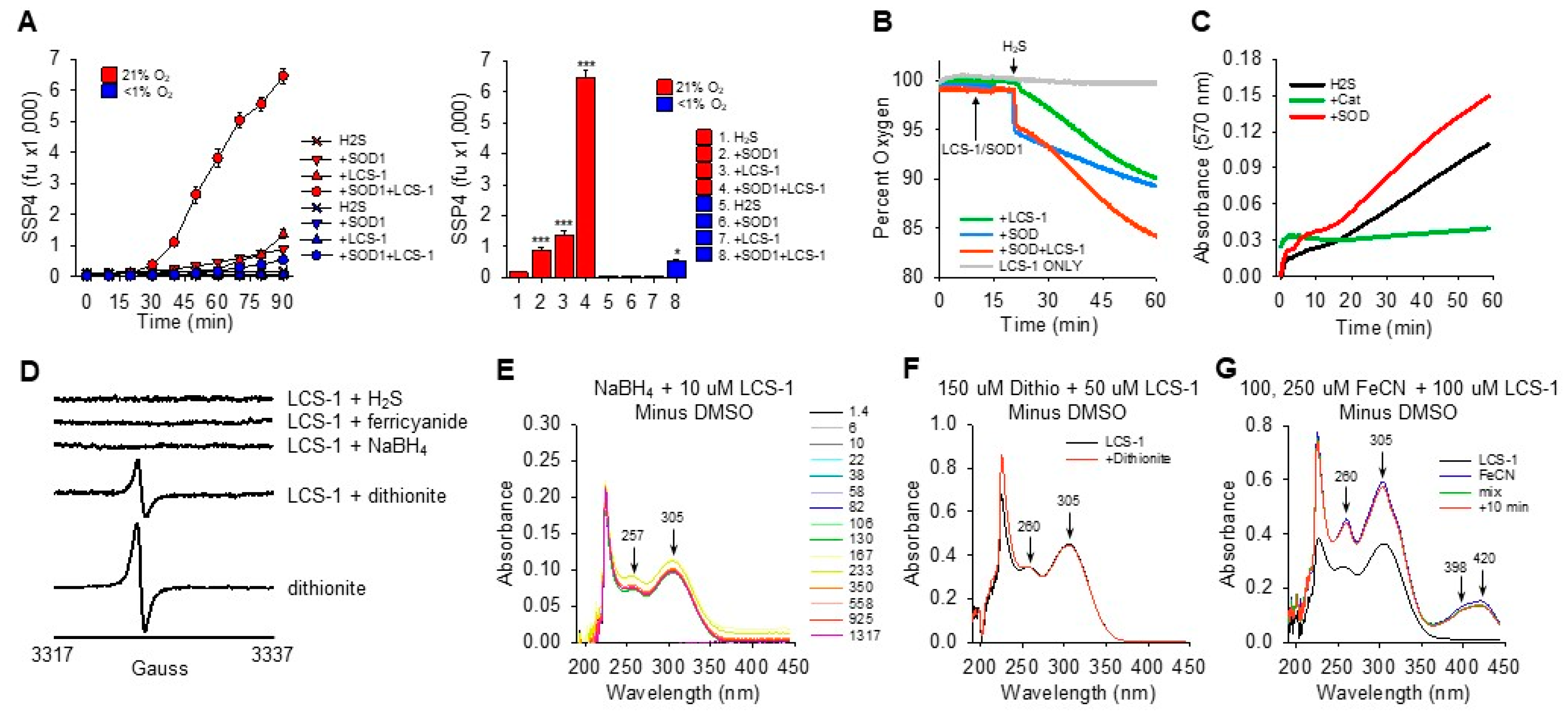
Oxygen dependency of H2S oxidation to polysulfides (SSP4 fluorescence) by SOD1 and LCS-1 was examined by sparging the reagents with nitrogen for 20 min prior to mixing and covering the 96-well plates with tape during counting. This reduced oxygen tension to <1%, prevented polysulfide formation by both SOD1 and LCS-1, and almost eliminated polysulfide production by SOD1 combined with LCS-1 (Figure 5A).
To confirm the oxygen dependency of these reactions, oxygen consumption was monitored following the addition of H2S to LCS-1, SOD1, and the combination of LCS-1 and SOD1. As shown in Figure 5B, the addition of H2S produced a slight, rapid decrease in oxygen tension followed by a steady decline. Adding H2S to SOD1 produced a rapid 5% decrease in oxygen tension, equivalent to a decrease of 13 μM, followed by a steady decline that was similar to that of H2S alone. H2S added to LCS-1 produced a slight (<1%) rapid decrease in oxygen, followed by what appeared to be a two-phase decrease that started slowly and then increased after 15 min. The rate of oxygen decrease after 15 min was approximately 0.23%, or 0.62 μmoles/min, and it exceeded the rate of H2S alone or H2S with SOD1. Adding H2S to SOD1 combined with LCS-1 produced a curve that exhibited characteristics of both SOD1 and LCS-1, although the net decrease in oxygen (24%, 63 μmoles) was greater than the sum of each of the two compounds (10% each, 53 μmoles total). The increase in oxygen consumption approximately 15 min after addition of H2S to SOD1 combined with LCS-1 is consistent with the delayed increase in polysulfide production observed in Figure 4.
Using the Amplex Red assay to detect H2O2, it was evident that addition of H2S to LCS-1 produced a progressive increase in absorbance that was inhibited by catalase, confirming that H2O2 was produced by H2S and LCS-1 (Figure 5C). The rate of H2O2 production from the H2S-LCS-1 reaction was further increased by SOD1, and in both the H2S-LCS-1 and H2S-LCS-1-SOD1 reactions, there was a noticeable increase in the rate of H2O2 production after approximately 15 min. This supports our observations of a synergistic interaction between LCS-1 and SOD1 that has an initial slow and a delayed, more rapid second phase.

3.5. SOD1-LCS-1 Oxidation of H2S Does Not Appear to Involve Redox Cycling of LCS-1

LCS-1-catalyzed oxidation of H2S and reduction of oxygen to hydrogen peroxide is similar to previous observations of redox cycling in H2S/oxygen reactions with naphthoquinones. Naphthoquinones undergo two consecutive single-electron reduction/oxidation reactions with H2S and molecular oxygen, respectively. This produces a characteristic semiquinone radical, detectable by EPR, and it changes the UV–vis spectrum characteristics from that of an oxidized to a fully reduced NQ [14,22,23]. These methods were employed to determine if similar processes were involved in H2S/oxygen reactions catalyzed by LCS-1.
Addition of 4 mM H2S to 4 mM LCS-1 did not produce any detectable EPR signal, nor did addition of 4 mM ferricyanide, NaBH4 crystals, or 4 mM dithionite (Figure 5D). The EPR signature in the LCS-1-dithionite reaction comes from the decomposition of dithionite to the SO2• radical, as shown in the bottom trace. These results suggest that either a radical species is not formed in LCS-1 H2S reactions or that it is too short-lived to be detected.
Similarly, no obvious changes were observed in UV–vis absorption spectra of reactions between 4 mM LCS-1 and 4 mM sodium borohydride (NaBH4), dithionite (S2O42−) crystals, and 4 mM ferric cyanide (Fe(CN)63−; Figure 5E–G). Absorption spectra of LCS-1 and reactions with H2S and Cys are shown in Supplemental Figure S1. H2S had two peaks at 209 nm and 229 nm. LCS-1 had two sharp peaks at 207 nm and 221 nm and two broad peaks at 257 nm and 306 nm. However, the sharp peaks were dominated by DMSO, and these disappeared when the DMSO spectrum was subtracted. The addition of 50 μM or 250 μM H2S to 50 μM LCS-1 produced a slight increase in the 221 nm peak, slightly red-shifted the downslope of the 221 nm peak and decreased the nadir at 245 nm. Higher concentrations of all reactants produced similar responses. The addition of SOD to LCS-1 and H2S produced a broad 305–322 nm peak that was assumed to be due to polysulfides. Cysteine (50 μM) did not affect the LCS-1 absorption spectrum. Collectively, these results suggest that, unlike NQs, there are no obvious redox-dependent changes in the LCS-1 spectrum that could be resolved within the time frame of our experiments.

3.6. Catalase Only Slightly Inhibits LCS-1-SOD1 Oxidation of H2S

The addition of catalase to the H2S-LCS-1-SOD1 reaction did not produce a consistent or substantial effect on the reaction products as measured by LC-MS/MS. Catalase slightly decreased H2S2-4 and H2SO3 and increased H2S2O3, whereas H2S, H2S5,6 and HS4,5OH were unaffected (Supplemental Figure S2). These results suggest that polysulfides and sulfoxides are primarily derived from the LCS-1-SOD1 oxidation of H2S and are not secondarily produced through H2O2 oxidation.

3.7. LCS-1 Forms Monothiol Adducts with SOD1 That Affect H2S Oxidation

GSH and Cys form adducts with NQs through Michael addition, and these adducts may affect NQ oxidation of H2S [14]. NQ-S adducts include mono- and di-thiol NQs and di-NQs connected to each other by NQ-Sn-NQ bridges (where S ≥ 1). However, LC-MS/MS examination showed that only H2S, glutathione, and cysteine monothiol S-adducts were formed with LCS-1, and this was through the replacement of one of the chloride atoms (Figure 1). While it was not possible to identify which chloride was involved, it has been reported that 4,5-dichloro-3(2H)-pyridazinones tend to react with sulfur nucleophiles at the 4-position (β to the carbonyl) under aqueous reaction conditions [24].
The effects of GSH and Cys adducts on H2S oxidation by LCS-1 and SOD1 were initially examined by incubating either thiol with LCS-1 and SOD1 for 30 min before adding SSP4 and H2S (Supplemental Figure S3A,B). Equimolar (10 μM) concentrations of LCS-1 and GSH increased polysulfide production, whereas 50 μM GSH and both 10 μM and 50 μM Cys decreased it. Neither GSH nor Cys affected polysulfide production when H2S was incubated with LCS-1 or SOD1 separately.
It is difficult to assess the effects of LCS-1-HS adducts on interactions with SOD1 because LCS-1 not only forms adducts with H2S but it also oxidizes H2S. Two sets of experiments were designed to address this issue. In the first set of experiments, 10 μM LCS-1 or 10 μM LCS-1 plus 0.1 μM SOD1 were pre-incubated for 10 min with either 10 μM or 50 μM H2S, then 300 μM H2S and SSP4 were added and counted for 140 min. These were compared to LCS-1 plus SOD pre-incubated without H2S for 0 or 10 min (Supplemental Figure S4A). Preincubation of LCS-1 with either 10 μM or 50 μM prevented polysulfide (SSP4 fluorescence) production. Simultaneous addition of LCS-1, SOD1, and 300 μM H2S produced a large increase in polysulfide production that was slightly but significantly greater than polysulfide production when LCS-1 and SOD1 were preincubated for 10 min before adding 300 μM H2S. Preincubation of LCS-1 or LCS-1 plus SOD1 with either 10 μM or 50 μM H2S consistently decreased polysulfide production from 75% to 40%, although 50 μM H2S was somewhat less efficacious than 10 μM H2S.
In the second set of experiments, LCS-1-SH adducts were prepared by pre-incubating low concentrations of H2S (10 μM or 50 μM) with 10 μM LCS-1, without or with 0.1 μM SOD1, for 30 min in closed containers to minimize H2S loss due to volatility. The containers were then opened for 1 h to allow volatilization of the unreacted H2S. The LCS-1-SH adduct without SOD1 was then incubated with SOD1 for another 30 min before adding H2S and SSP4. These samples were compared to controls preincubated for similar intervals but without H2S (Supplemental Figure S4B). There was no difference in polysulfide production between preincubating LCS-1 with SOD1 for 30 min prior to adding SSP4 and 300 μM H2S or adding SOD1 to LCS-1 immediately followed by SSP4 and 300 μM H2S. However, preincubation of LCS-1 with 10 μM H2S decreased polysulfide production by 35%, and preincubation of LCS-1 and SOD1 with 10 μM H2S decreased polysulfide production by 21%. With 50 μM H2S, these values were 21% and 19%, respectively. The increase in polysulfide production over time did not appear to be substantially different if LCS-1 was preincubated without or with SOD1 for any H2S treatment (red, green, and blue traces).
Collectively, these results suggest that LCS-1-SH adducts inhibit LCS-1 binding to SOD1, albeit less effectively than either LCS-1-GSH or LCS-1-Cys adducts. They also suggest that LCS-1 and SOD1 rapidly react with each other prior to augmenting H2S oxidation.
To confirm that there was little delay between addition of LCS-1 and SOD1 and enhancement of H2S oxidation, either 1 μM or 10 μM LCS-1 was incubated with 0.1 μM SOD1 0, 30, 60, 90, or 120 min before addition of H2S and SSP4 and fluorescence measured over an additional 130 min. As shown in Supplemental Figure S5A,B, the period of LCS-1 incubation with SOD1 did not affect either the onset or magnitude of polysulfide production. However, with 1 μM LCS-1, the delay before polysulfide production began to increase (approximately 70 min) was essentially twice that with 10 μM LCS-1 (~35 min). These results confirm that there is little, if any, delay between the addition of LCS-1 to SOD1 and the increased catalytic activity of the LCS-1-SOD1 complex.

3.8. SOD Inhibition by LCS-1 and ATN-244

Concentration-dependent inhibition of superoxide production by SOD1 using the nitro blue tetrazolium assay is shown in Supplemental Figure S6A. The approximate IC50 was 60 nM SOD. The effects of LCS-1 and the Cu-chelator ATN-244 on SOD activity are shown in Supplemental Figure S6B and the percent SOD1 inhibition in Supplemental Figure S6C. The addition of 10 μM ATN-244 nearly completely inhibited SOD1 activity, and the effects of ATN-244 were concentration-dependent. Conversely, all LCS-1 concentrations, from 0.1 μM to 100 μM, inhibited SOD1 by approximately 15%, and there was no apparent concentration-dependent effect. With the WST1 assay, 10 and 20 μM LCS-1 inhibited SOD1 by 22 and 35%, respectively, but these were not significantly different from each other (Supplemental Figure S6D). Collectively, it was not possible to inhibit more than 35% of SOD1 with LCS-1. This contrasts with an estimated 75% inhibition observed by Somwar et al. at 10 μM LCS-1 [2]. The reason for this large discrepancy is unknown, but the present experiments suggest that even with relatively high LCS-1 concentrations, SOD1 retains considerable dismutase activity.

3.9. Effects of LCS-1 on RSS in HEK293T Cells

As proof of principle, the effects of LCS-1 were examined in HEK293T cells. As shown in Figure 6, the effects of LCS-1 were more pronounced in 5% oxygen and with 1 μM LCS-1. Here, LCS-1 increased H2S, H2S2, H2S3, H2SO3, and GSSH and decreased GSH and Cys. The increases in inorganic polysulfides are generally consistent with LCS-1 oxidation of H2S in buffer, although they could also be the result of increased H2S due to decreased oxidation as observed with cells in 5% oxygen. Additional studies are underway to clarify these issues.

4. Discussion

Our experiments show that LCS-1 oxidizes H2S and that it acts synergistically with SOD1 to oxidize H2S to polysulfides with a preponderance of H2S3-5. These reactions have a slow onset but increase after 15–25 min, suggesting a chain reaction. H2S oxidation by the combination of LCS-1 and SOD1 consumes oxygen and produces hydrogen peroxide, but it does not appear to involve redox cycling of LCS-1. Nor does the hydrogen peroxide produced in these reactions appear to make a substantial contribution to polysulfide production. H2S oxidation is favored as the LCS-1:SOD1 ratio approaches or exceeds 10:1 or higher, and it is effective with as little as 0.01 μM SOD. LCS-1 forms monothiol adducts with H2S, GSH, and Cys, and these adducts inhibit LCS-1-SOD1 synergism. However, SOD1 was not completely inhibited by LCS-1. This suggests that in the observed reactions with H2S, SOD1 may have both oxidase and dismutase activities, but the relative proportions of each remain to be determined. In general, the LCS-1-SOD1 reactions observed in buffer were consistent with the effects of LCS-1 in cells, suggesting that some (if not many) of the effects of LCS-1 in biological systems may be mediated through thiol metabolism.
Quinones and naphthoquinones undergo consecutive one-electron reactions to oxidize H2S to polysulfides through redox cycling of the quinone with oxygen and H2S [14,22,23]. These reactions are also enhanced by SOD1. However, it is thought that in these reactions, SOD dismutation (and removal) of superoxide helps drive the otherwise unfavorable one-electron oxidation of the reduced hydroquinone by oxygen [25]. Since there was no evidence for LCS-1 redox cycling in the present studies, alternative catalytic mechanisms need to be considered.

4.1. H2S Oxidation by LCS-1

These experiments suggest that LCS-1 can oxidize H2S in reactions that consume H2S and molecular oxygen and produce polysulfides and hydrogen peroxide. There was no evidence that LCS-1 oxidation of H2S involved either one-electron production of LCS-1 radicals or a two-electron reduced quinone/hydroquinone-type molecule that could redox cycle with oxidized LCS-1. This could suggest that H2S oxidation by LCS-1 is distinct from redox cycling previously observed for quinones or naphthoquinones [14,22,23]. It is also possible that reduced LCS-1 was not detected due to rapid reoxidization. An S-adduct of LSC-1 formed through a Michael-type addition-elimination of Cl at the 4-position may also have redox activity. The structurally related herbicide chloridazon, which is a 4-amino (NH2) adduct of the LCS-1 scaffold (4-amino-5-chloro-3(2H)-pyridazinone), has useful redox properties [26], suggesting the potential for similar properties for the S-adduct of LCS-1.

4.2. Proposed Mechanism of LCS-1/SOD1 Synergism

The premise for LCS-1-SOD1 oxidation of H2S is based on previous observations that SOD1 can oxidize low molecular weight thiols such as Cys and, to a lesser extent, GSH. These reactions produce an oxidized dithiol and hydrogen peroxide Equation (1) but are independent of SOD1 dismutase activity [27].
2RSH + O2 + (SOD1) –> RSSR + H2O2
The reaction appears to be a two-step process. First, the low molecular weight thiol reduces the intramolecular Cys57-Cys146 disulfide bond in SOD1, which results in misfolded protein [28]. Misfolding opens the catalytic site, favors the loss of structural Zn2+, and, by increasing the oxidizing properties of Cu, changes SOD1 from a dismutase to an oxidase (Equation (2); [29,30]). In the second step, the misfolded SOD1 then catalytically oxidizes low molecular weight thiols and reduces molecular oxygen to H2O2. This reaction is especially efficient with Cys, where Cys readily reduces the intramolecular disulfide, and the resultant oxidized cystine is reduced back to Cys by GSH. The Cys keeps the intramolecular disulfide from reforming, i.e., a “cysteine-dependent redox short circuit” (Equation (3), [29]). The direct reaction of SOD1 with GSH is reportedly slow or non-existent [27,29], but its consumption through cystine reduction can deplete intracellular GSH and thereby exacerbate oxidative stress.
SOD1(Zn)(Cys57-Cys146) + 2Cys-S –> SOD1(Cys57, Cys146) + Cys-SS-Cys + Zn2+
Cys-SS-Cys + 2GSH –> 2Cys + GSSG
Thiol oxidation appears to result, via an intermediate Equation (4), from a two-electron process whereby oxygen reoxidizes Cu1+ to Cu2+ and forms a sulfenic acid, the latter then reacting with another thiol to form the oxidized dithiol. Superoxide is not required in the following reactions (Equations (5) and (6); where R = Cys, [27]):
SOD1-Cu2+ + RSH <–> [SOD1-Cu2+-RS <–> SOD1-Cu1+-RS•] + H+
[SOD1-Cu+1-RS•] + O2 + H+ + H2S –> SOD1-Cu2+ + RSOH + H2O2
RSH + RSOH –> RSSR + H2O
It is well known that LCS-1 binds to SOD1 [2] and increases intracellular ROS [3,4,6,7]. However, the mechanism of SOD1 inhibition by LCS-1 has not been resolved, i.e., there is no evidence that it directly binds to and inhibits redox-active Cu. The present studies suggest that LCS-1 forms an adduct with one (or both) of the intramolecular Cys57-Cys146 in SOD1, similar to Cys. By breaking this bond, SOD1 becomes an oxidase that enzymatically catalyzes the oxidation of H2S and reduces molecular oxygen as described for other thiols.
Collectively, the present experiments support the above hypothesis. LCS-1 and SOD1 act synergistically to oxidize H2S to polysulfides. This reaction consumes oxygen and produces ROS, but neither superoxide nor hydrogen peroxide appears to substantially contribute to H2S oxidation. This synergism is inhibited by preincubation of LCS-1 with GSH or Cys, presumably because the LCS-1-GSH or LCS-1-Cys adduct can no longer reduce the Cys57-Cys146 disulfide in SOD1. Furthermore, the sulfenic acid produced in this reaction can also react with polyhydrosulfides to produce longer polyhydrosulfides (Equation (7)).
H2Sn + HSOH –> H2S(n+1) + H2O

4.3. Chemical Reactivity of Products from H2S Oxidation

The hallmark of LCS-1 activity in cell toxicity is increased ROS production and depletion of GSH [2,3,4,7,8,29]. This is thought to occur somewhat paradoxically via an increase in hydrogen peroxide, although it could be due to GSH depletion. The primary reactive products of H2S oxidation by LCS-1/SOD1 are inorganic per- and polysulfides (H2S2 and H2S3-6), sulfite, and polysulfenic acids. How these act and interact in the context of biological systems is yet to be fully resolved, especially regarding their role(s) as cellular oxidants or reductants and in the context of homeostatic signaling molecules or cytotoxic compounds [31]. In arguably the most simplistic form, organic per- and polysulfides (where R is generally GSH or Cys and R’ may be GSH, Cys, or protein-Cys) are often considered in equilibrium with H2S or its anion HS (Equation (8)).
H2S(HS) + RSSR’ <–> RSSH(RSS) + R’SH
The biological outcome of this reaction will depend on what products are favored. Inorganic and organic hydroper- and hydropolysulfides may be either nucleophiles or electrophiles [32,33]. It has been argued that the reaction shown in eq. 8 is favored far to the left and that RSSH(RSS), if present, functions as potent electrophiles. This is based on thermodynamic grounds and practical considerations, i.e., protein integrity would be lost if their disulfide bonds were readily oxidized by H2S [33]. On the other hand, while acknowledging that the reaction in Equation (11) is favored to go to the left, it has also been posited that “unlike RSH, RSSH can be either reductants/nucleophiles or oxidants/electrophiles depending on the protonated state” [32].
The pKa1 and pKa2 of inorganic per- and polysulfides decrease as the number of sulfur atoms increases Table S1 Supplemental information [34]. This progressively increases the degree of ionization (Figure 7). At pH 7, half of S1 is fully protonated (H2S) and half is the hydrosulfide anion (HS), whereas with S2, less than 1% is protonated, nearly 83% is the hydrosulfide anion, and over 16% is the dianion. The pKa for small organic thiols also decreases below 6.0 upon persulfidation, e.g., 5.45 for GSSH and 5.2 for CysSSH, reportedly increasing their nucleophilicity and reactivity [35,36].
While the pH of intracellular organelles will affect the degree of ionization, it is evident that, except for the acidic lysosomes, essentially all intracellular S3-6 will be dianions and 20% of small organic thiols will be anions. This suggests an increased propensity for nucleophilic interactions, which would help maintain a reduced intracellular environment, but at the risk of destabilizing disulfide bridges. However, based on our LC-MS/MS analysis of the products of H2S oxidation by LCS-1 and LCS-1-SOD1, it also appears that the amount of polysulfides produced in these reactions decreases exponentially as the number of sulfur atoms increases. pH-specific intercompartmental equilibria of polysulfides, which favor more permeable, fully protonated species, will further complicate the issue. How these factors interact and affect the sulfur biome is a challenging and difficult task.

4.4. Biological Significance of H2S Oxidation by LCS-1

Here, we demonstrate that LCS-1 impacts low molecular weight RSS in HEK293T cells, and we show that these effects are consistent with H2S oxidation by LCS-1 and LCS-1/SOD1 in buffer. These off-target actions are likely to affect cellular sulfur metabolism and signaling, and they suggest alternative mechanisms need to be considered in evaluating the therapeutic efficacy of LCS-1. While the present work was designed as proof of principle, it clearly demonstrates the need for a reappraisal of the biological actions of LCS-1 in other cells and contexts.

Supplementary Materials

The following supporting information can be downloaded at: https://www.mdpi.com/article/10.3390/antiox13080991/s1.

Author Contributions

Conceptualization, K.R.O., K.J.C. and K.D.S.; formal analysis, K.R.O., T.T., K.J.C., Y.G., Z.M., K.M., T.A.K., P.J.J., J.F., G.W. and K.D.S.; funding acquisition, K.R.O.; investigation, T.T., K.J.C., Y.G., Z.M., E.P., K.M., T.A.K. and G.W.; methodology, K.R.O., T.T., K.J.C., Y.G., Z.M., P.J.J. and G.W.; project administration, K.R.O.; resources, K.R.O.; supervision, K.R.O.; validation, K.R.O., Y.G. and P.J.J.; visualization, K.R.O.; writing—original draft, K.R.O. and K.D.S.; writing—review and editing, K.R.O., T.T., K.J.C., T.A.K., P.J.J., J.F., G.W. and K.D.S. All authors have read and agreed to the published version of the manuscript.

Funding

This research was supported in part by United States National Science Foundation grant No. IOS2012106 (KRO), United States National Institutes of Health National Institute of Neurological Disorders and Stroke grant R01NS094535-07 (JMT, TAK), the Welch Foundation grant BE-0048 (TAK), and the Biomedical Research Foundation at Central Arkansas Veteran’s Healthcare System (KDS).

Institutional Review Board Statement

Not applicable.

Informed Consent Statement

Not applicable.

Data Availability Statement

The data presented in this study are available on request from the corresponding author.

Conflicts of Interest

The authors have no conflicts of interest.

References

  1. Somwar, R.; Shum, D.; Djaballah, H.; Varmus, H. Identification and preliminary characterization of novel small molecules that inhibit growth of human lung adenocarcinoma cells. J. Biomol. Screen. 2009, 14, 1176–1184. [Google Scholar] [CrossRef] [PubMed]
  2. Somwar, R.; Erdjument-Bromage, H.; Larsson, E.; Shum, D.; Lockwood, W.W.; Yang, G.; Sander, C.; Ouerfelli, O.; Tempst, P.J.; Djaballah, H.; et al. Superoxide dismutase 1 (SOD1) is a target for a small molecule identified in a screen for inhibitors of the growth of lung adenocarcinoma cell lines. Proc. Natl. Acad. Sci. USA 2011, 108, 16375–16380. [Google Scholar] [CrossRef] [PubMed]
  3. Du, T.; Song, Y.; Ray, A.; Chauhan, D.; Anderson, K.C. Proteomic analysis identifies mechanism(s) of overcoming bortezomib resistance via targeting ubiquitin receptor Rpn13. Leukemia 2021, 35, 550–561. [Google Scholar] [CrossRef] [PubMed]
  4. Ling, M.; Liu, Q.; Wang, Y.; Liu, X.; Jiang, M.; Hu, J. LCS-1 inhibition of superoxide dismutase 1 induces ROS-dependent death of glioma cells and degradates PARP and BRCA1. Front. Oncol. 2022, 12, 937444. [Google Scholar] [CrossRef]
  5. McAndrew, E.N.; Lepage, C.C.; McManus, K.J. The synthetic lethal killing of RAD54B-deficient colorectal cancer cells by PARP1 inhibition is enhanced with SOD1 inhibition. Oncotarget 2016, 7, 87417–87430. [Google Scholar] [CrossRef]
  6. Sajesh, B.V.; McManus, K.J. Targeting SOD1 induces synthetic lethal killing in BLM- and CHEK2-deficient colorectal cancer cells. Oncotarget 2015, 6, 27907–27922. [Google Scholar] [CrossRef]
  7. Papa, L.; Hahn, M.; Marsh, E.L.; Evans, B.S.; Germain, D. SOD2 to SOD1 switch in breast cancer. J. Biol. Chem. 2014, 289, 5412–5416. [Google Scholar] [CrossRef]
  8. Guo, T.; Wang, X.; Zhang, G.; Xia, T.; Zhu, R.; Tou, J. Dihydromyricetin functions as a tumor suppressor in hepatoblastoma by regulating SOD1/ROS pathway. Front. Oncol. 2023, 13, 1160548. [Google Scholar] [CrossRef]
  9. Steverding, D.; Barcelos, Y. Cytotoxic Activity of LCS-1 is not Only due to Inhibition of SOD1. Drug Res. 2020, 70, 57–60. [Google Scholar] [CrossRef]
  10. Switzer, C.H.; Kasamatsu, S.; Ihara, H.; Eaton, P. SOD1 is an essential H2S detoxifying enzyme. Proc. Natl. Acad. Sci. USA 2023, 120, e2205044120. [Google Scholar] [CrossRef]
  11. DeLeon, E.R.; Gao, Y.; Huang, E.; Arif, M.; Arora, N.; Divietro, A.; Patel, S.; Olson, K.R. A case of mistaken identity: Are reactive oxygen species actually reactive sulfide species? Am. J. Physiol.-Regul. Integr. Comp. Physiol. 2016, 310, R549–R560. [Google Scholar] [CrossRef] [PubMed]
  12. Greiner, R.; Palinkas, Z.; Basell, K.; Becher, D.; Antelmann, H.; Nagy, P.; Dick, T.P. Polysulfides link H2S to protein thiol oxidation. Antioxid. Redox Signal 2013, 19, 1749–1765. [Google Scholar] [CrossRef]
  13. Olson, K.R. Are Reactive Sulfur Species the New Reactive Oxygen Species? Antioxid. Redox Signal. 2020, 33, 1125–1142. [Google Scholar] [CrossRef] [PubMed]
  14. Olson, K.R.; Clear, K.J.; Gao, Y.; Ma, Z.; Cieplik, N.M.; Fiume, A.R.; Gaziano, D.J.; Kasko, S.M.; Luu, J.; Pfaff, E.; et al. Redox and Nucleophilic Reactions of Naphthoquinones with Small Thiols and Their Effects on Oxidization of H2S to Inorganic and Organic Hydropolysulfides and Thiosulfate. Int. J. Mol. Sci. 2023, 24, 7516. [Google Scholar] [CrossRef] [PubMed]
  15. Olson, K.R.; Clear, K.J.; Takata, T.; Gao, Y.; Ma, Z.; Pfaff, E.; Travlos, A.; Luu, J.; Wilson, K.; Joseph, Z.; et al. Reaction Mechanisms of H2S Oxidation by Naphthoquinones. Antioxidants 2024, 13, 619. [Google Scholar] [CrossRef] [PubMed]
  16. Bibli, S.I.; Luck, B.; Zukunft, S.; Wittig, J.; Chen, W.; Xian, M.; Papapetropoulos, A.; Hu, J.; Fleming, I. A selective and sensitive method for quantification of endogenous polysulfide production in biological samples. Redox Biol. 2018, 18, 295–304. [Google Scholar] [CrossRef]
  17. Olson, K.R.; Gao, Y. Effects of inhibiting antioxidant pathways on cellular hydrogen sulfide and polysulfide metabolism. Free Radic. Biol. Med. 2019, 135, 1–14. [Google Scholar] [CrossRef]
  18. Olson, K.R.; Gao, Y.; Arif, F.; Patel, S.; Yuan, X.; Mannam, V.; Howard, S.; Batinic-Haberle, I.; Fukuto, J.; Minnion, M.; et al. Manganese Porphyrin-Based SOD Mimetics Produce Polysulfides from Hydrogen Sulfide. Antioxidants 2019, 8, 639. [Google Scholar] [CrossRef]
  19. Kasamatsu, S.; Ida, T.; Koga, T.; Asada, K.; Motohashi, H.; Ihara, H.; Akaike, T. High-Precision Sulfur Metabolomics Innovated by a New Specific Probe for Trapping Reactive Sulfur Species. Antioxid. Redox Signal. 2021, 34, 1407–1419. [Google Scholar] [CrossRef]
  20. Ewing, J.F.; Janero, D.R. Microplate superoxide dismutase assay employing a nonenzymatic superoxide generator. Anal. Biochem. 1995, 232, 243–248. [Google Scholar] [CrossRef]
  21. Olson, K.R.; Gao, Y.; Arif, F.; Arora, K.; Patel, S.; DeLeon, E.R.; Sutton, T.R.; Feelisch, M.; Cortese-Krott, M.M.; Straub, K.D. Metabolism of hydrogen sulfide (H2S) and Production of Reactive Sulfur Species (RSS) by superoxide dismutase. Redox Biol. 2017, 15, 74–85. [Google Scholar] [CrossRef]
  22. Olson, K.R.; Clear, K.J.; Derry, P.J.; Gao, Y.; Ma, Z.; Wu, G.; Kent, T.A.; Straub, K.D. Coenzyme Q10 and related quinones oxidize H2S to polysulfides and thiosulfate. Free Radic. Biol. Med. 2022, 182, 119–131. [Google Scholar] [CrossRef] [PubMed]
  23. Olson, K.R.; Clear, K.J.; Derry, P.J.; Gao, Y.; Ma, Z.; Cieplik, N.M.; Fiume, A.; Gaziano, D.J.; Kasko, S.M.; Narloch, K.; et al. Naphthoquinones Oxidize H2S to Polysulfides and Thiosulfate, Implications for Therapeutic Applications. Int. J. Mol. Sci. 2022, 23, 13293. [Google Scholar] [CrossRef]
  24. Lyga, J.W. The reaction of 2-substituted-4,5-dichloro-3(2H)-pyridazinones with alkoxides and alkylthiolates. J. Heterocycl. Chem. 1988, 25, 1757–1760. [Google Scholar] [CrossRef]
  25. Song, Y.; Buettner, G.R. Thermodynamic and kinetic considerations for the reaction of semiquinone radicals to form superoxide and hydrogen peroxide. Free Radic. Biol. Med. 2010, 49, 919–962. [Google Scholar] [CrossRef]
  26. Zimpl, M.; Kotoucek, M.; Lemr, K.; Vesela, J.; Skopalova, J. Electrochemical reduction of chloridazon at mercury electrodes, and its analytical application. Fresenius J. Anal. Chem. 2001, 371, 975–982. [Google Scholar] [PubMed]
  27. Winterbourn, C.C.; Peskin, A.V.; Parsons-Mair, H.N. Thiol oxidase activity of copper, zinc superoxide dismutase. J. Biol. Chem. 2002, 277, 1906–1911. [Google Scholar] [CrossRef]
  28. Yamazaki, K.; Tahara, S.; Ohyama, T.; Kuroi, K.; Nakabayashi, T. SOD1 gains pro-oxidant activity upon aberrant oligomerization: Change in enzymatic activity by intramolecular disulfide bond cleavage. Sci. Rep. 2022, 12, 11750. [Google Scholar] [CrossRef] [PubMed]
  29. Bakavayev, S.; Chetrit, N.; Zvagelsky, T.; Mansour, R.; Vyazmensky, M.; Barak, Z.; Israelson, A.; Engel, S. Cu/Zn-superoxide dismutase and wild-type like fALS SOD1 mutants produce cytotoxic quantities of H2O2 via cysteine-dependent redox short-circuit. Sci. Rep. 2019, 9, 10826. [Google Scholar] [CrossRef]
  30. Nordlund, A.; Leinartaite, L.; Saraboji, K.; Aisenbrey, C.; Grobner, G.; Zetterstrom, P.; Danielsson, J.; Logan, D.T.; Oliveberg, M. Functional features cause misfolding of the ALS-provoking enzyme SOD1. Proc. Natl. Acad. Sci. USA 2009, 106, 9667–9672. [Google Scholar] [CrossRef]
  31. Akaike, T.; Morita, M.; Ogata, S.; Yoshitake, J.; Jung, M.; Sekine, H.; Motohashi, H.; Barayeu, U.; Matsunaga, T. New aspects of redox signaling mediated by supersulfides in health and disease. Free Radic. Biol. Med. 2024, 222, 539–551. [Google Scholar] [CrossRef] [PubMed]
  32. Fukuto, J.M. The chemistry of hydropersulfides (RSSH) as related to possible physiological functions. Arch. Biochem. Biophys. 2023, 743, 109659. [Google Scholar] [CrossRef]
  33. Switzer, C.H. How super is supersulfide?: Reconsidering persulfide reactivity in cellular biology. Redox Biol. 2023, 67, 102899. [Google Scholar] [CrossRef] [PubMed]
  34. Kamyshny, A., Jr.; Goifman, A.; Rizkov, D.; Lev, O. Formation of carbonyl sulfide by the reaction of carbon monoxide and inorganic polysulfides. Environ. Sci. Technol. 2003, 37, 1865–1872. [Google Scholar] [CrossRef]
  35. Benchoam, D.; Semelak, J.A.; Cuevasanta, E.; Mastrogiovanni, M.; Grassano, J.S.; Ferrer-Sueta, G.; Zeida, A.; Trujillo, M.; Moller, M.N.; Estrin, D.A.; et al. Acidity and nucleophilic reactivity of glutathione persulfide. J. Biol. Chem. 2020, 295, 15466–15481. [Google Scholar] [CrossRef]
  36. Benchoam, D.; Cuevasanta, E.; Roman, J.V.; Banerjee, R.; Alvarez, B. Acidity of persulfides and its modulation by the protein environments in sulfide quinone oxidoreductase and thiosulfate sulfurtransferase. J. Biol. Chem. 2024, 300, 107149. [Google Scholar] [CrossRef]
Figure 1. Structure of LCS-1, LCS-1-thiol adducts (R = H, GSH, or cysteine), and 2,3-dichloro-1,4-naphthoquinone.
Antioxidants 13 00991 g001
Figure 2. LCS-1 and SOD1, but not SOD2, synergistically consume H2S to produce polysulfides. (A) Both LCS-1 and SOD1 consume H2S (AzMC fluorescence), and when LCS-1 and SOD1 are combined, H2S consumption is further increased. H2S consumption was measured by incubating 100 μM H2S with 15 μM LCS-1 or with either 0.1 μM or 1 μM bovine SOD1 with or without 15 μM LCS-1 for 2 h; 25 μM AzMC was then added, and fluorescence was measured. Mean +SE, n = 4; ***, p < 0.001 compared to H2S; ###, p < 0.001 compared to respective SOD1. (B) Both LCS-1 and SOD1 produce polysulfides (SSP4 fluorescence), and when combined, polysulfide production is further increased. Polysulfide production was measured by incubating 25 μM SSP4 with 300 μM H2S and with 15 μM LCS-1 or with H2S and 0.1 μM or 1 μM SOD1 alone, or in combination with 15 μM LCS-1. SSP4 fluorescence was monitored at 10 min intervals for 120 min (left panel); right panel summarizes results at 180 min; mean +SE, n = 4; ***, p < 0.001 compared to H2S; ###, p < 0.001 compared to respective SOD1. (C) SOD2 does not synergize with 10 μM LCS-1 to oxidize 300 μM H2S to polysulfides (SSP4 fluorescence). SOD1 and SOD2 0.1 μM, mean + SE, *** p < 0.001 vs. H2S.
Antioxidants 13 00991 g002
Figure 3. (A) LC-MS/MS analysis (area under the curve, AUC) of reactive sulfur species in 100 μM H2S and after 20 min incubation of H2S with 1 μM SOD1, 10 μM LCS-1, and SOD1 with LCS-1. SOD1, LCS-1, and SOD1 with LCS-1 progressively decreased H2S; only SOD1 increased H2S2, whereas LCS-1 alone or in combination with SOD1 increased H2S3 and H2S4. Conversely, SOD1 alone and in combination with LCS-1 decreased sulfite (H2SO3), whereas LCS-1 had no effect. Thiosulfate (H2S2O3) was unaffected by any treatment. Sulfenic acids H2S4O and H2S5O were increased by SOD plus LCS-1, and SOD increased H2S5. Mean +SE, n = 3 replicates; *, p < 0.05; **, p < 0.01; ***, p < 0.001 vs. H2S; ##, p < 0.01; ###, p < 0.001 SOD1 vs. LCS-1 or SOD1 + LCS-1; †, p < 0.05; ††, p < 0.01; †††, p < 0.001 LCS-1 vs. SOD1 + LCS-1. (B) Fold increase in SOD1, LCS-1, and SOD1 plus LCS-1 compared to H2S control (e.g., AUCSOD1/AUCH2S). (C,D) Optical density (OD) at OD263 and OD650 following addition of 10 μM LCS-1, without or with 1 μM SOD and 300 μM H2S at approximately 300 s (arrows). All combinations produced a rapid increase in OD263 followed by a slow, linear decrease, there was no notable change in OD650 suggestive of little or no S8 production.
Antioxidants 13 00991 g003
Figure 4. Effects of LCS-1 and SOD1 concentration on synergistic oxidation of H2S to polysulfides (SSP4 fluorescence). (A) Incubation of 300 μM H2S with 10 μM LCS-1 or μM SOD1 or with various concentration ratios of SOD1 to LCS-1 ([SOD1]:[LCS-1]). LCS-1 alone did not affect polysulfides compared to H2S alone, whereas SOD1 increased it (p < 0.001). All combinations of SOD1 and LCS-1 SOD1 increased polysulfides compared to H2S with 10 μM LCS-1 (***, p < 0.001), and all but 3.0 μM SOD:3.0 μM LCS-1 increased polysulfides compared to 10 μM SOD1 (#, p < 0.05; ##, p < 0.01; ###, p < 0.001). The 0.1:1.0 SOD1:LCS-1 combination produced more (p < 0.001) polysulfides than any other combination of SOD1 and LCS-1. (B) Effects of 10 μM LCS-1 and variable SOD concentrations on polysulfides produced by 300 μM H2S compared to H2S and 10 μM LCS-1 or 10 μM SOD1. All combinations of SOD and LCS-1 produced more polysulfides than with either LCS-1 or SOD1 alone (p < 0.001). More polysulfides were produced by 0.1 μM SOD1 and 10 μM LCS-1 than by any other combination of SOD1 and LCS-1 (††, p < 0.01; †††, p < 0.001). (C) Effects of 10 μM SOD1 and variable LCS-1 concentrations on polysulfides produced by 300 μM H2S compared to H2S and either 10 μM SOD1 or 10 μM or 30 μM LCS-1 alone. Combinations of SOD and LCS-1 produced more polysulfides than with either LCS-1 or with SOD1 alone (p < 0.001). More polysulfides were produced by 0.1 μM SOD1 and either 10 μM or 30 μM LCS-1 than by any other combination of SOD1 and LCS-1; there was no difference between 0.1 μM SOD and either 10 μM or 30 μM LCS-1 (†††, p < 0.001; 0.1 μM SOD and 10 μM LCS-1 vs. other SOD-LCS-1 combinations). (D) Polysulfide production after incubation of 0.1 μM SOD1 and 300 μM H2S with either 1 μM or 10 μM LCS-1 for 24 h. The initial delay in polysulfide production was longer with 1 μM LCS-1 up to 6 h, but by 22 h there was no difference between 1 μM and 10 μM LCS-1; mean +SE, n = 4 replicates. (E) Polysulfide production by addition of 300 μM H2S to 3 μM 2,3-dichloro-1,4-naphthoquinone (DCNQ) without or with 0.1 μM SOD1. SOD increased polysulfide production, but there was no obvious lag period in either reaction. All samples are mean +SE, n = 4 wells; right panels summarize values at 90 min.
Antioxidants 13 00991 g004
Figure 5. Characteristics of H2S oxidation by SOD1 and LCS-1. (A) Oxygen dependency of polysulfide production (SSP4 fluorescence) on oxidation of 300 μM H2S by 0.1 μM SOD1 and 10 μM LCS-1 alone or in combination in 21% oxygen (room air) or <1% oxygen. No polysulfides were produced in <1% oxygen by H2S with SOD1 or LCS-1, and polysulfides were decreased more than ten-fold by SOD1 and LCS-1 together. Right panel shows 90 min mean +SE, n = 4 wells; * p < 0.05, ***, p < 0.001 compared to respective control. (B) Oxygen consumption following addition of 300 μM H2S to 1 μM SOD1 and 10 μM LCS-1 alone and in combination. Adding H2S to SOD1 produced a rapid decrease in O2, followed by a steady decline. H2S added to LCS-1 produced a slight, rapid decrease in O2, followed by what appeared to be a two-phase decrease that started slowly and then increased after 15 min. H2S added to LCS-1 with SOD1 produced a rapid drop in oxygen, followed by a two-phase decrease that consumed more oxygen than the sum of the individual H2S-LCS-1 and H2S-SOD1. No oxygen was consumed by LCS-1 alone. (C) Addition of 25 μM H2S to 25 μM LCS-1 continually produces H2O2. H2O2 production was inhibited by 0.028 μM bovine catalase but augmented by 0.1 μM SOD (Amplex Red assay; traces show absorbance at 570 nm). (D) EPR spectra of 4 mM LCS-1 added to 4 mM H2S, 4 mM ferricyanide, several crystals of NaBH4, 4 mM dithionite, or 4 mM dithionite alone. There was no evidence of LCS-1 radicals. (EG) Time-resolved absorbance spectra of LCS-1 reactions with reductants and oxidants; the spectrum of DMSO in solvent was subtracted for clarity. (E) Spectrum of 10 μM LCS-1 following addition of several crystals of sodium borohydride (NaBH4) at 22 s. (F) Spectrum before and after addition of 150 μM dithionite (S2O42−) to 50 μM LCS-1. (G) Spectra in tandem cuvettes, 100 μM LCS-1 only in cuvette #1 (black line), 150 μM ferricyanide (FeCN) only in cuvette #2 (blue line), immediately or 10 min after mixing cuvettes 1 and 2 (green and red lines, respectively). There was no effect of any compound on the LCS-1 spectrum.
Antioxidants 13 00991 g005
Figure 6. LC-MS/MS analysis of RSS in HEK293 cells after 48 h incubation in 21% (red bars) or 5% oxygen (blue bars) without or with 0.3 or 1 μM LCS-1. Mean + SE, n = 3 replicates; *, p < 0.05; ***, p < 0.001, 1 vs. 2 or 3 for either 25% or 5% oxygen; ##. p < 0.01; ###, p < 0.001 for 21% vs. respective 5% oxygen; †, p < 0.05; ††, p < 0.01; †††, p < 0.001 for 2 vs. 3.
Antioxidants 13 00991 g006
Figure 7. Effect of pH on the percent distribution of protonated (H2Sn), anionic (HSn-), and dianionic (Sn2−) sulfur species as a function of the number of catenated sulfur atoms (n). Dashed lines indicate the approximate pH of various cellular compartments and plasma.
Antioxidants 13 00991 g007
Disclaimer/Publisher’s Note: The statements, opinions and data contained in all publications are solely those of the individual author(s) and contributor(s) and not of MDPI and/or the editor(s). MDPI and/or the editor(s) disclaim responsibility for any injury to people or property resulting from any ideas, methods, instructions or products referred to in the content.