Article

Naringin Suppresses CoCl2-Induced Ferroptosis in ARPE-19 Cells

College of Chinese Herbal Medicine, Beijing University of Chinese Medicine, Beijing 102488, China
*
Authors to whom correspondence should be addressed.
Antioxidants 2025, 14(2), 236; https://doi.org/10.3390/antiox14020236
Submission received: 8 January 2025 / Revised: 31 January 2025 / Accepted: 6 February 2025 / Published: 18 February 2025
(This article belongs to the Section Natural and Synthetic Antioxidants)

Abstract

:
Hypoxic damage to retinal pigment epithelial (RPE) cells and subsequent neovascularization are key factors in the pathogenesis of branch retinal vein occlusion (BRVO). Naringin (NG), a naturally occurring flavanone glycoside, has demonstrated significant antioxidant and anti-neovascular activities. However, the regulatory effects and mechanisms of NG on ferroptosis in BRVO are yet to be explored. Our study aimed to investigate the protective effects of NG on RPE cells under hypoxic stress and to elucidate the underlying molecular mechanisms. Our findings revealed that NG significantly reduced cytotoxicity induced by cobaltous chloride (CoCl2) and also inhibited vascular proliferation in the retina, thereby attenuating choroidal neovascularization. NG pretreatment largely countered the overproduction of reactive oxygen species (ROS) and malondialdehyde (MDA) triggered by hypoxic damage, while also restoring levels of the antioxidants glutathione (GSH) and superoxide dismutase (SOD). Furthermore, NG pretreatment significantly activated the expression of hypoxia-inducible factor-1 alpha (HIF-1α) and its downstream heme oxygenase-1 (HO-1) and NADPH dehydrogenase (NQO1). In conclusion, NG not only inhibits neovascularization but also alleviates inflammation in RPE cells by modulating the HO-1/GPX4 pathway to inhibit ferroptosis. These findings highlight the potential of NG as a promising therapeutic agent for the treatment of BRVO.

Graphical Abstract

1. Introduction

Branch retinal vein occlusion (BRVO), a significant cause of irreversible vision loss, ranks among the most prevalent retinal vascular disorders in adults [1,2]. A frequent complication of BRVO, neovascularization, poses a serious threat to vision, often leading to severe impairment [3]. Retinal pigment epithelial (RPE) cells form a monolayer of metabolically active cells located on Bruch’s membrane, situated between the neurosensory retina and the vascular choroid [4]. These cells play a critical role in maintaining retinal health and supporting photoreceptor function [5].
The primary pathological changes associated with BRVO involve the disruption of the blood-retinal barrier (BRB) due to damage to RPE cells, as well as neovascularization [6]. As structural changes in the BRB contribute to hypoxia and dysregulation, along with the changes in its surrounding microenvironment, including hypoxia and inflammation, they are found to be associated with BRVO. As a result, understanding the mechanisms of RPE cell damage is crucial for unraveling BRVO pathology and advancing the development of novel therapeutic strategies. The activity of RPE cells is heavily reliant on an adequate energy supply, resulting in high reactive oxygen species (ROS) production [7]. Under normal circumstances, the cellular antioxidant system effectively neutralizes excessive ROS. However, in the case of BRVO, ROS production surges due to compromised antioxidant system functionality. Hypoxia, a primary contributing factor, further exacerbates the situation by diminishing the vitality and function of RPE cells [8]. Many studies have investigated RPE cell death mechanisms by inducing oxidative stress. Hypoxia triggers the stabilization of hypoxia-inducible factors (HIFs), which are cellular oxygen sensors that facilitate adaptation to hypoxic conditions by regulating many oxygen-dependent genes [9]. HIFs are protective in the short term, but chronic hypoxia, as observed in BRVO, can be detrimental due to HIF-dependent inflammation, neovascularization, metabolic shift, and impaired lipid transport [10]. Histopathological studies of BRVO suggest that hypoxia is accompanied by oxidative stress. Therefore, an in vitro RPE model combining oxidative stress and hypoxia would mimic the conditions observed in BRVO. Cobalt chloride (CoCl2) can substitute for ferrous ions in heme, causing structural alterations in the heme protein and impairing its function as an O2 sensor [11]. This, in turn, induces hypoxia and triggers DNA damage [12]. CoCl2 has been widely used as a hypoxia mimic in both in vivo and in vitro studies. Additionally, it amplifies ROS production in cells, exacerbating oxidative stress [13].
Ferroptosis, a novel form of cell death, is characterized by oxidative stress-induced ROS production and lipid peroxidation [14,15,16]. Recent studies have linked ferroptosis to the pathogenesis of eye diseases, including BRVO [17]. Notably, ferroptosis has been identified as a primary mechanism of hypoxia-induced cell death in retinal pigment epithelial (RPE) cells [18,19]. Thus, targeting ferroptosis pharmacologically has emerged as a promising therapeutic strategy for BRVO. Ferroptosis is characterized by the accumulation of lipid peroxides and ROS. Biochemically, heme oxygenase-1 (HO-1) and the intracellular glutamate/cystine antiporter system (Xc-system) are inhibited, glutathione (GSH) is depleted, and glutathione peroxidase 4 (GPX4) activity is reduced, leading to the accumulation of lipid peroxides and ROS, thereby promoting ferroptosis [20]. The Xc-system is a heterodimer composed of the light chain SLC7A11 (xCT) and the heavy chain SLC3A2 (4F2hc), which transports glutamate out of the cell while simultaneously importing cystine, which is involved in the synthesis of GSH [21]. GPX4 is the primary scavenger of intracellular lipid peroxides, converting GSH to oxidized glutathione while reducing toxic peroxides, making GPX4 a master regulator of ferroptosis [22].
In recent years, traditional Chinese medicine (TCM) has drawn considerable attention for its potential role in the prevention and treatment of BRVO [23,24,25]. Naringin (NG), a flavonoid with diverse biological properties, is widely present within citrus fruits [26]. It exhibits multiple beneficial activities, including anti-inflammatory, anti-neovascular, and antioxidant effects [27,28,29]. NG has the ability to inhibit the Fenton reaction of iron-ATP, possibly due to the presence of 4-ketone and 5-hydroxyl regions in its chemical structure, which facilitate iron chelation [30]. While the mechanisms underlying therapeutic effects on various diseases have become clearer in recent years, its potential application in retinal disease treatment remains largely unexplored.
This study aimed to investigate whether NG protects RPE cells from hypoxic stress injury, as well as its role in inhibiting neovascularization. To further assess its anti-neovascular properties, human umbilical vein endothelial cells (HUVECs) migration and lumen formation assays were performed. This study proposes a potential therapeutic strategy for BRVO treatment.

2. Materials and Methods

2.1. Reagents and Chemicals

Naringin (purity > 99%) was obtained from Shanghai Yuanye Bio-Technology Co., Ltd. (Shanghai, China). Fetal bovine serum (FBS), 0.05% trypsin, Dulbecco’s Modified Eagle Medium: Nutrient Mixture F-12 (DMEM/F-12), DMEM, and penicillin/streptomycin solutions were purchased from Gibco, Invitrogen (Carlsbad, CA, USA). Phosphate-buffered saline (PBS) and 3-(4,5-dimethyl thiazol-2-yl-)-2,5-diphenyl tetrazolium bromide (MTT) were obtained from Beijing Solarbio Science and Technology Co., Ltd. (Beijing, China). ROS, MDA, GSH, and SOD detection kits were purchased from Beyotime Biotechnology (Shanghai, China). The ELISA kits for IL-6, IL-1β, and TNF-α were purchased from Jianglai Biological (Shanghai, China), Shanghai. The basement membrane was purchased from Corning Incorporated (New York, NY, USA). The primary antibodies against NF-κB, ICAM-1, ACSL4, GPX4, HO-1, NQO-1, SLC7A11, and β-actin were all purchased from Abcam (Cambridge, UK).

2.2. Cell Cultures and Treatment

Human retinal pigment epithelial cells (ARPE-19) were purchased as frozen vials from Procell Life Science & Technology Co., Ltd. (Wuhan, China). The cells were cultured in DMEM/F-12 (supplemented with 10% FBS, 1% streptomycin/penicillin) at 37 °C in a humidified atmosphere containing 95% air and 5% CO2. Cells at 80–90% confluence were selected for subculture and subsequent experimentation. For CoCl2-induced apoptotic studies, the cells were cultured overnight in a serum-free medium, followed by exposure to CoCl2 (500 μM) for 24 h. HUVECs were purchased from iCell Bioscience Inc. (Shanghai, China). and cultured in DMEM supplemented with 10% (v/v) FBS and 1% penicillin/streptomycin at 37 °C in a humidified 5% CO2 incubator. The culture medium was refreshed every 1–2 days. All cell experiments were conducted using cells between the third and fifth passages.
Naringin, dissolved in DMSO, was stored at 4 °C. Based on our prior research, it was determined that a DMSO concentration below 0.1% does not induce cellular damage. Therefore, during the experimental conditions, this concentration was carefully maintained below the 0.1% threshold.

2.3. Cell Viability Assay and Morphology Examination

Cell viability was measured by MTT assay in the determination of the cytoprotective effect of NG on ARPE-19 cells. Overnight, the cells were plated in 96-well plates with a density of 4 × 10³ cells per well. After treatment with varying concentrations of NG (60–100 μM) for 24 h, CoCl2 (500 μM) was added, and treatment was continued for another 24 h. Cells without treatment were used as a control. After incubation, 100 μL of MTT working solution (0.5 mg/mL in new culture medium) was used to treat the cells at 37 °C for 4 h. Subsequently, 150 μL DMSO was added to each well to dissolve the water-insoluble formazan crystals formed. Cell viability was evaluated by measuring the absorbance of the formazan solutions at 590 nm using a microplate reader (Thermo, Multiskan, GO, USA). Morphological changes in ARPE-19 cells were observed and photographed under an inverted Olympus IX71 microscope (Olympus, Tokyo, Japan).

2.4. Transwell Assay

Cell migration was assessed with a 24-well transwell system containing 8 μm pore-sized filters without Matrigel. The HUVECs suspended in serum-free medium were added to the upper chambers, and cell culture medium was filled in the lower chambers. After 24 h of incubation, the filters were immobilized using 4% paraformaldehyde at room temperature. The migrated cells were subsequently stained using 0.1% crystal violet and observed under an inverted light microscope (Tokyo, Japan).

2.5. Tube Formation Assay

Tube formation assay was performed in Matrigel according to the manufacturer’s guidelines. After the Matrigel solidified, the cell suspension (1.5 × 10⁵ cells/well) was added, and the cells were treated with NG (60–100 μM) for 24 h. An inverted microscope was used to evaluate tube formation. Image-Pro Plus 6.0 software (Rockville, MD, USA) was used to determine the number of tubes.

2.6. Measurement of ROS Levels

DCFH-DA is a probe that freely passes through cell membranes and detects intracellular ROS. According to the manufacturer’s instructions, a diluted DCFH-DA fluorescent probe was added to the cells. The levels of ROS were determined in a CytoFLEX flow cytometer (BD Biosciences, Franklin Lakes, NJ, USA) and fluorescence microscopy (Olympus, Tokyo, Japan).

2.7. Measurement of GSH, MDA, and SOD Levels

The cells were lysed with ice-cold radio-immunoprecipitation assay (RIPA) buffer. The total protein concentrations of the lysate supernatants were determined using the Bradford protein assay kit (Biyuntian, Beijing, China). The levels of malondialdehyde (MDA), which is one of the important indicators, glutathione (GSH), an essential antioxidant substance, and superoxide dismutase (SOD), a key enzyme in antioxidant defense, in the supernatants were determined using their corresponding assay kits according to the manufacturers’ specifications. Data were calculated in terms of the protein concentration of each sample.

2.8. Western Blotting

The collected cells were lysed for 30 min on ice with RIPA buffer supplemented with protease inhibitors and then centrifuged at 10,000× g for 15 min, during which process the lysates were obtained. Following the determination of the protein concentration, all samples were subjected to boiling in loading buffer for 5 min in order to make the proteins denatured. Samples underwent SDS-PAGE and then were transferred onto PVDF membrane. The membranes, which were blocked with nonfat milk at room temperature for 2 h, were incubated with corresponding primary antibodies at 4 °C overnight. These membranes, which were then incubated with HRP-conjugated secondary antibody at room temperature for 2 h, were finally visualized by using chemiluminescence substrate. The intensity of all bands was measured by Image J (V1.51j8, NIH, Bethesda, MD, USA).

2.9. Quantitative Real-Time Polymerase Chain Reaction (qRT-PCR) Analysis

The methods of total RNA extraction and qRT-PCR have been described previously. [31]. Treat the ARPE-19 cells in the manner described previously. Then, the cells were collected, and the total RNA was isolated using TRIzol reagent (ThermoFisher, Waltham, MA, USA). Primer sequences of the targeted genes used in this study were as follows: HIF-1α(5′-CAGGAGCGAGACCCCACTAA-3′, forward; 5′-ATCACGCCACAGCTTTCCAG-3′, reverse) and VEGF(5′-TCACCAAGGCCAGCACAT-3′, forward; 5′-GGCTCCAGGGCATTAGACA-3′, reverse). All data were normalized to the mRNA expression level of GAPDH.

2.10. Statistics

Experiments were performed independently at least three times, and the results are expressed as mean ± standard deviation (SD). Statistical analysis was performed using GraphPad Prism 9.5 (GraphPad Software, Boston, MA, USA). The data were analyzed using Student’s t-test (statistical significance defined as *, p < 0.05; **, p < 0.01; ***, p < 0.001; ****, p < 0.0001) and one-way analysis of variance (ANOVA) followed by the Bonferroni/Dunn post hoc test (p < 0.05). Outliers were not identified or treated, and no transformations were applied to the data.

3. Results

3.1. Effects of CoCl2 and Naringin on Cell Viability

To determine the toxic effects of CoCl2 on ARPE-19 cells, the cells were incubated with varying concentrations of CoCl2 (50~600 μM) for 24 h, and their viability was determined using the MTT assay. As shown in Figure 1a, CoCl2 significantly inhibited the viability of ARPE-19 cells in a dose-dependent manner. The IC50 value of CoCl2 in ARPE-19 cells after 24 h of exposure was determined to be 500 μM, which was subsequently used in further experiments. As shown in Figure 1b, naringin, within the tested concentration range, exhibited no toxicity to RPE cells. Thus, naringin concentrations of 60, 80, and 100 μM were selected for subsequent studies. To evaluate the cytoprotective effect of naringin, the cells were pretreated with increasing concentrations of naringin (60~100 μM) for 24 h before exposure to 500 μM CoCl2 for 24 h. Compared to the CoCl2-treated group, naringin pretreatment enhanced the viability of ARPE-19 cells in a dose-dependent manner (Figure 1c). Furthermore, the protective effect of naringin on CoCl2-treated ARPE-19 cells was further confirmed by monitoring changes in cell number (Figure 1d).

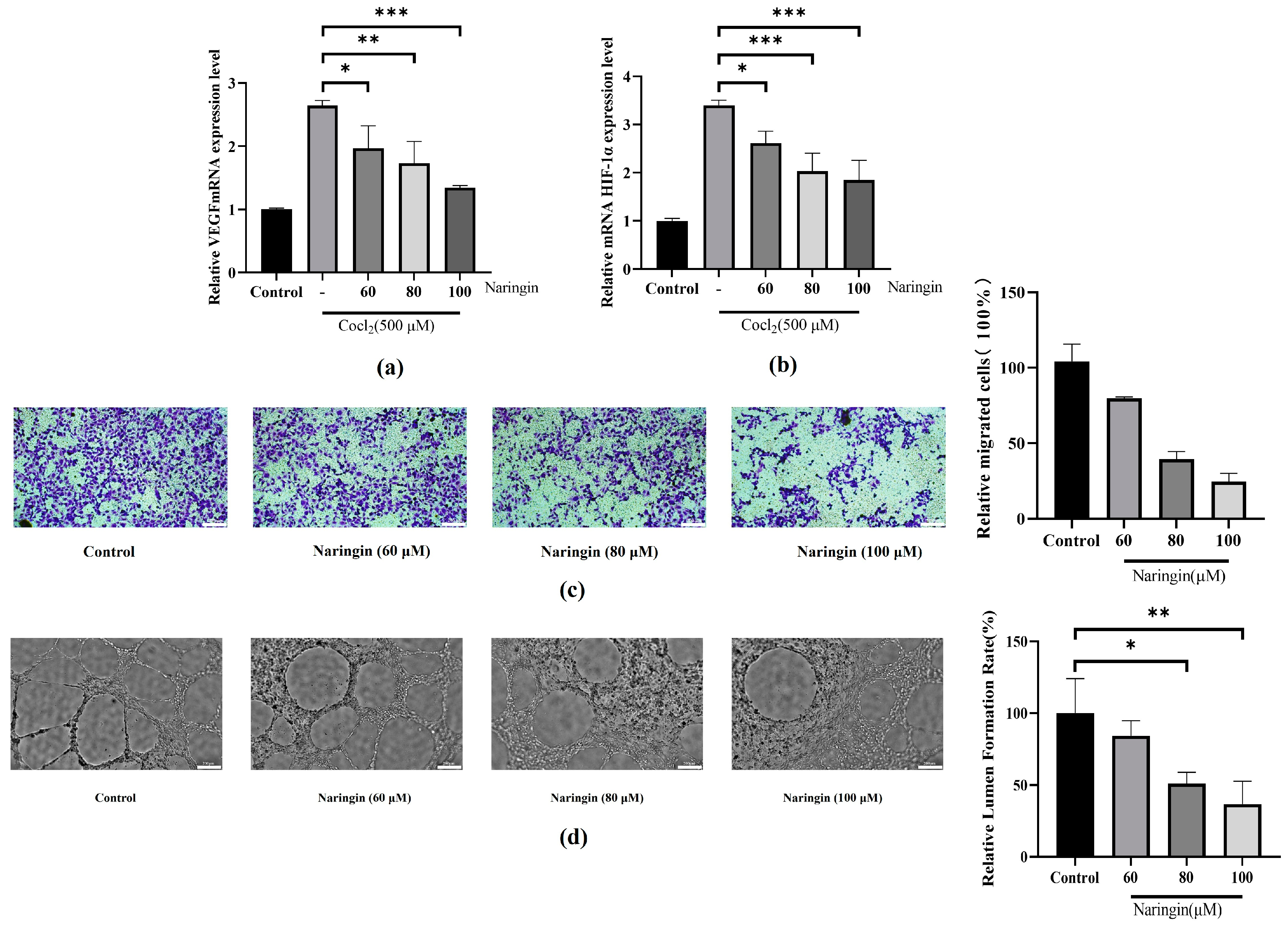
3.2. Naringin Pretreatment Attenuates Neovascularization in BRVO

During BRVO pathology, hypoxia stimulates neovascularization by influencing the regulation of the HIF-1α/VEGF signaling in RPE cells, which has a dramatic effect on neovascularization [32]. ARPE-19 cells treated with CoCl2 exhibited a marked increase in HIF-1α and vascular endothelial growth factor (VEGF) secretion compared to the control group (Figure 2a,b). These findings suggest that naringin may exert a therapeutic effect by inhibiting neovascularization. To further explore its anti-neovascular properties, we examined the impact of naringin on tube formation in HUVECs. The experimental results demonstrated that naringin not only suppressed the migration ability of HUVECs (Figure 2c) but also significantly reduced their tube formation capacity (Figure 2d). In summary, naringin inhibits the HIF-1α/VEGF signaling pathway, suggesting that naringin has anti-neovascular effects. The experiments further demonstrate that naringin can inhibit migration and tube formation in HUVECs, potentially offering a new strategy for BRVO treatment.

3.3. Naringin Pretreatment Attenuates CoCl2-Induced Inflammation and Oxidative Stress in ARPE-19

We have investigated the inflammatory response of ARPE-19 cells under hypoxic conditions by measuring the level of inflammatory mediators such as NF-κB and ICAM-1. As shown in Figure 3a. The level of NF-κB and ICAM-1, which was increased by more than three times in CoCl2-induced ARPE-19 cells, was compared to that in control cells. Treatment with naringin inhibited the upregulation stimulated by hypoxic conditions. Meanwhile, ICAM-1, a recognized promoter of neovascularization and migration, further verifies the inhibitory effect of naringin on neovascularization [33,34]. Likewise, ELISA was utilized to detect the expression of inflammatory cytokines, including IL-6, IL-1β, and TNF-α, in ARPE-19 cells. The results demonstrated a significant increase in the expression of IL-6, IL-1β, and TNF-α in the model group compared with the control group (Figure 3b). DCFH-DA (2′,7′-dichlorodihydrofluorescein diacetate) is a cell-permeable probe widely used to detect intracellular ROS, with high sensitivity to hydrogen peroxide (H2O2) and some peroxynitrite derivatives. We found that compared with the control group, the expression of ROS in the model group was significantly increased; after treatment with naringin, the expression levels of ROS were significantly reduced (Figure 3c,d).

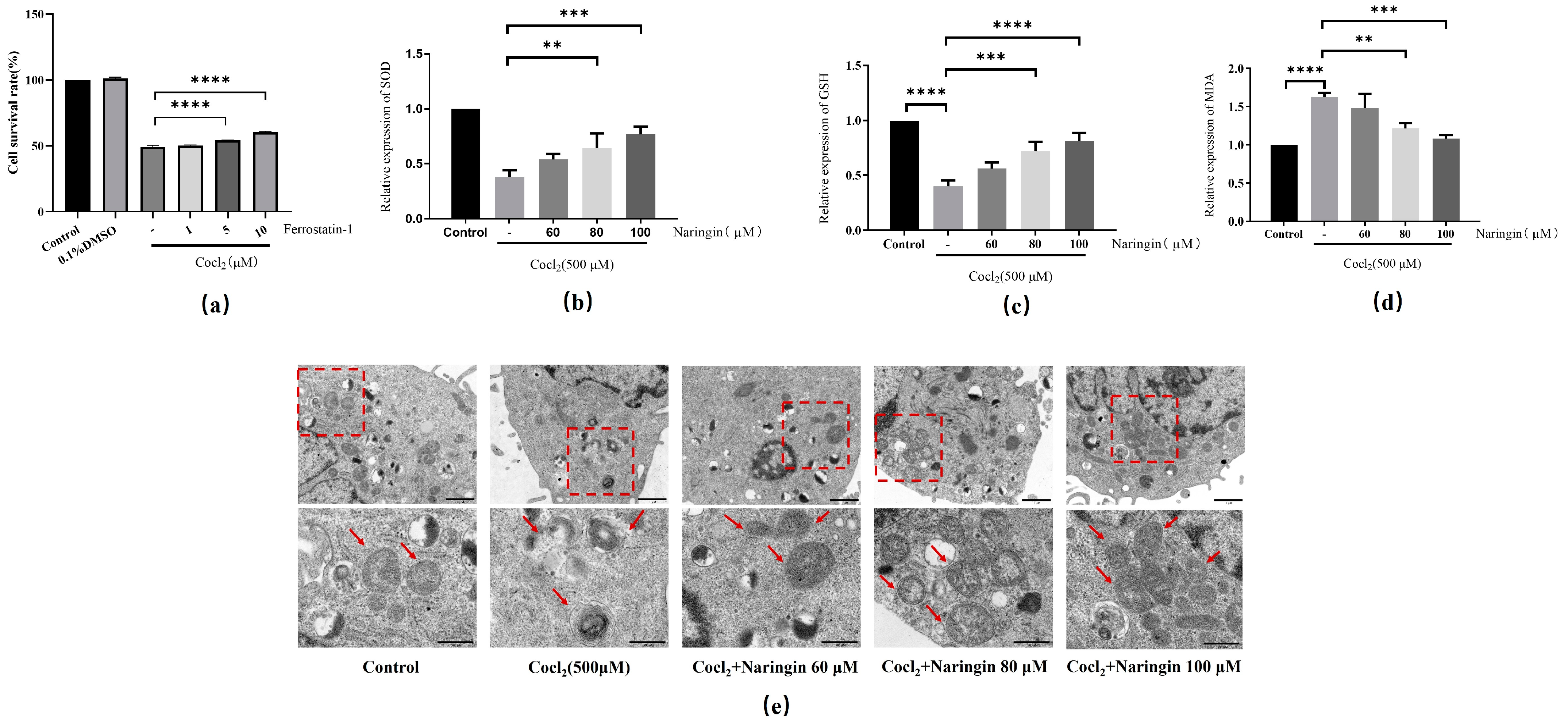
3.4. Naringin Inhibited CoCl2-Induced Ferroptosis of ARPE-19

Naringin has been shown to exert a protective effect against anoxic damage [35,36]. However, whether naringin has a protective effect on CoCl2-induced ferroptosis remains unclear. Overproduction of ROS is one of the critical indicators of oxidative stress that triggers ferroptosis signals [37]. Meanwhile, the use of Ferrostatin-1 can significantly protect against CoCl2-induced cell death, confirming the abnormal activation of ferroptosis in CoCl2-incubated ARPE-19 cells (Figure 4a). Next, we evaluated the effect of naringin on ferroptosis in CoCl2-induced ARPE-19 cells. As demonstrated in Figure 4b,c, the decreased SOD and GSH expression caused by CoCl2 stimulation could be rescued by naringin. Moreover, naringin treatment inhibited the accumulation of MDA in the CoCl2-induced cells (Figure 4d). Considering that mitochondrial morphological changes are a hallmark of ferroptosis [38], an ultrastructural analysis by TEM showed that naringin significantly improved CoCl2-induced mitochondrial crista reduction and even disappearance and outer membrane rupture in ARPE-19 cells (Figure 4e).

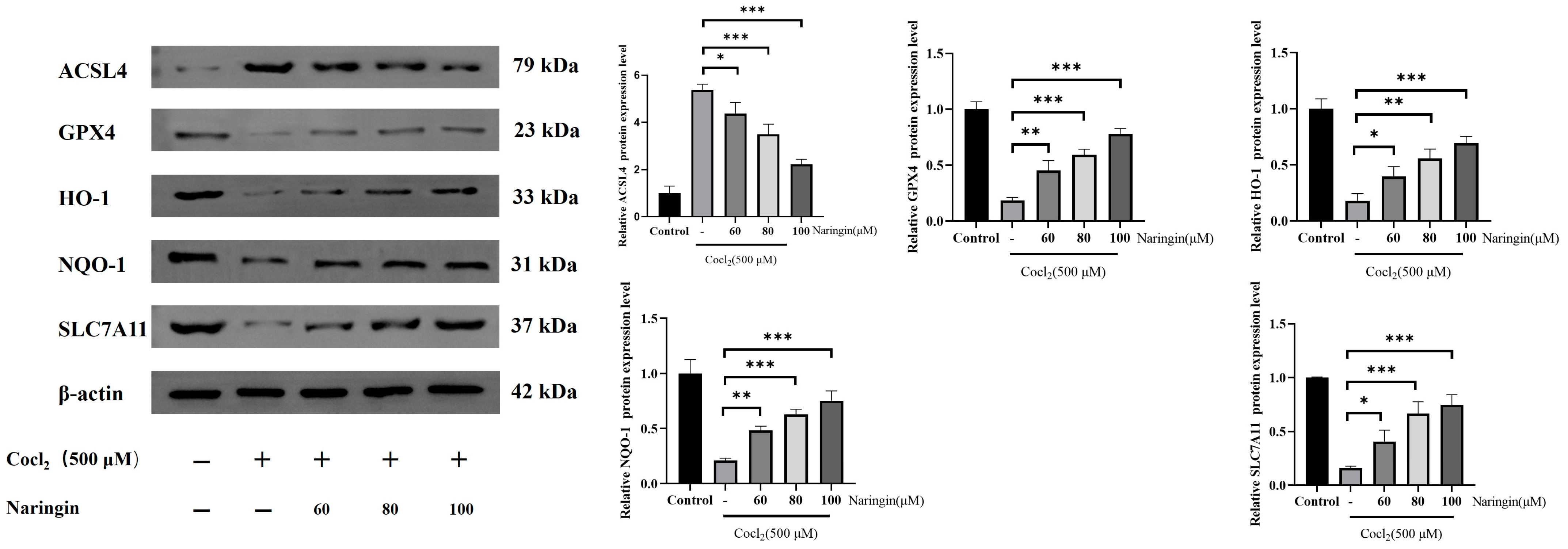
3.5. Naringin Adjusted HO-1/GPX4 Axis to Improve Ferroptosis

Under hypoxic conditions, the nuclear translocation of HIF-1α triggers the expression of antioxidant enzymes HO-1 and NQO1 [39]. The HO-1/GPX4 pathway is essential for regulating cellular oxidative stress resistance and ROS scavenging [40]. This signaling pathway is currently recognized as a key regulator of ferroptosis [41,42]. To assess ferroptosis-related protein expression, Western blot analysis was employed. CoCl2 treatment significantly reduced the protein expression of total GPX4, HO-1, NQO-1, and solute carrier family 7 member 11 protein (SLC7A11), while increasing Acyl-CoA synthetase long-chain family member 4 (ACSL4) protein levels. ACSL4 is a key regulator of lipid metabolism, closely linked to lipid peroxidation and ferroptosis [43]. These results suggest that ferroptosis occurred in CoCl2-incubated ARPE-19 cells (Figure 5). We propose that naringin modulates the HO-1/GPX4 axis to mitigate ferroptosis.

4. Discussion

The findings of this study demonstrate that naringin exerts protective effects on RPE cells under hypoxic conditions, primarily through the inhibition of ferroptosis. This is significant given the established role of ferroptosis in the pathogenesis of BRVO [44] and other retinal vascular diseases. The antioxidant properties of naringin likely contribute to its ability to alleviate oxidative stress, a key driver of ferroptosis [45]. Furthermore, our results suggest that naringin may also inhibit neovascularization, a critical factor in the progression of BRVO [46]. This dual mechanism of action—targeting both ferroptosis and neovascularization—positions naringin as a promising therapeutic candidate for BRVO treatment [47,48].
The observed reduction in hypoxia-induced cell death in RPE cells treated with naringin is consistent with previous studies demonstrating the compound’s protective effects in various oxidative stress and cell death models [49]. However, the specific pathways by which naringin inhibits ferroptosis in RPE cells have yet to be fully elucidated. Potential mechanisms may involve the modulation of lipid peroxidation, regulation of iron metabolism, or activation of antioxidant defense systems [50,51]. Future studies should focus on dissecting these pathways in greater detail to provide a more comprehensive understanding of naringin’s therapeutic potential.
Furthermore, naringin’s suppression of neovascularization suggests that it may also influence angiogenic signaling pathways, such as VEGF, which play a central role in BRVO [52]. This finding is particularly relevant considering the limitations of current anti-VEGF therapies, which, while effective, are associated with significant side effects and high treatment burdens. Naringin’s ability to target multiple pathological processes in BRVO could provide a more holistic treatment approach, potentially reducing the need for frequent intravitreal injections.
Therefore, this study provides compelling evidence for the therapeutic potential of naringin in BRVO, demonstrating its ability to protect RPE cells from hypoxia-induced ferroptosis and inhibit neovascularization. These findings emphasize the need for further research into the molecular mechanisms underlying naringin’s effects and its potential clinical applications. Future studies should also investigate the pharmacokinetics and safety profile of naringin to evaluate its viability as a therapeutic agent for BRVO and other retinal vascular diseases.
Firstly, cell viability assays demonstrated that naringin alleviates CoCl2-induced damage in ARPE-19 cells. Furthermore, naringin significantly reduces ROS production in RPE cells under hypoxic conditions, underscoring its protective role against hypoxic injury. While DCFH-DA detects a wide range of ROS, the observed fluorescence signal primarily reflects H2O2 accumulation, consistent with previous studies highlighting its key role in specific biological contexts, such as mitochondrial oxidative stress. While the DCFH-DA assay strongly suggests H2O2 as a major ROS, the specific contribution of H2O2 could be further validated using catalase treatment in future studies. These results highlight its potential as a therapeutic candidate for BRVO. Secondly, our study demonstrated that naringin effectively suppresses HIF-1α and VEGF mRNA expression in RPE cells while also inhibiting endothelial cell migration and tube formation. Under hypoxic conditions, RPE cells secrete large amounts of VEGF and other pro-neovascular factors [53]. VEGF is a pivotal driver of neovascularization, facilitating hypoxia-induced retinal vascular growth [54]. Retinal neovascularization extends along the retinal surface, heightening the risk of leakage and bleeding, which represent primary causes of blindness [55,56,57]. Therefore, our research suggests that naringin may inhibit retinal endothelial neovascularization associated with BRVO.
Subsequent findings revealed that pretreatment with naringin mitigated inflammation in hypoxia-damaged cells, as demonstrated by decreased levels of inflammatory factors, including IL-6, IL-1β, and TNF-α. Fer-1 treatment of ARPE-19 cells under hypoxic conditions significantly reduced cell death. We further observed that ARPE-19 cells exhibited ferroptosis-related pathological changes under hypoxia, including mitochondrial shrinkage and decreased inner mitochondrial cristae. Additionally, key markers of ferroptosis, including elevated MDA levels, along with reduced GSH and SOD levels, were also observed. Notably, pretreatment with naringin significantly ameliorated these cellular changes in ARPE-19 cells. Collectively, our results suggest that naringin protects RPE cells by downregulating inflammatory factors and inhibiting ferroptosis.
Finally, we conducted a preliminary investigation into the mechanism by which it inhibits cellular ferroptosis. HIF consists of a constitutive β-subunit and two oxygen-sensitive α-subunits (HIF-1α and HIF-2α). The activity of HIF is primarily regulated by oxygen-dependent proteolysis of the α-subunits [58,59,60,61]. HIF-1α plays a crucial role in metabolism, glucose transport, and cellular survival by modulating the expression of erythropoietin (EPO), VEGF, and HO-1 [62]. HO-1 serves as both a marker of ferroptosis and a critical antioxidant enzyme within the cellular defense system. Induction of HO-1 has been reported to exert beneficial effects on retinal degeneration [63]. For instance, the upregulation of HO-1 shields retinal endothelial cells from the stress caused by hyperglycemia [64]. NQO1 inhibits quinone redox cycling and ROS generation, thereby protecting cells from oxidative stress due to metabolic processes [65]. Solute carrier family 7 membrane 11 (SLC7A11) is an essential component of the system Xc-, and its inhibition can trigger ferroptosis [66]. GPX4 is a central regulator of ferroptosis, and its inactivation triggers ferroptosis through ROS accumulation resulting from lipid peroxidation [67]. ACSL4 is another crucial participant in lipid metabolism and ferroptosis [68]. Therefore, regulating the HO-1/GPX4 signaling pathway to inhibit lipid peroxidation and ferroptosis offers a viable therapeutic strategy. In this study, our results suggest that naringin inhibits CoCl2-induced ferroptosis and inflammation by regulating the HO-1/SLC7A11/GPX4 signaling pathway.

5. Conclusions

In summary, naringin alleviates neovascularization and mitigates hypoxic injury in RPE cells by regulating the HO-1/GPX4 signaling pathway, thereby suppressing ferroptosis. Our study suggests that ferroptosis may serve as a novel therapeutic target for BRVO, with naringin as a potential treatment option.

Author Contributions

Conceptualization, Y.Y.; methodology, M.L. and Y.Y.; software, J.B. and Y.Y.; validation, W.S. and Q.Z.; formal analysis, J.L. and Y.Y.; investigation, Z.W. and L.Y.; resources, X.Y. and X.D.; data curation, C.Q., J.N. and X.D.; writing—original draft preparation, Y.Y., J.N. and C.Q.; writing—review and editing, Y.Y., X.D. and C.Q.; visualization, Y.Y. and M.L.; supervision, J.N., X.D. and C.Q.; project administration, J.N. and C.Q.; funding acquisition, J.N. All authors have read and agreed to the published version of the manuscript.

Funding

This research received no external funding.

Institutional Review Board Statement

Not applicable.

Informed Consent Statement

Not applicable.

Data Availability Statement

The original contributions presented in the study are included in the article; further inquiries can be directed to the corresponding authors.

Conflicts of Interest

The authors declare no conflicts of interest.

References

  1. Scott, I.U.; Campochiaro, P.A.; Newman, N.J.; Biousse, V. Retinal vascular occlusions. Lancet 2020, 396, 1927–1940. [Google Scholar] [CrossRef] [PubMed]
  2. Romano, F.; Lamanna, F.; Gabrielle, P.H.; Teo, K.Y.C.; Battaglia Parodi, M.; Iacono, P.; Fraser-Bell, S.; Cornish, E.E.; Nassisi, M.; Viola, F.; et al. Update on Retinal Vein Occlusion. Asia Pac. J. Ophthalmol. 2023, 12, 196–210. [Google Scholar] [CrossRef] [PubMed]
  3. Wang, L.F.; Yan, Z.Y.; Li, Y.L.; Wang, Y.H.; Zhang, S.J.; Jia, X.; Lu, L.; Shang, Y.X.; Wang, X.; Li, Y.H.; et al. Inhibition of Obtusifolin on retinal pigment epithelial cell growth under hypoxia. Int. J. Ophthalmol. 2019, 12, 1539–1547. [Google Scholar] [CrossRef]
  4. Barresi, C.; Chhablani, J.; Dolz-Marco, R.; Gallego-Pinazo, R.; Berni, A.; Bandello, F.; Borrelli, E. Retinal neurodegeneration in age-related macular degeneration. Eur. J. Ophthalmol. 2024, 34, 624–630. [Google Scholar] [CrossRef]
  5. Ghosh, S.; Sharma, R.; Bammidi, S.; Koontz, V.; Nemani, M.; Yazdankhah, M.; Kedziora, K.M.; Stolz, D.B.; Wallace, C.T.; Yu-Wei, C.; et al. The AKT2/SIRT5/TFEB pathway as a potential therapeutic target in non-neovascular AMD. Nat. Commun. 2024, 15, 6150. [Google Scholar] [CrossRef]
  6. Huemer, J.; Khalid, H.; Wagner, S.K.; Nicholson, L.; Fu, D.J.; Sim, D.A.; Patel, P.J.; Balaskas, K.; Rajendram, R.; Keane, P.A. Phenotyping of retinal neovascularization in ischemic retinal vein occlusion using wide field OCT angiography. Eye 2021, 35, 2812–2819. [Google Scholar] [CrossRef]
  7. Liu, H.; Stepicheva, N.A.; Ghosh, S.; Shang, P.; Chowdhury, O.; Daley, R.A.; Yazdankhah, M.; Gupta, U.; Hose, S.L.; Valapala, M.; et al. Reducing Akt2 in retinal pigment epithelial cells causes a compensatory increase in Akt1 and attenuates diabetic retinopathy. Nat. Commun. 2022, 13, 6045. [Google Scholar] [CrossRef]
  8. Dong, Y.; Qian, C.; Yan, P.; Wan, G. YTHDF1-regulated ALOX5 in retinal pigment epithelial cells under hypoxia enhances VEGF expression and promotes viability, migration, and angiogenesis of vascular endothelial cells. Sci. Rep. 2024, 14, 23226. [Google Scholar] [CrossRef]
  9. Infantino, V.; Santarsiero, A.; Convertini, P.; Todisco, S.; Iacobazzi, V. Cancer Cell Metabolism in Hypoxia: Role of HIF-1 as Key Regulator and Therapeutic Target. Int. J. Mol. Sci. 2021, 22, 5703. [Google Scholar] [CrossRef]
  10. Qin, S.; Cao, G.; Tang, M.; Sun, S.; Dong, L. Baicalin alleviates the injury of human retinal pigment epithelium cells and improves branch retinal vein occlusion in rats by inhibiting the HIF-1α/VEGFA axis. Eur. J. Med. Res. 2024, 29, 564. [Google Scholar] [CrossRef]
  11. Muñoz-Sánchez, J.; Chánez-Cárdenas, M.E. The use of cobalt chloride as a chemical hypoxia model. J. Appl. Toxicol. JAT 2019, 39, 556–570. [Google Scholar] [CrossRef] [PubMed]
  12. Haley, M.J.; Bere, L.; Minshull, J.; Georgaka, S.; Garcia-Martin, N.; Howell, G.; Coope, D.J.; Roncaroli, F.; King, A.; Wedge, D.C.; et al. Hypoxia coordinates the spatial landscape of myeloid cells within glioblastoma to affect survival. Sci. Adv. 2024, 10, eadj3301. [Google Scholar] [CrossRef] [PubMed]
  13. Hou, Y.; Zhang, Y.; Jiang, S.; Xie, N.; Zhang, Y.; Meng, X.; Wang, X. Salidroside intensifies mitochondrial function of CoCl2-damaged HT22 cells by stimulating PI3K-AKT-MAPK signaling pathway. Phytomed. Int. J. Phytother. Phytopharm. 2023, 109, 154568. [Google Scholar] [CrossRef] [PubMed]
  14. Jang, S.K.; Ahn, S.H.; Kim, G.; Kim, S.; Hong, J.; Park, K.S.; Park, I.C.; Jin, H.O. Inhibition of VDAC1 oligomerization blocks cysteine deprivation-induced ferroptosis via mitochondrial ROS suppression. Cell Death Dis. 2024, 15, 811. [Google Scholar] [CrossRef]
  15. Chen, Y.; Fang, Z.M.; Yi, X.; Wei, X.; Jiang, D.S. The interaction between ferroptosis and inflammatory signaling pathways. Cell Death Dis. 2023, 14, 205. [Google Scholar] [CrossRef]
  16. Ye, Y.; Jiang, M.; Hong, X.; Fu, Y.; Chen, Y.; Wu, H.; Sun, Y.; Wang, X.; Zhou, E.; Wang, J.; et al. Quercetin Alleviates Deoxynivalenol-Induced Intestinal Damage by Suppressing Inflammation and Ferroptosis in Mice. J. Agric. Food Chem. 2023, 71, 10761–10772. [Google Scholar] [CrossRef]
  17. Wei, S.; Li, J.; Zhang, Y.; Li, Y.; Wang, Y. Ferroptosis in eye diseases: A systematic review. Eye 2024, 39, 18–27. [Google Scholar] [CrossRef]
  18. Henning, Y.; Blind, U.S.; Larafa, S.; Matschke, J.; Fandrey, J. Hypoxia aggravates ferroptosis in RPE cells by promoting the Fenton reaction. Cell Death Dis. 2022, 13, 662. [Google Scholar] [CrossRef]
  19. Liu, B.; Wang, W.; Shah, A.; Yu, M.; Liu, Y.; He, L.; Dang, J.; Yang, L.; Yan, M.; Ying, Y.; et al. Sodium iodate induces ferroptosis in human retinal pigment epithelium ARPE-19 cells. Cell Death Dis. 2021, 12, 230. [Google Scholar] [CrossRef]
  20. Zhan, S.; Liang, J.; Lin, H.; Cai, J.; Yang, X.; Wu, H.; Wei, J.; Wang, S.; Xian, M. SATB1/SLC7A11/HO-1 Axis Ameliorates Ferroptosis in Neuron Cells After Ischemic Stroke by Danhong Injection. Mol. Neurobiol. 2023, 60, 413–427. [Google Scholar] [CrossRef]
  21. Fantone, S.; Piani, F.; Olivieri, F.; Rippo, M.R.; Sirico, A.; Di Simone, N.; Marzioni, D.; Tossetta, G. Role of SLC7A11/xCT in Ovarian Cancer. Int. J. Mol. Sci. 2024, 25, 587. [Google Scholar] [CrossRef]
  22. Xie, Y.; Kang, R.; Klionsky, D.J.; Tang, D. GPX4 in cell death, autophagy, and disease. Autophagy 2023, 19, 2621–2638. [Google Scholar] [CrossRef] [PubMed]
  23. Long, P.; Yan, W.; Liu, J.; Li, M.; Chen, T.; Zhang, Z.; An, J. Therapeutic Effect of Traditional Chinese Medicine on a Rat Model of Branch Retinal Vein Occlusion. J. Ophthalmol. 2019, 2019, 9521379. [Google Scholar] [CrossRef] [PubMed]
  24. Zhao, Y.; Huang, Y. Quantitative Diagnosis of TCM Syndrome Types Based on Adaptive Resonant Neural Network. Comput. Intell. Neurosci. 2022, 2022, 2485089. [Google Scholar] [CrossRef] [PubMed]
  25. Zhou, D.; Wei, W.B.; Yang, C.X.; Ding, N.; Liu, Y.; He, M.L.; Hou, Z.J.; Zhou, J.Q. Treatment of retinal vein occlusion in rabbits with traditional Chinese medicine Fufang XueShuan Tong. Chin. Med. J. 2010, 123, 3293–3298. [Google Scholar]
  26. Shilpa, V.S.; Shams, R.; Dash, K.K.; Pandey, V.K.; Dar, A.H.; Ayaz Mukarram, S.; Harsányi, E.; Kovács, B. Phytochemical Properties, Extraction, and Pharmacological Benefits of Naringin: A Review. Molecules 2023, 28, 5623. [Google Scholar] [CrossRef]
  27. Heidary Moghaddam, R.; Samimi, Z.; Moradi, S.Z.; Little, P.J.; Xu, S.; Farzaei, M.H. Naringenin and naringin in cardiovascular disease prevention: A preclinical review. Eur. J. Pharmacol. 2020, 887, 173535. [Google Scholar] [CrossRef]
  28. Gu, L.; Wang, F.; Wang, Y.; Sun, D.; Sun, Y.; Tian, T.; Meng, Q.; Yin, L.; Xu, L.; Lu, X.; et al. Naringin protects against inflammation and apoptosis induced by intestinal ischemia-reperfusion injury through deactivation of cGAS-STING signaling pathway. Phytother. Res. 2023, 37, 3495–3507. [Google Scholar] [CrossRef]
  29. Guan, L.; Guo, L.; Zhang, H.; Liu, H.; Zhou, W.; Zhai, Y.; Yan, X.; Men, X.; Peng, L. Naringin Protects against Non-Alcoholic Fatty Liver Disease by Promoting Autophagic Flux and Lipophagy. Mol. Nutr. Food Res. 2024, 68, e2200812. [Google Scholar] [CrossRef]
  30. Benherlal, P.S.; Arumughan, C. Studies on modulation of DNA integrity in Fenton’s system by phytochemicals. Mutat. Res. 2008, 648, 1–8. [Google Scholar] [CrossRef]
  31. Livak, K.J.; Schmittgen, T.D. Analysis of relative gene expression data using real-time quantitative PCR and the 2(-Delta Delta C(T)) Method. Methods 2001, 25, 402–408. [Google Scholar] [CrossRef] [PubMed]
  32. Wu, J.; Ke, X.; Wang, W.; Zhang, H.; Ma, N.; Fu, W.; Zhao, M.; Gao, X.; Hao, X.; Zhang, Z. Aloe-emodin suppresses hypoxia-induced retinal angiogenesis via inhibition of HIF-1α/VEGF pathway. Int. J. Biol. Sci. 2016, 12, 1363–1371. [Google Scholar] [CrossRef] [PubMed]
  33. Deng, W.; Yi, C.; Pan, W.; Liu, J.; Qi, J.; Chen, J.; Zhou, Z.; Duan, Y.; Ning, X.; Li, J.; et al. Vascular Cell Adhesion Molecule-1 (VCAM-1) contributes to macular fibrosis in neovascular age-related macular degeneration through modulating macrophage functions. Immun. Ageing 2023, 20, 65. [Google Scholar] [CrossRef] [PubMed]
  34. Zhao, K.; Jiang, Y.; Zhang, J.; Shi, J.; Zheng, P.; Yang, C.; Chen, Y. Celastrol inhibits pathologic neovascularization in oxygen-induced retinopathy by targeting the miR-17-5p/HIF-1α/VEGF pathway. Cell Cycle 2022, 21, 2091–2108. [Google Scholar] [CrossRef]
  35. Cao, W.; Feng, S.J.; Kan, M.C. Naringin Targets NFKB1 to Alleviate Oxygen-Glucose Deprivation/Reoxygenation-Induced Injury in PC12 Cells Via Modulating HIF-1α/AKT/mTOR-Signaling Pathway. J. Mol. Neurosci. 2021, 71, 101–111. [Google Scholar] [CrossRef]
  36. Li, S.; Jiang, J.; Fang, J.; Li, X.; Huang, C.; Liang, W.; Wu, K. Naringin protects H9C2 cardiomyocytes from chemical hypoxia-induced injury by promoting the autophagic flux via the activation of the HIF-1α/BNIP3 signaling pathway. Int. J. Mol. Med. 2021, 47, 102. [Google Scholar] [CrossRef]
  37. Wang, B.; Wang, Y.; Zhang, J.; Hu, C.; Jiang, J.; Li, Y.; Peng, Z. ROS-induced lipid peroxidation modulates cell death outcome: Mechanisms behind apoptosis, autophagy, and ferroptosis. Arch. Toxicol. 2023, 97, 1439–1451. [Google Scholar] [CrossRef]
  38. Li, J.; Jia, Y.C.; Ding, Y.X.; Bai, J.; Cao, F.; Li, F. The crosstalk between ferroptosis and mitochondrial dynamic regulatory networks. Int. J. Biol. Sci. 2023, 19, 2756–2771. [Google Scholar] [CrossRef]
  39. Wu, Y.; Wang, J.; Zhao, T.; Chen, J.; Kang, L.; Wei, Y.; Han, L.; Shen, L.; Long, C.; Wu, S.; et al. Di-(2-ethylhexyl) phthalate exposure leads to ferroptosis via the HIF-1α/HO-1 signaling pathway in mouse testes. J. Hazard. Mater. 2022, 426, 127807. [Google Scholar] [CrossRef]
  40. Fu, C.; Wu, Y.; Liu, S.; Luo, C.; Lu, Y.; Liu, M.; Wang, L.; Zhang, Y.; Liu, X. Rehmannioside A improves cognitive impairment and alleviates ferroptosis via activating PI3K/AKT/Nrf2 and SLC7A11/GPX4 signaling pathway after ischemia. J. Ethnopharmacol. 2022, 289, 115021. [Google Scholar] [CrossRef]
  41. Lu, C.; Zhang, Z.; Fan, Y.; Wang, X.; Qian, J.; Bian, Z. Shikonin induces ferroptosis in osteosarcomas through the mitochondrial ROS-regulated HIF-1α/HO-1 axis. Phytomed. Int. J. Phytother. Phytopharm. 2024, 135, 156139. [Google Scholar] [CrossRef] [PubMed]
  42. Li, W.; Xiang, Z.; Xing, Y.; Li, S.; Shi, S. Mitochondria bridge HIF signaling and ferroptosis blockage in acute kidney injury. Cell Death Dis. 2022, 13, 308. [Google Scholar] [CrossRef] [PubMed]
  43. Doll, S.; Proneth, B.; Tyurina, Y.Y.; Panzilius, E.; Kobayashi, S.; Ingold, I.; Irmler, M.; Beckers, J.; Aichler, M.; Walch, A.; et al. ACSL4 dictates ferroptosis sensitivity by shaping cellular lipid composition. Nat. Chem. Biol. 2017, 13, 91–98. [Google Scholar] [CrossRef] [PubMed]
  44. Rogers, S.; McIntosh, R.L.; Cheung, N.; Lim, L.; Wang, J.J.; Mitchell, P.; Kowalski, J.W.; Nguyen, H.; Wong, T.Y.; International Eye Disease Consortium. The prevalence of retinal vein occlusion: Pooled data from population studies from the United States, Europe, Asia, and Australia. Ophthalmology 2010, 117, 313–319.e1. [Google Scholar] [CrossRef]
  45. Li, J.; Paulus, Y.M.; Shuai, Y.; Fang, W.; Liu, Q.; Yuan, S. New Developments in the Classification, Pathogenesis, Risk Factors, Natural History, and Treatment of Branch Retinal Vein Occlusion. J. Ophthalmol. 2017, 2017, 4936924. [Google Scholar] [CrossRef]
  46. Campochiaro, P.A.; Sophie, R.; Pearlman, J.; Brown, D.M.; Boyer, D.S.; Heier, J.S.; Marcus, D.M.; Feiner, L.; Patel, A.; RETAIN Study Group. Long-term outcomes in patients with retinal vein occlusion treated with ranibizumab: The RETAIN study. Ophthalmology 2014, 121, 209–219. [Google Scholar] [CrossRef]
  47. Driban, M.; Kedia, N.; Arora, S.; Chhablani, J. Novel pharmaceuticals for the management of retinal vein occlusion and linked disorders. Expert Rev. Clin. Pharmacol. 2023, 16, 1125–1139. [Google Scholar] [CrossRef]
  48. Hanada, N.; Iijima, H.; Sakurada, Y.; Imasawa, M. Recurrence of macular edema associated with branch retinal vein occlusion after intravitreal bevacizumab. Jpn. J. Ophthalmol. 2012, 56, 165–174. [Google Scholar] [CrossRef]
  49. Rivoira, M.A.; Rodriguez, V.; Talamoni, G.; Tolosa de Talamoni, N. New Perspectives in the Pharmacological Potential of Naringin in Medicine. Curr. Med. Chem. 2021, 28, 1987–2007. [Google Scholar] [CrossRef]
  50. Tang, G.; Pi, L.; Guo, H.; Hu, Z.; Zhou, C.; Hu, Q.; Peng, H.; Xiao, Z.; Zhang, Z.; Wang, M.; et al. Naringin Relieves Diabetic Cardiac Autonomic Neuropathy Mediated by P2Y14 Receptor in Superior Cervical Ganglion. Front. Pharmacol. 2022, 13, 873090. [Google Scholar] [CrossRef]
  51. Huang, Q.; Ru, Y.; Luo, Y.; Luo, X.; Liu, D.; Ma, Y.; Zhou, X.; Linghu, M.; Xu, W.; Gao, F.; et al. Identification of a targeted ACSL4 inhibitor to treat ferroptosis-related diseases. Sci. Adv. 2024, 10, eadk1200. [Google Scholar] [CrossRef] [PubMed]
  52. Yang, M.; Tsui, M.G.; Tsang, J.K.W.; Goit, R.K.; Yao, K.M.; So, K.F.; Lam, W.C.; Lo, A.C.Y. Involvement of FSP1-CoQ10-NADH and GSH-GPx-4 pathways in retinal pigment epithelium ferroptosis. Cell Death Dis. 2022, 13, 468. [Google Scholar] [CrossRef] [PubMed]
  53. Hwang, S.; Seong, H.; Ryu, J.; Jeong, J.Y.; Kang, T.S.; Nam, K.Y.; Seo, S.W.; Kim, S.J.; Kang, S.S.; Han, Y.S. Phosphorylation of STAT3 and ERBB2 mediates hypoxia-induced VEGF release in ARPE-19 cells. Mol. Med. Rep. 2020, 22, 2733–2740. [Google Scholar] [CrossRef]
  54. Yang, S.; Li, T.; Jia, H.; Gao, M.; Li, Y.; Wan, X.; Huang, Z.; Li, M.; Zhai, Y.; Li, X.; et al. Targeting C3b/C4b and VEGF with a bispecific fusion protein optimized for neovascular age-related macular degeneration therapy. Sci. Transl. Med. 2022, 14, eabj2177. [Google Scholar] [CrossRef] [PubMed]
  55. Lip, P.L.; Kolli, H.; Trivedi, D. Ultra-Widefield Fluorescein Angiographic Patterns, Retinal Microvascular Anomalies and Retinal Ischemic Index in Branch Retinal Vein Occlusions with Established Retinal Neovascularization. Clin. Ophthalmol. 2020, 14, 2965–2974. [Google Scholar] [CrossRef]
  56. Arrigo, A.; Aragona, E.; Bandello, F. VEGF-targeting drugs for the treatment of retinal neovascularization in diabetic retinopathy. Ann. Med. 2022, 54, 1089–1111. [Google Scholar] [CrossRef]
  57. Yang, Z.; Ni, B.; Zhou, T.; Huang, Z.; Zhou, H.; Zhou, Y.; Lin, S.; He, C.; Liu, X. HIF-1α Reduction by Lowering Intraocular Pressure Alleviated Retinal Neovascularization. Biomolecules 2023, 13, 1532. [Google Scholar] [CrossRef]
  58. Zhang, J.; Qin, Y.; Martinez, M.; Flores-Bellver, M.; Rodrigues, M.; Dinabandhu, A.; Cao, X.; Deshpande, M.; Qin, Y.; Aparicio-Domingo, S.; et al. HIF-1α and HIF-2α redundantly promote retinal neovascularization in patients with ischemic retinal disease. J. Clin. Investig. 2021, 131, e139202. [Google Scholar] [CrossRef]
  59. Huang, R.; Zhang, L.; Jin, J.; Zhou, Y.; Zhang, H.; Lv, C.; Lu, D.; Wu, Y.; Zhang, H.; Liu, S.; et al. Bruceine D inhibits HIF-1α-mediated glucose metabolism in hepatocellular carcinoma by blocking ICAT/β-catenin interaction. Acta Pharm. Sin. B 2021, 11, 3481–3492. [Google Scholar] [CrossRef]
  60. Qiu, B.; Yuan, P.; Du, X.; Jin, H.; Du, J.; Huang, Y. Hypoxia inducible factor-1α is an important regulator of macrophage biology. Heliyon 2023, 9, e17167. [Google Scholar] [CrossRef]
  61. Li, Z.L.; Ding, L.; Ma, R.X.; Zhang, Y.; Zhang, Y.L.; Ni, W.J.; Tang, T.T.; Wang, G.H.; Wang, B.; Lv, L.L.; et al. Activation of HIF-1α C-terminal transactivation domain protects against hypoxia-induced kidney injury through hexokinase 2-mediated mitophagy. Cell Death Dis. 2023, 14, 339. [Google Scholar] [CrossRef] [PubMed]
  62. Liu, Y.; Cui, H.; Mei, C.; Cui, M.; He, Q.; Wang, Q.; Li, D.; Song, Y.; Li, J.; Chen, S.; et al. Sirtuin4 alleviates severe acute pancreatitis by regulating HIF-1α/HO-1 mediated ferroptosis. Cell Death Dis. 2023, 14, 694. [Google Scholar] [CrossRef] [PubMed]
  63. Luo, P.; Liu, D.; Zhang, Q.; Yang, F.; Wong, Y.K.; Xia, F.; Zhang, J.; Chen, J.; Tian, Y.; Yang, C.; et al. Celastrol induces ferroptosis in activated HSCs to ameliorate hepatic fibrosis via targeting peroxiredoxins and HO-1. Acta Pharm. Sin. B 2022, 12, 2300–2314. [Google Scholar] [CrossRef]
  64. D’Agata, V.; D’Amico, A.G.; Maugeri, G.; Bucolo, C.; Rossi, S.; Giunta, S. Carnosol attenuates high glucose damage in human retinal endothelial cells through regulation of ERK/Nrf2/HO-1 pathway. J. Asian Nat. Prod. Res. 2023, 25, 783–795. [Google Scholar] [CrossRef]
  65. Wang, X.; Chen, J.; Tie, H.; Tian, W.; Zhao, Y.; Qin, L.; Guo, S.; Li, Q.; Bao, C. Eriodictyol regulated ferroptosis, mitochondrial dysfunction, and cell viability via Nrf2/HO-1/NQO1 signaling pathway in ovarian cancer cells. J. Biochem. Mol. Toxicol. 2023, 37, e23368. [Google Scholar] [CrossRef]
  66. Koppula, P.; Zhuang, L.; Gan, B. Cystine transporter SLC7A11/xCT in cancer: Ferroptosis, nutrient dependency, and cancer therapy. Protein Cell 2021, 12, 599–620. [Google Scholar] [CrossRef]
  67. Liang, D.; Feng, Y.; Zandkarimi, F.; Wang, H.; Zhang, Z.; Kim, J.; Cai, Y.; Gu, W.; Stockwell, B.R.; Jiang, X. Ferroptosis surveillance independent of GPX4 and differentially regulated by sex hormones. Cell 2023, 186, 2748–2764. [Google Scholar] [CrossRef]
  68. Miao, Z.; Tian, W.; Ye, Y.; Gu, W.; Bao, Z.; Xu, L.; Sun, G.; Li, C.; Tu, Y.; Chao, H.; et al. Hsp90 induces Acsl4-dependent glioma ferroptosis via dephosphorylating Ser637 at Drp1. Cell Death Dis. 2022, 13, 548. [Google Scholar] [CrossRef]
Figure 1. Naringin safeguarded RPE cells against damage induced by CoCl2. (a) ARPE-19 cells were first exposed to different concentrations of CoCl2 (50, 100, 200, 300, 400, 500, and 600 μM) for 24 h, and cell viability was determined with the MTT assay. (b) Effect of different concentrations of naringin on ARPE-19 cell viability. (c) Cytoprotective effect of naringin. ARPE-19 cells were pretreated with varying concentrations of naringin (60, 80, and 100 μM) for 24 h, and then CoCl2 (500 μM) was added, and the treatment was continued for 24 h. Cell viability was measured with the MTT assay. (d) Changes in the number of ARPE-19 cells were observed and presented. All the results are expressed as mean ± SD; ****, p < 0.0001; “ns” denotes non-significant (p ≥ 0.05).
Antioxidants 14 00236 g001
Figure 2. Naringin inhibits neovascularization. (a) Relative mRNA expression of HIF-1α. (b) Relative mRNA expression of VEGF. (c) Naringin inhibits the migration of HUVECs. (d) Naringin inhibits the tube formation of HUVECs. All the results are expressed as mean ± SD; *, p < 0.05; **, p < 0.01; ***, p < 0.001.
Antioxidants 14 00236 g002
Figure 3. Naringin exerted an anti-apoptotic protective effect on CoCl2-induced RPE cells. (a) The expression levels of related regulatory proteins in the cell cycle were detected by Western blot assay. Actin-1 was used as an internal control. Data are presented as mean ± SD of three independent experiments. (b) The expression levels of IL-6, IL-1β, and TNF-α were measured by using an ELISA kit (Jianglai Bio, Shanghai, China). (c) Use the DCFH-DA assay to quantitatively measure the production of ROS. (d) Use the DCFH-DA assay to qualitatively measure the production of ROS. All the results are expressed as mean ± SD; *, p < 0.05; **, p < 0.01; ***, p < 0.001; ****, p < 0.0001.
Antioxidants 14 00236 g003
Figure 4. Naringin inhibits CoCl2-induced ferroptosis in ARPE-19 cells. (a) Effect of different concentrations of Ferrostatin-1 on ARPE-19 cell viability. (b) The levels of cellular SOD were determined. (c) The levels of cellular GSH were determined. (d) The levels of cellular MDA were determined. (e) TEM was performed to evaluate the microscopic changes in mitochondria. The structure in the red box is the mitochondrion and red arrows denote mitochondria. All the results are expressed as mean ± SD; **, p < 0.01; ***, p < 0.001; ****, p < 0.0001.
Antioxidants 14 00236 g004
Figure 5. Naringin activated the SLC7A11 pathway in CoCl2-treated RPE cells. ARPE-19 cells were pretreated with or without naringin (60, 80, and 100 μM) for 24 h and then treated with 500 μM CoCl2 for 24 h. The expressions of ACSL4, GPX4, HO-1, NQO1, and SLC7A11 were measured with a Western blot assay. β-actin was used as an internal control for total protein. All the results are expressed as mean ± SD; *, p < 0.05; **, p < 0.01; ***, p < 0.001.
Antioxidants 14 00236 g005
Disclaimer/Publisher’s Note: The statements, opinions and data contained in all publications are solely those of the individual author(s) and contributor(s) and not of MDPI and/or the editor(s). MDPI and/or the editor(s) disclaim responsibility for any injury to people or property resulting from any ideas, methods, instructions or products referred to in the content.

