Article

Evaluation of the Efficacy of a Cholera Toxin-Based Staphylococcus aureus Vaccine against Bovine Intramammary Challenge

1
Department of Animal and Veterinary Science, University of Idaho, Moscow, ID 83844, USA
2
Biomolecular Sciences Graduate Program, Boise State University, Boise, ID 83725, USA
3
Department of Biological Sciences, Boise State University, Boise, ID 83725, USA
4
Biomolecular Research Center, Boise State University, Boise, ID 83725, USA
*
Author to whom correspondence should be addressed.
The authors contribute equally.
Vaccines 2021, 9(1), 6; https://doi.org/10.3390/vaccines9010006
Submission received: 24 November 2020 / Revised: 13 December 2020 / Accepted: 18 December 2020 / Published: 24 December 2020
(This article belongs to the Special Issue Evaluation of Vaccine Immunogenicity)

Abstract

:
Staphylococcus aureus (S. aureus) is a primary agent of bovine mastitis and a source of significant economic loss for the dairy industry. We previously reported antigen-specific immune induction in the milk and serum of dairy cows following vaccination with a cholera toxin A2 and B subunit (CTA2/B) based vaccine containing the iron-regulated surface determinant A (IsdA) and clumping factor A (ClfA) antigens of S. aureus (IsdA + ClfA-CTA2/B). The goal of the current study was to assess the efficacy of this vaccine to protect against S. aureus infection after intramammary challenge. Six mid-lactation heifers were randomized to vaccinated and control groups. On days 1 and 14 animals were inoculated intranasally with vaccine or vehicle control, and on day 20 animals were challenged with S. aureus. Clinical outcome, milk quality, bacterial shedding, and somatic cell count (SCC) were followed for ten days post-challenge. Vaccinated animals did not show signs of clinical S. aureus mastitis and had lower SCCs compared to control animals during the challenge period. Reductions in bacterial shedding were observed but were not significant between groups. Antibody analysis of milk and serum indicated that, upon challenge, vaccinated animals produced enhanced IsdA- and ClfA-CTA2/B specific immunoglobulin G (IgG) responses, while responses to CTA2/B alone were not different between groups. Responses after challenge were largely IgG1 against the IsdA antigen and mixed IgG1/IgG2 against the ClfA antigen. In addition, there was a significant increase in interferon gamma (IFN-γ) expression from blood cells in vaccinated animals on day 20. While preliminary, these findings support evidence of the induction of active immunity by IsdA + ClfA-CTA2/B, and further assessment of this vaccine is warranted.

1. Introduction

Mastitis, or inflammation of the udder, is one of the most economically-significant diseases affecting dairy cattle worldwide and is most often the result of a bacterial infection. Staphylococcus aureus (S. aureus), a main etiological agent, is highly contagious and can spread rapidly among herds. It is estimated that up to 70% of U.S. herds are positive for S. aureus, and this bacterium caused the highest overall annual yield losses among other mastitis pathogens in a recent Finnish study [1,2]. S. aureus infections are most commonly transmitted during the milking process and can impact animal welfare as well as milk yield and quality [3]. The ability of this bacterium to form biofilms and replicate intracellularly can promote subclinical colonization of the mammary gland, often leading to chronic infection, which is difficult to detect and is frequently the source of herd re-infection [4,5,6]. S. aureus is also commonly resistant to antimicrobial treatment and has a low expected cure rate during lactation [7]. While the impact of S. aureus infection is difficult to quantify, clinical mastitis caused by Gram-positive pathogens is reported to cost between $133 and $444 per case, or as much as USD 2 billion annually [8,9]. These costs include many factors such as milk loss, veterinary expenses, diagnostic testing, and loss of animals. Prevention of S. aureus mastitis with a cost-effective vaccine would improve animal welfare, reduce antibiotic use, and positively impact the economics and efficiency of milk production.
Previous approaches to S. aureus vaccination in cattle include whole-cell live and killed vaccines as well as purified antigens. Currently, two whole-cell inactivated vaccines are licensed for protection against S. aureus mastitis—Lysigin® (Boehringer Ingelheim, Duluth, GA, USA) and Startvac® (Hipra, Girona, Spain). While efficacy studies are somewhat conflicting, these vaccines have reported moderate decreases in the incidence of new S. aureus intramammary infection but are not in widespread use [10,11,12,13,14,15]. Recent studies have focused on the use of multiple purified surface adhesins and secreted virulence factors to develop a vaccine that offers more strain-to-strain cross-protection. Iron-regulated surface determinant A (IsdA) is a fibrinogen- and fibronectin-binding adhesin that contributes to iron sequestration and is a well-studied S. aureus vaccine candidate [16,17,18,19]. The presence of isdA is conserved among bovine S. aureus, and IsdA is expressed from these strains in milk [18,20,21,22,23]. The clumping factor A (ClfA) fibrinogen adhesin is also highly conserved, expressed from bovine clinical isolates, and a recognized vaccine candidate against mastitis [24,25,26,27,28,29,30]. The conservation, surface exposure, and importance in multiple mechanisms of pathogenesis supports the inclusion of the IsdA and ClfA antigens in a multivalent bovine vaccine. However, a number of additional antigens have been characterized and may be necessary to protect against multiple S. aureus serotypes.
While immune correlates of protection are not known, an understanding of immune responses is needed to inform antigen selection. The induction of both humoral and cellular immunity is essential to combating intracellular S. aureus infection [31,32,33]. Cellular subpopulations that play a central role in defense against S. aureus include neutrophils, CD8+ T lymphocytes, and CD4+ Th17 lymphocytes [34,35]. Cholera toxin (CT), produced by the bacterium Vibrio cholerae, and the homologous heat-labile toxin I (LTI), produced by the bacterium Escherichia coli, are gold standard vaccine adjuvants that can stimulate systemic immunity from mucosal and dermal sites (reviewed in [36]). The mechanism of adjuvanticity of these toxins depends upon active binding subunit targeting of dendritic cells and neutrophils, and has been attributed to enhanced antigen presentation, upregulation of surface molecules, and promotion of B-cell isotype switching to antigen-specific immunoglobulin A (IgA) and immunoglobulin G (IgG) [37,38,39,40,41]. CT and its non-toxic binding subunit (CTB) can also induce Th1, Th2, and Th17 responses [42,43,44].
The toxic A subunit of CT (CTA) is subdivided into an enzymatically-active domain (CTA1) and a linker domain (CTA2), which is non-covalently associated with the B subunit. CTA2/B chimeras were first described as a mechanism to make stable human vaccines with antigens coupled to the CTB subunit via the A2 linker domain [45,46]. These non-toxic molecules retain the adjuvanticity of CTB and possess additional advantages including ease of purification, direct association of antigen to adjuvant, and a holotoxin-like structure that retains binding and internalization motifs [47,48]. As reported previously, we have incorporated S. aureus IsdA and ClfA into a CTA2/B vaccine platform (IsdA + ClfA-CTA2/B). After two intranasal doses this vaccine was found to stimulate significant S. aureus antigen-specific humoral and cellular immunity in bovine blood and milk [49].
For this study we hypothesized that intranasal IsdA + ClfA-CTA2/B would be effective in reducing or eliminating S. aureus shedding and disease after intramammary challenge in cattle. We describe a preliminary trial to determine the efficacy of this mucosal enterotoxin-based vaccine to protect against acute S. aureus mastitis. While the vaccine did not prevent bacterial shedding after challenge, results indicate that IsdA + ClfA-CTA2/B induces antigen-specific immune responses that may contribute to a reduction in clinical severity and infiltration of leukocytes, or SCC, in infected animals.

2. Materials and Methods

2.1. Bacterial Strains, Plasmids, and Growth Conditions

S. aureus Newbould 305 was used for the cloning of isdA and clfA to construct IsdA + ClfA-CTA2/B and was also used for bacterial challenge [22,50]. E. coli ClearColi® (Lucigen, Madison, WI, USA) was used for protein expression (Table 1). The vector pARLDR19 expressing CTA2/B and containing a multiple cloning site was used to construct the plasmids pLR001 for Isd-CTA2/B expression and pLR003 for ClfA-CTA2/B expression (Figure 1A) as described previously [51]. For bacterial challenge, S. aureus Newbould 305 was prepared as described [10]. Briefly, Newbould 305 was grown at 37 °C with shaking to mid-log phase in brain–heart infusion and harvested by centrifugation at 3000× g for 15 min at 4 °C. The cell pellet was washed with phosphate-buffered saline (1X PBS, pH 7.2) and adjusted to an optical density (O.D.) of 0.2 at 600 nm. Serial dilutions were performed in 1X PBS to reach a bacterial concentration of 400 CFU/mL, as determined by plating on blood agar (BA).

2.2. Protein Expression and Purification

Chimeras were purified as previously described [49,51]. Briefly, to express IsdA-CTA2/B and ClfA-CTA2/B, ClearColi® with pLR001 or pLR003 were grown at 37 °C to an O.D. of 0.9 at 600 nm and induced for 24 h with 0.2% L-arabinose. Proteins were isolated from the periplasmic extract with 1 mg/mL polymyxin B and purified by affinity chromatography on immobilized D-galactose (Pierce™ D-Galactose Agarose, Thermo Fisher, Waltham, MA, USA). Vaccine proteins were dialyzed into sterile 5% glycerol + 1X PBS and concentrations were determined by bicinchoninic acid assay (BCA) (Pierce™ BCA, Thermo Fisher, Waltham, MA, USA). Sizes and purities of the vaccine chimeras were confirmed by sodium dodecyl sulphate-polyacrylamide gel electrophoresis (SDS-PAGE) prior to mixing at a final protein concentration of 600 µg/5 mL for vaccination (Figure 1B). Vaccines were tested to ensure endotoxin levels were below 0.05 EU/mL (LAL Endpoint Chromogenic, Lonza, Allendale, NJ, USA), plated for sterility on tryptic soy agar, and stored at −80 °C until use.

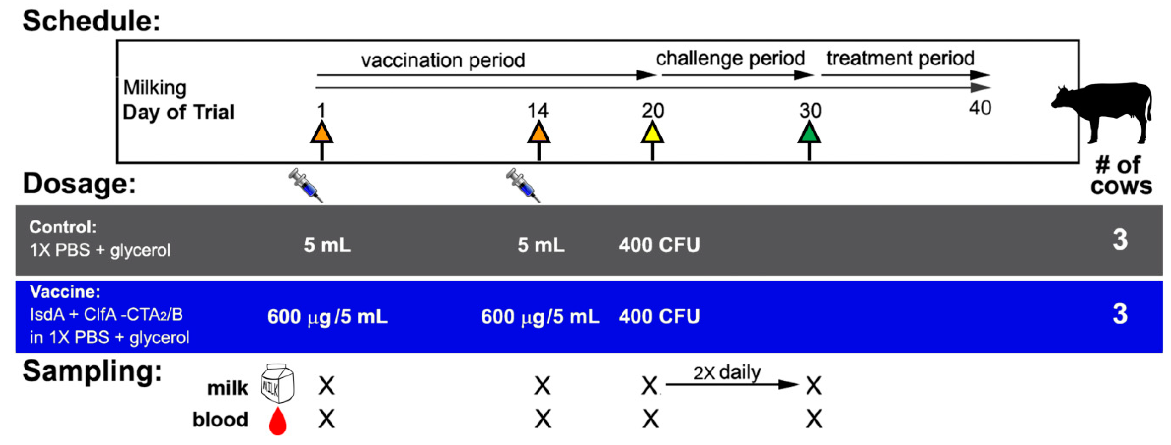
2.3. Animals, Vaccination, Challenge, and Clinical Assessment

All animal protocols were pre-approved by the University of Idaho Animal Care and Use Committee. Lactating healthy Holstein cows in the third or fourth lactation were pre-screened for inclusion as being those with two consecutive SCC readings below 200 × 103 cells/mL and no clinical evidence of mastitis. Further enrollment criteria were followed as described previously [49] and included: (1) no growth of S. aureus culture from milk as determined by plating on mannitol salt agar (MSA) and PCR with S. aureus nuc and isdA primers, (2) low baseline anti-IsdA responses as determined by enzyme-linked immunosorbent assay (ELISA) of milk and serum, and (3) no evidence of bovine leukemia virus infection (Washington Animal Disease Diagnostic Lab, WADDL, Pullman, WA, USA). Seven selected cows were ultimately randomized into vaccinated and control groups. Figure 2 shows the summary of trial design. Four vaccinated animals received a 600 µg intranasal dose of IsdA + ClfA-CTA2/B in 1X PBS + 5% glycerol on days 1 and 14 (orange arrows, blue bar), and a control group of three animals of similar age and lactation period received vehicle control (1X PBS + 5% glycerol) mock vaccination on days 1 and 14 (orange arrows, grey bar). The vaccine was delivered in 2.5 mL volumes into each nare using a nasal cannula (Merck & Co., Kenilworth, NJ, USA). On day 20, all animals were challenged in two quarters with 400 CFU in 1 mL of S. aureus Newbould 305 (yellow arrow). Quarters were identified as left front (LF), left rear (LR), right front (RF), and right rear (RR). The bacterial challenge was inoculated into two diagonal quarters of each vaccinated cow using teat cannulae (Valley Vet Supply, Marysville, KS, USA). Animals were monitored closely during the challenge period (days 20 to 30) and evaluated for the presence of clinical mastitis by assessment of rectal temperature (Figure 3), milk quality (Figure S2), and udder consistency (examination for edema, hardening, and/or swelling, Table S1) on days of milk sampling during the trial (Figure 2) [54,55]. Enrolled animals that developed pain and/or fever that exceeded 103 °F were administered painkillers (Banamine® and aspirin) as recommended by the attending veterinarian. Shortly after challenge (day 21), one vaccinated animal developed a severe Escherichia coli mastitis case in an unchallenged quarter (2779 LF) with systemic illness including septicemia. Thereafter, the other three quarters were involved and the animal developed clinical mastitis due to Staphylococcus aureus. The animal was euthanized on day 5 post-challenge. Results from this animal are not included in the data in this report and resulting sample size was n = 3 per group, as represented in Figure 2. On day 30 all other animals began treatment with Spectramast (Zoetis, Parsippany, NJ, USA) until consecutive negative cultures were indicative of safe release to herd as determined by the attending veterinarian.

2.4. Sample Collection and Milk Culture

Blood and milk were sampled on day −2 for screening and then on days 1, 14, 20, and 30, and milk was sampled twice daily during the challenge period (Figure 2). Blood was collected from the tail vein and allowed to coagulate at room temperature (RT) for 1 h prior to centrifugation and resuspension into 1:10 inhibitor solution (IS, 1X HALT™ protease inhibitor and 5% glycerol in 1X PBS). On day 20, whole blood was also collected in vacutainer tubes for peripheral blood mononuclear cell (PBMC) isolation (Becton Dickinson, Franklin Lakes, NJ, USA). Milk was collected aseptically as 50 mL quarter samples after washing teat ends with 70% ethanol and was aliquoted into three equal tubes for culture, SCC, and ELISA. For SCC, milk was fixed prior to shipping and analysis was performed using the California Mastitis Test (WADDL, Pullman, WA, USA). For ELISA, milk was centrifuged at 700× g for 20 min at 4 °C to remove fat. Skim milk was collected and centrifuged at 20,000× g for 30 min at 4 °C. Whey was collected and stored in 1:10 IS. Equal volumes of diluted whey from each quarter were pooled and stored at −20 °C prior to analysis. For milk culture, 100 µL and 10 µL of tenfold serially-diluted quarter milk was plated on MSA, BA, and MP2 agar (Udder Health Systems, Inc., Meridian, ID, USA) to determine the number of colony-forming units per mL (CFU/mL). The presence of larger yellow colonies with yellow zones on MSA, beta-hemolysis on BA, or small, white, esculin-negative colonies on MP2 was considered presumptive S. aureus. These colonies were isolated and confirmed by a positive coagulase test or a PCR test using nuc or isdA primers [23]. CFU by quarter data, based upon final quantitation on MSA, was determined once daily on days −2, 1, 14, and 20 prior to challenge and twice daily (AM/PM) during the challenge period. Quarter data were combined and total CFU/mL by cow was reported for six animals (n = 3 per group).

2.5. IgG, IgG1, IgG2, and IgA Enzyme-Linked Immunosorbent Assay (ELISA)

IsdA- and ClfA-specific immune responses in serum and milk were detected using ELISA as described [51]. Briefly, 96-well microtiter plates (Nunc, Thermo Fisher, Waltham, MA, USA) were coated with 10 µg of either IsdA-CTA2/B, ClfA-CTA2/B, or CTA2/B in 1X PBS and incubated overnight at 4 °C. Coated plates were blocked for 2 h at 37 °C in 1% goat milk + 1X PBS. After washing, plates were incubated with two-fold dilutions of either bovine serum (dilutions initiated at 1:200 concentration) or pooled quarter milk (dilutions initiated at a 1:10 concentration). Plates were incubated at 4 °C overnight. After washing, plates were incubated with horseradish peroxidase (HRP)-conjugated anti-bovine IgG, IgG1, IgG2, or IgA (1:10,000 Bethyl Laboratories, Montgomery, TX, USA) at 37 °C for 1 h. Plates were developed with tetramethylbenzidine (PromegaTM TMB One, Thermo Fisher, Waltham, MA, USA) and read at 370 nm per TMB manufacturer’s instruction. ELISA results from serum or pooled quarter milk were reported by cow (n = 3) and presented as the ratio of results (day X/day 1) of the O.D. (370 nm) from a representative antibody dilution in the linear part of the curve (1:1600 serum, 1:160 milk). Results are the average of three independent assays.

2.6. Peripheral Blood Mononuclear Cell (PBMC) Isolation and Cytokine qPCR

PBMCs were isolated from whole bovine blood on day 20 for cytokine analysis. PBMCs were isolated using a density gradient established by layering whole blood diluted 1:2 with 1X PBS on Histopaque®-1077 (Sigma-Aldrich, St. Louis, MO, USA). Blood samples were centrifuged at 800× g for 30 min at RT. The buffy coat was removed and washed three times by centrifugation with Hank’s Balanced Salt Solution for 10 min at 400× g at RT, and cells were counted with 0.2% trypan blue. For cytokine assays, total RNA from PBMCs from each cow (n = 3 per group) was extracted (RNeasy, Qiagen, Germantown, MD, USA) with an additional Dnase I (Promega, Madison, WI, USA) digestion. cDNA was reverse transcribed per manufacturer’s instructions (High-Capacity RNA-to-cDNA™ Kit, Thermo Fisher, Waltham, MA, USA). qRT-PCR was conducted using SYBR fast (Kapa Biosystems, Thermo Fisher, Waltham, MA, USA) on interferon gamma (IFN-γ), interleukin-6 (IL-6), and interleukin-10 (IL-10) primers, using bovine glyceraldehyde 3-phosphate dehydrogenase (GAPDH) as a reference gene (primers, Table 1). Results are presented as relative gene expression 2−ΔΔCt [56]. All qRT-PCR experiments were performed in triplicate per cow PBMC sample.

2.7. Sample Size, Statistical Methods, and Analysis

Sample size was estimated prior to study by power analysis based upon predicted SCC and CFU/mL in milk and using the assumption that quarters are independent, as has been reported [57,58]. A sample size of 13 quarters per group was predicted to provide, at a 95% level of confidence, 80% power to detect a difference in logged SCC. Resulting quarter bacterial counts and SCC data from this study were analyzed by (1) assuming independent quarters and (2) as the combined average of quarters by cow. Outcomes were not different, thus results are reported as the average by cow and assuming quarters are not independent. The log-base 10 values of CFU, SCC, temperature, and serum and milk anti-IsdA, ClfA, and CTB antibodies were analyzed using repeated measures analysis of variance (ANOVA) with time as the within-subjects variable and group as the between-subjects variable. Within-subjects correlation was modeled with either first-order autoregressive or compound symmetric structure, depending on Akaike’s Information Criterion [59]. Comparisons of interest were identified prior to modeling and were examined regardless of the significance of main effects or interaction. First, we explicitly compared the outcome at each study time point. Second, we examined the change in outcome within group, comparing days and adjusting the paired comparisons using false discovery rate [60]. Cytokine analysis was performed using a two-group t-test between vaccinated and control animals. Statistical analyses were conducted using JMP and SAS software (Cary, NC). p-values are reported as p ≤ 0.05(*), p ≤ 0.01(**), or p ≤ 0.0001(****) and reflect two-sided tests.

3. Results

3.1. Bacterial Culture and Clinical Assessment

Quantification of S. aureus was determined after plating milk that had been sampled once daily on days −2, 1, 14, and 20 prior to challenge and twice daily during the challenge period (Figure 2). Prior to challenge no animals were found to be shedding S. aureus, and immediately after challenge all animals shed high levels of S. aureus from infected quarters (Figure 3A). Results revealed a rapid decline in bacterial shedding from all animals within 24 h and then a slow decline beginning in the middle of the challenge period. Between days 2 and 10 of the challenge period (days 22 and 30 of trial) control animals shed a total of 1.08 × 10⁶ CFU/mL and vaccinated animals shed 7.53 × 10⁵ CFU/mL. Differences between treatment groups were observed on days 21, 29, and 30 during the challenge period (p = 0.029, 0.011, and 0.018, respectively), however, after adjusting for multiple comparisons, these results are not significant. S. aureus was isolated from all challenged quarters in both treatment groups, and all animals continued to shed S. aureus throughout the trial. Animals did not shed from uninfected quarters. While one vaccinated animal was culture negative at two time points late in the challenge period (day 29 AM and day 30 AM), no animals were consistently sterile of S. aureus by the end of the challenge period. Analysis of positive quarters indicated that there were more days showing a lower percentage of infected quarters for vaccinated animals (Supplementary Figure S1).
SCC taken once daily before the challenge period and twice daily during challenge is shown in Figure 3B. Results show a consistently reduced SCC from vaccinated animals beginning 48 h post-challenge (day 22). While individual days were not significant after adjustment, across and after the challenge period (days 21 to 39) unvaccinated animals had significantly-higher SCC than vaccinated animals (model main effect of treatment group, p = 0.002). SCCs of individual cows throughout the trial are shown in Supplementary Figure S3B.
The average rectal temperature per group during the challenge period is shown in Figure 3C. Temperatures at 72 h post-challenge (day 23 AM) showed an average of 102.7 °F for control and 101.5 °F for vaccinated animals, however there was no statistically-significant difference in temperature between groups on any day during this period. In addition, no differences in temperature between groups occurred within 24 h after vaccination (days 1 and 14).
Clinical assessments indicated that animals did not show signs of systemic illness, loss of appetite, or adverse local reactions due to the vaccine, and no animals had clinical evidence of mastitis prior to challenge (day 20). Clinical results are summarized in Supplementary Table S1. Clinical mastitis due to S. aureus was observed in challenged quarters of control cows 2767 (LF and RF) and 2830 (LR) throughout the evaluation period. The latter cow developed a persistent mastitis starting on day 23 with apparent milk changes that included clots and flakes in the LR quarter. Clinical mastitis in this animal included persistent udder swelling in addition to pain, heat, and sensation of the affected teat until the end of the challenge period. Temporary enlargement of the supramammary lymph node was noted in one of the vaccinated cows (2823) on day 24, and persistent enlargement observed in one control animal (2830).
Milk quality assessments indicated that while the fat, protein, lactose, and solids-not-fat (SNF) percentages were frequently higher in vaccinated animals, these differences were not statistically significant (Supplementary Figure S2).

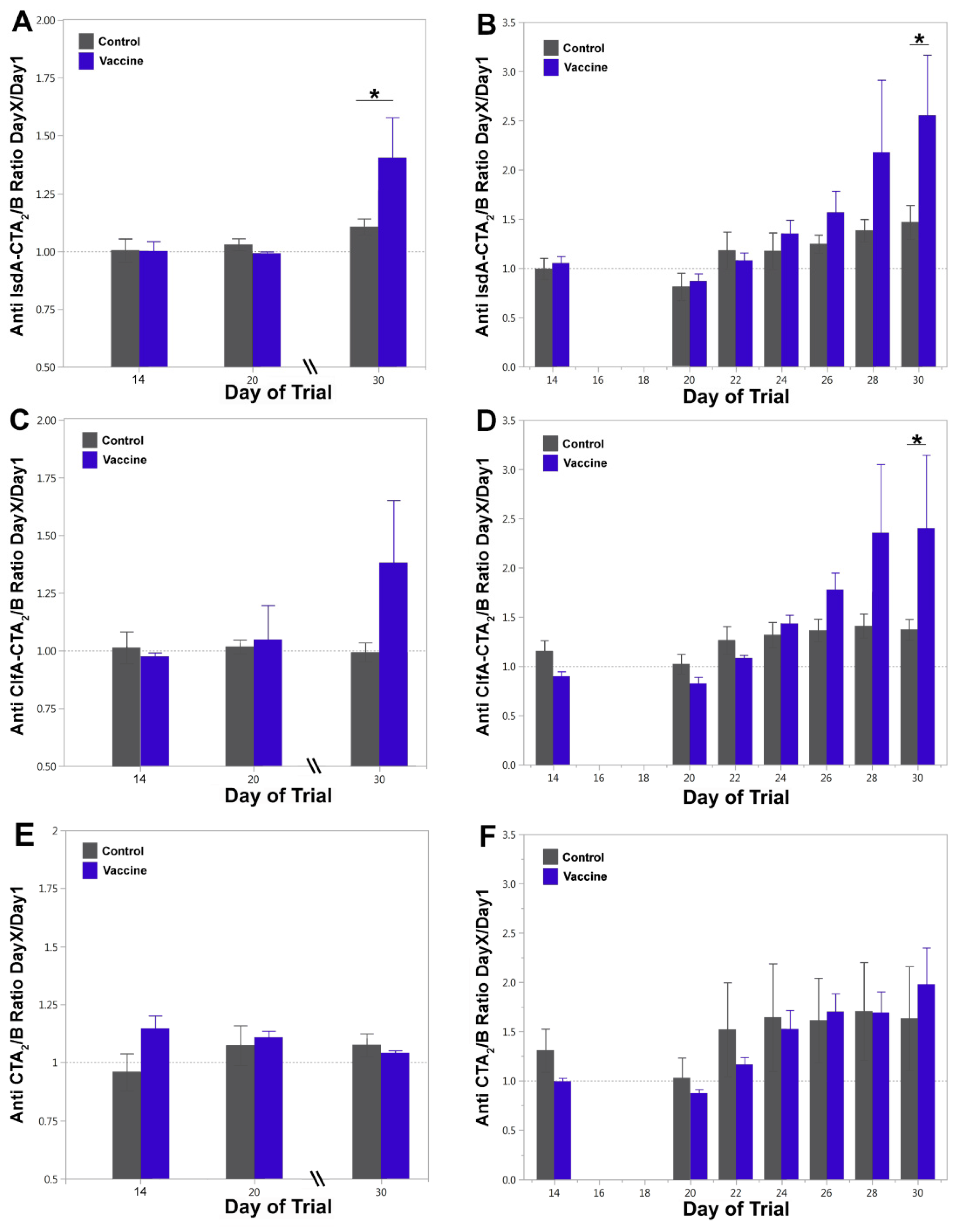
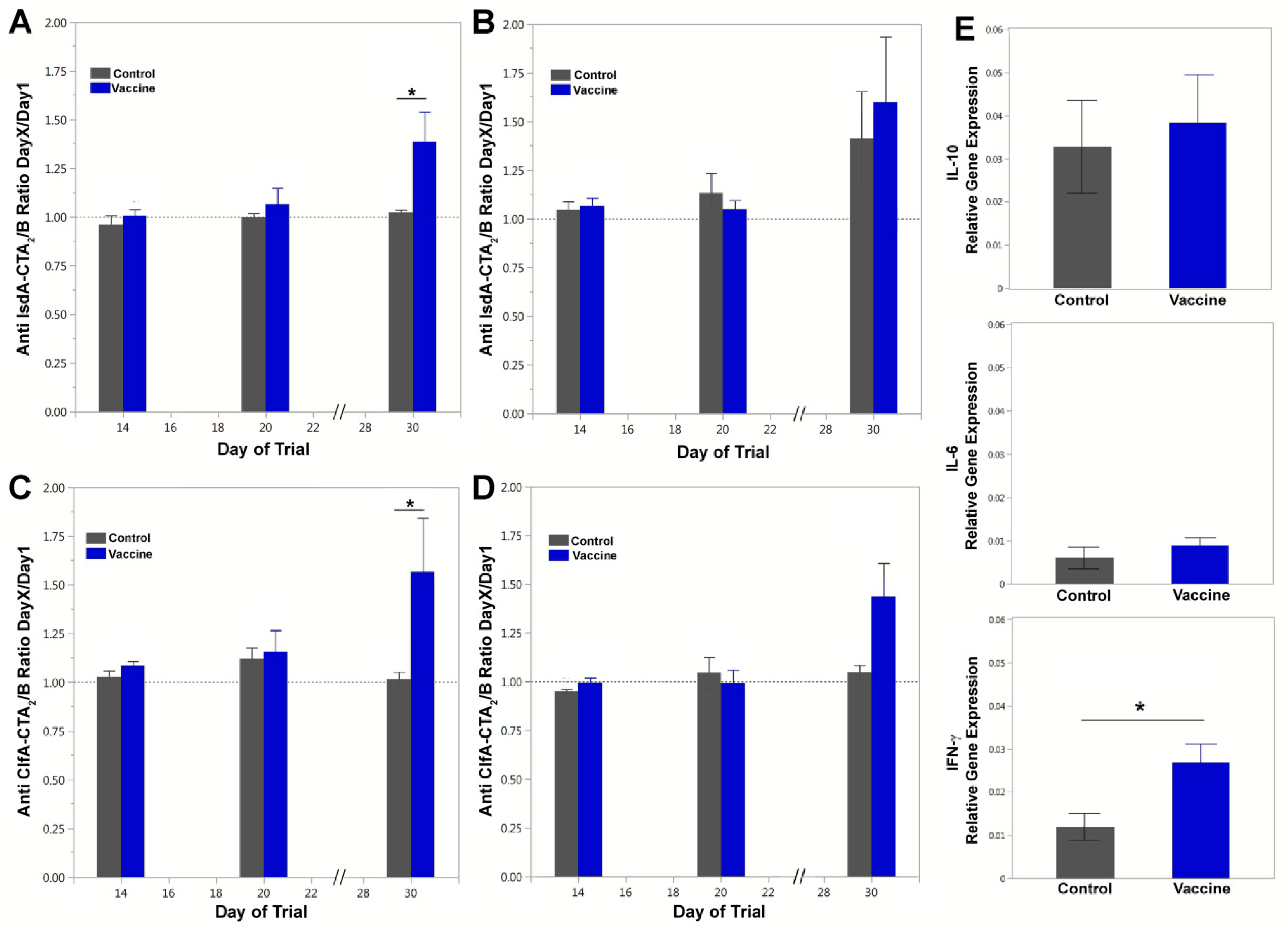
3.2. Vaccine-Specific Antibody Responses in Blood and Milk

Antigen-specific humoral responses were quantified by ELISA from blood and milk. Anti-IgG responses in serum on days 14, 20 and 30, relative to day 1, are shown in Figure 4A–C and E. Vaccinated animals (blue bars) showed a significant IsdA-CTA2/B-specific IgG response in serum after challenge on day 30 relative to days 14 and 20 (padj = 0.008 for both) and on day 30 relative to control animals (p = 0.030 *) (Figure 4A). Vaccinated animals showed a similar, but non-significant, anti-ClfA-CTA2/B-specific IgG responses in serum on day 30 relative to days 14 and 20 (padj = 0.120) as well as on day 30 relative to control animals (p = 0.079) (Figure 4C). Anti-CTA2/B-specific IgG responses in serum remained low and non-significant between groups throughout and after challenge (day 30) (Figure 4E).
Anti-IgG responses in milk on days 14, 22, 24, 26, 28, and 30, relative to day 1, are shown in Figure 4B,D–F. IsdA-CTA2/B-specific IgG responses in milk increased over the challenge period in vaccinated animals, with values significantly higher on day 30 relative to days 20 to 26 (adjusted p-values all <0.05) and on day 30 relative to control cows (p = 0.030 *) (Figure 4B). The anti-IsdA-CTA2/B differences between days for control cows were non-significant after day 20. Anti-ClfA-CTA2/B-specific IgG responses in milk were significant on day 30 relative to days 20–24 (adjusted p-values all <0.05) for the vaccinated group and on day 30 relative to unvaccinated cows (p = 0.043 *) (Figure 4D). Milk anti-CTA2/B-specific IgG responses increased moderately during the challenge period in both vaccinated and control animals with significant increases on day 30 relative to days 14 and 20 in the vaccinated group and no change in the control group (adjusted p-values all <0.05). The differences in anti-CTA2B responses between vaccine and control groups were non-significant on all days tested (Figure 4F).
Serum IgG subtype (IgG1 and IgG2) responses were evaluated to further define the T helper immune response (Figure 5A–D). Vaccinated animals exhibited increases in IgG1 and IgG2 responses on day 30 for both the IsdA- and ClfA-CTA2/B antigens. The serum anti-IsdA-CTA2/B IgG1 response on day 30 relative to days 14 and 20 was significant for vaccinated animals (blue bars, padj = 0.004 and padj = 0.007, respectively), and the difference between groups was significant on day 30 (p = 0.033 *) (Figure 5A). For anti-IsdA-CTA2/B IgG2 responses, day 30 was higher than days 14 and 20 for both vaccinated and control groups (padj = 0.045 for both comparisons) with no significant differences between groups on day 30 (Figure 5B). For serum anti-ClfA-CTA2/B IgG1, vaccinated animals showed an increase on day 30 compared to day 14 (padj = 0.023) and day 20 (padj = 0.029), and the difference between groups was significant on day 30 (p = 0.029 *) (Figure 5C). For anti-ClfA-CTA2/B IgG2 responses, day 30 was higher than days 14 and 20 for both groups as well (padj = 0.015 for both comparisons), however the difference between vaccinated and control groups on day 30 was non-significant after adjustment (p = 0.050) (Figure 5D). Assessment of milk anti-IsdA-CTA2/B and anti-ClfA-CTA2/B IgG1, IgG2 and IgA responses was also performed, and while results indicated an increase on day 30 for both IgG1 and IgA, they were non-significant between vaccine and control groups on the days (14, 20, and 30) tested (data not shown).
Combined, ELISA analysis shows an induction of antigen-specific humoral responses in the milk and serum after intranasal IsdA + ClfA-CTA2/B vaccination, as evidenced by a significant booster effect upon bacterial challenge. Antibody subtyping indicated that both antigens stimulated a Th2-type response, with ClfA potentially inducing a mixed Th1/Th2 response. Lastly, there was no significant antibody response to the CTA2/B adjuvant vector alone.

3.3. Cytokine Assay

The stimulation of cellular cytokine responses was assessed by quantitative RT-PCR using PBMCs isolated from vaccinated and control cows on day 20 (Figure 5E). IL-12, TNF-α, and IL-4 levels were not significantly different between vaccinated and control animals (data not shown). Vaccinated cows showed a slight but significant increase in IFN-γ expression (p = 0.048 *) but no significant difference in IL-10 or IL-6 expression (Figure 5E).

4. Discussion

This report describes the outcomes of a small bovine challenge trial to assess the efficacy of the IsdA + ClfA-CTA2/B mucosal S. aureus mastitis vaccine. We hypothesized that vaccination would prevent or reduce bacterial shedding from the udder after intramammary challenge and reduce disease outcomes. Animals were vaccinated intranasally during milking and challenged in two quarters with the homologous S. aureus Newbould 305 vaccine strain. An averaged reduction in CFU/mL from combined quarters of vaccinated compared to unvaccinated animals was observed beginning 24 h after challenge to the end of the challenge period, however, this difference was not significant on specific days during the challenge period. Analysis of bacteriology using independent quarters did not change data interpretations, however, a lower percentage of infected quarters was observed on multiple days after challenge. Analysis of SCC revealed that vaccinated animals had lower numbers of cells on the majority of days during the challenge period of the trial, and this decrease was significant between vaccinated and control animals during the whole of the period. These results were also consistent with the evidence of reduced clinical mastitis in vaccinated animals.
The assessment of humoral immune responses in milk and serum in this report showed induction of IsdA- and ClfA-CTA2/B specific IgG antibodies in vaccinated animals after S. aureus challenge indicating that vaccination induced antigen-specific responses that were amplified by bacterial challenge. In contrast to previous studies, no significant increase in antigen-specific humoral responses was detected in the serum directly after vaccination and boost, despite the same vaccine dose and schedule [49].The lower sample size in this trial compared to previous trials with IsdA + ClfA-CTA2/B may have contributed to this outcome, and larger trials will be essential to advance this vaccine candidate. In addition, animals were vaccinated during milking for this study instead of during dry-off, which is a period of higher susceptibility to mastitis and changes in immune function that may explain observed differences in immunogenicity. As with previous trials, antibody analysis revealed that not all vaccinated animals responded well to the same vaccine preparation and dosage. Variations in host genetics or inconsistencies in administration can cause these disparities, and larger trials will help to exclude them. Other vaccination routes, or alternate prime-boost strategies, may also promote vaccination consistency and efficacy. These routes were not explored in this early study to enable a narrow focus on mucosal delivery, but intramuscular, subcutaneous, and transdermal routes are all effective for CT-adjuvanted vaccines and could be explored. Lastly, in this study we maintained a short dosage interval of only 14 days to align with previous trials, however, a longer interval between doses may improve responses and will be explored in the future.
Animals were vaccinated during milking to permit bacterial quantification and limit the potential for systemic or chronic infection. Despite this, one vaccinated animal was euthanized shortly after challenge due to an E. coli infection that rapidly became systemic. While little has been reported about the effects of co-infection on the severity of E. coli mastitis, the cow immune status is a key factor, and S. aureus is known for the production of virulence factors that modulate the immune response [61]. Specifically the S. aureus superantigens (SAgs) can activate specific T-cell subsets, resulting in inflammation, tissue damage, and potential T-cell anergy [62,63,64]. S. aureus Newbould 305 strain was chosen for these studies because it induces mild and chronic mastitis, has been utilized before in vaccine challenge trials, and contains a limited repertoire of SAgs [50,65,66]. It is recognized, however, that immune dysregulation likely occurred upon challenge and, despite vaccination, contributed to the enhanced spread and systemic infection in this animal. The potential for co-infection and the ability of the vaccine to protect against heterologous S. aureus isolates that may induce more severe disease will both need to be addressed in future studies.
As described above, CT and its non-toxic B subunit can induce humoral and cellular immune responses to co-delivered antigens. CTA2/B molecules retain much of the well-characterized adjuvanticity of CTB to induce both humoral and cellular responses. The IgG1 and IgG2 profiles we observed in the serum of vaccinated animals on day 30 were consistent with our previous studies indicating that CTA2/B chimeras promote a largely Th2-type cellular response [49,51]. In the current study, however, the responses to IsdA were more clearly polarized toward Th2, while the anti-ClfA responses are supportive of a potential mixed Th1/Th2 response. Cytokine expression analysis in the current study, performed on day 20 prior to challenge, showed no effect on IL-10 and IL-6, but an increase in IFN-γ in vaccinated animals. Cytokine analysis from previous immunogenicity studies using the IsdA + ClfA-CTA2/B vaccine largely supported a Th2-type response and did not indicate IFN-γ upregulation [49,51]. This apparent contradiction may be due to differences in the timing of analysis (6 days after vaccination in the current study versus 45 days after vaccination in previous studies) and the methods used (unstimulated versus stimulated PBMCs). Reports indicate that while CTB more commonly induces Th2-type responses, it can induce a mixed Th2/Th1 response with enhanced IFN-γ secretion, depending upon the antigen and route of delivery [42,67,68,69,70,71]. Similar to CTB, vaccination with CTA2/B chimeras may promote early macrophage or dendritic cell activation and antigen presentation through IFN-γ upregulation. In this study there was not a clear early effect on the inflammatory and pro-inflammatory balance of serum IL-6 and IL-10, however, others have reported anti-inflammatory properties in CT and its derivatives. These properties may be advantageous for the prevention of S. aureus udder colonization and are consistent with our observed reduction in SCC after challenge [72,73,74].
Lastly, in this study we determined if animals responded to the vaccine adjuvant alone by producing anti-CTA2/B humoral responses. Results showed no significant differences between vaccinated and control groups. While S. aureus challenge was not expected to induce anti-CT antibodies, previous studies have reported the undesirable effect of significant anti-CT antibodies after use of this adjuvant for mucosal vaccination [75]. The low adjuvant-specific antibody response observed here, combined with the reduced recruitment of somatic cells, provides support for the utility of CTA2/B-based vaccines.
These studies indicate that IsdA + ClfA-CTA2/B may be effective in the reduction of S. aureus colonization and clinical outcome, as evidenced by reduced SCC, but do not provide evidence of complete protection or elimination. Both vaccinated and unvaccinated animals shed high levels of S. aureus Newbould 305 immediately after challenge, and all animals in the study were found to shed the challenge strain during the entire 10-day challenge period. This outcome may be the result of a high bacterial dose and the artificial nature of intramammary challenge. The use of a lower challenge dose, a different method of challenge, and/or focus on natural transmission in a larger field trial will better determine efficacy to prevent infection. In addition, studies are needed that utilize heterologous isolates, compare outcomes with current vaccines, and assess alternate routes of immunization. IsdA and ClfA are established and highly-conserved antigens from bovine S. aureus, however, the incorporation of additional antigens, including toxins and anti-immune factors, may also be necessary to promote strain cross-protection and control immune modulation.

5. Conclusions

Results indicate vaccine efficacy in reducing SCC and improving clinical outcome and support further exploration of the IsdA + ClfA-CTA2/B vaccine to prevent bovine mastitis. The development of an effective vaccine to prevent mastitis caused by S. aureus would have many positive impacts on animal health and food production and may decrease overall antibiotic use in the industry. Needle-free vaccination of cattle would also be beneficial by reducing the transmission of disease, inducing mucosal immunity, and promoting vaccine distribution and use. This study provides important preliminary results of a cholera-toxin-based intranasal vaccine in a mastitis challenge model and supports the continued exploration of this antigen-adjuvant platform to prevent bovine disease.

Supplementary Materials

The following are available online at https://www.mdpi.com/2076-393X/9/1/6/s1, Figure S1: Percent S. aureus Newbould 305 infected quarters from vaccinated and control groups during the challenge period, Figure S2: Milk quality assessment during the trial period, Figure S3: Individual cow bacterial shedding and SCC during trial, Table S1: Clinical outcomes after challenge on day 20.

Author Contributions

All authors were blinded throughout the trial with the exception of J.K.T. H.A.A. performed trial coordination at the University of Idaho as well as vaccination, sampling, and cow monitoring. D.S. performed whole-blood PBMC extraction and cytokine analysis as well as milk DNA qPCR. E.O. performed milk and serum ELISA. J.E.W. performed sample processing, trial coordination, and shipments. O.M.M. purified and quality tested vaccines. G.D. processed milk and blood samples and performed colony counts at Boise State University. L.B. aided with study design and post-trial statistical analysis. M.A.M. was co-lead investigator at the University of Idaho. J.K.T. was co-lead investigator at Boise State. All authors have read and agreed to the published version of the manuscript.

Funding

This research was funded by a 2013 USDA AFRI standard grant (#2013-01189, PI-Tinker, Co-PI McGuire), and a faculty seed grant to J.K.T. from an Institutional Development Award (IDeA) from the National Institute of General Medical Sciences of the National Institutes of Health (#P20GM103408 and P20GM109095). We also acknowledge support from The Biomolecular Research Center at Boise State with funding from the National Science Foundation, Grants # 0619793 and #0923535, the MJ Murdock Charitable Trust, and the Idaho State Board of Education.

Acknowledgments

We would like to thank Neha Misra, Brian Mitchell, Laura Rogers, Jim Schroeder, Edgar Ayala Tapia, and Aurora Thomson-Vogel, for technical support and discussion, as well as Michael Jobling and Randall Holmes for kind donation of the pARLDR19 vector.

Conflicts of Interest

J.K.T. holds an unlicensed patent for the use of cholera toxin chimera as a staphylococcal vaccine (Tinker, U.S. Pat. No. 8,834,898).

References

  1. Heikkila, A.M.; Liski, E.; Pyorala, S.; Taponen, S. Pathogen-specific production losses in bovine mastitis. J. Dairy Sci 2018, 101, 9493–9504. [Google Scholar] [CrossRef] [Green Version]
  2. APHIS, U. Dairy 2014. Milk Quality, Milking Procedures, and Mastitis on U.S. Dairies; United States Department of Agriculture: Washington, DC, USA, 2014; Volume 2016. [Google Scholar]
  3. Roberson, J.R.; Fox, L.K.; Hancock, D.D.; Gay, J.M.; Besser, T.E. Sources of intramammary infections from Staphylococcus aureus in dairy heifers at first parturition. J. Dairy Sci. 1998, 81, 687–693. [Google Scholar] [CrossRef]
  4. Hebert, A.; Sayasith, K.; Senechal, S.; Dubreuil, P.; Lagace, J. Demonstration of intracellular Staphylococcus aureus in bovine mastitis alveolar cells and macrophages isolated from naturally infected cow milk. FEMS Microbiol. Lett. 2000, 193, 57–62. [Google Scholar] [CrossRef]
  5. Sacco, S.C.; Velazquez, N.S.; Renna, M.S.; Beccaria, C.; Baravalle, C.; Pereyra, E.A.L.; Monecke, S.; Calvinho, L.F.; Dallard, B.E. Capacity of two Staphylococcus aureus strains with different adaptation genotypes to persist and induce damage in bovine mammary epithelial cells and to activate macrophages. Microb. Pathog. 2020, 142, 104017. [Google Scholar] [CrossRef] [PubMed]
  6. Zaatout, N.; Ayachi, A.; Kecha, M. Staphylococcus aureus persistence properties associated with bovine mastitis and alternative therapeutic modalities. J. Appl. Microbiol. 2020. [Google Scholar] [CrossRef]
  7. Sol, J.; Sampimon, O.C.; Snoep, J.J.; Schukken, Y.H. Factors associated with bacteriological cure during lactation after therapy for subclinical mastitis caused by Staphylococcus aureus. J. Dairy Sci. 1997, 80, 2803–2808. [Google Scholar] [CrossRef]
  8. Rollin, E.; Dhuyvetter, K.C.; Overton, M.W. The cost of clinical mastitis in the first 30 days of lactation: An economic modeling tool. Prev. Vet. Med. 2015, 122, 257–264. [Google Scholar] [CrossRef] [Green Version]
  9. Cha, E.; Bar, D.; Hertl, J.A.; Tauer, L.W.; Bennett, G.; Gonzalez, R.N.; Schukken, Y.H.; Welcome, F.L.; Grohn, Y.T. The cost and management of different types of clinical mastitis in dairy cows estimated by dynamic programming. J. Dairy Sci. 2011, 94, 4476–4487. [Google Scholar] [CrossRef] [Green Version]
  10. Middleton, J.R.; Ma, J.; Rinehart, C.L.; Taylor, V.N.; Luby, C.D.; Steevens, B.J. Efficacy of different Lysigin formulations in the prevention of Staphylococcus aureus intramammary infection in dairy heifers. J. Dairy Res. 2006, 73, 10–19. [Google Scholar] [CrossRef]
  11. Schukken, Y.H.; Bronzo, V.; Locatelli, C.; Pollera, C.; Rota, N.; Casula, A.; Testa, F.; Scaccabarozzi, L.; March, R.; Zalduendo, D.; et al. Efficacy of vaccination on Staphylococcus aureus and coagulase-negative staphylococci intramammary infection dynamics in 2 dairy herds. J. Dairy Sci. 2014, 97, 5250–5264. [Google Scholar] [CrossRef] [Green Version]
  12. Bradley, A.J.; Breen, J.E.; Payne, B.; White, V.; Green, M.J. An investigation of the efficacy of a polyvalent mastitis vaccine using different vaccination regimens under field conditions in the United Kingdom. J. Dairy Sci. 2015, 98, 1706–1720. [Google Scholar] [CrossRef] [PubMed] [Green Version]
  13. Piepers, S.; Prenafeta, A.; Verbeke, J.; De Visscher, A.; March, R.; De Vliegher, S. Immune response after an experimental intramammary challenge with killed Staphylococcus aureus in cows and heifers vaccinated and not vaccinated with Startvac, a polyvalent mastitis vaccine. J. Dairy Sci. 2017, 100, 769–782. [Google Scholar] [CrossRef] [PubMed] [Green Version]
  14. Landin, H.; Mork, M.J.; Larsson, M.; Waller, K.P. Vaccination against Staphylococcus aureus mastitis in two Swedish dairy herds. Acta Vet. Scand. 2015, 57, 81. [Google Scholar] [CrossRef] [PubMed] [Green Version]
  15. Freick, M.; Frank, Y.; Steinert, K.; Hamedy, A.; Passarge, O.; Sobiraj, A. Mastitis vaccination using a commercial polyvalent vaccine or a herd-specific Staphylococcus aureus vaccine. Results of a controlled field trial on a dairy farm. Tierarztl Prax Ausg G Grosstiere Nutztiere 2016, 44, 219–229. [Google Scholar] [CrossRef]
  16. Kim, H.K.; DeDent, A.; Cheng, A.G.; McAdow, M.; Bagnoli, F.; Missiakas, D.M.; Schneewind, O. IsdA and IsdB antibodies protect mice against Staphylococcus aureus abscess formation and lethal challenge. Vaccine 2010, 28, 6382–6392. [Google Scholar] [CrossRef] [Green Version]
  17. Clarke, S.R.; Andre, G.; Walsh, E.J.; Dufrene, Y.F.; Foster, T.J.; Foster, S.J. Iron-regulated surface determinant protein A mediates adhesion of Staphylococcus aureus to human corneocyte envelope proteins. Infect. Immun. 2009, 77, 2408–2416. [Google Scholar] [CrossRef] [Green Version]
  18. Stapleton, M.; Wright, L.; Clarke, S.; Moseby, H.; Tarkowski, A.; Vendrengh, M.; Foster, S. Identification of Conserved Antigens from Staphylococcal and Streptococcal Pathogens. J. Med. Microbiol. 2012. [Google Scholar] [CrossRef] [Green Version]
  19. Clarke, S.R.; Brummell, K.J.; Horsburgh, M.J.; McDowell, P.W.; Mohamad, S.A.; Stapleton, M.R.; Acevedo, J.; Read, R.C.; Day, N.P.; Peacock, S.J.; et al. Identification of in vivo-expressed antigens of Staphylococcus aureus and their use in vaccinations for protection against nasal carriage. J. Infect. Dis. 2006, 193, 1098–1108. [Google Scholar] [CrossRef] [Green Version]
  20. Wolf, C.; Kusch, H.; Monecke, S.; Albrecht, D.; Holtfreter, S.; von Eiff, C.; Petzl, W.; Rainard, P.; Broker, B.M.; Engelmann, S. Genomic and proteomic characterization of Staphylococcus aureus mastitis isolates of bovine origin. Proteomics 2011, 11, 2491–2502. [Google Scholar] [CrossRef]
  21. Herron-Olson, L.; Fitzgerald, J.R.; Musser, J.M.; Kapur, V. Molecular Correlates of Host Specialization in Staphylococcus aureus. PLoS ONE 2007, 2, e1120. [Google Scholar] [CrossRef]
  22. Bouchard, D.; Peton, V.; Almeida, S.; Le Maréchal, C.; Miyoshi, A.; Azevedo, V.; Berkova, N.; Rault, L.; François, P.; Schrenzel, J.; et al. Genome sequence of Staphylococcus aureus Newbould 305, a strain associated with mild bovine mastitis. J. Bacteriol. 2012, 194, 6292–6293. [Google Scholar] [CrossRef] [PubMed] [Green Version]
  23. Misra, N.; Wines, T.F.; Knopp, C.L.; McGuire, M.A.; Tinker, J.K. Expression, immunogenicity and variation of iron-regulated surface protein A from bovine isolates of Staphylococcus aureus. FEMS Microbiol. Lett. 2017, 364. [Google Scholar] [CrossRef] [PubMed] [Green Version]
  24. Xu, H.; Hu, C.; Gong, R.; Chen, Y.; Ren, N.; Xiao, G.; Xie, Q.; Zhang, M.; Liu, Q.; Guo, A.; et al. Evaluation of a novel chimeric B cell epitope-based vaccine against mastitis induced by either Streptococcus agalactiae or Staphylococcus aureus in mice. Clin. Vaccine Immunol. 2011, 18, 893–900. [Google Scholar] [CrossRef] [PubMed] [Green Version]
  25. Castagliuolo, I.; Piccinini, R.; Beggiao, E.; Palu, G.; Mengoli, C.; Ditadi, F.; Vicenzoni, G.; Zecconi, A. Mucosal genetic immunization against four adhesins protects against Staphylococcus aureus-induced mastitis in mice. Vaccine 2006, 24, 4393–4402. [Google Scholar] [CrossRef]
  26. Gong, R.; Hu, C.; Xu, H.; Guo, A.; Chen, H.; Zhang, G.; Shi, L. Evaluation of clumping factor A binding region A in a subunit vaccine against Staphylococcus aureus-induced mastitis in mice. Clin. Vaccine Immunol. 2010, 17, 1746–1752. [Google Scholar] [CrossRef] [Green Version]
  27. Maira-Litran, T.; Bentancor, L.V.; Bozkurt-Guzel, C.; O’Malley, J.M.; Cywes-Bentley, C.; Pier, G.B. Synthesis and Evaluation of a Conjugate Vaccine Composed of Staphylococcus aureus Poly-N-Acetyl-Glucosamine and Clumping Factor A. PLoS ONE 2012, 7, e43813. [Google Scholar] [CrossRef]
  28. Hawkins, J.; Kodali, S.; Matsuka, Y.V.; McNeil, L.K.; Mininni, T.; Scully, I.L.; Vernachio, J.H.; Severina, E.; Girgenti, D.; Jansen, K.U.; et al. A recombinant Clumping factor A containing vaccine induces functional antibodies to Staphylococcus aureus that are not observed after natural exposure. Clin. Vaccine Immunol. 2012. [Google Scholar] [CrossRef] [Green Version]
  29. Creech, C.B.; Frenck, R.W.; Fiquet, A.; Feldman, R.; Kankam, M.K.; Pathirana, S.; Baber, J.; Radley, D.; Cooper, D.; Eiden, J.; et al. Persistence of Immune Responses Through 36 Months in Healthy Adults After Vaccination with a Novel. Open Forum Infect. Dis. 2020, 7, ofz532. [Google Scholar] [CrossRef] [Green Version]
  30. Fluit, A.C.; Terlingen, A.M.; Andriessen, L.; Ikawaty, R.; van Mansfeld, R.; Top, J.; Cohen Stuart, J.W.; Leverstein-van Hall, M.A.; Boel, C.H. Evaluation of the DiversiLab system for detection of hospital outbreaks of infections by different bacterial species. J. Clin. Microbiol. 2010, 48, 3979–3989. [Google Scholar] [CrossRef] [Green Version]
  31. Gomez, M.I.; Sordelli, D.O.; Buzzola, F.R.; Garcia, V.E. Induction of cell-mediated immunity to Staphylococcus aureus in the mouse mammary gland by local immunization with a live attenuated mutant. Infect. Immun. 2002, 70, 4254–4260. [Google Scholar] [CrossRef] [Green Version]
  32. Lee, J.W.; O’Brien, C.N.; Guidry, A.J.; Paape, M.J.; Shafer-Weaver, K.A.; Zhao, X. Effect of a trivalent vaccine against Staphylococcus aureus mastitis lymphocyte subpopulations, antibody production, and neutrophil phagocytosis. Can. J. Vet. Res. 2005, 69, 11–18. [Google Scholar] [PubMed]
  33. Camussone, C.M.; Veaute, C.M.; Porporatto, C.; Morein, B.; Marcipar, I.S.; Calvinho, L.F. Immune response of heifers against a Staphylococcus aureus CP5 whole cell vaccine formulated with ISCOMATRIX adjuvant. J. Dairy Res. 2012, 1–9. [Google Scholar] [CrossRef]
  34. Riollet, C.; Rainard, P.; Poutrel, B. Cell subpopulations and cytokine expression in cow milk in response to chronic Staphylococcus aureus infection. J. Dairy Sci 2001, 84, 1077–1084. [Google Scholar] [CrossRef]
  35. Lin, L.; Ibrahim, A.S.; Xu, X.; Farber, J.M.; Avanesian, V.; Baquir, B.; Fu, Y.; French, S.W.; Edwards, J.E., Jr.; Spellberg, B. Th1-Th17 cells mediate protective adaptive immunity against Staphylococcus aureus and Candida albicans infection in mice. PLoS Pathog. 2009, 5, e1000703. [Google Scholar] [CrossRef]
  36. Snider, D.P. The Mucosal Adjuvant Activities of ADP-Ribosylating Bacterial Enterotoxins. Crit Rev. Immunol. 2017, 37, 499–530. [Google Scholar] [CrossRef] [PubMed]
  37. George-Chandy, A.; Eriksson, K.; Lebens, M.; Nordstrom, I.; Schon, E.; Holmgren, J. Cholera toxin B subunit as a carrier molecule promotes antigen presentation and increases CD40 and CD86 expression on antigen-presenting cells. Infect. Immun. 2001, 69, 5716–5725. [Google Scholar] [CrossRef] [Green Version]
  38. Schnitzler, A.C.; Burke, J.M.; Wetzler, L.M. Induction of cell signaling events by the cholera toxin B subunit in antigen-presenting cells. Infect. Immun. 2007, 75, 3150–3159. [Google Scholar] [CrossRef] [Green Version]
  39. Nashar, T.O.; Hirst, T.R.; Williams, N.A. Modulation of B-cell activation by the B subunit of Escherichia coli enterotoxin: Receptor interaction up-regulates MHC class II, B7, CD40, CD25 and ICAM-1. Immunology 1997, 91, 572–578. [Google Scholar] [CrossRef]
  40. Bromander, A.K.; Kjerrulf, M.; Holmgren, J.; Lycke, N. Cholera toxin enhances antigen presentation. Adv. Exp. Med. Biol. 1995, 371B, 1501–1506. [Google Scholar]
  41. Cong, Y.; Weaver, C.T.; Elson, C.O. The mucosal adjuvanticity of cholera toxin involves enhancement of costimulatory activity by selective up-regulation of B7.2 expression. J. Immunol. 1997, 159, 5301–5308. [Google Scholar]
  42. Eriksson, K.; Fredriksson, M.; Nordstrom, I.; Holmgren, J. Cholera toxin and its B subunit promote dendritic cell vaccination with different influences on Th1 and Th2 development. Infect. Immun. 2003, 71, 1740–1747. [Google Scholar] [CrossRef] [PubMed] [Green Version]
  43. Xu-Amano, J.; Jackson, R.J.; Fujihashi, K.; Kiyono, H.; Staats, H.F.; McGhee, J.R. Helper Th1 and Th2 cell responses following mucosal or systemic immunization with cholera toxin. Vaccine 1994, 12, 903–911. [Google Scholar] [CrossRef]
  44. Mattsson, J.; Schon, K.; Ekman, L.; Fahlen-Yrlid, L.; Yrlid, U.; Lycke, N.Y. Cholera toxin adjuvant promotes a balanced Th1/Th2/Th17 response independently of IL-12 and IL-17 by acting on Gsalpha in CD11b(+) DCs. Mucosal Immunol. 2015, 8, 815–827. [Google Scholar] [CrossRef] [Green Version]
  45. Jobling, M.G.; Holmes, R.K. Fusion proteins containing the A2 domain of cholera toxin assemble with B polypeptides of cholera toxin to form immunoreactive and functional holotoxin-like chimeras. Infect. Immun. 1992, 60, 4915–4924. [Google Scholar] [CrossRef] [PubMed] [Green Version]
  46. Hajishengallis, G.; Hollingshead, S.K.; Koga, T.; Russell, M.W. Mucosal immunization with a bacterial protein antigen genetically coupled to cholera toxin A2/B subunits. J. Immunol. 1995, 154, 4322–4332. [Google Scholar]
  47. Martin, M.; Hajishengallis, G.; Metzger, D.J.; Michalek, S.M.; Connell, T.D.; Russell, M.W. Recombinant antigen-enterotoxin A2/B chimeric mucosal immunogens differentially enhance antibody responses and B7-dependent costimulation of CD4(+) T cells. Infect. Immun. 2001, 69, 252–261. [Google Scholar] [CrossRef] [Green Version]
  48. Sultan, F.; Jin, L.L.; Jobling, M.G.; Holmes, R.K.; Stanley, S.L., Jr. Mucosal immunogenicity of a holotoxin-like molecule containing the serine-rich Entamoeba histolytica protein (SREHP) fused to the A2 domain of cholera toxin. Infect. Immun. 1998, 66, 462–468. [Google Scholar] [CrossRef] [Green Version]
  49. Misra, N.; Wines, T.F.; Knopp, C.L.; Hermann, R.; Bond, L.; Mitchell, B.; McGuire, M.A.; Tinker, J.K. Immunogenicity of a Staphylococcus aureus-cholera toxin A. Vaccine 2018, 36, 3513–3521. [Google Scholar] [CrossRef]
  50. Prasad, L.B.; Newbould, F.H. Inoculation of the bovine teat duct with Staph. aureus: The relationship of teat duct lenght, milk yield and milking rate to development of intramammary infection. Can. Vet. J. 1968, 9, 107–115. [Google Scholar]
  51. Arlian, B.M.; Tinker, J.K. Mucosal immunization with a Staphylococcus aureus IsdA-cholera toxin A2/B chimera induces antigen-specific Th2-type responses in mice. Clin. Vaccine Immunol. 2011, 18, 1543–1551. [Google Scholar] [CrossRef] [Green Version]
  52. Gonzales, V.K.; de Mulder, E.L.; de Boer, T.; Hannink, G.; van Tienen, T.G.; van Heerde, W.L.; Buma, P. Platelet-rich plasma can replace fetal bovine serum in human meniscus cell cultures. Tissue Eng. Part. C Methods 2013, 19, 892–899. [Google Scholar] [CrossRef] [PubMed]
  53. Coussens, P.M.; Verman, N.; Coussens, M.A.; Elftman, M.D.; McNulty, A.M. Cytokine gene expression in peripheral blood mononuclear cells and tissues of cattle infected with Mycobacterium avium subsp. paratuberculosis: Evidence for an inherent proinflammatory gene expression pattern. Infect. Immun. 2004, 72, 1409–1422. [Google Scholar] [CrossRef] [PubMed] [Green Version]
  54. Wenz, J.R.; Garry, F.B.; Barrington, G.M. Comparison of disease severity scoring systems for dairy cattle with acute coliform mastitis. J. Am. Vet. Med. Assoc. 2006, 229, 259–262. [Google Scholar] [CrossRef] [PubMed] [Green Version]
  55. Atalla, H.; Gyles, C.; Wilkie, B.; Leslie, K.; Mallard, B. Somatic cell scores and clinical signs following experimental intramammary infection of dairy cows with a Staphylococcus aureus small colony variant (S. aureus SCV) in comparison to other bovine strains. Vet. Microbiol. 2009, 137, 326–334. [Google Scholar] [CrossRef]
  56. Livak, K.J.; Schmittgen, T.D. Analysis of relative gene expression data using real-time quantitative PCR and the 2(-Delta Delta C(T)) Method. Methods 2001, 25, 402–408. [Google Scholar] [CrossRef]
  57. Shkreta, L.; Talbot, B.G.; Diarra, M.S.; Lacasse, P. Immune responses to a DNA/protein vaccination strategy against Staphylococcus aureus induced mastitis in dairy cows. Vaccine 2004, 23, 114–126. [Google Scholar] [CrossRef]
  58. Leitner, G.; Lubashevsky, E.; Glickman, A.; Winkler, M.; Saran, A.; Trainin, Z. Development of a Staphylococcus aureus vaccine against mastitis in dairy cows. I. Challenge trials. Vet. Immunol. Immunopathol. 2003, 93, 31–38. [Google Scholar] [CrossRef]
  59. Burnham, K.P.; Anderson, D.R. Model Selection and Multimodel Inference: A Practical Information-Theoretic Approach, 2nd ed.; Springer: New York, NY, USA, 2002; p. 488. [Google Scholar]
  60. Benjamini, Y.; Hochberg, Y. Controlling the False Discovery Rate—A Practical and Powerful Approach to Multiple Testing. J. R. Stat. Soc. Ser. B Stat. Methodol. 1995, 57, 289–300. [Google Scholar] [CrossRef]
  61. Burvenich, C.; Van Merris, V.; Mehrzad, J.; Diez-Fraile, A.; Duchateau, L. Severity of E. coli mastitis is mainly determined by cow factors. Vet. Res. 2003, 34, 521–564. [Google Scholar] [CrossRef] [Green Version]
  62. Proctor, R.A. Immunity to Staphylococcus aureus: Implications for Vaccine Development. Microbiol. Spectr. 2019, 7. [Google Scholar] [CrossRef]
  63. Wilson, G.J.; Tuffs, S.W.; Wee, B.A.; Seo, K.S.; Park, N.; Connelley, T.; Guinane, C.M.; Morrison, W.I.; Fitzgerald, J.R. Bovine Staphylococcus aureus Superantigens Stimulate the Entire T Cell Repertoire of Cattle. Infect. Immun. 2018, 86. [Google Scholar] [CrossRef] [PubMed] [Green Version]
  64. Gunther, J.; Petzl, W.; Bauer, I.; Ponsuksili, S.; Zerbe, H.; Schuberth, H.J.; Brunner, R.M.; Seyfert, H.M. Differentiating Staphylococcus aureus from Escherichia coli mastitis: S. aureus triggers unbalanced immune-dampening and host cell invasion immediately after udder infection. Sci Rep. 2017, 7, 4811. [Google Scholar] [CrossRef] [PubMed]
  65. Schukken, Y.H.; Mallard, B.A.; Dekkers, J.C.; Leslie, K.E.; Stear, M.J. Genetic impact on the risk of intramammary infection following Staphylococcus aureus challenge. J. Dairy Sci. 1994, 77, 639–647. [Google Scholar] [CrossRef]
  66. Kim, Y.; Atalla, H.; Mallard, B.; Robert, C.; Karrow, N. Changes in Holstein cow milk and serum proteins during intramammary infection with three different strains of Staphylococcus aureus. BMC Vet. Res. 2011, 7, 51. [Google Scholar] [CrossRef] [PubMed] [Green Version]
  67. Liu, T.; Wei, Y.; Liu, G.; Shi, B.; Giovanni, S.; Peterson, J.W.; Chopra, A.K. A mutated cholera toxin without the ADP-ribosyltransferase activity induces cytokine production and inhibits apoptosis of splenocytes in mice possibly via toll-like receptor-4 signaling. Mol. Immunol. 2016, 75, 21–27. [Google Scholar] [CrossRef] [PubMed]
  68. Wiedinger, K.; Pinho, D.; Bitsaktsis, C. Utilization of cholera toxin B as a mucosal adjuvant elicits antibody-mediated protection against S. pneumoniae infection in mice. Ther. Adv. Vaccines 2017, 5, 15–24. [Google Scholar] [CrossRef] [PubMed] [Green Version]
  69. Anjuere, F.; George-Chandy, A.; Audant, F.; Rousseau, D.; Holmgren, J.; Czerkinsky, C. Transcutaneous immunization with cholera toxin B subunit adjuvant suppresses IgE antibody responses via selective induction of Th1 immune responses. J. Immunol. 2003, 170, 1586–1592. [Google Scholar] [CrossRef]
  70. Albu, D.I.; Jones-Trower, A.; Woron, A.M.; Stellrecht, K.; Broder, C.C.; Metzger, D.W. Intranasal vaccination using interleukin-12 and cholera toxin subunit B as adjuvants to enhance mucosal and systemic immunity to human immunodeficiency virus type 1 glycoproteins. J. Virol. 2003, 77, 5589–5597. [Google Scholar] [CrossRef] [Green Version]
  71. Baldauf, K.J.; Royal, J.M.; Hamorsky, K.T.; Matoba, N. Cholera toxin B: One subunit with many pharmaceutical applications. Toxins 2015, 7, 974–996. [Google Scholar] [CrossRef] [Green Version]
  72. Holmgren, J.; Harandi, A.M.; Czerkinsky, C. Mucosal adjuvants and anti-infection and anti-immunopathology vaccines based on cholera toxin, cholera toxin B subunit and CpG DNA. Expert Rev. Vaccines 2003, 2, 205–217. [Google Scholar] [CrossRef]
  73. Royal, J.M.; Matoba, N. Therapeutic Potential of Cholera Toxin B Subunit for the Treatment of Inflammatory Diseases of the Mucosa. Toxins 2017, 9. [Google Scholar] [CrossRef] [PubMed] [Green Version]
  74. Zhang, L.; Huang, Y.; Lin, Y.; Shan, Y.; Tan, S.; Cai, W.; Li, H.; Zhang, B.; Men, X.; Lu, Z. Anti-inflammatory effect of cholera toxin B subunit in experimental stroke. J. Neuroinflammation 2016, 13, 147. [Google Scholar] [CrossRef] [PubMed] [Green Version]
  75. Russell, M.W.; Moldoveanu, Z.; White, P.L.; Sibert, G.J.; Mestecky, J.; Michalek, S.M. Salivary, nasal, genital, and systemic antibody responses in monkeys immunized intranasally with a bacterial protein antigen and the Cholera toxin B subunit. Infect. Immun. 1996, 64, 1272–1283. [Google Scholar] [CrossRef] [PubMed] [Green Version]
Figure 1. S. aureus cholera toxin A2/B (CTA2/B) chimeric mucosal vaccines. (A) pLR001 for expression of IsdA-CTA2/B, and pLR003 for expression of ClfA-CTA2/B, and (B) SDS-PAGE of purified IsdA-CTA2/B (1, IsdA-CTA2~38 kD, CTB~11 kD) and ClfA-CTA2/B (2, ClfA-CTA2~37 kD, CTB~11 kD).
Vaccines 09 00006 g001
Figure 2. Trial design summary. Animals (n = 3 per group, #) were vaccinated intranasally on day 1 and boosted on day 14 with 5 mL of either phosphate-buffered saline (PBS) + 5% glycerol vehicle control or 600 µg IsdA + ClfA-CTA2/B vaccine (orange arrows). On day 20 animals were challenged once with 400 colony-forming units (CFU) of S. aureus Newbould 305 in two quarters (yellow arrow) and on day 30, animals were treated (end of challenge period, green arrow). Samples of blood were taken on days 1, 14, 20, and 30 (X). Samples of milk were taken on days 1 and 14 (X), and every day for ten days over the challenge period (days 20–30, X→X).
Vaccines 09 00006 g002
Figure 3. Vaccination outcomes during the trial period. (A) Quantification of bacterial shedding by cows during the challenge period. Log10 of CFU/mL of Staphylococcus aureus on mannitol salt agar (MSA). Mean ± standard error, n = 3 per group, and analyzed using repeated measures analysis of variance (ANOVA). No significance after false discovery rate (FDR) adjustment for multiple comparisons. (B) Somatic cell count (SCC) (×1000 cells/mL) by cow. Mean ± standard error, n = 3 per group, and analyzed using repeated measures ANOVA. During the challenge period, control cows uniformly had higher SCC than vaccinated cows (main model effect p = 0.002). (C) Rectal temperature in degrees Fahrenheit (°F). Mean ± standard error, n = 3 per group, and analyzed using repeated measures ANOVA showing no significance between groups. Orange arrows indicate day of booster vaccination (14), yellow arrows indicate day of bacterial challenge (20) and green arrows indicates the last day of challenge (30).
Vaccines 09 00006 g003
Figure 4. Immunoglobulin G (IgG) antibody responses in serum and milk as determined by enzyme-linked immunosorbent assay (ELISA). Anti-IsdA-CTA2/B IgG responses in (A) serum and (B) milk, anti-ClfA-CTA2/B IgG responses in (C) serum and (D) milk, and anti-CTA2/B IgG responses in (E) serum and (F) milk. Serum was analyzed on days 14, 20, and 30 and milk on days 14, 20, 22, 24, 26, 28, and 30 during the trial period. Results are reported as ELISA ratios of day X/day 1 at O.D. 370 at serum dilutions of 1:1600 and milk dilutions of 1:160. Shown are mean and standard error by treatment with control (gray) and vaccinated (blue) (n = 3 per group). Significant differences between groups are represented as p ≤ 0.05 (*). The log10 of the values were analyzed using repeated measures analysis of variance (ANOVA) with a compound symmetric covariance structure for cows across days. Model-based estimates were compared between groups within days and adjusted for multiple comparisons.
Vaccines 09 00006 g004
Figure 5. Serum IgG1, IgG2, and cytokine expression analysis. (A) Anti-IsdA-CTA2/B IgG1, (B) anti-IsdA-CTA2/B IgG2, (C) anti-ClfA-CTA2/B IgG1, and (D) anti-ClfA-CTA2/B IgG2 responses in serum on days 14, 20, and 30. Results are reported as ELISA ratios of day X/day 1 at O.D. 370 at serum dilutions of 1:1600. Shown are mean and standard error by treatment with control (gray) and vaccinated (blue) (n = 3 per group). The log10 of the values were analyzed using analysis of variance (ANOVA), with a compound symmetric covariance structure for cows across days. Model-based estimates were compared between groups within days and adjusted for multiple comparisons. (E) IL-10, IL-6, and IFN-γ expression as determined by quantitative RT-PCR of peripheral blood mononuclear cells (PBMCs) isolated from whole blood after boost on day 20. Results are shown as relative gene expression to GAPDH (2−ΔΔCt). Data are presented as mean and standard error of control (gray) and vaccinated (blue) showing median and range (n = 3 per group). Data were analyzed using a two-group t-test between vaccinated and control. Significant differences between groups are represented as p ≤ 0.05 (*).
Vaccines 09 00006 g005
Table 1. Bacterial strains, plasmids, and primers used in this study.
Bacterial StrainsGenotype or CharacteristicsSource
E. coli ClearColi®BL21(DE3)Lucigen, Madison, WI
S. aureus Newbould 305Bovine clinical isolate[50]
PlasmidsGeneVectorSource
pLR001isdA (Newbould)pARLDR19[49]
pLR003clfA (Newbould)pARLDR19[49]
Bovine Cytokine qPCR PrimersGeneAmpliconSource
FW 5′-GCATCGTGGAGGGACTTATGA-3′GAPDH67[52]
RV 5′-GGGCCATCCACAGTCTTCTG-3′
FW 5′-CTTGTCGGAAATGATCCAGTTTT-3′IL-1066[53]
RV 5′-TCAGGCCCGTGGTTCTCA-3′
FW 5′-CAGAAAGCGGAAGAGAAGTCAGA-3′IFN-γ72[52]
RV 5′-TGCAGGCAGGAGGACCAT-3′
FW 5′-GGCTCCCATGATTGTGGTAGTT-3′IL-664[53]
RV 5′-GCCCAGTGGACAGGTTTCTG-3′
Publisher’s Note: MDPI stays neutral with regard to jurisdictional claims in published maps and institutional affiliations.

