

The Role of Outer Membrane Proteins in UPEC Antimicrobial Resistance: A Systematic Review
 ,
,  ,
,  ,
,
Abstract
:1. Introduction
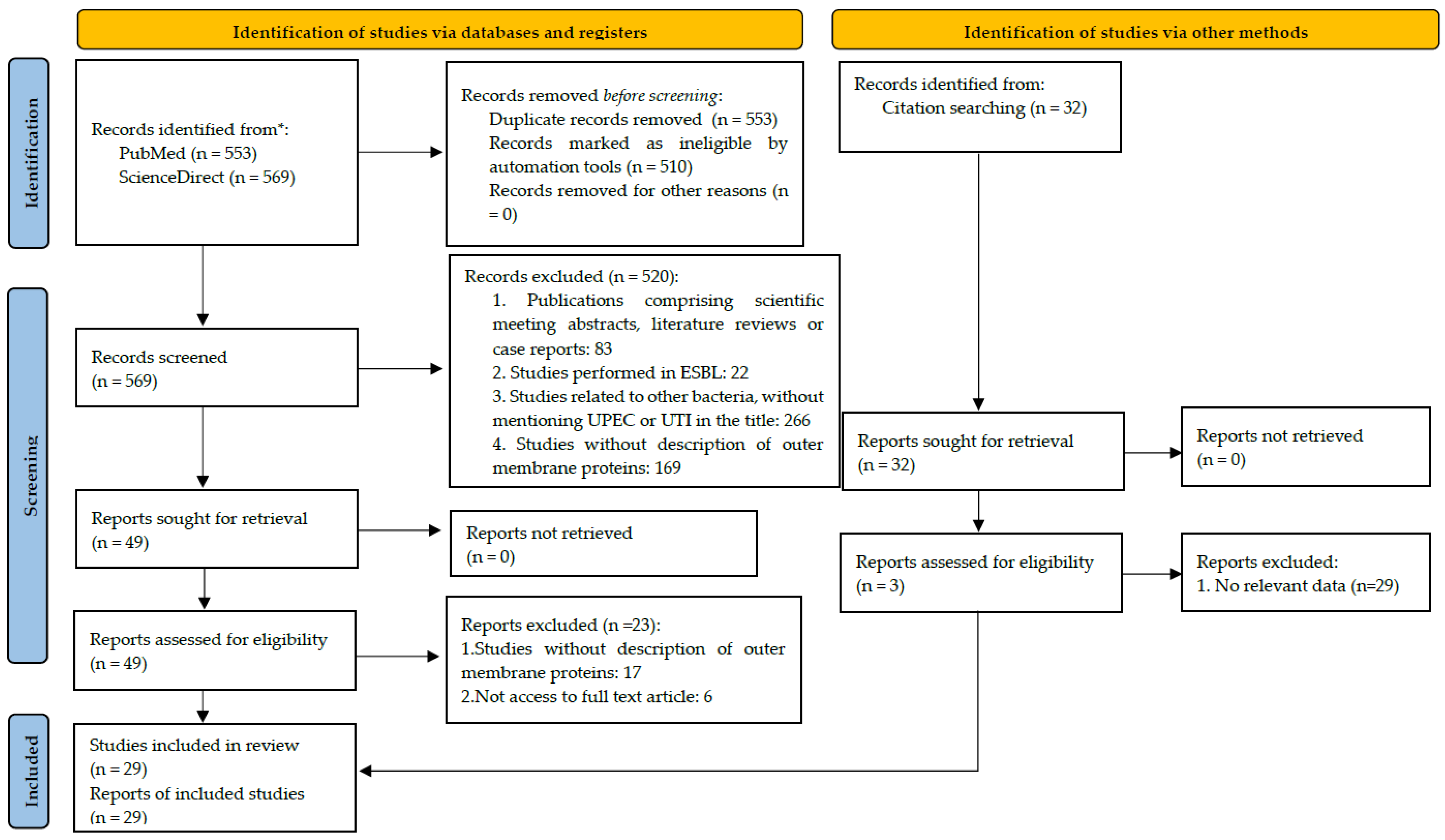
2. Materials and Methods
2.1. Selection Strategy
2.2. Selection Process and Data Extraction
- Title, authors and outer(s) membrane(s) detected;
- Samples origin and type;
- Method used for the detection of outer membrane protein;
- Group of outer membrane protein;
- Description of outer membrane protein;
- Function of outer membrane protein.
2.3. Quality Assessment
3. Results
3.1. Description of Studies
3.2. Quality Assessment
3.3. Characteristics of Studies and Outcomes Measures
3.4. Characterization of Outer Membrane Proteins of UPEC
3.5. Association to Antimicrobial Resistance
4. Discussion
5. Conclusions
Author Contributions
Funding
Institutional Review Board Statement
Data Availability Statement
Acknowledgments
Conflicts of Interest
References
- Nittayasut, N.; Yindee, J.; Boonkham, P.; Yata, T.; Suanpairintr, N.; Chanchaithong, P. Multiple and High-Risk Clones of Extended-Spectrum Cephalosporin-Resistant and blaNDM-5-Harbouring Uropathogenic Escherichia coli from Cats and Dogs in Thailand. Antibiotics 2021, 10, 1374. [Google Scholar] [CrossRef]
- Terlizzi, M.E.; Gribaudo, G.; Maffei, M.E. UroPathogenic Escherichia coli (UPEC) Infections: Virulence Factors, Bladder Responses, Antibiotic, and Non-antibiotic Antimicrobial Strategies. Front. Microbiol. 2017, 8, 1566. [Google Scholar] [CrossRef] [Green Version]
- Mysorekar, I.U.; Hultgren, S.J. Mechanisms of uropathogenic Escherichia coli persistence and eradication from the urinary tract. Proc. Natl. Acad. Sci. USA 2006, 103, 14170–14175. [Google Scholar] [CrossRef] [Green Version]
- Huang, W.-C.; Hashimoto, M.; Shih, Y.-L.; Wu, C.-C.; Lee, M.-F.; Chen, Y.-L.; Wu, J.-J.; Wang, M.-C.; Lin, W.-H.; Hong, M.-Y.; et al. Peptidoglycan Endopeptidase Spr of Uropathogenic Escherichia coli Contributes to Kidney Infections and Competitive Fitness During Bladder Colonization. Front. Microbiol. 2020, 11, 586214. [Google Scholar] [CrossRef]
- Wurpel, D.J.; Moriel, D.G.; Totsika, M.; Easton, D.M.; Schembri, M.A. Comparative analysis of the uropathogenic Escherichia coli surface proteome by tandem mass-spectrometry of artificially induced outer membrane vesicles. J. Proteom. 2015, 115, 93–106. [Google Scholar] [CrossRef]
- Yep, A.; McQuade, T.; Kirchhoff, P.; Larsen, M.; Mobley, H.L.T. Inhibitors of TonB Function Identified by a High-Throughput Screen for Inhibitors of Iron Acquisition in Uropathogenic Escherichia coli CFT073. mBio 2014, 5, e01089-13. [Google Scholar] [CrossRef] [Green Version]
- Zogg, A.L.; Zurfluh, K.; Schmitt, S.; Nüesch-Inderbinen, M.; Stephan, R. Antimicrobial resistance, multilocus sequence types and virulence profiles of ESBL producing and non-ESBL producing uropathogenic Escherichia coli isolated from cats and dogs in Switzerland. Veter- Microbiol. 2018, 216, 79–84. [Google Scholar] [CrossRef] [Green Version]
- ECDC. Surveillance of antimicrobial resistance in Europe. Br. Med. J. 2018, 317, 614–615. [Google Scholar]
- Ghosh, A.; Mukherjee, M. Incidence of multidrug resistance, pathogenicity island markers, and pathoadaptive FimH mutations in uropathogenic Escherichia coli isolated from asymptomatic hospitalized patients. Folia Microbiol. 2019, 64, 587–600. [Google Scholar] [CrossRef]
- Kalas, V.; Hibbing, M.E.; Maddirala, A.R.; Chugani, R.; Pinkner, J.S.; Mydock-McGrane, L.K.; Conover, M.S.; Janetka, J.W.; Hultgren, S.J. Structure-Based Discovery of Glycomimetic FmlH Ligands As Inhibitors of Bacterial Adhesion During Urinary Tract Infection. Proc. Natl. Acad. Sci. USA 2018, 115, E2819–E2828. [Google Scholar] [CrossRef] [Green Version]
- Matin, F.Z.; Rezatofighi, S.E.; Ardakani, M.R.; Akhoond, M.R.; Mahmoodi, F. Virulence characterization and clonal analysis of uropathogenic Escherichia coli metallo-beta-lactamase-producing isolates. Ann. Clin. Microbiol. Antimicrob. 2021, 20, 50. [Google Scholar] [CrossRef]
- Bélanger, L.; Garenaux, A.; Harel, J.; Boulianne, M.; Nadeau, E.; Dozois, C.M. Escherichia coli from animal reservoirs as a potential source of human extraintestinal pathogenic E. coli. FEMS Immunol. Med. Microbiol. 2011, 62, 1–10. [Google Scholar] [CrossRef] [Green Version]
- Salgado-Caxito, M.; Benavides, J.A.; Adell, A.D.; Paes, A.C.; Moreno-Switt, A.I. Global prevalence and molecular characterization of extended-spectrum β-lactamase producing-Escherichia coli in dogs and cats—A scoping review and meta-analysis. One Health 2021, 12, 100236. [Google Scholar] [CrossRef]
- Piras, C.; Soggiu, A.; Greco, V.; Martino, P.A.; Del Chierico, F.; Putignani, L.; Urbani, A.; Nally, J.E.; Bonizzi, L.; Roncada, P. Mechanisms of antibiotic resistance to enrofloxacin in uropathogenic Escherichia coli in dog. J. Proteom. 2015, 127, 365–376. [Google Scholar] [CrossRef]
- Matamoros-Recio, A.; Franco-Gonzalez, J.F.; Forgione, R.E.; Torres-Mozas, A.; Silipo, A.; Martín-Santamaría, S. Understanding the Antibacterial Resistance: Computational Explorations in Bacterial Membranes. ACS Omega 2021, 6, 6041–6054. [Google Scholar] [CrossRef]
- Silhavy, T.J.; Kahne, D.; Walker, S. The Bacterial Cell Envelope. Cold Spring Harb. Perspect. Biol. 2010, 2, 1–16. Available online: https://www.ncbi.nlm.nih.gov/pmc/articles/PMC2857177/pdf/cshperspect-PRK-a000414.pdf (accessed on 4 September 2022). [CrossRef]
- Dehghani, B.; Mottamedifar, M.; Khoshkharam-Roodmajani, H.; Hassanzadeh, A.; Zomorrodian, K.; Rahimi, A. SDS-PAGE Analysis of the Outer Membrane Proteins of Uropathogenic Escherichia coli Isolated from Patients in Different Wards of Nemazee Hospital, Shiraz, Iran. Iran. J. Med. Sci. 2016, 41, 399–405. [Google Scholar]
- Alteri, C.; Mobley, H.L.T. Quantitative Profile of the Uropathogenic Escherichia coli Outer Membrane Proteome during Growth in Human Urine. Infect. Immun. 2007, 75, 2679–2688. [Google Scholar] [CrossRef] [Green Version]
- Page, M.J.; McKenzie, J.E.; Bossuyt, P.M.; Boutron, I.; Hoffmann, T.C.; Mulrow, C.D.; Shamseer, L.; Tetzlaff, J.M.; Akl, E.A.; Brennan, S.E.; et al. The PRISMA 2020 statement: An updated guideline for reporting systematic reviews. BMJ 2021, 372, n71. [Google Scholar] [CrossRef]
- Wells, G.; Shea, B.; O’Connel, D.; Peterson, J.; Welch, V.; Losos, M.; Tugwell, P. The Newcastle-Ottawa Scale (NOS) for assessing the quality of nonrandomised studies in meta-analyses. 2021. Available online: https://www.ohri.ca//programs/clinical_epidemiology/oxford.asp (accessed on 25 August 2022).
- Beck, C.M.; Willett, J.L.E.; Cunningham, D.A.; Kim, J.J.; Low, D.A.; Hayes, C.S. CdiA Effectors from Uropathogenic Escherichia coli Use Heterotrimeric Osmoporins as Receptors to Recognize Target Bacteria. PLoS Pathog. 2016, 12, e1005925. [Google Scholar] [CrossRef] [Green Version]
- Brannon, J.R.; Thomassin, J.L.; Desloges, I.; Gruenheid, S.; Le Moual, H. Role of uropathogenic Escherichia coli OmpT in the resistance against human cathelicidin LL-37. FEMS Microbiol. Lett. 2013, 345, 64–71. [Google Scholar] [CrossRef] [Green Version]
- Choi, U.; Lee, C.-R. Distinct Roles of Outer Membrane Porins in Antibiotic Resistance and Membrane Integrity in Escherichia coli. Front. Microbiol. 2019, 10, 953. [Google Scholar] [CrossRef]
- Conover, M.S.; Ruer, S.; Taganna, J.; Kalas, V.; De Greve, H.; Pinkner, J.; Dodson, K.W.; Remaut, H.; Hultgren, S.J. Inflammation-Induced Adhesin-Receptor Interaction Provides a Fitness Advantage to Uropathogenic E. coli during Chronic Infection. Cell Host. Microbe. 2016, 176, 498–503. [Google Scholar] [CrossRef] [Green Version]
- Desloges, I.; Taylor, J.A.; Leclerc, J.M.; Brannon, J.R.; Portt, A.; Spencer, J.D.; Dewar, K.; Marczynski, G.T.; Manges, A.; Gruenheid, S.; et al. Identification and characterization of OmpT-like proteases in uropathogenic Escherichia coli clinical isolates. Microbiologyopen 2019, 8, e915. [Google Scholar] [CrossRef] [Green Version]
- Diao, J.; Bouwman, C.; Yan, D.; Kang, J.; Katakam, A.; Liu, P.; Pantua, H.; Xu, M.; Kapadia, S. Peptidoglycan Association of Murein Lipoprotein is required for KpsD-Dependent Group 2 Capsular Polysaccharide Expression and Serum Resistance in a Uropathogenic Escherichia coli Isolate. Am. Soc. Fir. Microbiol. 2018, 200, 500–502. [Google Scholar] [CrossRef] [Green Version]
- Geibel, S.; Procko, E.; Hultgren, S.J.; Baker, D.; Waksman, G. Structural and energetic basis of folded-protein transport by the FimD usher. Nature 2013, 496, 243–246. [Google Scholar] [CrossRef] [Green Version]
- Greene, S.E.; Hibbing, M.E.; Janetka, J.; Chen, S.; Hultgren, S.J. Human Urine Decreases Function and Expression of Type 1 Pili in Uropathogenic Escherichia coli. mBio 2015, 6, e00820-15. [Google Scholar] [CrossRef] [Green Version]
- He, X.L.; Wang, Q.; Peng, L.; Qu, Y.-R.; Puthiyakunnon, S.; Liu, X.-L.; Hui, C.Y.; Boddu, S.; Cao, H.; Huang, S.-H. Role of uropathogenic Escherichia coli outer membrane protein T in pathogenesis of urinary tract infection. Pathog. Dis. 2015, 73, ftv006. [Google Scholar] [CrossRef] [Green Version]
- Henderson, N.S.; Thanassi, D.G. Purification of the Outer Membrane Usher Protein and Periplasmic Chaperone-Subunit Complexes from the P and Type 1 Pilus Systems. Methods Mol. Biol. 2013, 966, 37–52. [Google Scholar] [CrossRef]
- Hirakawa, H.; Suzue, K.; Kurabayashi, K.; Tomita, H. The Tol-Pal System of Uropathogenic Escherichia coli Is Responsible for Optimal Internalization Into and Aggregation Within Bladder Epithelial Cells, Colonization of the Urinary Tract of Mice, and Bacterial Motility. Front. Microbiol. 2019, 10, 1827. [Google Scholar] [CrossRef] [Green Version]
- Nielsen, D.W.; Ricker, N.; Barbieri, N.L.; Allen, H.K.; Nolan, L.K.; Logue, C.M. Outer membrane protein A (OmpA) of extraintestinal pathogenic Escherichia coli. BMC Res. Notes 2020, 13, 51. [Google Scholar] [CrossRef] [Green Version]
- Pantel, A.; Dunyach-Remy, C.; Essebe, C.N.; Mesureur, J.; Sotto, A.; Pagès, J.-M.; Nicolas-Chanoine, M.-H.; Lavigne, J.-P. Modulation of Membrane Influx and Efflux in Escherichia coli Sequence Type 131 Has an Impact on Bacterial Motility, Biofilm Formation, and Virulence in a Caenorhabditis elegans Model. Antimicrob. Agents Chemother. 2016, 60, 2901–2911. [Google Scholar] [CrossRef]
- Pantua, H.; Skippington, E.; Braun, M.-G.; Noland, C.L.; Diao, J.; Peng, Y.; Gloor, S.L.; Yan, D.; Kang, J.; Katakam, A.K.; et al. Unstable Mechanisms of Resistance to Inhibitors of Escherichia coli Lipoprotein Signal Peptidase. mBio 2020, 11, e02018-20. [Google Scholar] [CrossRef]
- Ribeiro, C.D.S.; Martins, W.M.B.D.S.; da Silva, A.A.; Gales, A.C.; Rando, D.G.G.; Minarini, L.A.D.R. Exposure to sub-inhibitory ciprofloxacin and nitrofurantoin concentrations increases recA gene expression in uropathogenic Escherichia coli: The role of RecA protein as a drug target. Eur. J. Pharm. Sci. 2020, 146, 105268. [Google Scholar] [CrossRef]
- Tavio, M.M.; Aquili, V.D.; Poveda, J.B.; Antunes, N.T.; Sanchez-Cespedes, J.; Vila, J. Quorum-sensing regulator sdiA and marA overexpression is involved in in vitro-selected multidrug resistance of Escherichia coli. J. Antimicrob. Chemother. 2010, 65, 1178–1186. [Google Scholar] [CrossRef]
- Wurpel, D.J.; Totsika, M.; Allsopp, L.; Webb, R.I.; Moriel, D.G.; Schembri, M.A. Comparative proteomics of uropathogenic Escherichia coli during growth in human urine identify UCA-like (UCL) fimbriae as an adherence factor involved in biofilm formation and binding to uroepithelial cells. J. Proteom. 2016, 131, 177–189. [Google Scholar] [CrossRef] [Green Version]
- Xicohtencatl-Cortes, J.; Cruz-Córdova, A.; Cázares-Domínguez, V.; Escalona-Venegas, G.; Zavala-Vega, S.; Arellano-Galindo, J.; Romo-Castillo, M.; Hernández-Castro, R.; Ochoa, S.A.; Luna-Pineda, V.M. Uropathogenic Escherichia coli strains harboring tosA gene were associated to high virulence genes and a multidrug-resistant profile. Microb. Pathog. 2019, 134, 103593. [Google Scholar] [CrossRef]
- Zalewska-Piątek, B.; Wilkanowicz, S.; Bruździak, P.; Piątek, R.; Kur, J. Biochemical characteristic of biofilm of uropathogenic Escherichia coli Dr + strains. Microbiol. Res. 2013, 168, 367–378. [Google Scholar] [CrossRef]
- Zalewska-Piątek, B.M.; Piatek, R.; Olszewski, M.; Kur, J. Identification of antigen Ag43 in uropathogenic Escherichia coli Dr+ strains and defining its role in the pathogenesis of urinary tract infections. Microbiology 2015, 161, 1034–1049. [Google Scholar] [CrossRef]
- Zude, I.; Leimbach, A.; Dobrindt, U. Prevalence of autotransporters in Escherichia coli: What is the impact of phylogeny and pathotype? Int. J. Med. Microbiol. 2014, 304, 243–256. [Google Scholar] [CrossRef]
- Martins da Costa, P.; Loureiro, L.; Matos, A.J.F. Transfer of Multidrug-Resistant Bacteria Between Intermingled Ecological Niches: The Interface Between Humans, Animals and the Environment. Int. J. Environ. Res. Public Health 2013, 10, 278–294. [Google Scholar] [CrossRef] [Green Version]
- Collignon, P.J.; McEwen, S.A. One Health—Its Importance in Helping to Better Control Antimicrobial Resistance. Trop. Med. Infect. Dis. 2019, 4, 22. [Google Scholar] [CrossRef]
- Pagès, J.-M.; James, C.E.; Winterhalter, M. The porin and the permeating antibiotic: A selective diffusion barrier in Gram-negative bacteria. Nat. Rev. Microbiol. 2008, 6, 893–903. [Google Scholar] [CrossRef] [Green Version]
- Worthington, R.J.; Melander, C. Overcoming Resistance to β-Lactam Antibiotics. J. Org. Chem. 2014, 78, 4207–4213. [Google Scholar] [CrossRef] [Green Version]
- Munita, J.; Arias, C. Mechanisms of Antibiotic Resistance. Microbiol. Spectr. 2016, 17, 119–127. [Google Scholar]
- Cardoso, M.H.; Almeida, K.C.D.; Cândido, E.D.S.; Murad, A.M. Comparative NanoUPLC-MS E analysis between magainin I-susceptible and -resistant Escherichia coli strains. Sci. Rep. 2017, 7, 4197. [Google Scholar] [CrossRef] [Green Version]
- Liu, Y.-F.; Yan, J.-J.; Lei, H.-Y.; Teng, C.-H.; Wang, M.-C.; Tseng, C.-C.; Wu, J.-J. Loss of Outer Membrane Protein C in Escherichia coli Contributes to Both Antibiotic Resistance and Escaping Antibody-Dependent Bactericidal Activity. Infect. Immun. 2012, 80, 1815–1822. [Google Scholar] [CrossRef] [Green Version]
- Kishii, R.; Takei, M. Relationship between the expression of ompF and quinolone resistance in Escherichia coli. J. Infect. Chemother. 2009, 15, 361–366. [Google Scholar] [CrossRef]
- Fernández, L.; Hancock, R.E.W. Adaptive and Mutational Resistance: Role of Porins and Efflux Pumps in Drug Resistance. Clin. Microbiol. Rev. 2012, 25, 661–681. [Google Scholar] [CrossRef] [Green Version]
- Chowdhury, N.; Suhani, S.; Purkaystha, A.; Begum, M.K.; Raihan, T.; Alam, J. Identification of AcrAB-TolC Efflux Pump Genes and Detection of Mutation in Efflux Repressor AcrR from Omeprazole Responsive Multidrug-Resistant Escherichia coli Isolates Causing Urinary Tract Infections. Microbiol. Insights 2019, 12, 1178636119889629. [Google Scholar] [CrossRef] [Green Version]
- Ochoa, S.A.; Cruz-Córdova, A.; Luna-Pineda, V.M.; Reyes, J.P.; Cázares-Domínguez, V.; Escalona, G.; Sepúlveda-González, M.E.; López-Montiel, F.; Arellano-Galindo, J.; López-Martínez, B.; et al. Multidrug- and Extensively Drug-Resistant Uropathogenic Escherichia coli Clinical Strains: Phylogenetic Groups Widely Associated with Integrons Maintain High Genetic Diversity. Front. Microbiol. 2016, 7, 2042. [Google Scholar] [CrossRef] [Green Version]
- Contreras-Alvarado, L.M.; Zavala-Vega, S.; Cruz-Córdova, A.; Reyes-Grajeda, J.P.; Escalona-Venegas, G.; Flores, V.; Alcázar-López, V.; Arellano-Galindo, J. Molecular Epidemiology of Multidrug-Resistant Uropathogenic Escherichia coli O25b Strains Associated with Complicated Urinary Tract Infection in Children. Microorganisms 2021, 9, 2299. [Google Scholar] [CrossRef]
- Ebbensgaard, A.E.; Mordhorst, H.; Aarestrup, F.; Hansen, E.B. The Role of Outer Membrane Proteins and Lipopolysaccharides for the Sensitivity of Escherichia coli to Antimicrobial Peptides. Front. Microbiol. 2018, 9, 2153. [Google Scholar] [CrossRef]
- Mathelié-Guinlet, M.; Asmar, A.T.; Collet, J.-F.; Dufrêne, Y.F. Lipoprotein Lpp regulates the mechanical properties of the E. coli cell envelope. Nat. Commun. 2020, 11, 1789. [Google Scholar] [CrossRef] [Green Version]
- Asmar, A.T. Lpp, the Braun lipoprotein, turns 50—Major achievements and remaining issues. FEMS Microbiol. Lett. 2018, 365, fny199. [Google Scholar] [CrossRef] [Green Version]
- Poirel, L.; Cattoir, V.; Nordmann, P. Plasmid-Mediated Quinolone Resistance; Interactions between Human, Animal, and Environmental Ecologies. Front. Microbiol. 2012, 3, 24. [Google Scholar] [CrossRef] [Green Version]
- Iacovelli, R.; Sokolova, N.; Haslinger, K. Striving for Sustainable Biosynthesis: Discovery, Diversification, and Production of Antimicrobial Drugs in Escherichia coli; Portland Press Ltd.: London, UK, 2022; p. 6. [Google Scholar]
- Kot, B. Antibiotic Resistance Among Uropathogenic Escherichia coli. Pol. J. Microbiol. 2019, 68, 403–415. [Google Scholar] [CrossRef] [Green Version]
- Peterson, J.H.; Doyle, M.T.; Bernstein, H.D. Small Molecule Antibiotics Inhibit Distinct Stages of Bacterial Outer Membrane Protein Assembly. mBio 2022, e02286-22. [Google Scholar] [CrossRef]
- Karam, M.R.A.; Habibi, M.; Bouzari, S. Urinary tract infection: Pathogenicity, antibiotic resistance and development of effective vaccines against Uropathogenic Escherichia coli. Mol. Immunol. 2019, 108, 56–67. [Google Scholar] [CrossRef]
- Piatek, R.; Zalewska-Piatek, B.; Dzierzbicka, K.; Makowiec, S.; Pilipczuk, J.; Szemiako, K.; Cyranka-Czaja, A.; Wojciechowski, M. Pilicides inhibit the FGL chaperone/usher assisted biogenesis of the Dr fimbrial polyadhesin from uropathogenic Escherichia coli. BMC Microbiol. 2013, 13, 131. [Google Scholar] [CrossRef] [Green Version]
- Chahales, P.; Hoffman, P.S.; Thanassi, D.G. Nitazoxanide Inhibits Pilus Biogenesis by Interfering with Folding of the Usher Protein in the Outer Membrane. Antimicrob. Agents Chemother. 2016, 60, 2028–2038. [Google Scholar] [CrossRef]

| Author | Year of Publication | Origin of Isolates | Sample Type | Method | Risk of Bias | OMP Detected | Reference |
|---|---|---|---|---|---|---|---|
| Alteri et al. | 2008 | Human | Urine and blood | 2D-DIGE, MS/MS, SDS-PAGE | Low | IutA | [18] |
| Beck et al. | 2016 | Human | Urine, blood and faecal | Adhesion assays, immunoblot analysis | Moderate | BamA OmpC OmpF | [21] |
| Brannon et al. | 2013 | Human | Urine, blood and fecal | Membrane integrity assays, proteolytic activity, SDS-PAGE, WB | Low | OmpT | [22] |
| Choi et al. | 2020 | Human | Urine and blood | Membrane integrity assays | Low | OmpA OmpC OmpF | [23] |
| Conover et al. | 2016 | Human | Urine and blood | Crystallization, structure determination and further vaccination of an infected mouse | Low | FimIH | [24] |
| Dehghani et al. | 2016 | Human | Urine | SDS-PAGE | Low | OmpA OmpC OmpF | [17] |
| Desloges et al. | 2019 | Human | Urine, blood and fecal | Proteolytic activity, SDS-PAGE, WB | Low | OmpP OmpT | [25] |
| Diao et al. | 2018 | Human | Urine and blood | LC-MS/MS, SDS-PAGE, WB | Low | Bam A BamD BamE c1765 c2482 c3153 chuA FimD IreA KpsD lolB Lpp nmpC OmpA OmpC OmpF OmpX Slp SurA tolC | [26] |
| Geibel et al. | 2013 | Non-specified origin | - | Crystallization, structure determination, computational methods | Moderate | Complex FimD–FimC–FimF–FimG–FimH | [27] |
| Ghosh et al. | 2019 | Human | Urine | Polymorphism study | Low | FimH | [9] |
| Greene et al. | 2015 | Human and non-specified origin | Urine and blood | Electron microscopy, HA, immunoblot analysis | Moderate | Type 1 pili | [28] |
| He et al. | 2015 | Human and non-specified origin | Urine and blood | Adhesion and invasion assays, HA, proteolytic activity | Moderate | OmpT | [29] |
| Hederson & Thanassi | 2013 | Non-specified origin | - | FPLC | Moderate | FimC–FimH chaperone–adhesin Type 1 pilus usher FimD P pilus usher PapC PapD–PapG chaperone–adhesin | [30] |
| Hirakawa et al. | 2019 | Human | Urine and blood | Adhesion and biofilm assays, HA, motility assays, SDS-PAGE | Low | TolB | [31] |
| Huang et al. | 2020 | Human | Urine | LC-MS/MS, invasion assay, macrophage and serum survival methods, SDS-PAGE, survival in SDS and low pH conditions assays, WB | Low | FliC | [4] |
| Kalas et al. | 2018 | Human | Urine and blood | Crystallization, structure determination, in silico docking, immunofluorescence microscopy | Moderate | Type 1 pili F9 pili | [10] |
| Nielsen et al. | 2020 | Human and avian | Fecal, vaginal and CSF | In silico analysis | Moderate | OmpA | [32] |
| Pantel et al. | 2016 | Human | Urine and blood | Biofilm, nematode-killing and motility assays, immunoblot analysis, SDS-PAGE | Low | AcrA-AcrB-TolC OmpC OmpF | [33] |
| Pantua et al. | 2022 | Human and non-specified origin | Urine and blood | SDS-PAGE, WB | Low | Lpp | [34] |
| Piras et al. | 2015 | Dog | Urine | 2D-DIGE, nLC-MS/MS | Low | OmpW | [14] |
| Ribeiro et al. | 2020 | Human | Urine | Molecular docking studies | Moderate | QepA OqxAB | [35] |
| Tavio et al. | 2010 | Human | Urine | SDS-PAGE | Low | AcrA–AcrB–TolC | [36] |
| Wurpel et al. | 2015 | Human | Urine and blood | nLC-MS/MS | Moderate | BamA BamB BamC BtuB ChuA CirA CjrC FadL FepA FhuA FhuE FitA Fiu Flu FyuA GdhA Iha IreA IroN IutA Lpp LptD LptE MalB NlpD NmpC OmpA OmpC OmpF OmpT OmpX Pal RcsF SmpA TraT Tsh Tsx Slp SlyB UTI89_CC1129 UTI89_C4946 VacJ Wzi YbhC YddB YeaF YgeR | [5] |
| Wurpel et al. | 2016 | Human | Urine and blood | Adhesin and biofilm assays, nLC-MS/MS, TEM | Low | BtuB ChuA CirA CjrC FepA FhuA Fiu Flu FyuA IutA Iha IreA IroN NmpC OmpA OmpC OmpF OmpT OmpW OmpX UidC UTI89C2234 SlyB | [37] |
| Xicohtencatl-Cortes et al. | 2019 | Human | Urine | Adhesion assays, SDS-PAGE, WB | Low | TosA | [38] |
| Yep et al. | 2014 | Human | Urine and blood | Bacteriophage adsorption assay, chelator assay, dose-response analysis, HTS | Moderate | RecA | [6] |
| Zalewska-Piatek et al. | 2013 | Human and non-specified origin | Urine | Biofilm assays | Moderate | Dr Fimbriae | [39] |
| Zalewska-Piatek et al. | 2015 | Human and non-specified origin | Urine | Autoaggregation assay, biofilm, adhesion, invasion assays, immunofluorescence microscopy, SDS-PAGE, WB | Moderate | DraD DraE | [40] |
| Zude et al. | 2014 | Non-specified origin | - | Autoaggregation and biofilm assays, extracellular matrix protein binding method, immunofluorescence microscopy, WB | Moderate | Ag43 | [41] |
| Group of OMP | OMP | Description | Function | Association to Antimicrobial Resistance |
|---|---|---|---|---|
| Autotransporter | Ag43 | Phase-variable antigen 43 autotransporter protein | Autotransporter | - |
| BAM complex | BamA BamB BamC BamD BamE | Outer membrane protein assembly factor A Outer membrane protein assembly factor B Outer membrane protein assembly factor C Outer membrane protein assembly factor D Outer membrane protein assembly factor E | OMP assembly OMP assembly OMP assembly OMP assembly OMP assembly | - - - - - |
| Efflux pumps | TolC | Outer membrane protein ToC | Substance efflux | Yes |
| Fimbriae | DraE DraD FimA FimC FimD FimF FimG FimH FimIH PapC PapD PapG | Dr fimbriae subunit Dr fimbriae subunit tip Type 1 fimbrial protein, A chain Type 1 pilus chaperone, FimC Outer membrane usher protein FimD Outer membrane protein FimF Outer membrane protein FimG Type 1 pilus adhesin, FimH Pili FimH-like adhesin P pilus protein PapC P pilus chaperone PapD P pilus protein PapG | Dr fimbriae Adhesin Type 1 pili structure Type 1 pili structure Type 1 pili structure Type 1 pili structure Type 1 pili structure Adhesin Adhesin P pili structure P pili structure Adhesin | - - - - - - - - - - - - |
| Flagella | FliC | Flagellin | Flagella struture | - |
| Iron receptor and siderophores | c2482 ChuA CirA CjrC FepA FhuA FhuE FitA Fiu FyuA Iha IreA IroN IutA TonB–ExbB–ExbD UTI89_C1129 UTI89C2234 | Putative outer membrane receptor for iron or colicin Heme/hemoglobin receptor Colicin I receptor and iron receptor Putative siderophore receptor Ferrienterobactin receptor Ferrichrome–iron receptor Ferri–rhodotorulic siderophore receptor Ferrichrome iron transport receptor Catecholate siderophore recetor Yersiniabactin receptor Siderophore receptor/adhesion Putative siderophore receptor Samochelin receptor Ferric aerobactin receotor TonB system Putative heme/hemoglobin receptor Putative iron compound receptor | Metal ion receptor and transport Metal ion receptor Metal ion receptor and transport Metal ion receptor Metal ion receptor Metal ion receptor Metal ion receptor Metal ion receptor Metal ion receptor Metal ion receptor Metal ion receptor Metal ion receptor Metal ion receptor Metal ion receptor Metal ion receptor and transport Metal ion receptor Metal ion receptor | - - - - - - - - - - - - - - - - - |
| Lipoproteins | lolB NlpD Lpp LptD LptE Slp SlyB VacJ Wzi YbhC YgeR | Outer-membrane lipoprotein LolB Lipoprotein Murein lipoprotein Putative uncharacterized protein LPS assembly lipoprotein Outer membrane protein Outer membrane protein Lipoprotein Outer membrane protein Putative pectinesterase Lipoprotein | LPS organization/synthesis LPS organization/synthesis LPS organization/synthesis LPS organization/synthesis LPS organization/synthesis Unknown Unknown Unknown LPS organization/synthesis LPS organization/synthesis LPS organization/synthesis | - - Yes - - - - - - - - |
| Other | c1765 c3153 Flu GdhA KpsD RcsF SmpA TosA TraT Tsh SurA UTI89_C4946 YeaF | Partial Putative outer membrane channel protein Putative outer membrane protein of prophage Antigen 43 AT NADP-specific glutamate dehydrogenase Putative outer membrane translocon for export of group 2 capsular polysaccharides Regulator in colanic acid synthesis Small protein A Putative repeats-in-toxin protein Conjugal transfer surface exclusion protein Temperature sensitive hemagglutinin AT Outer membrane protein chaperone Putative filamentous hemagglutinin Putative LPS scaffolding protein | Unknown Unknown Autoaggregatiom Aminoacid metabolismo Transport Unknown OMP assembly Nonfimbrial adhesin Conjugation Unknown OMP assembly Unknown Unknown | - - - - - - Yes - - - - - |
| Porin | MalB NmpC/c2348 OmpA OmpC OmpF OmpP OmpT OmpW OmpX Tsx UidC YddB | Maltose-inducible porin Outer membrane porin protein Outer membrane protein A Outer membrane protein C Outer membrane protein F Outer membrane protein P Outer membrane protein T, protéase VII Outer membrane protein W Outer membrane protein X Nucleoside-specific channel forming protein Outer membrane porin protein Putative porin protein | Porin Porin Porin Porin Porin Porin Proteolysis Unknown Unknown Porin Porin Porin | - - - Yes Yes - - Yes - - - Yes |
| Tol–Pal System | Pal | Peptidoglycan-associated lipoprotein | Membrane integrity | - |
| Transporters | BtuB FadL | Vitamin B-12 receptor Bifunctional long-chain fatty acid transporter | Transporter Transporter | - - |
Publisher’s Note: MDPI stays neutral with regard to jurisdictional claims in published maps and institutional affiliations. |
© 2022 by the authors. Licensee MDPI, Basel, Switzerland. This article is an open access article distributed under the terms and conditions of the Creative Commons Attribution (CC BY) license (https://creativecommons.org/licenses/by/4.0/).
Share and Cite
Rodrigues, I.C.; Rodrigues, S.C.; Duarte, F.V.; Costa, P.M.d.; Costa, P.M.d. The Role of Outer Membrane Proteins in UPEC Antimicrobial Resistance: A Systematic Review. Membranes 2022, 12, 981. https://doi.org/10.3390/membranes12100981
Rodrigues IC, Rodrigues SC, Duarte FV, Costa PMd, Costa PMd. The Role of Outer Membrane Proteins in UPEC Antimicrobial Resistance: A Systematic Review. Membranes. 2022; 12(10):981. https://doi.org/10.3390/membranes12100981
Chicago/Turabian StyleRodrigues, Inês C., Sílvia C. Rodrigues, Filipe V. Duarte, Paula M. da Costa, and Paulo M. da Costa. 2022. "The Role of Outer Membrane Proteins in UPEC Antimicrobial Resistance: A Systematic Review" Membranes 12, no. 10: 981. https://doi.org/10.3390/membranes12100981
APA StyleRodrigues, I. C., Rodrigues, S. C., Duarte, F. V., Costa, P. M. d., & Costa, P. M. d. (2022). The Role of Outer Membrane Proteins in UPEC Antimicrobial Resistance: A Systematic Review. Membranes, 12(10), 981. https://doi.org/10.3390/membranes12100981