Review

Membrane Applications in Autologous Cell Therapy

1
Department of Engineering Science, Institute of Biomedical Engineering, University of Oxford, Oxford OX3 7DQ, UK
2
Oxford Suzhou Centre for Advanced Research (OSCAR), University of Oxford, Suzhou 215123, China
*
Authors to whom correspondence should be addressed.
These authors contributed equally to this work.
Membranes 2022, 12(12), 1182; https://doi.org/10.3390/membranes12121182
Submission received: 3 October 2022 / Revised: 3 November 2022 / Accepted: 7 November 2022 / Published: 24 November 2022
(This article belongs to the Special Issue Honorary Issue for Professor Anthony Fane)

Abstract

:
Stem cell and cell therapies, particularly autologous cell therapies, are becoming a common practice. However, in order for these technologies to achieve wide-scale clinical application, the prohibitively high cost associated with these therapies must be addressed through creative engineering. Membranes can be a disruptive technology to reshape the bioprocessing and manufacture of cellular products and significantly reduce the cost of autologous cell therapies. Examples of successful membrane applications include expansions of CAR-T cells, various human stem cells, and production of extracellular vesicles (EVs) using hollow fibre membrane bioreactors. Novel membranes with tailored functions and surface properties and novel membrane modules that can accommodate the changing needs for surface area and transport properties are to be developed to fulfil this key role.

1. Introduction

Through the manipulation, expansion, processing, and subsequent re-implantation of a patient’s own cells, autologous cell therapies are now used in the treatment of otherwise incurable diseases and degenerative conditions. As the cells used in these therapies are derived from the patient themselves, complications associated with immunological rejection may be negated. Consequently, autologous therapies are considered to be more readily achievable than other technologies in the field of regenerative medicine. To date, successful autologous cell therapies in clinical practice include:
  • Autologous chondrocyte implantation (ACI) for cartilage repair [1].
  • Autologous cell therapy for treatment of burns [2],
  • Autologous stem cell transplantation for the treatment of multiple myeloma and multiple scoliosis [3], and
  • Chimeric antigen receptor T-Cell (CAR-T) therapy for the treatment of blood cancers [4].
Autologous cell therapies, such as chondrocyte implantation therapy, are multi-step processes [5] as shown in the block flow diagram in Figure 1. In such a process, cells are initially obtained from the patient either through biopsy or a blood sample. Consequently, in contrast to traditional therapies, each treatment is bespoke to an individual patient. Processes developed for the production of autologous therapies are therefore not based on “scale up”, but rather on the parallel “scale out” using single use disposable platforms.
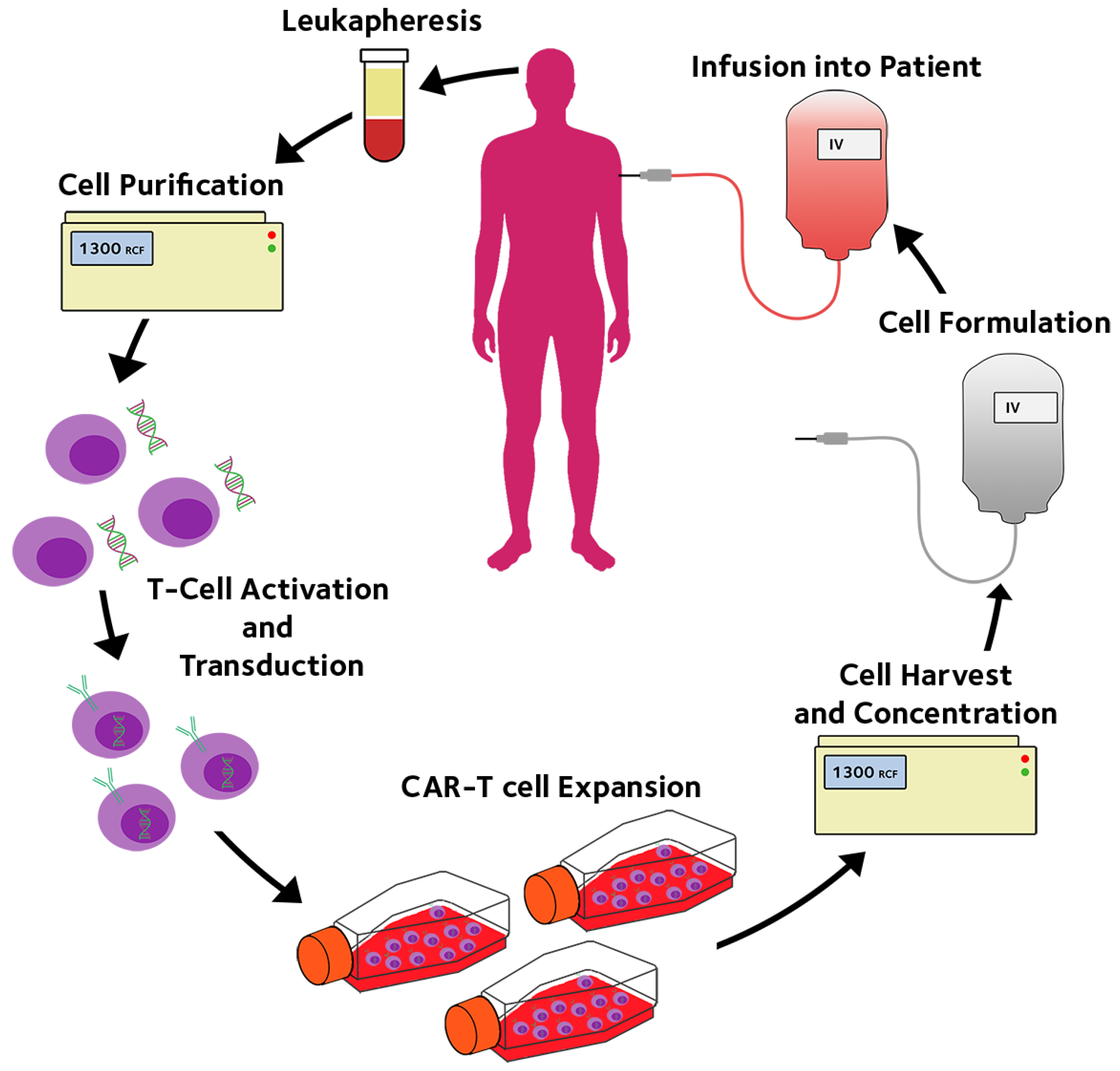
One of the main challenges to autologous cell therapies is their high cost. For example, CAR-T therapies approved for use by the NHS in the United Kingdom, tisagenlecleucel or Kymriah, come with the steep price tag of GBP 282,000 per treatment, with studies revealing the real cost being even higher when considering post-treatment care [6]. Bioprocessing and manufacture of the cellular products account for a large fraction of the cost of goods. Current processing technologies are largely based on mimicking laboratory cell manipulation and culture with different degrees of automation [7] as shown in the process flow diagram Figure 2. Infrastructure investment to create ultraclean environments (good manufacture practice, or GMP) is necessary for the “open operation” where the product or component are exposed to the operational environment. There is an urgent need for novel bioprocessing technologies, new equipment, and new devices, to form a fully enclosed, fully automatic, and single use system, to perform all the processing steps in hospitals without the need for dedicated GMP facilities. In this way, membranes can play an important role.
Membranes offer a physical barrier (excluding liquid membranes) with selective transport properties and different surface properties. With the desirable properties, membranes can be used to solve some key issues, in a genius way, for the bioprocessing and manufacture of autologous cellular products. Herein, we review the application of membrane technologies towards, CAR-T therapy, the culture of adherent cell types, expansion of stem cells, and isolation of extracellular vesicles (EVs).

2. Hollow Fibre Membrane Bioreactors for CAR-T Immunotherapy

It has long been understood that the immune system plays an essential role in cancer development and growth [9], constant “immunosurveillance” by the immune system seeking and eradicating potential cancer cells [10]. Further studies [11] demonstrated that specific T lymphocyte populations, killer T cells, are heavily involved in this natural cancer prevention through the hunting down and killing precancerous and cancerous cells [12]. These T cells have an innate “guidance system” in the form of T Cell Receptors (TCRs), which they use to recognize cancerous and precancerous cells [13]. These TCRs are sensitive enough to distinguish single amino acid changes in antigenic peptide sequences [14]. However, to avoid the development of autoimmunity, it is estimated that more than 90% of all immature T cells and all the T cells with self-reacting TCRs are destroyed before they reach maturity [15]. Thus, most T cells in the blood stream are nonreactive or only weakly reactive against mutated self-antigens on tumours. It is necessary to circumvent this problem to effectively use T cells as a form of cancer therapy.
The first form of T cell therapy utilised Tumour-Infiltrating Lymphocytes (TILs) that are found inside tumour tissues [16]. The rational for this was that T cells inside tumour will have a higher chance of reacting with mutated self-antigens on tumours. However, in spite of this TIL therapy has enjoyed only limited success [17].
A breakthrough in the effectiveness of this therapy was achieved by combining the low-reactivity high-cytotoxicity T cell therapy with Monoclonal antibody (mAb) therapy, which on its own has high reactivity but low cytotoxicity. This was achieved through genetic modification of T cells, enabling researchers to attach high affinity cancer specific antibodies called Chimeric Antigen Receptors (CAR) onto the surface of highly cytotoxic T cells to create the first CAR-T cells [18]. Through multiple clinical trials, it was demonstrated that these newly created CAR-T cells were exceptionally good at treating certain types of cancer [19]. In particular, the anti-CD19 CAR-T cell therapy for the treatment of B-cell lymphoma had been extremely successful [20] and subsequently six therapies have been commercially approved by the FDA including, ABECMA (idecabtagene vicleucel, Bristol Myers Squibb, New York, NY, USA), CARVYKTITM (ciltacabtagene autoleucel, Janssen Biotech, Inc., Horsham, PA, USA), TECARTUSTM (brexucabtagene autoleucel, Kite Pharma, Inc., Los Angeles, CA, USA), Kymria™ (tisagenlecleucel, Novatis, Basel, Switzerland), Yescarta™ (axicabtagene ciloleucel, Kite Pharma, Inc., Los Angeles, CA, USA), and Breyanzi (lisocabtagene maraleucel, Bristol Myers Squibb, New York, NY, USA) [21,22,23,24,25,26].
A key technical step in CAR-T therapy is to transduce and expand the CAR-T cells to about 1 billion cells for a couple of treatments. This is usually done using traditional cell culture flasks or wave-bag bioreactors. It should be pointed out the wave-bags are made of gas permeable membranes to allow gas exchanges during the culture. CAR-T therapy had been immensely successful in clinical trials and was generally regarded as “ground-breaking”. However, its clinical implementation and uptake has been slow [27] and has been plagued by problems such as the high cost and risk of associated side effects [28,29]. CAR-T cell expansion has been identified as an important challenge to improve the efficiency, uniformity, and controllability of the cell products.
As illustrated in Figure 2, all the currently approved CAR-T therapies use autologous peripheral blood or apheresis as the cell source [21,22,23,24,25,26]. Traditionally in pharmaceutical production cost is reduced through economy of scale during scale up. By using bigger bioreactors and producing bigger batches, the unit cost of the product can be lowered [30]. However, the need for autologous cells means that doses must be made-to-order as opposed to made-to-stock, which ultimately leads to higher unit cost. Not only does this increase the patient-to-patient curative effect variation [31] but also limits the scale of production. Thus, CAR-T manufacture capacity may only be increased by scaling out, increasing capacity by adding more equivalently functional production units, rather than scaling up [8]. Consequently, most CAR-T manufacturers still use largely manual flask or bag-based culture systems, these flask or bag based culture systems can be easily scaled out by adding more flasks or bags. However manual flask or bag-based cultures require large centralized GMP facilities with a lot of support equipment and space to maintain a sterile environment, which makes them very space inefficient and more important costly.
To negate these issues, standalone closed bioreactor units have been developed to replace flask or bag-based cultures, examples of which may be seen in Figure 3 and a comparison of which may be seen in Table 1. Several clinical CAR-T trials have used systems such as CliniMACS Prodigy from Miltenyi Biotec for their production [32,33,34], which provides an end-to-end platform for the expansion of both adherent and suspension cell types. The Cocoon platform from Lonza is another similar system. Each of these platforms is supplied with a touch screen interface and software which enables protocol design, online process monitoring and logging. The Xuri cell expansion system [35], which is based on the WAVE bioreactor platform, uses a large culture bag which is sufficient to expand large numbers of CAR-T cells with semi-feed of fresh medium. None of these systems can maintain ‘chemostat’ and uniform environment for the cultured cells [34].
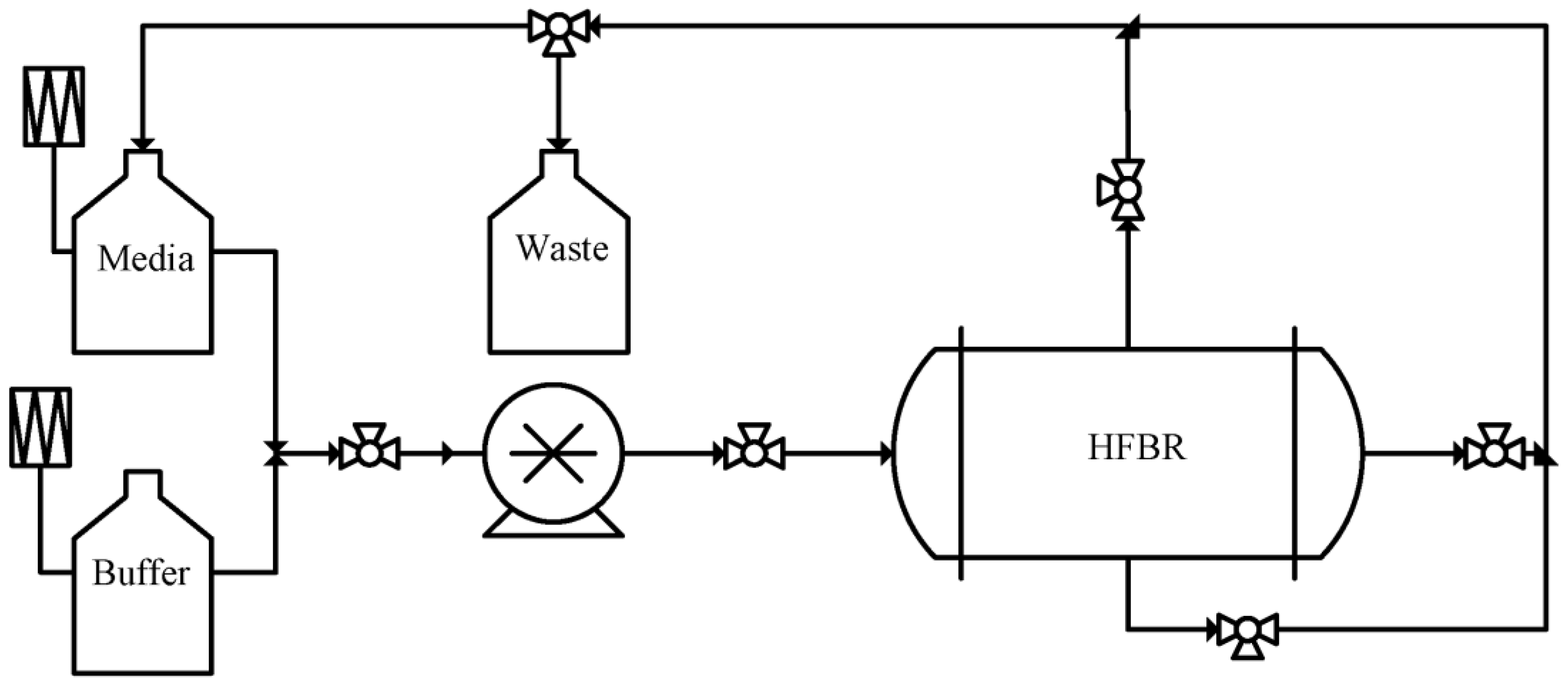
Hollow fibre membrane bioreactors (HFBRs), with continuous perfusion, can precisely control the extracellular environment and control the cell culture process. Since their first development in 1972, HFBRs have been touted as a means to overcome many of the issues presented by vessel and wave bag based platforms, by offering a closed system with automated temperature, gas concentration, and inlet media flow rate control [39]. Typically consisting of a controller, positive displacement pump, HFBR cartridge, waste, media, and buffer reservoirs, connected in a continuous loop [40], as illustrated by Figure 4, HFBRs effectively facilitate the removal of waste products while simultaneously providing fresh nutrients directly to the cells, which ultimately allows for the support a much higher cell densities [41,42,43]. Membranes used in HFBR cartridges are typically produced through immersion precipitation phase inversion in which a polymer solution is extruded through a spinneret into a precipitation bath, in which asymmetric hollow fibre are formed with a pore density gradient in the radial direction, before being rinsed and stored [44]. Polymers commonly used for cartridge production include, Polyvinylidene fluoride, Polysulfone, and cellulose [45].
Depending on the cell type and the intended product, HFBRs may be configured such that cells are grown in the lumen of the membranes or in the extra capillary space (ECS) in between the hollow fibres (HFs). Suspended cells, such as CAR-T cells, are ideally grown in the ECS, in which a near fluid shear stress-free environment is maintained. In this configuration, high cell numbers may be maintained as membrane bound advective mass transfer facilitates the maintenance of high transmembrane concentration gradients for efficient nutrient transfer. Cells may also be grown in the HF lumen, while being exposed to fluid shear forces to induce mechanical stimuli. The use of membrane bioreactors for cell culture and bioprocessing is not new, and the advantages in retaining cells while removing secreted products and metabolic wastes have been explored for a wide range of bioprocessing applications in both research and commercial settings.
HFBRs can be made into an automatic system with single use functionally closed cell culture consumable set, such as the Quantum Cell Expansion System. Studies have shown that the Quantum Cell Expansion System can produce large quantities of functional T-cells at clinical dosage levels [46] with increased potency [47].
Chang et al. developed a single use modular hollow fibre membrane bioreactor system, specifically for CAR-T cell expansion. The system is fully closed, fully automated, and integrated with all necessary steps involved including T cells transduction and activation, expansion, product formulation, volume reduction and final harvest. It might be possible that such a system can be placed at the hospital bedside without GMP bioprocessing facilities and cell transportation, reducing the cost of CAR-T cell processing significantly.
The membrane provides a physical barrier to enable a fully closed system. This minimises risk of contamination. As terminal sterilization techniques such as gamma radiation ethylene oxide or UV sterilization cannot be used on living cells, strict aseptic processing protocols must be followed to prevent contamination. When using traditional flask or bag-based cell manufacturing processes, due to the prevalence of open processes involved there is a higher microorganism exposure risk [48,49]. This is because, when using static flask-based cell cultures or rocking motion-based bag cell cultures, these processes involve multiple open processes such as, adding media or cell harvesting [50,51].
Traditional culture platforms in contrast to HFBR systems are very manual labour intensive. This in turn increases the costs associated with the operation of such processes due to the reliance on a large highly skilled labour force. In addition to the costs this also increases the chances of human error and batch variability, a phenomena realised in the increased rate of failure rate of commercial CAR-T products compared to other pharmaceutical products [52].
An outstanding advantage of the HFBR system developed by Chang et al. is to integrate all processing steps into the fully closed and automated system, with T cells genetically modified so that they express the chimeric antigen receptor, activated to unlock their proliferation potential, expanded, concentrated, formulated before being infused back to the patient [52,53]. Without the HFBR system, these multiple step manipulations mandate the use of multiple pieces of equipment for each step and technician’s operations which is very labour intensive and introduces potential batch to batch variability and risk of contamination.

3. Membrane Bioreactor for Autologous Cell Expansion

Autologous cell therapies, either using primary cells or various stem cells, involve expansion of the target cells from biopsy to a large number of cells (millions to billions) of desired quality and functions. Initial protocols use manual operations following the procedures in the laboratory research. Bioreactors for cell expansion soon appeared as a key requirement in autologous cell therapy. While multiplate, spheroid, stirred tank and packed bed bioreactor systems go some way in achieving this goal, the dependency of these systems on labour intensive tissue culture practices and low cell yields inhibits their wide spread commercial success [54].
Since their first use in their expansion of human choriocarcinoma cells, HFBRs have been used to culture many adherent cell types, for research and clinical applications, including, but not exclusively, endothelial cells (ECs), satellite cells, leukaemia cells, stromal cells, human cord blood derived mononuclear cells, and erythroid cells.
ECs were first cultured in the lumen of HF membranes in 1995 [55]. It was noted that when exposed to hydrodynamic shear stress that endothelial cells developed a phenotype more akin to that found in vivo and may even explain the heterogeneity of ECs throughout the body [56]. Through the compartmentalisation of cells grown in the lumen and ECS, HFBRs have been used to recapitulate more complex cellular models. By co-culturing of ECs with various other cell types, models have been developed for the blood brain barrier [57], and leukemic lymphocytic metastasis.
HFBRs have shown great promise for stem cell expansion. Autologous stem cells, including mesenchymal (MSCs) or hematopoietic stem cells (HSCs), as well as induced pluripotent stem cells (iPSCs), have enormous medical potential owing to their potential to differentiate into specific cell linages [56]. HFBRs have been demonstrated as a means by which iPSCs may be expanded, similar to embryonic stem cells [57,58,59], although it was noted that further process refinement was still required [60]. HFBRs have also shown promise [60,61] in culturing MSC, the stem cells more readily for clinical applications. While shear stress may be used to induce differentiation of MSCs, when expanding MSCs the minimisation of shear stress is desirable, and to this end HFBRs excel [58,62]. Consequently, HFBRs have been used for MSC expansion in large scale automated systems for a wide range of derived tissues including neural [63,64], skeletal [65], and for immunomodulatory effect [66].
HFBRs proved effective in producing red blood cells. It is known that the hematopoietic cellular niche plays an integral role in the regulation of haematopoiesis. Owing to their rigid mechanical properties and the relatively hydrodynamic shear stress free environment in the ECS, HFBRs have gained attention as a platform which may support ex-vivo haematopoiesis. Through the co-culture of human stromal cell line (HS-5) and human cord blood derived mononuclear cells a population of CD 34+ progenitor cells were expanded in serum free conditions. Further examination revealed the deposition of ECM proteins between the membranes, thus demonstrating the three-dimensional (3D) nature of HFBR cultures [67]. This promising result garnered much attention, alluding to the potential of ex-vivo red cell production. Subsequently a tertiary polyurethane scaffold was employed in the ECS to mimic the structure of trabecular bone. Following the seeding of umbilical cord blood mononuclear cells, hematopoietic populations were supported for 28 days allowing for the continuous harvest of enucleated red cells in serum free conditions [68,69]. Consequently, HFBRs offer a means by which red blood cells may be produced ex-vivo at greatly reduced cost and potentially alleviate the dependency of donations [70].
The clear need for HFBRs for regenerative medicine has naturally drawn great attention from industry and various commercial systems have been developed. For example, TERUMO, fibreCellSystems, The CellCultureCompany, and Oxford Mestar now offer a range HFBR solutions for bioprocessing applications [45,71,72,73,74]. Each of these companies has targeted slightly different markets with; TERUMO providing a wholistic end to end cell culture solution for the expansion of cells in the form of the Quantum platform. With full control of media flow rate, temperature and gas compositions afforded by the automatically fed cabinet, the Quantum system facilitates reduced manual operations thereby reducing the chance of contamination while improving reproducibility [73]. To date the platform has been used for, the expansion of CD3+ T-cell [75,76], MSCs [46,77], adipose stromal cells [78], neural stem cells [54], and iPSCs [79]. The DUET pump platform developed by FibreCellSystems is intended for laboratory scale HFBR systems. Depending on the intended application a range of different membrane modules may be used, each produced with a range of membrane materials and pore sizes. It is the relatively low cost and flexibility, compared to complete cell expansion systems, associated with the FibreCellSystems platform that make it so attractive to researchers. The DUET platform has consequently be used in the investigation of immune regulatory effect of MSCs [66], for the production of EVs at a GMP standard [80], the interaction of ECs and myeloid leukaemia [81], and even in the investigation of stem cell health in microgravity conditions onboard the international space station [82]. The CellCultureCompany has produced several platforms suitable for a wide range of applications and scales of operation. At the smallest scale, intended for use in a standard tissue culture incubator, the HF Primer system is most suitable for research applications. Increasing in size the AutovaxID is a standalone system intended as a pilot scale platform to bridge the gap between R&D and production scale. Finally, the AcuSyst-Maximizer and AcuSyst-Xcellerator provide a scale-up pathway intended for production scale applications of between 500–2000 L fed-bed equivalent in size, while fitting in a cabinet the size of a refrigerator. The RegenMed Solution system developed by Oxford Mestar is a modular system integrating cell seeding, expansion, harvest, and final formulation together, and has proved successful in CAR-T culture and human MSC expansion.

4. Membrane applications in EV Production

Membrane technology can play an important role in extracellular vesicle (EV) production. EVs are a type of lipid bilayer membrane-bound nanovesicles secreted by cells. EVs produced by stem cells [83,84,85,86,87] are an important secretory product that might be responsible for various therapeutic effects of stem cell therapy [88,89,90], and hence can be an important therapeutic product for anti-aging as an example. However, research and application of this potential cellular secretome product is largely hindered by its low productivity and the challenges of isolation and purification.
As EVs are secreted products of cells, the yield of EV products is partially dependent on the ability of cell expansion [91]. Current production of EVs is mainly based on traditional flask-based cell culture processes, which is largely limited by scale, highly manual labour-intensive and time consuming, while also being associated with high chances of human error and batch variability. As previously discussed HFBRs may yield large numbers of adherent stem cells in a small 3D space, and consequently, may be used to increase the yield of EVs. A key practical advantage that HFBRs systems offer is that they may also function as a part of the downstream process. Owing to the semi-permeable nature of the membranes in an HFBR, membranes with suitable pore size allow the mass transfer of nutrients and waste through continuous perfusion while retaining EVs that are produced. Through this means, EV products can accumulate and be preliminarily concentrated within the cell culture compartment of an HFBR. This in turn is beneficial for downstream purification processes as the smaller start volume makes subsequent liquid handling easier [92]. Recently, Mendt et al. demonstrated the Terumo Quantum platform may be used as an effective platform for the consistent expansion of exosomes, a type of EVs. The exosomes produced in turn were shown to increase the rate of survival from pancreatic cancer [93]. Furthermore, Williams et al. and Potter et al. demonstrated that MSC-derived exosomes could be successfully obtained from Quantum cell expansion system, while preserving their therapeutic effects [94,95]. Similarly, Cobin et al. demonstrated that EVs could be produced efficiently and reproducibly with MSCs derived from multiple human donors with a HFBR platform from FibreCellSystems, while preserving the phenotype and functionality of the cells [80]. It should be noted that EVs reflect the state of the cells from which they originate as their content can be considered as a fingerprint of the type and status of their parent cells. Thus, in order to ensure that the EVs harvested from ana HFBR are of desired type, quality, and function, it is vital to continuously monitor the status of the cells in the bioreactor as previously mentioned. Currently, sterile sampling of metabolic products and macromolecular biomarkers is readily achievable to monitor the cell condition through the use of membrane probes. However, it is very challenging to achieve direct online monitoring of the status and functions of the parent cells, and there is currently no existing method that would allow direct characterization of EVs within the bioreactors during the cell culture, due to the limitation of current characterization method. Therefore, it is also essential to characterize both the phenotype of their parent cells after the culture in the bioreactor and the isolated EVs sample itself in terms of size, morphology, expression of EV markers and functional potency, prior to the next stage research and application.
EVs are nanoparticles with a size range mainly from 30–1000 nm. They are usually spherical, carrying cargoes including proteins, RNA species, DNAs, and lipids. Several common methods are available for EV isolation and purification, the pros and cons of which may be seen in Table 2. Among these EV isolation methods, ultracentrifugation, immunoaffinity capture, precipitation, and size exclusion chromatography have limitations of poor scalability, high cost, introduce unwanted agents and requiring extra isolation steps, respectively [90]. In contrast, membrane filtration is capable for high throughput, is more readily scalable, requires less capital investment for equipment, is faster, and less labour intensive. Consequently, ultrafiltration (UF) is a very promising EV separation method for large-scale manufacture of EVs intended for clinical applications.
Microfiltration (MF) and UF can readily be used for EV isolation and purification. MF using membranes with pore sizes ranging between 0.1–0.8 µm is normally used to remove larger particles from the target EV fraction [96]. Track-etched polycarbonate membranes with pore sizes from 30–600 nm have been utilized for fast EV isolation or detection in specialized filtration devices, e.g., a cyclic tangential flow filtration (TFF) system [97], a TFF based microfluidic chip [98], an integrated double-filtration microfluidic device [99], an exosome total isolation chip [100], a lab-on-a-disc integrated with two nanofilters [101]. Commercial centrifugal ultrafiltration filters with multiple membrane materials and molecular weight cut-off (MWCO) ranging from 10–300 kDa are used for volume reduction of a large amount of biofluids and conditioned medium from cell culture before EV isolation [102], or as an additional concentration step for the relatively dilute EVs after other isolation steps, for example, size exclusion chromatography (SEC) [103]. It was reported by Vergauwen et al. that the most popular membrane type for EV concentration is regenerated cellulose with MWCO of 100 kDa, based on the records in the EV-TRACK knowledgebase [104].
UF-TFF can also be applied to EV isolation and purification. In TFF with a suitable choice of membrane MWCO, EVs larger than the pore sizes can be retained, concentrated and recirculated in the capillary space where biofluid or conditioned cell culture media is continuously pumped in, while smaller molecules, such as some small proteins, salts, and solvents can travel through the pores on the membrane and be removed in the permeate [105,106]. In addition, to increase the purity of EV products, diafiltration can be operated in TFF systems. Small molecules are further removed, buffer exchanged, or lowered by adding fresh exchange buffer at the same rate as the permeate flow rate. Compared to conventional batch-wise dead-end filtration, TFF is superior as it reduces the formation of filter cake and can operate large-scale EV isolation and purification in a continuous process. Choi et al. successfully isolated EVs from human adipose-derived stem cells by TFF with a 500 kDa MWCO membrane filter capsule, confirming the functional recovery in photo-damaged human dermal fibroblasts after EV treatments [107]. Higher purity might be obtained by combining different techniques with UF, although UF alone is applicable for EV isolation and purification [108]. EVs were reported to have been concentrated 50 fold through TFF with a 100 kDa MWCO membrane, followed by purification using chromatography column and further concentrated by 100 kDa MWCO centrifugal filter [109]. Finally, Hydrostatic filtration dialysis, a filtration technique which consists of filtration, concentration and dialysis using hydrostatic pressure of the fluid, has also been employed to isolate EVs from urine using dialysis membranes with 1000 kDa MWCO [110].

5. Needs for Research

Membrane bioreactors have an important role to play. When developing a platform for the production of cells or cellular products, such as with EVs, there are crucial considerations, such as, process conditions, regulatory approval, cost of device, quality control, membrane area, and product recovery. First, it is vital to establish uniform bioreactor conditions. This is necessary to ensure a homogeneous and reproducible product as cells are sensitive to the local environment. This is particularly important to the culture of stem cells as they are more susceptible to environmental perturbations.
There is always a need for new membranes that are biocompatible for single use medical devices. Unlike other industrial applications of membranes, the cellular product will be implanted or injected directly into the human body. Consequently, any material that is in contact with the product stream such as the membrane, housing case, potting material or glue must be approved for use in medical devices and produced in from pyrogen-free materials [111]. Regulatory issues should not be overlooked during the research and development of new membranes and modules which provide inevitable hurdles in the path to commercialisation. To prevent complications associated with cleaning, systems are commonly developed to be single use [112]. Consequently, the cost of such membrane device must be low, e.g., at 10 USD per square meter of membrane area or lower. Finally, the device will have to be pre-sterilised, usually with gamma radiation. This adds an additional selection criteria that needs to be considered when selecting membrane and housing materials [113].
Membrane biosensors may find a wider use in cell therapy. To meet regulatory requirements, it is necessary to have robust methods to monitor the process for quality control purposes. Currently, the sterile sampling of metabolic products and macromolecular biomarkers is readily achievable through the use of membrane probes and various sampling techniques like microdialysis [114]. In contrast, cell function can only be assessed with biomarkers and complicated assays. It is therefore challenging to achieve the online monitoring of cell function. However, as human cells do not grow quickly, offline assays offer a practical means of measurement although their use increases the risk of contamination. In addition, the direct characterization and quality control method for the production of cellular products such as EVs is still at its infancy. Specialized biosensors that can be integrated to membrane bioreactors are therefore required.
Novel membrane modules with changing and adjustable membrane areas are needed. Throughout the culture period of adhering cells if the area is insufficient, cell growth will be inhibited by contact inhibition. However, if the area is too large initially, the cell density on the surface will be low and the cells will not proliferate. This is also true for culturing suspended cells with an optimal cell density in the membrane reactor. The development of a membrane bioreactor which may increase the area of membrane exposed throughout culture, to maintain exponential cell growth, is therefore required.
Upon culture completion, the cellular product must be harvested, concentrated, resuspended, and formulated for cryopreservation and clinical application. Usually, the cell suspension is reduced to a volume that fits into a syringe for administration. Current protocols for harvest and formulation rely on labour- intensive multi-step processes. As with the culture process, through the use of an HFBR platform, it is possible to replace these manual steps with a membrane device capable of buffer exchange via dialysis, concentration, final formulation, and volume reduction. Such a device would be a closed system and may be fully automized, thereby greatly reducing the risk of contamination.
Single use and low cost membrane oxygenator for gas–liquid oxygen exchange is welcomed. Further to the use of membranes in bioreactors, membrane technologies may be used to optimize bioprocessing more generally. Membrane oxygenators have been widely used in the culture of shear sensitive cells and microbes, which was stemmed out from Extracorporeal Membrane Oxygenation (ECMO) [115]. The challenges with membrane oxygenators are primarily the cost of the device and suitability as a medical device. In addition, the use of an oxygenator increases the liquid holdup and should therefore be minimized as the culture media are expensive.
Cell separation using membranes to replace flow cytometry would be a big step forward. Currently, upon culture completion cell sorting and purification for stem and T-cell production is achieved through flow cytometry. The development of a flow through device for pre-treatment or cell sorting would represent a huge increase in efficiency. Such a device would likely be based on the difference in the surface properties of cells, and therefore operate upon affinity instead of size difference. Another application for affinity or ion exchange membranes would be the removal of macromolecules secreted by the cells, or from lysed cells, which otherwise negatively affect cell growth. The designing of such a membrane will need detailed understanding on the specific proteins produced by the particular cell type.
The separation and purification of EVs using membranes have great potential. Although membrane-based techniques are widely used for EV isolation, the interaction between EVs and membranes with different parameters, such as MWCO, materials, charged or not, and any kind of modifications, are rarely investigated to increase the selectivity and reduce the losses from EV binding to membranes. The process of filtration should also be studied and optimized to increase the efficiency and yield of EV isolation. Deformation, disruption, or loss of properties and function of EVs caused during membrane separation requires further investigation [96]. Additionally, while there are several commercial membrane-based separation products available, these have been originally developed for different processes and subsequently adapted to EV separations. Specialized membranes for EV isolation and purification based on the properties of EVs and EV-membrane interaction research are therefore required. Integration of stem cell expansion and EV production would increase the EV productivity enormously as the cells can be retained and cultured for a longer period of time while EVs are still produced.

6. Conclusions

As the age of personalized medicine dawns, many engineering challenges come with the realization of new technologies. For these technologies to reach full fruition, these challenges must be overcome. No technology better exemplifies this than CAR-T therapies. While, broadly considered to be ground-breaking, CAR-T therapies have failed to reach widespread clinical application, predominantly due to their prohibitively expensive cost per treatment. To date, the cost of pharmaceuticals has been minimized through the economy of scales. However, the bespoke nature of personalized medicine prevents this approach. Instead, cost effective scale-out, rather than scale-up, methods are imperative for success, as is an alternative business model.
Autologous cell therapies mandate the growth if a patient own cells for re-implantation back to the patient. Traditional flask and wave bag culture techniques adopted in scale-out processes are costly, labour intensive, and prone to contamination. HFBRs, developed for the culture of various cell types in both research and commercial settings, may offer an automated, cost-effective, closed system alternative. However, to implement HFBRs into a clinical setting, rigorous regulatory approval of commercial devices, quality control, and validation methods are necessary to ensure success, as there normally is not a second chance.
Membrane technologies represent a relatively untapped market in bioprocessing of autologous cells and cellular products. There are some interesting and challenging problems such as cell sorting and EV purification that provides an exciting research opportunity through the development of specialized affinity membranes. New membrane modules, e.g., those with adjustable membrane areas, would be interesting to develop too.

Author Contributions

Conceptualization, H.Y. and Z.C.; writing—original draft preparation, R.M., R.L., Y.Z., J.Z., and H.C.; writing—review and editing, R.M., H.Y., and Z.C.; supervision, H.Y. and Z.C.; funding acquisition, H.Y. and Z.C. All authors have read and agreed to the published version of the manuscript.

Funding

This research was funded by the Engineering and Physical Sciences Research Council (EPSRC) grant number EP/R513295/1, EPSRC DTP grant, Jiangsu Industrial Technology Research Institute, and Innovate UK.

Institutional Review Board Statement

Not applicable.

Acknowledgments

This article is dedicated to A. G. Fane. In the last 25 years, Tony has been a great mentor, collaborator, and supporter to Cui, and above all, has been a true friend. He has been a great collaborator and friend to the Oxford Membrane Research Group. The authors are grateful for research support to the UK Engineering and Physical Sciences Research Council (EPSRC) University of Oxford Engineering Science DPT grant, Jiangsu Industrial Technology Research Institute, and Innovate UK.

Conflicts of Interest

The authors declare no conflict of interest.

Abbreviations

3DThree-dimensional
ACIAutologous Chondrocyte Implantation
CARChimeric Antigen Receptors
CAR-TChimeric Antigen Receptor T Cell
ECMOExtracorporeal Membrane Oxygenation
ECSExtra Capillary Space
ECsEndothelial Cells
EVsExtracellular Vesicles
GMPGood Manufacture Practice
HFMBsHollow Fibre Membrane Bioreactors
HFsHollow Fibres
HSCsHematopoietic Stem Cells
IPSCsInduced Pluripotent Stem Cells
mAbMonoclonal Antibody
MFMicrofiltration
MSCsMesenchymal Stem Cells
MWCOMolecular Weight Cut-Off
SECSize Exclusion Chromatography
TCRsT Cell Receptors
TFFTangential Flow Filtration
TILsTumour-Infiltrating Lymphocytes
UFUltrafiltration

References

  1. Brittberg, M.; Lindahl, A.; Nilsson, A.; Ohlsson, C.; Isaksson, O.; Peterson, L. Treatment of Deep Cartilage Defects in the Knee with Autologous Chondrocyte Transplantation. N. Engl. J. Med. 1994, 331, 889–895. [Google Scholar] [CrossRef]
  2. Wood, F.; Martin, L.; Lewis, D.; Rawlins, J.; McWilliams, T.; Burrows, S.; Rea, S. A Prospective Randomised Clinical Pilot Study to Compare the Effectiveness of Biobrane® Synthetic Wound Dressing, with or without Autologous Cell Suspension, to the Local Standard Treatment Regimen in Paediatric Scald Injuries. Burns 2012, 38, 830–839. [Google Scholar] [CrossRef] [PubMed]
  3. Mancardi, G.; Saccardi, R. Autologous Haematopoietic Stem-Cell Transplantation in Multiple Sclerosis. Lancet. Neurol. 2008, 7, 626–636. [Google Scholar] [CrossRef] [PubMed]
  4. Braendstrup, P.; Levine, B.L.; Ruella, M. The Long Road to the First FDA-Approved Gene Therapy: Chimeric Antigen Receptor T Cells Targeting CD19. Cytotherapy 2020, 22, 57–69. [Google Scholar] [CrossRef]
  5. Carticel. Available online: https://www.maci.com/patients/?carticel (accessed on 4 April 2022).
  6. Cost Effectiveness and Budget Impact of Tisagenlecleucel. PharmacoEconomics Outcomes News 2018, 817, 12. [CrossRef]
  7. Moutsatsou, P.; Ochs, J.; Schmitt, R.H.; Hewitt, C.J.; Hanga, M.P. Automation in Cell and Gene Therapy Manufacturing: From Past to Future. Biotechnol. Lett. 2019, 41, 1245–1253. [Google Scholar] [CrossRef] [Green Version]
  8. Lam, C.; Meinert, E.; Yang, A.; Cui, Z. Comparison between Centralized and Decentralized Supply Chains of Autologous Chimeric Antigen Receptor T-Cell Therapies: A UK Case Study Based on Discrete Event Simulation. Cytotherapy 2021, 23, 433–451. [Google Scholar] [CrossRef]
  9. Decker, W.K.; da Silva, R.F.; Sanabria, M.H.; Angelo, L.S.; Guimarães, F.; Burt, B.M.; Kheradmand, F.; Paust, S. Cancer Immunotherapy: Historical Perspective of a Clinical Revolution and Emerging Preclinical Animal Models. Front. Immunol. 2017, 8. [Google Scholar] [CrossRef] [Green Version]
  10. Dunn, G.P.; Old, L.J.; Schreiber, R.D. The Three Es of Cancer Immunoediting. Annu. Rev. Immunol. 2004, 22, 329–360. [Google Scholar] [CrossRef]
  11. Clemente, C.G.; Mihm, M.C.; Bufalino, R.; Zurrida, S.; Collini, P.; Cascinelli, N. Prognostic Value of Tumor Infiltrating Lymphocytes in the Vertical Growth Phase of Primary Cutaneous Melanoma. Cancer 1996, 77, 1303–1310. [Google Scholar] [CrossRef]
  12. Borst, J.; Ahrends, T.; Bąbała, N.; Melief, C.J.M.; Kastenmüller, W. CD4+ T Cell Help in Cancer Immunology and Immunotherapy. Nat. Rev. Immunol. 2018, 18, 635–647. [Google Scholar] [CrossRef]
  13. Neefjes, J.; Jongsma, M.L.M.; Paul, P.; Bakke, O. Towards a Systems Understanding of MHC Class I and MHC Class II Antigen Presentation. Nat. Rev. Immunol. 2011, 11, 823–836. [Google Scholar] [CrossRef]
  14. Kalergis, A.M.; Ono, T.; Wang, F.; DiLorenzo, T.P.; Honda, S.; Nathenson, S.G. Single Amino Acid Replacements in an Antigenic Peptide Are Sufficient to Alter the TCR V Beta Repertoire of the Responding CD8+ Cytotoxic Lymphocyte Population. J. Immunol. 1999, 162, 7263–7270. [Google Scholar]
  15. Egerton, M.; Scollay, R.; Shortman, K. Kinetics of Mature T-Cell Development in the Thymus. Proc. Natl. Acad. Sci. USA 1990, 87, 2579–2582. [Google Scholar] [CrossRef] [Green Version]
  16. Rosenberg, S.A.; Spiess, P.; Lafreniere, R. A New Approach to the Adoptive Immunotherapy of Cancer with Tumor-Infiltrating Lymphocytes. Science 1986, 233, 1318–1321. [Google Scholar] [CrossRef]
  17. Wang, S.; Sun, J.; Chen, K.; Ma, P.; Lei, Q.; Xing, S.; Cao, Z.; Sun, S.; Yu, Z.; Liu, Y.; et al. Perspectives of Tumor-Infiltrating Lymphocyte Treatment in Solid Tumors. BMC Med. 2021, 19, 140. [Google Scholar] [CrossRef]
  18. Eshhar, Z.; Waks, T.; Gross, G.; Schindler, D.G. Specific Activation and Targeting of Cytotoxic Lymphocytes through Chimeric Single Chains Consisting of Antibody-Binding Domains and the Gamma or Zeta Subunits of the Immunoglobulin and T-Cell Receptors. Proc. Natl. Acad. Sci. USA 1993, 90, 720–724. [Google Scholar] [CrossRef] [Green Version]
  19. Kalos, M.; Levine, B.L.; Porter, D.L.; Katz, S.; Grupp, S.A.; Bagg, A.; June, C.H. T Cells with Chimeric Antigen Receptors Have Potent Antitumor Effects and Can Establish Memory in Patients with Advanced Leukemia. Sci. Transl. Med. 2011, 3, 95ra73. [Google Scholar] [CrossRef] [Green Version]
  20. Hollyman, D.; Stefanski, J.; Przybylowski, M.; Bartido, S.; Borquez-Ojeda, O.; Taylor, C.; Yeh, R.; Capacio, V.; Olszewska, M.; Hosey, J.; et al. Manufacturing Validation of Biologically Functional T Cells Targeted to CD19 Antigen for Autologous Adoptive Cell Therapy. J. Immunother. 2009, 32, 169–180. [Google Scholar] [CrossRef] [Green Version]
  21. FDA Okays Second CAR-T for Kite. Nat. Biotechnol. 2020, 38, 1012. [CrossRef]
  22. Mullard, A. FDA Approves First CAR T Therapy. Nat. Rev. Drug Discov. 2017, 16, 669. [Google Scholar] [CrossRef] [PubMed]
  23. Mullard, A. Second Anticancer CAR T Therapy Receives FDA Approval. Nat. Rev. Drug Discov. 2017, 16, 818. [Google Scholar] [CrossRef]
  24. Mullard, A. FDA Approves Fourth CAR-T Cell Therapy. Nat. Rev. Drug Discov. 2021, 20, 166. [Google Scholar] [CrossRef] [PubMed]
  25. Mullard, A. FDA Approves First BCMA-Targeted CAR-T Cell Therapy. Nat. Rev. Drug Discov. 2021, 20, 332. [Google Scholar] [CrossRef] [PubMed]
  26. Mullard, A. FDA Approves Second BCMA-Targeted CAR-T Cell Therapy. Nat. Rev. Drug Discov. 2022, 21, 249. [Google Scholar] [CrossRef]
  27. Rafiq, S.; Hackett, C.S.; Brentjens, R.J. Engineering Strategies to Overcome the Current Roadblocks in CAR T Cell Therapy. Nat. Rev. Clin. Oncol. 2020, 17, 147–167. [Google Scholar] [CrossRef]
  28. Lyman, G.H.; Nguyen, A.; Snyder, S.; Gitlin, M.; Chung, K.C. Economic Evaluation of Chimeric Antigen Receptor T-Cell Therapy by Site of Care Among Patients With Relapsed or Refractory Large B-Cell Lymphoma. JAMA Netw. Open 2020, 3, e202072. [Google Scholar] [CrossRef]
  29. Karschnia, P.; Rejeski, K.; Winkelmann, M.; Schöberl, F.; Bücklein, V.L.; Blumenberg, V.; Schmidt, C.; Blobner, J.; von Bergwelt-Baildon, M.; Tonn, J.-C.; et al. Toxicities and Response Rates of Secondary CNS Lymphoma After Adoptive Immunotherapy With CD19-Directed Chimeric Antigen Receptor T Cells. Neurology 2022, 98, 884–889. [Google Scholar] [CrossRef]
  30. Mahal, H.; Branton, H.; Farid, S.S. End-to-end Continuous Bioprocessing: Impact on Facility Design, Cost of Goods, and Cost of Development for Monoclonal Antibodies. Biotechnol. Bioeng. 2021, 118, 3468–3485. [Google Scholar] [CrossRef]
  31. Jiang, J.; Ahuja, S. Addressing Patient to Patient Variability for Autologous CAR T Therapies. J. Pharm. Sci. 2021, 110, 1871–1876. [Google Scholar] [CrossRef]
  32. Castella, M.; Caballero-Baños, M.; Ortiz-Maldonado, V.; González-Navarro, E.A.; Suñé, G.; Antoñana-Vidósola, A.; Boronat, A.; Marzal, B.; Millán, L.; Martín-Antonio, B.; et al. Point-Of-Care CAR T-Cell Production (ARI-0001) Using a Closed Semi-Automatic Bioreactor: Experience From an Academic Phase I Clinical Trial. Front. Immunol. 2020, 11. [Google Scholar] [CrossRef]
  33. Fernández, L.; Fernández, A.; Mirones, I.; Escudero, A.; Cardoso, L.; Vela, M.; Lanzarot, D.; de Paz, R.; Leivas, A.; Gallardo, M.; et al. GMP-Compliant Manufacturing of NKG2D CAR Memory T Cells Using CliniMACS Prodigy. Front. Immunol. 2019, 10. [Google Scholar] [CrossRef] [Green Version]
  34. Jackson, Z.; Roe, A.; Sharma, A.A.; Lopes, F.B.T.P.; Talla, A.; Kleinsorge-Block, S.; Zamborsky, K.; Schiavone, J.; Manjappa, S.; Schauner, R.; et al. Automated Manufacture of Autologous CD19 CAR-T Cells for Treatment of Non-Hodgkin Lymphoma. Front. Immunol. 2020, 11. [Google Scholar] [CrossRef]
  35. Smith, T.A. CAR-T Cell Expansion in a Xuri Cell Expansion System W25; Humana: New York, NY, USA, 2020; pp. 151–163. ISBN 978-1-0716-0145-7. [Google Scholar]
  36. Cytiva Xuri Cell Expansion System. Available online: https://www.cytivalifesciences.com/en/us/shop/cell-therapy/systems/xuri-cell-expansion-system-w25-p-06192 (accessed on 24 October 2022).
  37. Biotec, M. CliniMACS Prodigy. Available online: https://www.miltenyibiotec.com/GB-en/products/clinimacs-prodigy.html#200-075-301 (accessed on 24 October 2022).
  38. Lonza The Cocoon. Available online: https://demo-pharma.lonza.com/technologies-products/cocoon-platform (accessed on 24 October 2022).
  39. Knazek, R.A.; Gullino, P.M.; Kohler, P.O.; Dedrick, R.L. Cell Culture on Artificial Capillaries: An Approach to Tissue Growth in Vitro. Science 1972, 178, 65–67. [Google Scholar] [CrossRef]
  40. Sohaib, Q.; Kalakech, C.; Charmette, C.; Cartier, J.; Lesage, G.; Mericq, J.-P. Hollow-Fiber Membrane Contactor for Biogas Recovery from Real Anaerobic Membrane Bioreactor Permeate. Membranes 2022, 12, 112. [Google Scholar] [CrossRef]
  41. Kleinstreuer, C.; Agarwal, S.S. Analysis and Simulation of Hollow-Fiber Bioreactor Dynamics. Biotechnol. Bioeng. 1986, 28, 1233–1240. [Google Scholar] [CrossRef]
  42. Tharakan, J.P.; Chau, P.C. A Radial Flow Hollow Fiber Bioreactor for the Large-Scale Culture of Mammalian Cells. Biotechnol. Bioeng. 1986, 28, 329–342. [Google Scholar] [CrossRef]
  43. Storm, M.P.; Sorrell, I.; Shipley, R.; Regan, S.; Luetchford, K.A.; Sathish, J.; Webb, S.; Ellis, M.J. Hollow Fiber Bioreactors for In Vivo-like Mammalian Tissue Culture. J. Vis. Exp. 2016, e53431. [Google Scholar] [CrossRef] [Green Version]
  44. Ladewig, B.; Al-Shaeli, M.N.Z. Fundamentals of Membrane Bioreactors. In Springer Transactions in Civil and Environmental Engineering; Springer: Singapore, 2017; ISBN 978-981-10-2013-1. [Google Scholar]
  45. FiberCellSystems. Available online: https://www.fibercellsystems.com/products/cartridges/ (accessed on 25 October 2022).
  46. Nold, P.; Brendel, C.; Neubauer, A.; Bein, G.; Hackstein, H. Good Manufacturing Practice-Compliant Animal-Free Expansion of Human Bone Marrow Derived Mesenchymal Stroma Cells in a Closed Hollow-Fiber-Based Bioreactor. Biochem. Biophys. Res. Commun. 2013, 430, 325–330. [Google Scholar] [CrossRef]
  47. Yoo, S.M.; Lau, V.W.C.; Aarts, C.; Bojovic, B.; Steinberg, G.; Hammill, J.A.; Dvorkin-Gheva, A.; Ghosh, R.; Bramson, J.L. Manufacturing T Cells in Hollow Fiber Membrane Bioreactors Changes Their Programming and Enhances Their Potency. Oncoimmunology 2021, 10. [Google Scholar] [CrossRef]
  48. Wahlig, S.; Peh, G.S.L.; Adnan, K.; Ang, H.-P.; Lwin, C.N.; Morales-Wong, F.; Ong, H.S.; Lovatt, M.; Mehta, J.S. Optimisation of Storage and Transportation Conditions of Cultured Corneal Endothelial Cells for Cell Replacement Therapy. Sci. Rep. 2020, 10, 1681. [Google Scholar] [CrossRef] [PubMed]
  49. Massie, I.; Selden, C.; Hodgson, H.; Fuller, B. Storage Temperatures for Cold-Chain Delivery in Cell Therapy: A Study of Alginate-Encapsulated Liver Cell Spheroids Stored at −80 °C or −170 °C for up to 1 Year. Tissue Eng. Part C. Methods 2013, 19, 189–195. [Google Scholar] [CrossRef] [PubMed] [Green Version]
  50. Namdaroğlu, S.; Tekgündüz, E.; Bozdağ, S.C.; Durgun, G.; Sarıca, A.; Demiriz, I.Ş.; Koçubaba, Ş.; İskender, G.; Kayıkçı, Ö.; Altuntaş, F. Microbial Contamination of Hematopoietic Progenitor Cell Products. Transfus. Apher. Sci. 2013, 48, 403–406. [Google Scholar] [CrossRef] [PubMed]
  51. Kozlowska-Skrzypczak, M.; Bembnista, E.; Kubiak, A.; Matuszak, P.; Schneider, A.; Komarnicki, M. Microbial Contamination of Peripheral Blood and Bone Marrow Hematopoietic Cell Products and Environmental Contamination in a Stem Cell Bank: A Single-Center Report. Transplant. Proc. 2014, 46, 2873–2876. [Google Scholar] [CrossRef] [PubMed]
  52. Bersenev, A. CAR-T Cell Manufacturing: Time to Put It in Gear. Transfusion 2017, 57, 1104–1106. [Google Scholar] [CrossRef] [PubMed] [Green Version]
  53. Wang, X.; Rivière, I. Clinical Manufacturing of CAR T Cells: Foundation of a Promising Therapy. Mol. Ther. Oncolytics 2016, 3, 16015. [Google Scholar] [CrossRef] [Green Version]
  54. Tirughana, R.; Metz, M.Z.; Li, Z.; Hall, C.; Hsu, D.; Beltzer, J.; Annala, A.J.; Oganesyan, D.; Gutova, M.; Aboody, K.S. GMP Production and Scale-Up of Adherent Neural Stem Cells with a Quantum Cell Expansion System. Mol. Ther. Methods Clin. Dev. 2018, 10, 48–56. [Google Scholar] [CrossRef] [Green Version]
  55. Ballermann, B.J.; Ott, M.J. Adhesion and Differentiation of Endothelial Cells by Exposure to Chronic Shear Stress: A Vascular Graft Model. Blood Purif. 1995, 13, 125–134. [Google Scholar] [CrossRef]
  56. Redmond, E.M.; Cahill, P.A.; Sitzmann, J. V Perfused Transcapillary Smooth Muscle and Endothelial Cell Co-Culture—A Novel in Vitro Model. In Vitro Cell. Dev. Biol. Anim. 1995, 31, 601–609. [Google Scholar] [CrossRef]
  57. Cucullo, L.; Hossain, M.; Tierney, W.; Janigro, D. A New Dynamic In Vitro Modular Capillaries-Venules Modular System: Cerebrovascular Physiology in a Box. BMC Neurosci. 2013, 14, 18. [Google Scholar] [CrossRef] [Green Version]
  58. Jossen, V.; Pörtner, R.; Kaiser, S.C.; Kraume, M.; Eibl, D.; Eibl, R. Mass Production of Mesenchymal Stem Cells—Impact of Bioreactor Design and Flow Conditions on Proliferation and Differentiation. In Cells and Biomaterials in Regenerative Medicine; InTech: London, UK, 2014. [Google Scholar]
  59. Knöspel, F.; Freyer, N.; Stecklum, M.; Gerlach, J.C.; Zeilinger, K. Periodic Harvesting of Embryonic Stem Cells from a Hollow-Fiber Membrane Based Four-Compartment Bioreactor. Biotechnol. Prog. 2016, 32, 141–151. [Google Scholar] [CrossRef]
  60. Matsushita, S.; Kajiwara, T.; Mizumoto, H. Expansion and Differentiation of Human IPS Cells in a Three-Dimensional Culture Using Hollow Fibers and Separation of the Specific Population by Magnetic-Activated Cell Sorting. J. Biosci. Bioeng. 2019, 128, 480–486. [Google Scholar] [CrossRef]
  61. Stachelscheid, H.; Wulf-Goldenberg, A.; Eckert, K.; Jensen, J.; Edsbagge, J.; Björquist, P.; Rivero, M.; Strehl, R.; Jozefczuk, J.; Prigione, A.; et al. Teratoma Formation of Human Embryonic Stem Cells in Three-Dimensional Perfusion Culture Bioreactors. J. Tissue Eng. Regen. Med. 2013, 7, 729–741. [Google Scholar] [CrossRef] [Green Version]
  62. Mizukami, A.; Pereira Chilima, T.D.; Orellana, M.D.; Neto, M.A.; Covas, D.T.; Farid, S.S.; Swiech, K. Technologies for Large-Scale Umbilical Cord-Derived MSC Expansion: Experimental Performance and Cost of Goods Analysis. Biochem. Eng. J. 2018, 135, 36–48. [Google Scholar] [CrossRef]
  63. Frank, N.D.; Jones, M.E.; Vang, B.; Coeshott, C. Evaluation of Reagents Used to Coat the Hollow-Fiber Bioreactor Membrane of the Quantum® Cell Expansion System for the Culture of Human Mesenchymal Stem Cells. Mater. Sci. Eng. C 2019, 96, 77–85. [Google Scholar] [CrossRef]
  64. Vymetalova, L.; Kucirkova, T.; Knopfova, L.; Pospisilova, V.; Kasko, T.; Lejdarova, H.; Makaturova, E.; Kuglik, P.; Oralova, V.; Matalova, E.; et al. Large-Scale Automated Hollow-Fiber Bioreactor Expansion of Umbilical Cord-Derived Human Mesenchymal Stromal Cells for Neurological Disorders. Neurochem. Res. 2020, 45, 204–214. [Google Scholar] [CrossRef]
  65. De Napoli, I.E.; Scaglione, S.; Giannoni, P.; Quarto, R.; Catapano, G. Mesenchymal Stem Cell Culture in Convection-Enhanced Hollow Fibre Membrane Bioreactors for Bone Tissue Engineering. J. Memb. Sci. 2011, 379, 341–352. [Google Scholar] [CrossRef]
  66. Allen, A.; Vaninov, N.; Li, M.; Nguyen, S.; Singh, M.; Igo, P.; Tilles, A.W.; O’Rourke, B.; Miller, B.L.K.; Parekkadan, B.; et al. Mesenchymal Stromal Cell Bioreactor for Ex Vivo Reprogramming of Human Immune Cells. Sci. Rep. 2020, 10, 10142. [Google Scholar] [CrossRef]
  67. Xue, C.; Kwek, K.Y.C.; Chan, J.K.Y.; Chen, Q.; Lim, M. The Hollow Fiber Bioreactor as a Stroma-Supported, Serum-Free Ex Vivo Expansion Platform for Human Umbilical Cord Blood Cells. Biotechnol. J. 2014, 9, 980–989. [Google Scholar] [CrossRef]
  68. Allenby, M.C.; Tahlawi, A.; Morais, J.C.F.; Li, K.; Panoskaltsis, N.; Mantalaris, A. Ceramic Hollow Fibre Constructs for Continuous Perfusion and Cell Harvest from 3D Hematopoietic Organoids. Stem Cells Int. 2018, 2018. [Google Scholar] [CrossRef] [Green Version]
  69. Allenby, M.C.; Panoskaltsis, N.; Tahlawi, A.; Dos Santos, S.B.; Mantalaris, A. Dynamic Human Erythropoiesis in a Three-Dimensional Perfusion Bone Marrow Biomimicry. Biomaterials 2019, 188, 24–37. [Google Scholar] [CrossRef] [PubMed]
  70. Misener, R.; Allenby, M.C.; Fuentes-Garí, M.; Gupta, K.; Wiggins, T.; Panoskaltsis, N.; Pistikopoulos, E.N.; Mantalaris, A. Stem Cell Biomanufacturing under Uncertainty: A Case Study in Optimizing Red Blood Cell Production. AIChE J. 2018, 64, 3011–3022. [Google Scholar] [CrossRef] [PubMed] [Green Version]
  71. The CellCultureCompany. Available online: https://cellculturecompany.com/ (accessed on 4 April 2022).
  72. Flocel. Available online: https://www.flocel.com/ (accessed on 10 April 2022).
  73. Terumo. Terumo Quantum. Available online: https://www.terumobct.com/quantum/ (accessed on 31 March 2022).
  74. Oxford Mestar Ltd. Bioreactor Systems by Oxford Mestar. Available online: http://www.oxford-mestar.com/ (accessed on 31 March 2022).
  75. Coeshott, C.; Vang, B.; Jones, M.; Nankervis, B. Large-Scale Expansion and Characterization of CD3+ T-Cells in the Quantum® Cell Expansion System. J. Transl. Med. 2019, 17, 258. [Google Scholar] [CrossRef] [PubMed]
  76. Jones, M.; Nankervis, B.; Roballo, K.S.; Pham, H.; Bushman, J.; Coeshott, C. A Comparison of Automated Perfusion- and Manual Diffusion-Based Human Regulatory T Cell Expansion and Functionality Using a Soluble Activator Complex. Cell Transplant. 2020, 29, 096368972092357. [Google Scholar] [CrossRef] [PubMed]
  77. Hanley, P.J.; Mei, Z.; Durett, A.G.; da Graca Cabreira-Harrison, M.; Klis, M.; Li, W.; Zhao, Y.; Yang, B.; Parsha, K.; Mir, O.; et al. Efficient Manufacturing of Therapeutic Mesenchymal Stromal Cells with the Use of the Quantum Cell Expansion System. Cytotherapy 2014, 16, 1048–1058. [Google Scholar] [CrossRef] [PubMed] [Green Version]
  78. Haack-Sørensen, M.; Follin, B.; Juhl, M.; Brorsen, S.K.; Søndergaard, R.H.; Kastrup, J.; Ekblond, A. Culture Expansion of Adipose Derived Stromal Cells. A Closed Automated Quantum Cell Expansion System Compared with Manual Flask-Based Culture. J. Transl. Med. 2016, 14, 319. [Google Scholar] [CrossRef] [Green Version]
  79. Paccola Mesquita, F.C.; Hochman-Mendez, C.; Morrissey, J.; Sampaio, L.C.; Taylor, D.A. Laminin as a Potent Substrate for Large-Scale Expansion of Human Induced Pluripotent Stem Cells in a Closed Cell Expansion System. Stem Cells Int. 2019, 2019, 9704945. [Google Scholar] [CrossRef] [Green Version]
  80. Gobin, J.; Muradia, G.; Mehic, J.; Westwood, C.; Couvrette, L.; Stalker, A.; Bigelow, S.; Luebbert, C.C.; Bissonnette, F.S.-D.; Johnston, M.J.W.; et al. Hollow-Fiber Bioreactor Production of Extracellular Vesicles from Human Bone Marrow Mesenchymal Stromal Cells Yields Nanovesicles That Mirrors the Immuno-Modulatory Antigenic Signature of the Producer Cell. Stem Cell Res. Ther. 2021, 12, 127. [Google Scholar] [CrossRef]
  81. Usuludin, S.B.M.; Cao, X.; Lim, M. Co-Culture of Stromal and Erythroleukemia Cells in a Perfused Hollow Fiber Bioreactor System as an in Vitro Bone Marrow Model for Myeloid Leukemia. Biotechnol. Bioeng. 2012, 109, 1248–1258. [Google Scholar] [CrossRef]
  82. Blaber, E.; Sato, K.; Almeida, E.A.C. Stem Cell Health and Tissue Regeneration in Microgravity. Stem Cells Dev. 2014, 23, 73–78. [Google Scholar] [CrossRef] [Green Version]
  83. Madrigal, M.; Rao, K.S.; Riordan, N.H. A Review of Therapeutic Effects of Mesenchymal Stem Cell Secretions and Induction of Secretory Modification by Different Culture Methods. J. Transl. Med. 2014, 12, 260. [Google Scholar] [CrossRef]
  84. Sagaradze, G.; Grigorieva, O.; Nimiritsky, P.; Basalova, N.; Kalinina, N.; Akopyan, Z.; Efimenko, A. Conditioned Medium from Human Mesenchymal Stromal Cells: Towards the Clinical Translation. Int. J. Mol. Sci. 2019, 20, 1656. [Google Scholar] [CrossRef] [Green Version]
  85. Shojaei, F.; Rahmati, S.; Banitalebi Dehkordi, M. A Review on Different Methods to Increase the Efficiency of Mesenchymal Stem Cell-based Wound Therapy. Wound Repair Regen. 2019, 27, 661–671. [Google Scholar] [CrossRef]
  86. Wu, Y.; Chen, L.; Scott, P.G.; Tredget, E.E. Mesenchymal Stem Cells Enhance Wound Healing Through Differentiation and Angiogenesis. Stem Cells 2007, 25, 2648–2659. [Google Scholar] [CrossRef] [Green Version]
  87. Chen, L.; Tredget, E.E.; Wu, P.Y.G.; Wu, Y. Paracrine Factors of Mesenchymal Stem Cells Recruit Macrophages and Endothelial Lineage Cells and Enhance Wound Healing. PLoS One 2008, 3, e1886. [Google Scholar] [CrossRef] [Green Version]
  88. Yew, T.-L.; Hung, Y.-T.; Li, H.-Y.; Chen, H.-W.; Chen, L.-L.; Tsai, K.-S.; Chiou, S.-H.; Chao, K.-C.; Huang, T.-F.; Chen, H.-L.; et al. Enhancement of Wound Healing by Human Multipotent Stromal Cell Conditioned Medium: The Paracrine Factors and P38 MAPK Activation. Cell Transplant. 2011, 20, 693–706. [Google Scholar] [CrossRef]
  89. Pawitan, J.A. Prospect of Stem Cell Conditioned Medium in Regenerative Medicine. Biomed Res. Int. 2014, 2014, 1–14. [Google Scholar] [CrossRef] [Green Version]
  90. Phan, J.; Kumar, P.; Hao, D.; Gao, K.; Farmer, D.; Wang, A. Engineering Mesenchymal Stem Cells to Improve Their Exosome Efficacy and Yield for Cell-Free Therapy. J. Extracell. Vesicles 2018, 7, 1522236. [Google Scholar] [CrossRef]
  91. Colao, I.L.; Corteling, R.; Bracewell, D.; Wall, I. Manufacturing Exosomes: A Promising Therapeutic Platform. Trends Mol. Med. 2018, 24, 242–256. [Google Scholar] [CrossRef] [Green Version]
  92. Wen, Y.-T.; Chang, Y.-C.; Lin, L.-C.; Liao, P.-C. Collection of in Vivo-like Liver Cell Secretome with Alternative Sample Enrichment Method Using a Hollow Fiber Bioreactor Culture System Combined with Tangential Flow Filtration for Secretomics Analysis. Anal. Chim. Acta 2011, 684, 81–88. [Google Scholar] [CrossRef]
  93. Mendt, M.; Kamerkar, S.; Sugimoto, H.; McAndrews, K.M.; Wu, C.-C.; Gagea, M.; Yang, S.; Blanko, E.V.R.; Peng, Q.; Ma, X.; et al. Generation and Testing of Clinical-Grade Exosomes for Pancreatic Cancer. JCI Insight 2018, 3, e99263. [Google Scholar] [CrossRef] [PubMed]
  94. Williams, A.M.; Dennahy, I.S.; Bhatti, U.F.; Halaweish, I.; Xiong, Y.; Chang, P.; Nikolian, V.C.; Chtraklin, K.; Brown, J.; Zhang, Y.; et al. Mesenchymal Stem Cell-Derived Exosomes Provide Neuroprotection and Improve Long-Term Neurologic Outcomes in a Swine Model of Traumatic Brain Injury and Hemorrhagic Shock. J. Neurotrauma 2019, 36, 54–60. [Google Scholar] [CrossRef] [PubMed]
  95. Potter, D.R.; Miyazawa, B.Y.; Gibb, S.L.; Deng, X.; Togaratti, P.P.; Croze, R.H.; Srivastava, A.K.; Trivedi, A.; Matthay, M.; Holcomb, J.B.; et al. Mesenchymal Stem Cell-Derived Extracellular Vesicles Attenuate Pulmonary Vascular Permeability and Lung Injury Induced by Hemorrhagic Shock and Trauma. J. Trauma Acute Care Surg. 2018, 84, 245–256. [Google Scholar] [CrossRef] [PubMed]
  96. Konoshenko, M.Y.; Lekchnov, E.A.; Vlassov, A.V.; Laktionov, P.P. Isolation of Extracellular Vesicles: General Methodologies and Latest Trends. Biomed Res. Int. 2018, 2018, 8545347. [Google Scholar] [CrossRef] [PubMed] [Green Version]
  97. Kim, K.; Park, J.; Jung, J.-H.; Lee, R.; Park, J.-H.; Yuk, J.M.; Hwang, H.; Yeon, J.H. Cyclic Tangential Flow Filtration System for Isolation of Extracellular Vesicles. APL Bioeng. 2021, 5, 016103. [Google Scholar] [CrossRef] [PubMed]
  98. Han, Z.; Peng, C.; Yi, J.; Zhang, D.; Xiang, X.; Peng, X.; Su, B.; Liu, B.; Shen, Y.; Qiao, L. Highly Efficient Exosome Purification from Human Plasma by Tangential Flow Filtration Based Microfluidic Chip. Sens. Actuators B Chem. 2021, 333, 129563. [Google Scholar] [CrossRef]
  99. Liang, L.-G.; Sheng, Y.-F.; Zhou, S.; Inci, F.; Li, L.; Demirci, U.; Wang, S. An Integrated Double-Filtration Microfluidic Device for Detection of Extracellular Vesicles from Urine for Bladder Cancer Diagnosis. In Extracellular Vesicles; Humana Press: New York, NY, USA, 2017; pp. 355–364. [Google Scholar]
  100. Liu, F.; Vermesh, O.; Mani, V.; Ge, T.J.; Madsen, S.J.; Sabour, A.; Hsu, E.-C.; Gowrishankar, G.; Kanada, M.; Jokerst, J.V.; et al. The Exosome Total Isolation Chip. ACS Nano 2017, 11, 10712–10723. [Google Scholar] [CrossRef]
  101. Woo, H.-K.; Sunkara, V.; Park, J.; Kim, T.-H.; Han, J.-R.; Kim, C.-J.; Choi, H.-I.; Kim, Y.-K.; Cho, Y.-K. Exodisc for Rapid, Size-Selective, and Efficient Isolation and Analysis of Nanoscale Extracellular Vesicles from Biological Samples. ACS Nano 2017, 11, 1360–1370. [Google Scholar] [CrossRef]
  102. Gardiner, C.; Di Vizio, D.; Sahoo, S.; Théry, C.; Witwer, K.W.; Wauben, M.; Hill, A.F. Techniques Used for the Isolation and Characterization of Extracellular Vesicles: Results of a Worldwide Survey. J. Extracell. Vesicles 2016, 5, 32945. [Google Scholar] [CrossRef]
  103. Welton, J.L.; Webber, J.P.; Botos, L.-A.; Jones, M.; Clayton, A. Ready-Made Chromatography Columns for Extracellular Vesicle Isolation from Plasma. J. Extracell. Vesicles 2015, 4, 27269. [Google Scholar] [CrossRef]
  104. Vergauwen, G.; Dhondt, B.; Van Deun, J.; De Smedt, E.; Berx, G.; Timmerman, E.; Gevaert, K.; Miinalainen, I.; Cocquyt, V.; Braems, G.; et al. Confounding Factors of Ultrafiltration and Protein Analysis in Extracellular Vesicle Research. Sci. Rep. 2017, 7, 2704. [Google Scholar] [CrossRef]
  105. McNamara, R.P.; Caro-Vegas, C.P.; Costantini, L.M.; Landis, J.T.; Griffith, J.D.; Damania, B.A.; Dittmer, D.P. Large-Scale, Cross-Flow Based Isolation of Highly Pure and Endocytosis-Competent Extracellular Vesicles. J. Extracell. Vesicles 2018, 7, 1541396. [Google Scholar] [CrossRef] [Green Version]
  106. Busatto, S.; Vilanilam, G.; Ticer, T.; Lin, W.-L.; Dickson, D.; Shapiro, S.; Bergese, P.; Wolfram, J. Tangential Flow Filtration for Highly Efficient Concentration of Extracellular Vesicles from Large Volumes of Fluid. Cells 2018, 7, 273. [Google Scholar] [CrossRef] [Green Version]
  107. Choi, J.S.; Cho, W.L.; Choi, Y.J.; Kim, J.D.; Park, H.-A.; Kim, S.Y.; Park, J.H.; Jo, D.-G.; Cho, Y.W. Functional Recovery in Photo-Damaged Human Dermal Fibroblasts by Human Adipose-Derived Stem Cell Extracellular Vesicles. J. Extracell. Vesicles 2019, 8, 1565885. [Google Scholar] [CrossRef] [Green Version]
  108. Liangsupree, T.; Multia, E.; Riekkola, M.-L. Modern Isolation and Separation Techniques for Extracellular Vesicles. J. Chromatogr. A 2021, 1636, 461773. [Google Scholar] [CrossRef]
  109. Arslan, F.; Lai, R.C.; Smeets, M.B.; Akeroyd, L.; Choo, A.; Aguor, E.N.E.; Timmers, L.; van Rijen, H.V.; Doevendans, P.A.; Pasterkamp, G.; et al. Mesenchymal Stem Cell-Derived Exosomes Increase ATP Levels, Decrease Oxidative Stress and Activate PI3K/Akt Pathway to Enhance Myocardial Viability and Prevent Adverse Remodeling after Myocardial Ischemia/Reperfusion Injury. Stem Cell Res. 2013, 10, 301–312. [Google Scholar] [CrossRef] [Green Version]
  110. Musante, L.; Tataruch, D.; Gu, D.; Benito-Martin, A.; Calzaferri, G.; Aherne, S.; Holthofer, H. A Simplified Method to Recover Urinary Vesicles for Clinical Applications and Sample Banking. Sci. Rep. 2015, 4, 7532. [Google Scholar] [CrossRef] [Green Version]
  111. FDA Pyrogens, Still a Danger. Available online: https://www.fda.gov/inspections-compliance-enforcement-and-criminal-investigations/inspection-technical-guides/pyrogens-still-danger (accessed on 27 August 2022).
  112. Eibl, R.; Werner, S.; Eibl, D. Bag Bioreactor Based on Wave-Induced Motion: Characteristics and Applications. Adv. Biochem. Eng. Biotechnol. 2009, 115, 55–87. [Google Scholar] [CrossRef]
  113. Baudequin, T.; Nyland, R.; Ye, H. Objectives, Benefits and Challenges of Bioreactor Systems for the Clinical-Scale Expansion of T Lymphocyte Cells. Biotechnol. Adv. 2021, 49, 107735. [Google Scholar] [CrossRef]
  114. Boubriak, O.A.; Urban, J.P.; Cui, Z. Monitoring of Metabolite Gradients in Tissue-Engineered Constructs. J. R. Soc. Interface 2006, 3, 637–648. [Google Scholar] [CrossRef] [Green Version]
  115. Allen, S.; Holena, D.; McCunn, M.; Kohl, B.; Sarani, B. A Review of the Fundamental Principles and Evidence Base in the Use of Extracorporeal Membrane Oxygenation (ECMO) in Critically Ill Adult Patients. J. Intensive Care Med. 2011, 26, 13–26. [Google Scholar] [CrossRef] [PubMed]
Figure 1. The processing steps of the approved ACI procedures based on the commercial process [5].
Membranes 12 01182 g001
Figure 2. Simplified schematic of the processing steps in CAR-T manufacture for immunotherapy of cancers [8].
Membranes 12 01182 g002
Figure 3. The (A) CliniMACS Prodigy, (B) Xuri cell, and (C) Cocoon, standalone closed bioreactor systems, edited from [36,37,38].
Membranes 12 01182 g003
Figure 4. Block flow diagram of a typical hollow fibre bioreactor configuration consisting of a positive displacement pump, hollow fibre cartridge, media, buffer, and waste reservoirs.
Membranes 12 01182 g004
Table 1. Comparison of platforms developed for the manufacturing CAR-T cell therapies.
NameCompanyPrincipleVolume/Surface AreaScalabilityGene
Editing?
Temperature ControlFinal formulation
CliniMACS ProdigyMiltenyi BiotecCentriCultUnit or
External culture vessel
low/dependant on
external vessel size
lowIn placeRequires external temperature control when external vessel used. No reagent temperature controlFill and finish capable
CocoonLonzaCustomizable cassette460 mL—lowmediumIn placeDuel environmental control for reagents and cell growthFill and finish capable
Xuri cellCytivaWavebag0.3–25 L—mediummediumIn placeIntegrated tray heater and sensorsExternal finishing required
Duet PumpFibreCellSystemsHollow fibre membrane80 cm2–1.2 m2mediumNoRequires CO2 IncubatorExternal finishing required
QuantumTERUMOHollow fibre membrane1.7–2.1 m2HighNoContinuous control of temperatureExternal finishing required
HF PrimerCellCultureCompanyHollow fibre membrane1.5 m2mediumNoRequires CO2 IncubatorConcentrates harvest but external finishing required
AutovaxIDCellCultureCompanyHollow fibre membrane80–100 L equivalentHighNoAutomated control of temperatureIntegrated refrigerator for continuous harvest
AcuSyst-MaximizerCellCultureCompanyHollow fibre membrane80–200 L equivalentHighNoAutomated control of temperatureIntegrated refrigerator for continuous harvest and In-line filter for harvest clarification to reduce downstream processing
AcuSyst-XcelleratorCellCultureCompanyHollow fibre membrane500–2000 L equivalentHighNoAutomated control of temperatureIntegrated refrigerator for continuous harvest and In-line filter for harvest clarification to reduce downstream processing
Table 2. Pros and cons of common extracellular vesicle isolation methods.
MethodPrincipleThroughputScalabilityCostOperationEffects on EVs
UltracentrifugationSequential centrifugation step, separated EVs based on size and densityLargeLowHigh equipment costManual labour intensive, time intensive, batch variabilityMechanical damage
ImmunoaffinityCapture EVs based on their surface markersLowMediumHigh cost for antibodiesRequire a pre-concentration step, time consumingReversible step required
PrecipitationUse precipitating agent to induce the pelleting of EVsLowMediumMedium/LowFurther purification required to remove the precipitating agentsIntroduction of synthetic precipitating agents to EVs
Size exclusion chromatographySeparated EVs based on size with a packed column of with fine, porous beadsMediumMediumMedium Require concentration step before and afterMinimal detrimental effects on EVs
Membrane filtrationSeparated EVs based on size with filtersLargeHighMedium/LowTime-efficientLess detrimental effects on EVs
Publisher’s Note: MDPI stays neutral with regard to jurisdictional claims in published maps and institutional affiliations.