
Imaging Review of Pediatric Benign Osteocytic Tumors and Latest Updates on Management


Abstract
:1. Introduction
2. Clinical Evaluation and Imaging Approach
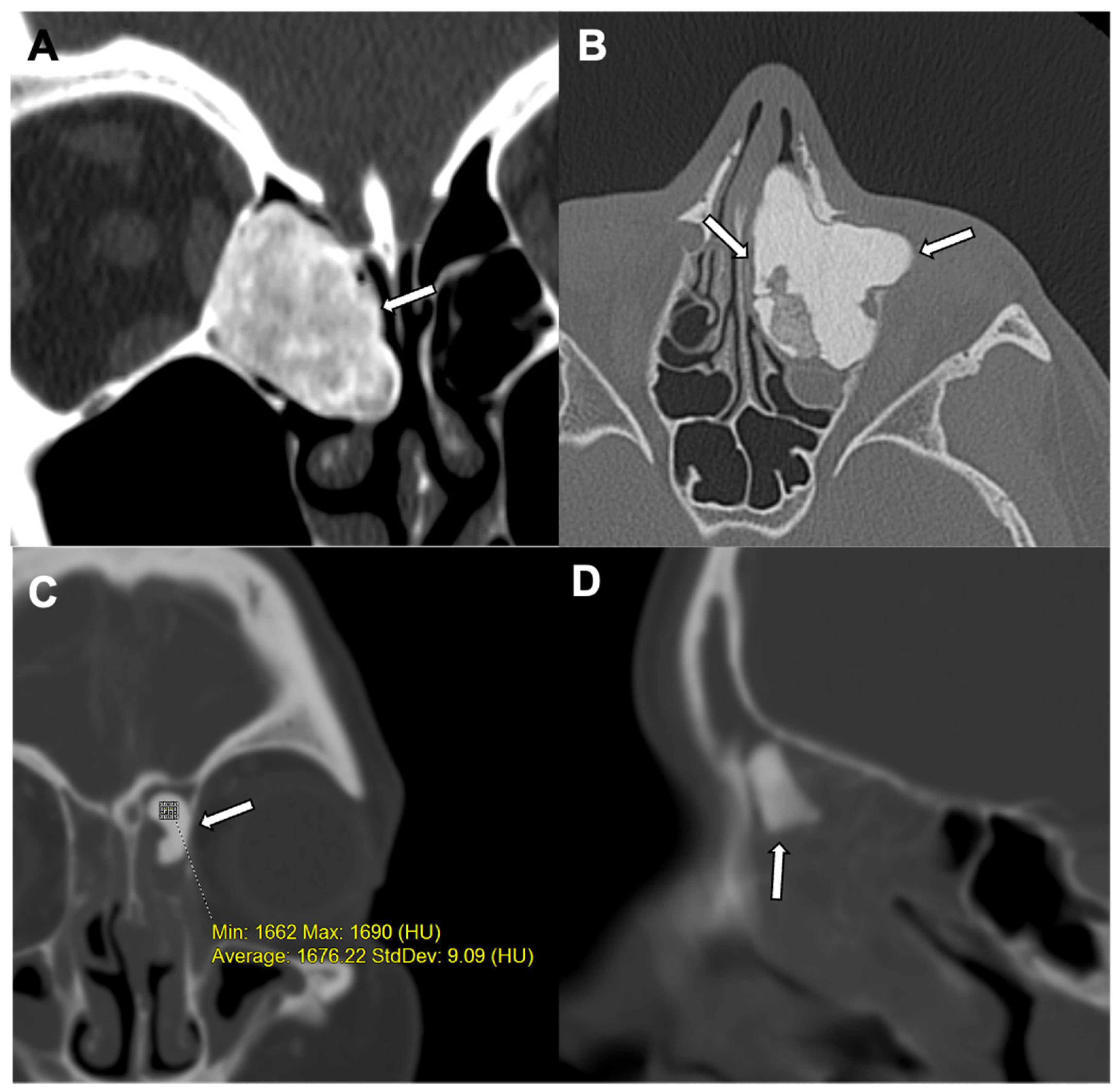
3. Osteoma
- Ivory osteoma (also known as eburnated osteoma) contains dense bone without a haversian system.
- Mature osteoma (also known as osteoma spongiosum) is histologically similar to normal bone. Mature osteoma is composed of trabecular bone, often with marrow.
- Mixed osteoma contains features of both ivory and mature osteoma.
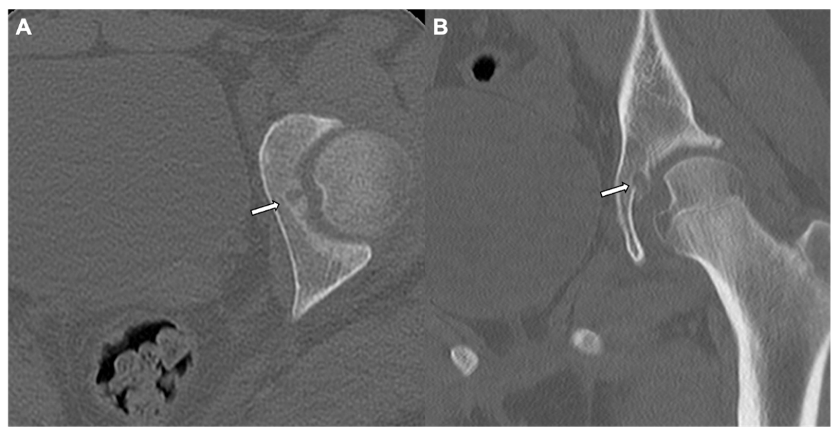
4. Enostosis (Bone Island)
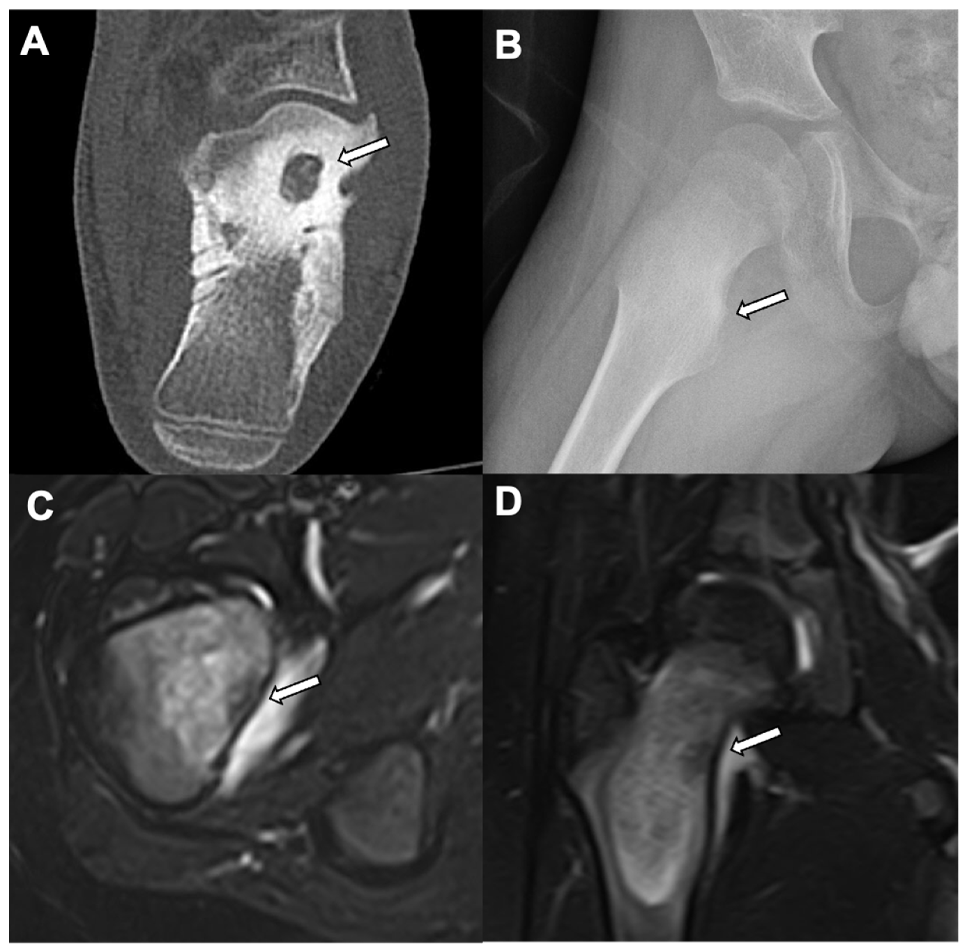
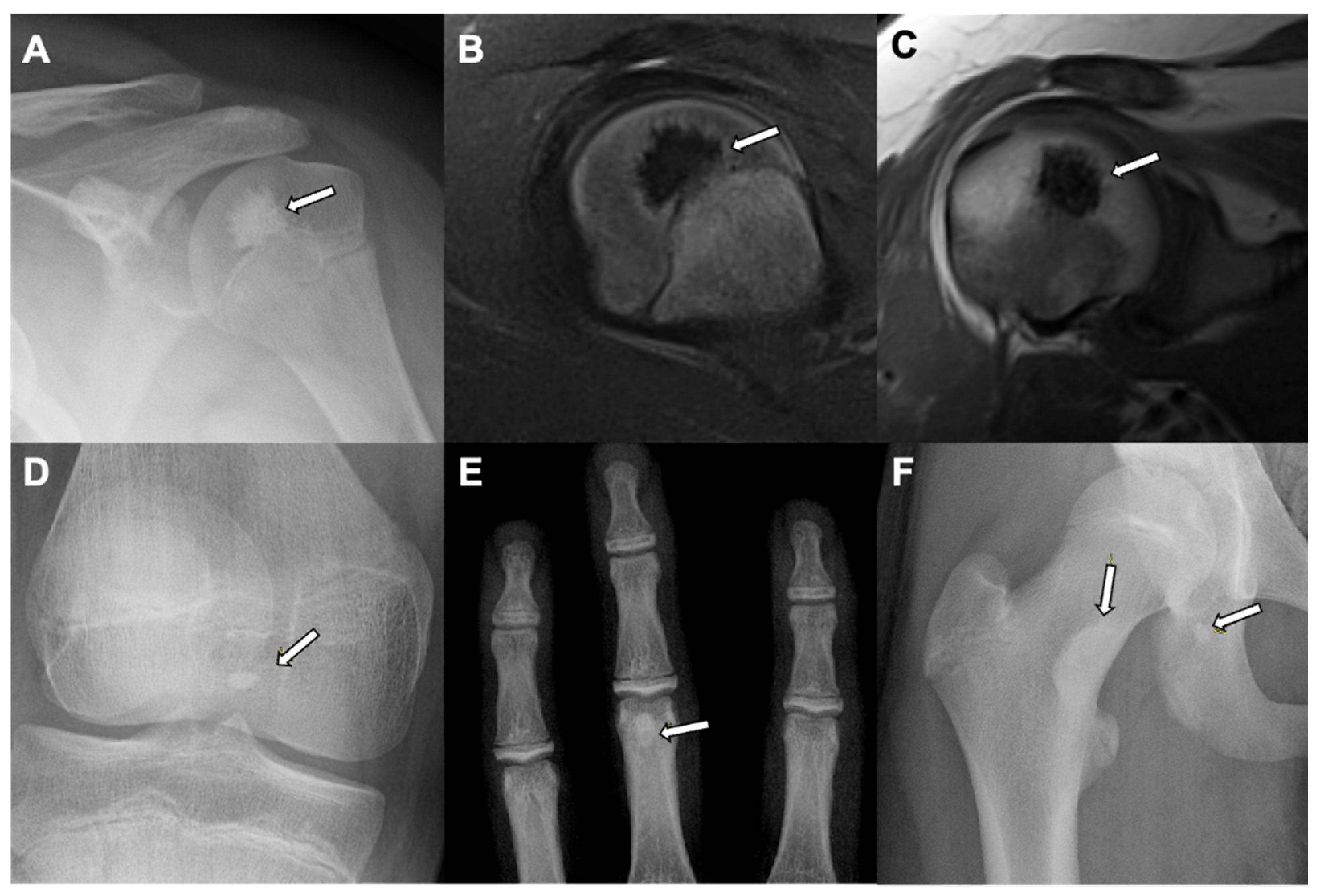
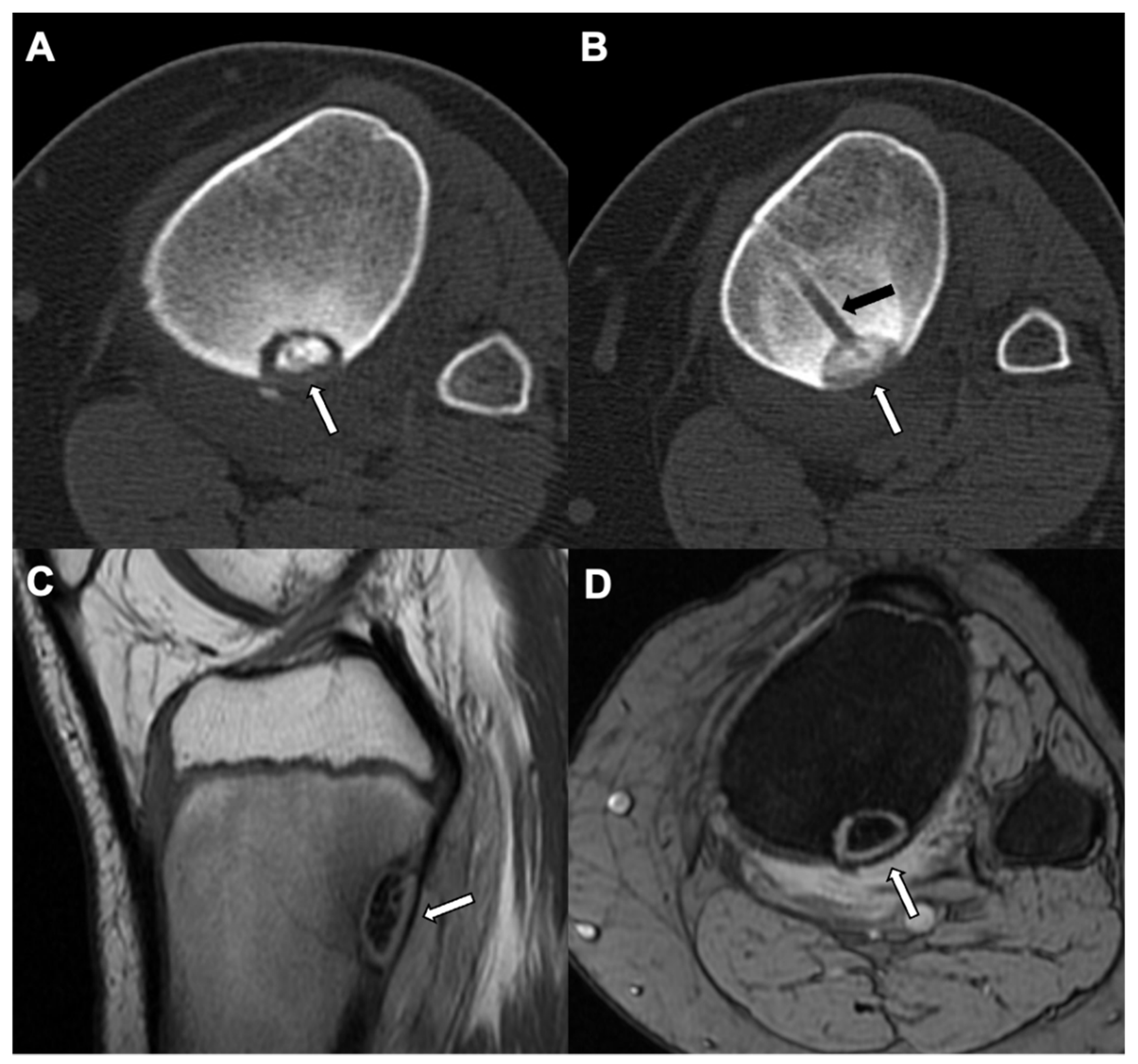
5. Osteoid Osteoma
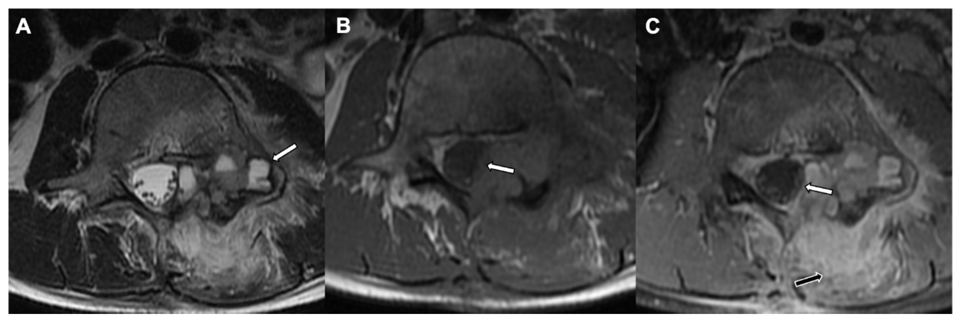
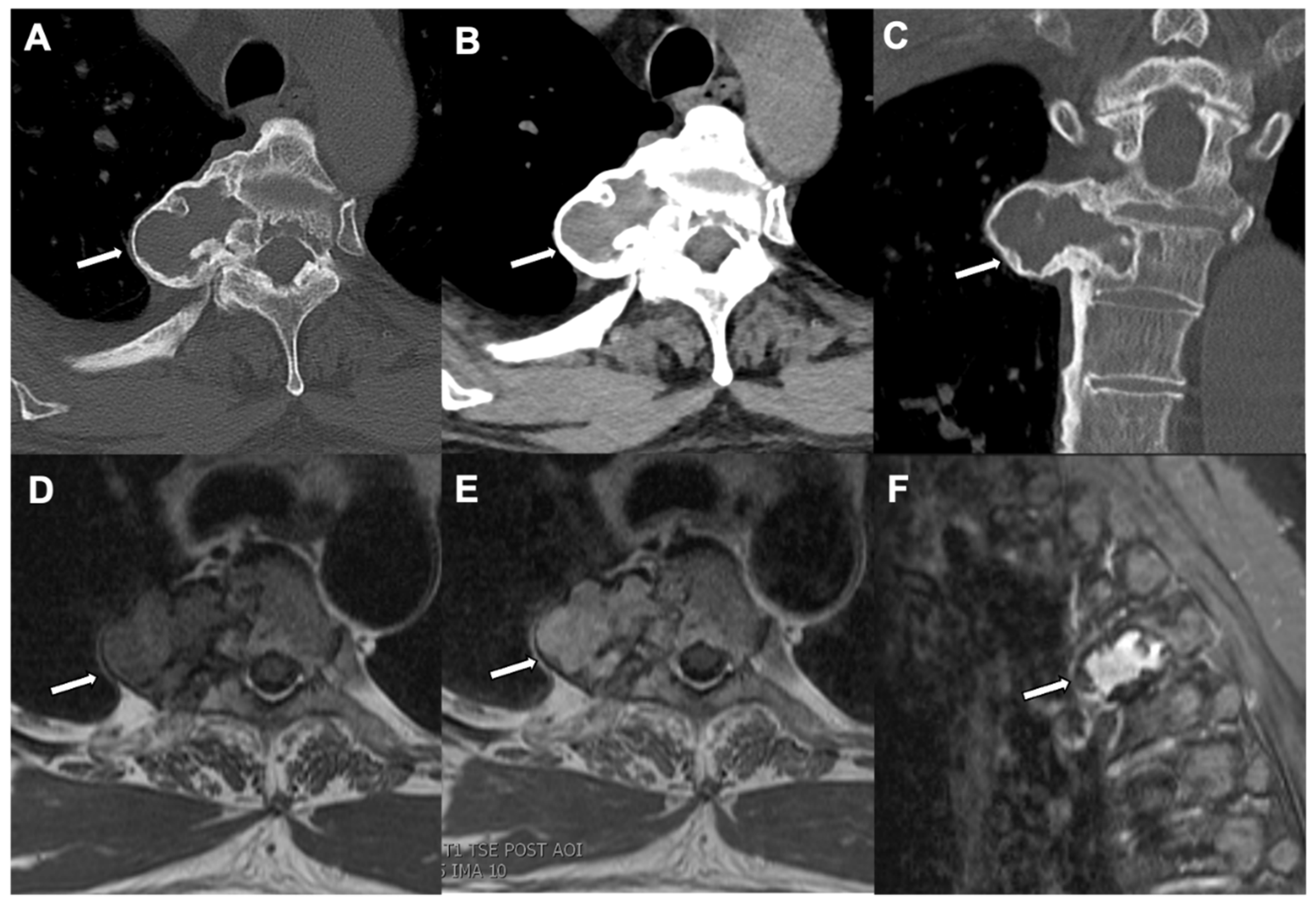
6. Osteoblastoma
7. Conclusions
Funding
Conflicts of Interest
References
- Shah, J.N.; Cohen, H.L.; Choudhri, A.F.; Gupta, S.; Miller, S.F. Pediatric Benign Bone Tumors: What Does the Radiologist Need to Know?: Pediatric Imaging. Radiographics 2017, 37, 1001–1002. [Google Scholar] [CrossRef] [PubMed] [Green Version]
- Stacy, G.S.; Mahal, R.S.; Peabody, T.D. Staging of Bone Tumors: A Review with Illustrative Examples. Am. J. Roentgenol. 2006, 186, 967–976. [Google Scholar] [CrossRef]
- Umer, M.; Hasan, O.H.; Khan, D.; Uddin, N.; Noordin, S. Systematic approach to musculoskeletal benign tumors. Int. J. Surg. Oncol. 2017, 2, e46. [Google Scholar] [CrossRef] [PubMed] [Green Version]
- Sayan, N.B.; Üçok, C.; Karasu, H.A.; Günhan, Ö. Peripheral osteoma of the oral and maxillofacial region: A study of 35 new cases. J. Oral Maxillofac. Surg. 2002, 60, 1299–1301. [Google Scholar] [CrossRef]
- Chattopadhyay, C.P.K.; Chander, M.G.M. Peripheral Osteoma of the Maxillofacial Region Diagnosis and Management: A Study of 06 Cases. J. Maxillofac. Oral Surg. 2012, 11, 425–429. [Google Scholar] [CrossRef] [Green Version]
- Colas, L.; Caron, S.; Cotten, A. Skull Vault Lesions: A Review. Am. J. Roentgenol. 2015, 205, 840–847. [Google Scholar] [CrossRef]
- Gomez, C.K.; Schiffman, S.R.; Bhatt, A. Radiological review of skull lesions. Insights Imaging 2018, 9, 857–882. [Google Scholar] [CrossRef] [PubMed] [Green Version]
- Motamedi, K.; Seeger, L.L. Benign Bone Tumors. Radiol. Clin. North Am. 2011, 49, 1115–1134. [Google Scholar] [CrossRef] [PubMed]
- Amaral, L.; Chiurciu, M.; Almeida, J.R.; Ferreira, N.F.; Mendonça, R.; Lima, S.S. MR imaging for evaluation of lesions of the cranial vault: A pictorial essay. Arq. de Neuro-Psiquiatria 2003, 61, 521–532. [Google Scholar] [CrossRef] [Green Version]
- Becelli, R.; Santamaria, S.; Saltarel, A.; Carboni, A.; Iannetti, G. Endo-Orbital Osteoma: Two Case Reports. J. Craniofacial Surg. 2002, 13, 493–496. [Google Scholar] [CrossRef]
- Chae, S.Y.; Sim, H.B.; Kim, M.J.; Jang, Y.H.; Lee, S.-J.; Kim, D.W.; Lee, W.J. Button Osteoma: A Review of Ten Cases. Ann. Dermatol. 2015, 27, 394–397. [Google Scholar] [CrossRef] [PubMed]
- Oyer, S.L.; Patel, K.G. Endoscopic brow approach for frontal osteoma in a pediatric patient. Int. J. Pediatr. Otorhinolaryngol. 2012, 76, 1211–1213. [Google Scholar] [CrossRef] [PubMed]
- Ulano, A.; Bredella, M.A.; Burke, P.; Chebib, I.; Simeone, F.J.; Huang, A.J.; Torriani, M.; Chang, C.Y. Distinguishing Untreated Osteoblastic Metastases From Enostoses Using CT Attenuation Measurements. Am. J. Roentgenol. 2016, 207, 362–368. [Google Scholar] [CrossRef] [PubMed]
- Bernard, S.; Walker, E.; Raghavan, M. An Approach to the Evaluation of Incidentally Identified Bone Lesions Encountered on Imaging Studies. Am. J. Roentgenol. 2017, 208, 960–970. [Google Scholar] [CrossRef]
- Sala, F.; Dapoto, A.; Morzenti, C.; Firetto, M.C.; Valle, C.; Tomasoni, A.; Sironi, S. Bone islands incidentally detected on computed tomography: Frequency of enostosis and differentiation from untreated osteoblastic metastases based on CT attenuation value. Br. J. Radiol. 2019, 92, 20190249. [Google Scholar] [CrossRef] [PubMed]
- Hong, J.H.; Jung, J.-Y.; Jo, A.; Nam, Y.; Pak, S.; Lee, S.-Y.; Park, H.; Lee, S.E.; Kim, S. Development and Validation of a Radiomics Model for Differentiating Bone Islands and Osteoblastic Bone Metastases at Abdominal CT. Radiology 2021, 299, 626–632. [Google Scholar] [CrossRef] [PubMed]
- Fonseca, E.K.U.N.; Castro, A.D.A.E.; Kubo, R.S.; Miranda, F.C.; Taneja, A.K.; Santos, D.D.C.B.; Rosemberg, L.A. Musculoskeletal “don’t touch” lesions: Pictorial essay. Radiol. Bras. 2019, 52, 48–53. [Google Scholar] [CrossRef]
- Boscainos, P.J.; Cousins, G.R.; Kulshreshtha, R.; Oliver, T.B.; Papagelopoulos, P.J. Osteoid Osteoma. Orthopedics 2013, 36, 792–800. [Google Scholar] [CrossRef]
- Iyer, R.S.; Chapman, T.; Chew, F.S. Pediatric Bone Imaging: Diagnostic Imaging of Osteoid Osteoma. Am. J. Roentgenol. 2012, 198, 1039–1052. [Google Scholar] [CrossRef]
- Chen, Y.-L.; Jiang, W.-Y.; Ma, W.-H. Osteoid osteoma: Lower back pain combined with scoliosis. J. Int. Med. Res. 2020, 48, 300060520903873. [Google Scholar] [CrossRef] [Green Version]
- Noordin, S.; Allana, S.; Hilal, K.; Nadeem, N.; Lakdawala, R.; Sadruddin, A.; Uddin, N. Osteoid osteoma: Contemporary management. Orthop. Rev. 2018, 10, 7496. [Google Scholar] [CrossRef] [Green Version]
- Roach, P.J.; Connolly, L.P.; Zurakowski, D.; Treves, S.T. Osteoid osteoma: Comparative utility of high-resolution planar and pinhole magnification scintigraphy. Pediatr. Radiol. 1996, 26, 222–225. [Google Scholar] [CrossRef] [PubMed]
- Rodallec, M.H.; Feydy, A.; Larousserie, F.; Anract, P.; Campagna, R.; Babinet, A.; Zins, M.; Drapé, J.-L. Diagnostic Imaging of Solitary Tumors of the Spine: What to Do and Say. Radiographics 2008, 28, 1019–1041. [Google Scholar] [CrossRef] [Green Version]
- Liu, P.T.; Chivers, F.S.; Roberts, C.; Schultz, C.J.; Beauchamp, C.P. Imaging of Osteoid Osteoma with Dynamic Gadolinium-enhanced MR Imaging. Radiology 2003, 227, 691–700. [Google Scholar] [CrossRef]
- Harish, S.; Saifuddin, A. Imaging features of spinal osteoid osteoma with emphasis on MRI findings. Eur. Radiol. 2005, 15, 2396–2403. [Google Scholar] [CrossRef] [PubMed]
- Motamedi, K.; Ilaslan, H.; Seeger, L.L. Imaging of the Lumbar Spine Neoplasms. Semin. Ultrasound CT MRI 2004, 25, 474–489. [Google Scholar] [CrossRef] [PubMed] [Green Version]
- Barile, A.; Arrigoni, F.; Zugaro, L.; Zappia, M.; Cazzato, R.L.; Garnon, J.; Ramamurthy, N.; Brunese, L.; Gangi, A.; Masciocchi, C. Minimally invasive treatments of painful bone lesions: State of the art. Med. Oncol. 2017, 34, 53. [Google Scholar] [CrossRef]
- Rosenthal, D.I.; Alexander, A.; Rosenberg, A.E.; Springfield, D. Ablation of osteoid osteomas with a percutaneously placed electrode: A new procedure. Radiology 1992, 183, 29–33. [Google Scholar] [CrossRef] [PubMed]
- Rosenthal, D.I.; Hornicek, F.J.; Torriani, M.; Gebhardt, M.C.; Mankin, H.J. Osteoid Osteoma: Percutaneous Treatment with Radiofrequency Energy. Radiology 2003, 229, 171–175. [Google Scholar] [CrossRef]
- Rosenthal, D.I.; Marota, J.J.A.; Hornicek, F.J. Osteoid Osteoma: Elevation of Cardiac and Respiratory Rates at Biopsy Needle Entry into Tumor in 10 Patients. Radiology 2003, 226, 125–128. [Google Scholar] [CrossRef] [PubMed]
- Pinto, C.H.; Taminiau, A.H.M.; Vanderschueren, G.M.; Hogendoorn, P.C.W.; Bloem, J.L.; Obermann, W.R. Technical Considerations in CT-Guided Radiofrequency Thermal Ablation of Osteoid Osteoma: Tricks of the Trade. Am. J. Roentgenol. 2002, 179, 1633–1642. [Google Scholar] [CrossRef] [PubMed] [Green Version]
- Rosenthal, D.I.; Hornicek, F.J.; Wolfe, M.W.; Jennings, L.C.; Gebhardt, M.C.; Mankin, H.J. Decreasing Length of Hospital Stay in Treatment of Osteoid Osteoma. Clin. Orthop. Relat. Res. 1999, 361, 186–191. [Google Scholar] [CrossRef]
- Tomasian, A.; Jennings, J. Percutaneous Radiofrequency Ablation of Spinal Osteoid Osteomas Using a Targeted Navigational Bipolar Electrode System. Am. J. Neuroradiol. 2018, 39, 2385–2388. [Google Scholar] [CrossRef] [PubMed] [Green Version]
- Arrigoni, F.; Barile, A.; Zugaro, L.; Splendiani, A.; di Cesare, E.; Caranci, F.; Ierardi, A.M.; Floridi, C.; Angileri, A.S.; Reginelli, A.; et al. Intra-articular benign bone lesions treated with Magnetic Resonance-guided Focused Ultrasound (MRgFUS): Imaging follow-up and clinical results. Med. Oncol. 2017, 34, 55. [Google Scholar] [CrossRef]
- Arrigoni, F.; Bruno, F.; Gianneramo, C.; Palumbo, P.; Zugaro, L.; Zoccali, C.; Barile, A.; Masciocchi, C. Evolution of the imaging features of osteoid osteoma treated with RFA or MRgFUS during a long-term follow-up: A pictorial review with clinical correlations. La Radiol. Med. 2020, 125, 578–584. [Google Scholar] [CrossRef]
- Mishra, A.; Pruthi, N.; Nandeesh, B.; Shukla, D. Cervical Spine Osteoblastoma with an Aneurysmal Bone Cyst in a 2-Year-Old Child: A Case Report. Pediatr. Neurosurg. 2019, 54, 46–50. [Google Scholar] [CrossRef]
- Avadhanam, P.K.; Vuyyur, S.; Panigrahi, M.K. A rare occurrence of osteoblastoma in a child. J. Pediatric Neurosci. 2010, 5, 153–156. [Google Scholar] [CrossRef] [PubMed]
- Kroon, H.M.; Schurmans, J. Osteoblastoma: Clinical and radiologic findings in 98 new cases. Radiology 1990, 175, 783–790. [Google Scholar] [CrossRef]
- Lucas, D.R.; Unni, K.K.; McLeod, R.A.; O’Connor, M.I.; Sim, F.H. Osteoblastoma: Clinicopathologic study of 306 cases. Hum. Pathol. 1994, 25, 117–134. [Google Scholar] [CrossRef]
- Galgano, M.A.; Goulart, C.R.; Iwenofu, H.; Chin, L.S.; Lavelle, W.; Mendel, E. Osteoblastomas of the spine: A comprehensive review. Neurosurg. Focus 2016, 41, E4. [Google Scholar] [CrossRef] [PubMed] [Green Version]
- Saccomanni, B. Retracted article: Osteoid osteoma and osteoblastoma of the spine: A review of the literature. Curr. Rev. Musculoskelet. Med. 2009, 2, 65–67. [Google Scholar] [CrossRef] [PubMed]
- Ma, W.; Quan, Z.; Wang, J.; Li, X.; Li, G. The one-in-all diagnostic value of 99mTc-MDP bone scan combining with single-photon emission tomography (SPECT)/CT imaging in spinal osteoblastoma. J. Orthop. Surg. Res. 2020, 15, 1–9. [Google Scholar] [CrossRef]
- Trübenbach, J.; Nägele, T.; Bauer, T.; Ernemann, U. Preoperative Embolization of Cervical Spine Osteoblastomas: Report of Three Cases. Am. J. Neuroradiol. 2006, 27, 1910–1912. [Google Scholar] [PubMed]
- Wu, M.; Xu, K.; Xie, Y.; Yan, F.; Deng, Z.; Lei, J.; Cai, L. Diagnostic and Management Options of Osteoblastoma in the Spine. Med. Sci. Monit. 2019, 25, 1362–1372. [Google Scholar] [CrossRef] [PubMed]
- Cazzato, R.L.; Auloge, P.; Dalili, D.; De Marini, P.; Di Marco, A.; Garnon, J.; Gangi, A. Percutaneous Image-Guided Cryoablation of Osteoblastoma. Am. J. Roentgenol. 2019, 213, 1157–1162. [Google Scholar] [CrossRef] [PubMed]


| Osteoma | Enostosis | Osteoid Osteoma | Osteoblastoma | |
|---|---|---|---|---|
| Age | Between 3rd and 5th decades | No age predilection | Between 5 and 25 years of age | Between 10 and 30 years of age |
| Presentation | Usually asymptomatic; may present with headache and sinusitis | Most commonly incidental on imaging | Worsening night pain, relieved by non-steroidal anti-inflammatory drugs | Dull pain, not relieved by non-steroidal anti-inflammatory drugs |
| Lesion Location | Paranasal sinuses, mainly frontal and ethmoidal | Axial skeleton (spine, pelvis, and ribs); long bones | Long bones, mainly femur | Spine and long bones |
| Radiographic/CT features | Juxtacortical, well-circumscribed homogenous sclerotic lesion | Homogenous intra-medullary sclerotic focus with spiculated margins | Small (less than 2 cm) cortical lucency with extensive surrounding sclerosis | Expansile large (more than 2 cm) lucent lesion with matrix mineralization |
| Treatment | None if asymptomatic; excision if complications related to mass effect | None | Percutaneous CT guided radiofrequency ablation | Surgery or percutaneous CT guided ablation |
Publisher’s Note: MDPI stays neutral with regard to jurisdictional claims in published maps and institutional affiliations. |
© 2021 by the authors. Licensee MDPI, Basel, Switzerland. This article is an open access article distributed under the terms and conditions of the Creative Commons Attribution (CC BY) license (https://creativecommons.org/licenses/by/4.0/).
Share and Cite
Shah, J.; Gandhi, D.; Chauhan, A.; Gupta, S. Imaging Review of Pediatric Benign Osteocytic Tumors and Latest Updates on Management. J. Clin. Med. 2021, 10, 2823. https://doi.org/10.3390/jcm10132823
Shah J, Gandhi D, Chauhan A, Gupta S. Imaging Review of Pediatric Benign Osteocytic Tumors and Latest Updates on Management. Journal of Clinical Medicine. 2021; 10(13):2823. https://doi.org/10.3390/jcm10132823
Chicago/Turabian StyleShah, Jignesh, Darshan Gandhi, Ankita Chauhan, and Saurabh Gupta. 2021. "Imaging Review of Pediatric Benign Osteocytic Tumors and Latest Updates on Management" Journal of Clinical Medicine 10, no. 13: 2823. https://doi.org/10.3390/jcm10132823
APA StyleShah, J., Gandhi, D., Chauhan, A., & Gupta, S. (2021). Imaging Review of Pediatric Benign Osteocytic Tumors and Latest Updates on Management. Journal of Clinical Medicine, 10(13), 2823. https://doi.org/10.3390/jcm10132823