
Ruling Out Coronavirus Disease 2019 in Patients with Pneumonia: The Role of Blood Cell Count and Lung Ultrasound

 ,
,
Abstract
:1. Introduction
2. Materials and Methods
Statistics
3. Results
4. Discussion
Author Contributions
Funding
Institutional Review Board Statement
Informed Consent Statement
Data Availability Statement
Conflicts of Interest
References
- Musher, D.M.; Thorner, A.R. Community-Acquired Pneumonia. N. Engl. J. Med. 2014, 371, 1619–1628. [Google Scholar] [CrossRef] [PubMed]
- Wang, D.; Hu, B.; Hu, C.; Zhu, F.; Liu, X.; Zhang, J.; Wang, B.; Xiang, H.; Cheng, Z.; Xiong, Y.; et al. Clinical Characteristics of 138 Hospitalized Patients With 2019 Novel Coronavirus–Infected Pneumonia in Wuhan, China. JAMA 2020, 323, 1061. [Google Scholar] [CrossRef]
- Corradini, E.; Ventura, P.; Ageno, W.; Cogliati, C.B.; Muiesan, M.L.; Girelli, D.; Pirisi, M.; Gasbarrini, A.; Angeli, P.; Querini, P.R.; et al. Clinical factors associated with death in 3044 COVID-19 patients managed in internal medicine wards in Italy: Results from the SIMI-COVID-19 study of the Italian Society of Internal Medicine (SIMI). Intern. Emerg. Med. 2021, 16, 1005–1015. [Google Scholar] [CrossRef]
- Lieberman, J.A.; Pepper, G.; Naccache, S.N.; Huang, M.-L.; Jerome, K.R.; Greninger, A.L. Comparison of Commercially Available and Laboratory-Developed Assays for In Vitro Detection of SARS-CoV-2 in Clinical Laboratories. J. Clin. Microbiol. 2020, 58, e00821-20. [Google Scholar] [CrossRef] [PubMed]
- Tali, S.H.S.; LeBlanc, J.J.; Sadiq, Z.; Oyewunmi, O.D.; Camargo, C.; Nikpour, B.; Armanfard, N.; Sagan, S.M.; Jahanshahi-Anbuhi, S. Tools and Techniques for Severe Acute Respiratory Syndrome Coronavirus 2 (SARS-CoV-2)/COVID-19 Detection. Clin. Microbiol. Rev. 2021, 34, e00228-20. [Google Scholar] [CrossRef]
- Hanson, K.E.; Altayar, O.; Caliendo, A.M.; Arias, C.A.; Englund, J.A.; Hayden, M.K.; Lee, M.J.; Loeb, M.; Patel, R.; El Alayli, A.; et al. The Infectious Diseases Society of America Guidelines on the Diagnosis of COVID-19: Antigen Testing. Clin. Infect. Dis. 2021. [Google Scholar] [CrossRef]
- Weissleder, R.; Lee, H.; Ko, J.; Pittet, M.J. COVID-19 diagnostics in context. Sci. Transl. Med. 2020, 12, eabc1931. [Google Scholar] [CrossRef] [PubMed]
- Long, D.R.; Gombar, S.; Hogan, C.A.; Greninger, A.L.; O’Reilly-Shah, V.; Bryson-Cahn, C.; Stevens, B.; Rustagi, A.; Jerome, K.R.; Kong, C.S.; et al. Occurrence and Timing of Subsequent Severe Acute Respiratory Syndrome Coronavirus 2 Reverse-transcription Polymerase Chain Reaction Positivity Among Initially Negative Patients. Clin. Infect. Dis. 2020, 72, 323–326. [Google Scholar] [CrossRef]
- Mair, M.D.D.; Hussain, M.; Siddiqui, S.; Das, S.; Baker, A.; Conboy, P.; Valsamakis, T.; Uddin, J.; Rea, P. A systematic review and meta-analysis comparing the diagnostic accuracy of initial RT-PCR and CT scan in suspected COVID-19 patients. Br. J. Radiol. 2021, 94, 20201039. [Google Scholar] [CrossRef]
- Islam, N.; Salameh, J.-P.; Leeflang, M.M.; Hooft, L.; McGrath, T.A.; Van Der Pol, C.B.; Frank, R.A.; Kazi, S.; Prager, R.; Hare, S.S.; et al. Thoracic imaging tests for the diagnosis of COVID-19. Cochrane Database Syst. Rev. 2020, 11, CD013639. [Google Scholar] [CrossRef]
- Finance, J.; Zieleskewicz, L.; Habert, P.; Jacquier, A.; Parola, P.; Boussuges, A.; Bregeon, F.; Eldin, C. Low Dose Chest CT and Lung Ultrasound for the Diagnosis and Management of COVID-19. J. Clin. Med. 2021, 10, 2196. [Google Scholar] [CrossRef]
- Bar, S.; Lecourtois, A.; Diouf, M.; Goldberg, E.; Bourbon, C.; Arnaud, E.; Domisse, L.; Dupont, H.; Gosset, P. The association of lung ultrasound images with COVID-19 infection in an emergency room cohort. Anaesthesia 2020, 75, 1620–1625. [Google Scholar] [CrossRef] [PubMed]
- Brahier, T.; Meuwly, J.-Y.; Pantet, O.; Vez, M.-J.B.; Donnet, H.G.; Hartley, M.-A.; Hugli, O.; Boillat-Blanco, N. Lung Ultrasonography for Risk Stratification in Patients with Coronavirus Disease 2019 (COVID-19): A Prospective Observational Cohort Study. Clin. Infect. Dis. 2020. [Google Scholar] [CrossRef]
- Kirschner, J.; Hunter, B. In the ED, LUS and CT did not differ for sensitivity or specificity for diagnosing COVID-19 pneumonia. Ann. Intern. Med. 2021, 174, JC57. [Google Scholar] [CrossRef]
- Volpicelli, G.; Gargani, L.; Perlini, S.; Spinelli, S.; Barbieri, G.; Lanotte, A.; Casasola, G.G.; Nogué-Bou, R.; Lamorte, A.; Agricola, E.; et al. Lung ultrasound for the early diagnosis of COVID-19 pneumonia: An international multicenter study. Intensiv. Care Med. 2021, 47, 444–454. [Google Scholar] [CrossRef]
- Rodriguez, L.; Pekkarinen, P.T.; Lakshmikanth, T.; Tan, Z.; Consiglio, C.R.; Pou, C.; Chen, Y.; Mugabo, C.H.; Nguyen, N.A.; Nowlan, K.; et al. Systems-Level Immunomonitoring from Acute to Recovery Phase of Severe COVID-19. Cell Rep. Med. 2020, 1, 100078. [Google Scholar] [CrossRef] [PubMed]
- Martens, R.J.H.; van Adrichem, A.J.; Mattheij, N.J.A.; Brouwer, C.G.; van Twist, D.J.L.; Broerse, J.J.C.R.; Magro-Checa, C.; van Dongen, C.M.P.; Mostard, R.L.M.; Ramiro, S.; et al. Hemocytometric characteristics of COVID-19 patients with and without cytokine storm syndrome on the sysmex XN-10 hematology analyzer. Clin. Chem. Lab. Med. 2020, 59, 783–793. [Google Scholar] [CrossRef]
- Thell, R.; Zimmermann, J.; Szell, M.; Tomez, S.; Eisenburger, P.; Haugk, M.; Kreil, A.; Spiel, A.; Blaschke, A.; Klicpera, A.; et al. Standard blood laboratory values as a clinical support tool to distinguish between SARS-CoV-2 positive and negative patients. Sci. Rep. 2021, 11, 1–9. [Google Scholar] [CrossRef] [PubMed]
- Perlini, S.; Ciprandi, G.; Castagnoli, R.; Licari, A.; Marseglia, G.L. Eosinopenia could be a relevant prognostic biomarker in patients with coronavirus disease 2019. Allergy Asthma Proc. 2020, 41, e80–e82. [Google Scholar] [CrossRef] [PubMed]
- Lin, A.; He, Z.-B.; Zhang, S.; Zhang, J.-G.; Zhang, X.; Yan, W.-H. Early Risk Factors for the Duration of Severe Acute Respiratory Syndrome Coronavirus 2 Viral Positivity in Patients with Coronavirus Disease 2019. Clin. Infect. Dis. 2020, 71, 2061–2065. [Google Scholar] [CrossRef]
- Qin, C.; Zhou, L.; Hu, Z.; Zhang, S.; Yang, S.; Tao, Y.; Xie, C.; Ma, K.; Shang, K.; Wang, W.; et al. Dysregulation of Immune Response in Patients with Coronavirus 2019 (COVID-19) in Wuhan, China. Clin. Infect. Dis. 2020, 71, 762–768. [Google Scholar] [CrossRef] [PubMed]
- Rosenberg, H.F.; Foster, P.S. Eosinophils and COVID-19: Diagnosis, prognosis, and vaccination strategies. Semin. Immunopathol. 2021, 43, 383–392. [Google Scholar] [CrossRef] [PubMed]
- González, M.M.; Gonzalo, E.S.; Lopez, I.C.; Fernández, F.A.; Pérez, J.L.B.; Monge, D.M.; Núñez, J.A.V.; Fenoll, R.G.; Fernández, C.S.; Castro, S.J.F.; et al. The Prognostic Value of Eosinophil Recovery in COVID-19: A Multicentre, Retrospective Cohort Study on Patients Hospitalised in Spanish Hospitals. J. Clin. Med. 2021, 10, 305. [Google Scholar] [CrossRef] [PubMed]
- Huang, R.; Xie, L.; He, J.; Dong, H.; Liu, T. Association between the peripheral blood eosinophil counts and COVID-19. Med. 2021, 100, e26047. [Google Scholar] [CrossRef] [PubMed]
- Sun, S.; Cai, X.; Wang, H.; He, G.; Lin, Y.; Lu, B.; Chen, C.; Pan, Y.; Hu, X. Abnormalities of peripheral blood system in patients with COVID-19 in Wenzhou, China. Clin. Chim. Acta 2020, 507, 174–180. [Google Scholar] [CrossRef]
- Volpicelli, G.; Elbarbary, M.; Blaivas, M.; Lichtenstein, D.A.; Mathis, G.; Kirkpatrick, A.W.; Melniker, L.; Gargani, L.; Noble, V.E.; et al.; International Liaison Committee on Lung Ultrasound (ILC-LUS) for the International Consensus Conference on Lung Ultrasound (ICC-LUS) International evidence-based recommendations for point-of-care lung ultrasound. Intensiv. Care Med. 2012, 38, 577–591. [Google Scholar] [CrossRef] [Green Version]
- Quan, H.; Li, B.; Couris, C.M.; Fushimi, K.; Graham, P.; Hider, P.; Januel, J.-M.; Sundararajan, V. Updating and Validating the Charlson Comorbidity Index and Score for Risk Adjustment in Hospital Discharge Abstracts Using Data From 6 Countries. Am. J. Epidemiol. 2011, 173, 676–682. [Google Scholar] [CrossRef] [Green Version]
- Lim, W.S.; Van Der Eerden, M.M.; Laing, R.; Boersma, W.G.; Karalus, N.; Town, G.I.; Lewis, S.A.; Macfarlane, J.T. Defining community acquired pneumonia severity on presentation to hospital: An international derivation and validation study. Thorax 2003, 58, 377–382. [Google Scholar] [CrossRef] [Green Version]
- Outh, R.; Boutin, C.; Gueudet, P.; Suzuki, M.; Saada, M.; Aumaître, H. Eosinopenia <100/μL as a marker of active COVID-19: An observational prospective study. J. Microbiol. Immunol. Infect. 2021, 54. [Google Scholar] [CrossRef]
- Pivetta, E.; Goffi, A.; Tizzani, M.; Locatelli, S.M.; Porrino, G.; Losano, I.; Leone, D.; Calzolari, G.; Vesan, M.; Steri, F.; et al. Lung Ultrasonography for the Diagnosis of SARS-CoV-2 Pneumonia in the Emergency Department. Ann. Emerg. Med. 2021, 77, 385–394. [Google Scholar] [CrossRef]
- Chen, J.; Han, T.; Huang, M.; Yang, Y.; Shang, F.; Zheng, Y.; Zhao, W.; Luo, L.; Han, X.; Lin, A.; et al. Clinical characteristics of asymptomatic carriers of novel coronavirus disease 2019: A multi-center study in Jiangsu Province. Virulence 2020, 11, 1557–1568. [Google Scholar] [CrossRef]
- Song, L.; Liang, E.-Y.; Wang, H.-M.; Shen, Y.; Kang, C.-M.; Xiong, Y.-J.; He, M.; Fu, W.-J.; Ke, P.-F.; Huang, X.-Z. Differential diagnosis and prospective grading of COVID-19 at the early stage with simple hematological and biochemical variables. Diagn. Microbiol. Infect. Dis. 2021, 99, 115169. [Google Scholar] [CrossRef] [PubMed]
- Luo, Y.; Yuan, X.; Xue, Y.; Mao, L.; Lin, Q.; Tang, G.; Song, H.; Liu, W.; Hou, H.; Wang, F.; et al. Using a diagnostic model based on routine laboratory tests to distinguish patients infected with SARS-CoV-2 from those infected with influenza virus. Int. J. Infect. Dis. 2020, 95, 436–440. [Google Scholar] [CrossRef] [PubMed]
- Chen, J.; Pan, Y.; Li, G.; Xu, W.; Zhang, L.; Yuan, S.; Xia, Y.; Lu, P.; Zhang, J. Distinguishing between COVID-19 and influenza during the early stages by measurement of peripheral blood parameters. J. Med. Virol. 2020, 93, 1029–1037. [Google Scholar] [CrossRef]
- Asch, D.A.; Sheils, N.E.; Islam, N.; Chen, Y.; Werner, R.M.; Buresh, J.; Doshi, J.A. Variation in US Hospital Mortality Rates for Patients Admitted With COVID-19 During the First 6 Months of the Pandemic. JAMA Intern. Med. 2021, 181, 471. [Google Scholar] [CrossRef] [PubMed]
- Fan, G.; Yang, Z.; Lin, Q.; Zhao, S.; Yang, L.; He, D. Decreased Case Fatality Rate of COVID-19 in the Second Wave: A study in 53 countries or regions. Transbound. Emerg. Dis. 2020, 68, 213–215. [Google Scholar] [CrossRef] [PubMed]
- Domingo, P.; Pomar, V.; Mur, I.; Castellví, I.; Corominas, H.; de Benito, N. Not all COVID-19 pandemic waves are alike. Clin. Microbiol. Infect. 2021, 27, 1040.e7–1040.e10. [Google Scholar] [CrossRef]
- Berlin, D.; Gulick, R.; Martinez, F.J. Severe Covid-19. N. Engl. J. Med. 2020, 383, 2451–2460. [Google Scholar] [CrossRef]
- Meyerowitz, E.A.; Sen, P.; Schoenfeld, S.R.; Neilan, T.G.; Frigault, M.J.; Stone, J.H.; Kim, A.Y.; Mansour, M.K.; CIG (COVID-19 Immunomodulatory Group). Immunomodulation as Treatment for Severe Coronavirus Disease 2019: A Systematic Review of Current Modalities and Future Directions. Clin. Infect. Dis. 2020, 72, e1130–e1143. [Google Scholar] [CrossRef] [PubMed]


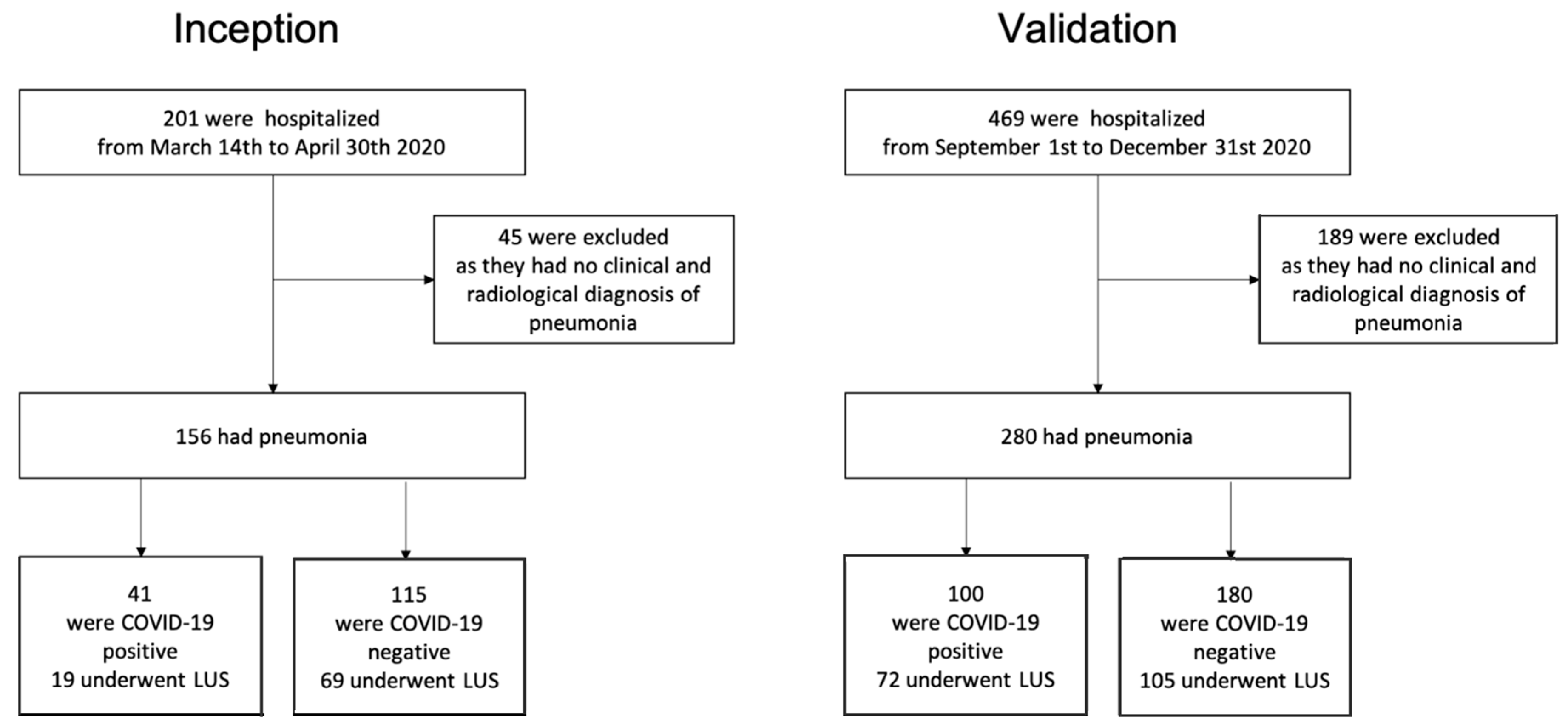
| Inception Cohort | All (n = 156) | non-COVID-19 (n = 115) | COVID-19 (n = 41) |
|---|---|---|---|
| Age, median (IQR)—years | 81 (73–88) | 82 (73–88) | 80 (74–86) |
| Female sex—no. (%) | 69 (44) | 49 (43) | 20 (49) |
| Charlson Comorbidity Index | 3 (1–4) | 3 (1–4) | 3 (2–5) |
| CURB-65 | 3 (2–3) | 3 (2–4) | 3 (2–3) |
| 10-day mortality, no. (%) | 26 (17) | 20 (17) | 6 (15) |
| 30-day mortality, no. (%) | 49 (31) | 30 (26) | 19 (46) * |
| C-reactive protein, mg/L | 79 (34–165) | 78 (37–163) | 79 (20–167) |
| Creatinine | 0.98 (0.79–1.45) | 0.99 (0.79–1.45) | 0.98 (0.80–1.34) |
| Bilirubine | 0.84 (0.57–1.23) | 0.93 (0.60–1.29) | 0.73 (0.46–1.06) |
| Hemoglobin, g/dL | 11.7 (10.4–13.2) | 11.7 (10.5–13.0) | 12.3 (10.5–13.9) |
| Platelet count, per mm3 | 224,000 (178,000–304,000) | 218,000 (172,000–302,000) | 242,000 (184,000–321,000) |
| White-cell count, per mm3 | 9485 (7155–12,978) | 9550 (7340–12,770) | 9120 (6260–14,310) |
| Neutrophil count, per mm3 | 7159 (5078–10,167) | 7146 (5197–10212) | 7366 (4919–9673) |
| Lymphocyte count, per mm3 | 1044 (659–1656) | 1138 (682–1848) | 914 (615–1216) * |
| Monocyte count, per mm3 | 683 (465–916) | 762 (509–1035) | 524 (299–652) * |
| Eosinophil count, per mm3 | 19 (0–108) | 43 (0–135) | 0 (0–2) * |
| Basophil count, per mm3 | 31 (12–48) | 38 (21–51) | 12 (7–29) * |
| Validation Cohort | All (n= 280) | non-COVID-19 (n= 180) | COVID-19 (n= 100) |
| Age, median (IQR)—years | 80 (71–87) | 81 (74–89) | 76 (63–85) * |
| Female sex—no. (%) | 124 (44) | 84 (47) | 40 (40) |
| Charlson Comorbidity Index | 2 (1–3) † | 2 (1–3) | 2 (1–3) |
| CURB-65 | 2 (1–3) † | 2 (1–3) | 2 (1–3) |
| 10-day mortality, no. (%) | 24 (9) | 17 (9) | 7 (7) |
| 30-day mortality, no. (%) | 62 (22) † | 41 (23) | 21 (21) |
| C-reactive protein, mg/L | 72 (27–140) | 87 (26–176) | 60 (31–116) |
| Creatinine, mg/dL | 0.99 (0.76–1.36) | 0.98 (0.74–1.34) | 1.01 (0.80–1.37) |
| Bilirubine, mg/dL | 0.69 (0.52–1.08) † | 0.78 (0.55–1.18) | 0.63 (0.49–0.81) * |
| Hemoglobin, g/dL | 12.3 (10.5–13.9) | 11.8 (10.2–13.2) | 13.5 (11.6–14.9) * |
| Platelet count, per mm3 | 217,000 (163,000–286,000) | 230,000 (167,000–286,000) | 201,000 (158,000–284,000) |
| White-cell count, per mm3 | 8780 (6795–12,635) | 9225 (6875–13,155) | 8540 (5390–11,410) |
| Neutrophil count, per mm3 | 7169 (4796–10,651) | 7107 (4988–11,145) | 6918 (4192–9457) |
| Lymphocyte count, per mm3 | 957 (569–1359) † | 1084 (670–1477) | 740 (490–1036) * |
| Monocyte count, per mm3 | 592 (352–836) † | 681 (450–940) | 474 (250–628) * |
| Eosinophil count, per mm3 | 0 (0–70) † | 23 (0–133) | 0 (0–0) * |
| Basophil count, per mm3 | 20 (10–39) † | 31 (14–51) | 11 (6–19) * |
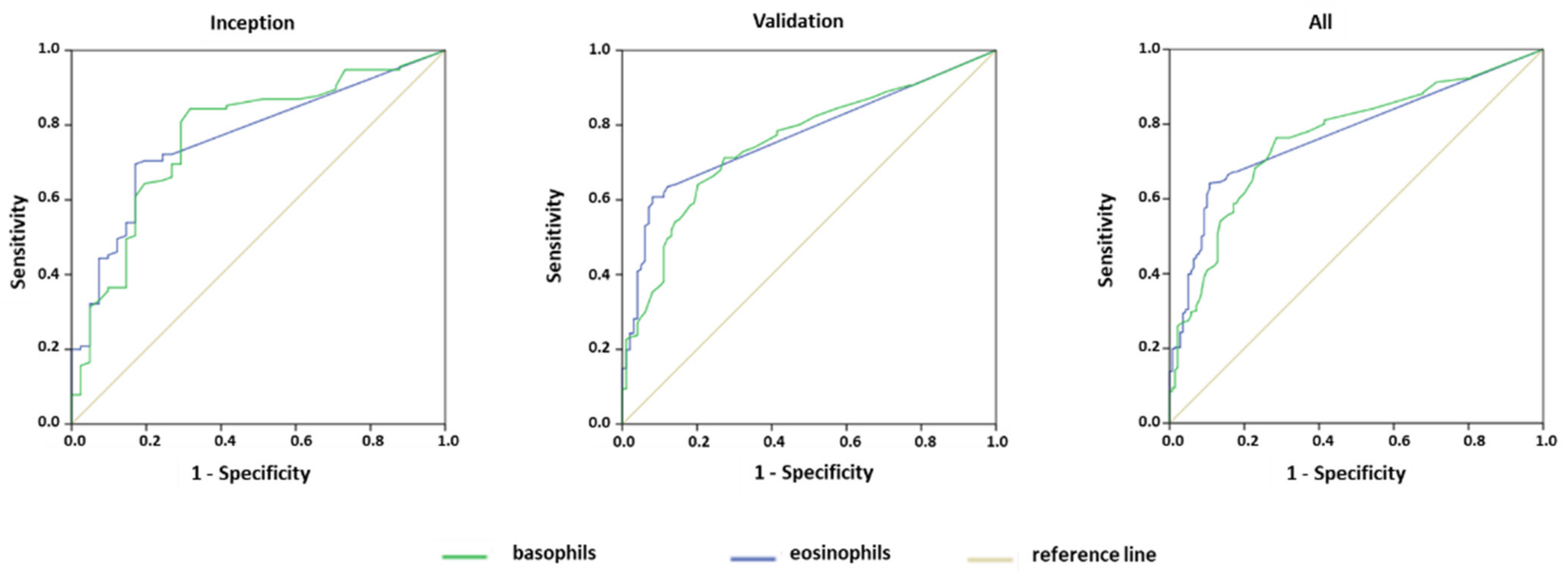
| Positive/Negative | AUC | CI 95% | p | Cuoffs | ||
|---|---|---|---|---|---|---|
| Basophils | Inception | 41/115 | 0.77 | 0.68–0.86 | <0.0001 | 17.5 |
| Validation | 100/180 | 0.75 | 0.70–0.81 | <0.0001 | 17.5 | |
| All | 141/295 | 0.76 | 0.72–0.81 | <0.0001 | 17.5 | |
| Eosinophils | Inception | 41/115 | 0.77 | 0.68–0.84 | <0.0001 | 10.5 |
| Validation | 100/180 | 0.77 | 0.72–0.83 | <0.0001 | 10.5 | |
| All | 141/295 | 0.77 | 0.73–0.82 | <0.0001 | 10.5 |
| Sensitivity | Specificity | PPV | NPV | PLR | NLR | ||
|---|---|---|---|---|---|---|---|
| Basophils ≤ 17 cells/mm3 | Inception | 68 (52–82) | 84 (76–90) | 61 (49–71) | 88 (83–92) | 4.36 (2.72–7.00) | 0.38 (0.24–0.59) |
| Validation | 73 (63–81) | 72 (65–78) | 59 (52–65) | 83 (77–87) | 2.58 (1.98–3.35) | 0.38 (0.27–0.53) | |
| All | 72 (63–79) | 77 (71–81) | 59 (54–65) | 85 (81–88) a,b | 3.06 (2.43–3.86) | 0.37 (0.28–0.48) | |
| Eosinophils ≤ 10 cells/mm3 | Inception | 81 (65–91) | 70 (60–78) | 49 (41–56) | 91 (84–95) | 2.72 (2.00–3.71) | 0.25 (0.12–0.49) |
| Validation | 92(85–97) | 61 (54–68) | 57 (52–61) | 93 (88–96) | 2.37 (1.95–2.87) | 0.13 (0.07–0.26) | |
| All | 89(83–94) | 64 (59–70) | 55 (51–59) | 93 (89–95) a,c | 2.51(2.13–2.96) | 0.37(0.28–0.48) | |
| Basophils ≤ 17 cells/mm3 and Eosinophils ≤ 10 cells/mm3 | Inception | 56 (40–72) | 87 (79–93) | 61 (47–73) | 85 (80–89) | 4.30 (2.50–4.71) | 0.50 (0.35–0.72) |
| Validation | 66 (56–75) | 80 (73–86) | 65 (57–72) | 81 (76–85) | 3.30 (2.39–4.56) | 0.42 (0.32–0.56) | |
| All | 63 (55–71) | 83 (78–87) | 64 (57–70) a | 82 (79–85) d | 3.65 (2.76–4.83) | 0.45 (0.36–0.56) | |
| Basophils ≤ 17 cells/mm3 and/or Eosinophils ≤ 10 cells/mm3 | Inception | 95 (84–99) | 67 (58–75) | 51 (44–57) | 98 (91–99) | 2.88 (2.20–3.77) | 0.07 (0.02–0.28) |
| Validation | 99 (95–100) | 53 (45–60) | 54 (50–58) | 99 (93–100) | 2.10 (1.79–2.45) | 0.02 (0.00–0.13) | |
| All | 98 (94–100) | 58 (53–64) | 53 (50–56) a | 98 (94–99) b,c,d | 2.35 (2.05–2.69) | 0.04 (0.01–0.11) |
| Sensitivity | Specificity | PPV | NPV | PLR | NLR | |
|---|---|---|---|---|---|---|
| B-lines | 86(77–92) a,b,c,d | 44(36–51) a,b,c,d | 44(41–48) a,b,c,d | 85(78–91) a | 1.52(1.30–1.78) | 0.33(0.19–0.56) |
| Basophils ≤ 17 cells/mm3 and/or Eosinophils ≤ 10 cells/mm3 | 99(94–100) a,e,f | 58(50–65) a,e | 55(51–59) a,e | 99(93–100) a,b,c | 2.33(1.95–2.77) | 0.02(0.00–0.13) |
| Basophils ≤ 17 cells/mm3 and Eosinophils ≤ 10 cells/mm3 | 69(59–79) b,e,g | 79(73–85) b,f | 64(56–71) b | 83(78–87) b | 3.35(2.43–4.62) | 0.39(0.28–0.53) |
| B-lines and Basophils ≤ 17 cells/mm3 and Eosinophils ≤ 10 cells/mm3 | 60(50–71) c,f,h | 88(82–92) c | 72(63–80) c,e,f | 81(77–85) c | 5.01(3.24–7.73) | 0.45(0.35–0.58) |
| B-lines and/or Basophils ≤ 17 cells/mm3 and/or Eosinophils ≤ 10 cells/mm3 | 100(96–100) d,g,h | 57(50–65) d,e,f | 55(51–59) d,f | 100 | 2.35(1.98–2.79) | 0 |
Publisher’s Note: MDPI stays neutral with regard to jurisdictional claims in published maps and institutional affiliations. |
© 2021 by the authors. Licensee MDPI, Basel, Switzerland. This article is an open access article distributed under the terms and conditions of the Creative Commons Attribution (CC BY) license (https://creativecommons.org/licenses/by/4.0/).
Share and Cite
Biolo, G.; Fiotti, N.; Cominotto, F.; Di Girolamo, F.G.; Panizon, E.; Altamura, N.; Casarsa, C.; Pipoli, A.; Giordano, M.; Torelli, L.; et al. Ruling Out Coronavirus Disease 2019 in Patients with Pneumonia: The Role of Blood Cell Count and Lung Ultrasound. J. Clin. Med. 2021, 10, 3481. https://doi.org/10.3390/jcm10163481
Biolo G, Fiotti N, Cominotto F, Di Girolamo FG, Panizon E, Altamura N, Casarsa C, Pipoli A, Giordano M, Torelli L, et al. Ruling Out Coronavirus Disease 2019 in Patients with Pneumonia: The Role of Blood Cell Count and Lung Ultrasound. Journal of Clinical Medicine. 2021; 10(16):3481. https://doi.org/10.3390/jcm10163481
Chicago/Turabian StyleBiolo, Gianni, Nicola Fiotti, Franco Cominotto, Filippo Giorgio Di Girolamo, Emiliano Panizon, Nicola Altamura, Chiara Casarsa, Alessandro Pipoli, Mauro Giordano, Lucio Torelli, and et al. 2021. "Ruling Out Coronavirus Disease 2019 in Patients with Pneumonia: The Role of Blood Cell Count and Lung Ultrasound" Journal of Clinical Medicine 10, no. 16: 3481. https://doi.org/10.3390/jcm10163481
APA StyleBiolo, G., Fiotti, N., Cominotto, F., Di Girolamo, F. G., Panizon, E., Altamura, N., Casarsa, C., Pipoli, A., Giordano, M., Torelli, L., Mearelli, F., & Vinci, P. (2021). Ruling Out Coronavirus Disease 2019 in Patients with Pneumonia: The Role of Blood Cell Count and Lung Ultrasound. Journal of Clinical Medicine, 10(16), 3481. https://doi.org/10.3390/jcm10163481