Article

Lessons Learned from 30 Years of Transverse Myocutaneous Gracilis Flap Breast Reconstruction: Historical Appraisal and Review of the Present Literature and 300 Cases

by
Laurenz Weitgasser
1,*,
Maximilian Mahrhofer
1,
Karl Schwaiger
2,
Kathrin Bachleitner
1,
Elisabeth Russe
2,
Gottfried Wechselberger
2 and
Thomas Schoeller
1
1
Department of Plastic and Reconstructive Surgery, Marienhospital Stuttgart, Teaching Hospital of the Eberhard Karls University, 72074 Tübingen, Germany
2
Department of Plastic and Reconstructive Surgery, Hospital of the Brothers of St. John of God (Barmherzige Brüder), Paracelsus Medical University, 5020 Salzburg, Austria
*
Author to whom correspondence should be addressed.
J. Clin. Med. 2021, 10(16), 3629; https://doi.org/10.3390/jcm10163629
Submission received: 28 June 2021 / Revised: 27 July 2021 / Accepted: 16 August 2021 / Published: 17 August 2021
(This article belongs to the Special Issue Challenges and Innovation in Breast Reconstruction)

Abstract

:
Background: Happy 30th birthday to the transverse myocutaneous gracilis (TMG) flap. Since 1991 the TMG flap has been used to reconstruct a wide variety of defects and became a workhorse flap and reliable alternative to the deep inferior epigastric perforator (DIEP) flap in many breast reconstruction services worldwide. This manuscript sheds light on the history and success of the TMG flap by critically reviewing the present literature and a series of 300 patients receiving a breast reconstruction. Patients and Methods: The present literature and history of the TMG flap was reviewed and a retrospective double center cohort study of 300 free TMG free flaps for autologous breast reconstruction was conducted. Patient demographics, perioperative data, and post-operative complications were recorded and compared with literature findings. Results: Mean flap weight was 320 g. Mean pedicle length was 70 mm. Complications included 19 (6.3%) flap loss. 10 patients (3.3%) had postoperative cellulitis and 28 (9.3%) wound healing disturbance of the donor site. Conclusion: Recipient and donor site complications were comparable to other free flaps used for breast reconstruction. A low BMI or the lack of an abdominal based donor site do not represent a limitation for breast reconstruction and can be overcome using the TMG flap.

1. Introduction

Yousif et al. first published the detailed anatomy of the transverse myocutaneous gracilis (TMG) flap and clinical applications in the Annals of Plastic Surgery Journal in December 1991 [1]. However, due to problems of partial skin paddle necrosis with the gracilis myocutaneous flap it did not receive as much attention in the following years [2,3]. It was not until a series of publications by the senior authors of this study, Gottfried Wechselberger and Thomas Schoeller, between 2001 and 2004 that the TMG flap suddenly became a popular second option to the deep inferior epigastric perforator (DIEP) flap for autologous breast reconstructions [4,5,6]. In patients who do not offer an adequate lower abdominal donor site due to prior abdominal surgeries or a lack of soft tissue, the TMG flap can often be used alternatively due to the presence of excess soft tissue in the upper thigh. It was found that even in very athletic and low Body Mass Index (BMI) patients the TMG flap most often offers enough soft tissue for the autologous reconstruction of a small cup A to B breast.
Consequently numerous studies have further elaborated the TMG flap and the surgical techniques have been extensively refined further. Thereby offering save and reliable reconstructive results together with a low donor site morbidity comparable to abdominally-based flaps [7,8,9,10].With increasing experience the TMG flap became a powerful solution for immediate as well as delayed secondary and tertiary, uni- and bilateral breast reconstruction purposes [11].
In the last three decades countless patients worldwide have now benefited from this versatile and innovative myocutaneous flap and it represents a very well established option for breast reconstruction with known limitations in terms of volume and pedicle length.
Since the introduction of the fasciocutaneous profunda artery perforator flap in 2012 by Allen et al. which uses a similar angiosome for breast reconstruction, the TMG flap has faded from the spotlight a bit, but still represents an easy to raise and reliable workhorse flap in many breast reconstructive services [12,13]. This can be explained by the constant anatomy and relatively easy and convenient flap harvest compared to the PAP flap and the familiarity of many plastic surgeons with the gracilis flap itself. However, sacrifice of one of the adductor muscles poses a noteworthy drawback in the modern era of perforator based flap reconstructions.
This manuscript pays tribute to the 30th anniversary and summarizes the history and discusses the success of the TMG-flap. Moreover, a historical appraisal a review of the present literature focusing on the TMG flap for the use of breast reconstruction was conducted and 300 consecutive cases were retrospectively analyzed. Results were compared with literature findings of alternative popular free flap options to evaluate the current role of the TMG flap for autologous breast reconstruction today.

2. Patients and Methods

The present available literature from the first publication in the year 1991 until today was analyzed and the history and evolution of the TMG flap through this time period was critically evaluated. The senior authors (Gottfried Wechselberger and Thomas Schoeller) who were among the forerunners in TMG flap breast reconstruction back in the late 1990s and published numerous important manuscripts about its use share their knowledge and were asked to critically judge and discuss the current role of the TMG flap compared to its alternatives. Three hundred patients who had a unilateral breast reconstruction with a TMG flap at the senior authors institutions between September 2010 and October 2020 were reviewed in a retrospective double center cohort study. Detailed patient and flap characteristics as well as complications were categorized and recorded. Complications were divided into major and minor complications according to a modified classification system of Neaman et al. (Table 1) [14].
All patients were treated according to a standardized two-team approach of simultaneous flap harvest and recipient site dissection. An optimized surgical technique has been published recently [15]. Both senior authors have implemented identical pre-, intra- and postoperative protocols in each respective unit thus offering a large group of patients without compromising the data set. Data were checked for consistency in terms of typing errors, and ranges were inspected for validity. The study was conducted in accordance with the Ethical Principles for Medical Research involving Human Subjects of the Declaration of Helsinki. Intraoperative surgical technique of a unilateral breast reconstruction using the TMG flap is shown in Figure 1, Figure 2, Figure 3 and Figure 4. Pre- and postoperative images after completion of two-stage bilateral reconstruction are shown in Figure 5, Figure 6 and Figure 7.

3. Results

3.1. Patient Characteristics

Mean patient age was 48 years (18–77 range); 19.3% (58/300) of patients received a primary, 41.3% (124/300) a secondary, and 39.4% (118/300) a tertiary unilateral breast reconstruction. 38.6% (116/300) of patients underwent preoperative radiotherapy. Mean BMI was 23 kg/m2 (SD 3.1). Mean follow up time was 21.4 months (SD 20.9). 55% (165/300) of patients received postoperative lipofilling to improve breast symmetry. Detailed patient characteristics are listed in Table 2.

3.2. Flap Characteristics

The mean flap weight was 320 g (155–600 g range). The mean skin island diameter was 9 cm (7–13 cm range) in width and 31 cm (25 to 36 cm range) in length. Anastomosis to the mammary artery and vein was feasible in 92% of patients. The mean size of the venous coupler used was 2.5 mm (1.5 to 3.5 range). The mean pedicle length was 70 mm (43 mm to 110 mm range). Overall, 8.0% (24/300) of flaps needed to be anastomosed to the thoracodorsal artery and vein after failed or insufficient mammary artery anastomosis. Detailed flap characteristics are listed in Table 3.

3.3. Overall Complications

Overall complication rate was calculated to be 49% (147/300); 51% (153/300) of patients had no complication, 27% (83/300) had a major complication and 23% (70/300) had a minor complication (minor and major complications were assessed separately, double count possible). Flap take-backs to theatre due to venous or arterial insufficiency were recorded in 14.0% of patients (42/300). Flap loss was observed in 6.3% (19/300) of patients. Detailed complications are listed in Table 4.

3.4. Recipient Site Complications

Forty four patients (14.6%) had a hematoseroma of the recipient site; 15 patients (5.0%) demonstrated a postoperative fat necrosis; 16 patients (5.3%) suffered a cellulitis of the recipient site postoperatively; 16 patients (5.3%) had a wound healing disturbance of the recipient site which needed surgical management; 15 patients (5.0%) had to undergo later revision to remove a fat necrosis.

3.5. Donor Site Complications

Twenty one (7.0%) patients had a hematoseroma of the donor site; 10 (3.3%) patients had a cellulitis of the donor site which needed surgical management; 28 (9.3%) patients had a wound healing disturbance of the donor site which needed surgical management; 3 (1.0%) patients needed scar revision of the donor site. No patients had pain from a cluneal nerve neuroma postoperatively. No lymphocele or lymphedema was observed postoperatively in 300 reviewed flaps.

4. Discussion

The history of the TMG flap started in 1991 and it offers some controversy in terms of its denomination and popularity. After being first described by Yousif et al. it seems that surgeons and scientists were not aware of its potential at first and for the next 10 years no further studies on this interesting new flap were published [1]. The senior authors of this study started using it for breast-, head- and neck as well as extremity reconstruction and published their preliminary experience between 2001 and 2002 [6]. This sparked further interest in the TMG flap and more studies were published by Hallock and Arnez et al. in 2004 [7,16].
While the oldest studies used the name TMG flap, Arnez et al. came up with the name transverse upper gracilis (TUG) flap instead which led to utilization of both names in the surgical and scientific community. A current literature search in PUBMED is able to identify 35 studies using the name TMG flap while 40 studies use TUG flap today. This disparity was pointed out by Georg Huemer in 2013 who recommended a uniform denomination using TMG to improve future scientific communications, comparisons and referencing [17]. Besides the up to date present controversy regarding the flap’s name, it is also remarkable that a review of the current literature in PUBMED only revealed a total of 75 studies when searching for TMG or TUG flap. A graph listing the number of publications on the TMG flap between 2004 and today is shown in Figure 8.
In comparison, a total of 1251 studies on the DIEP flap could be identified. Although the TMG flap still serves as a popular secondary, and in some patients even primary, option for breast reconstruction this discrepancy points out a certain lack of research interest in the TMG flap. This can be explained in many ways. Compared to the DIEP flap which offers a variable angiosome depending on the perforator location and quality the TMG flap offers a constant angiosome and vascular anatomy which makes it a less exciting flap to study, but also a simple and reliable solution for breast reconstruction [5,18]. Numerous studies evaluated the complexity of the DIEP flap angiosome and were able to decode its variability depending on a lateral or medial perforator row location [19]. Similar to venous supercharging in DIEP flaps the TMG flap can be supercharged using the distal end of the great saphenous vein [5,20]. We found supercharging of TMG flaps rarely necessary since flap sizes are lower compared to DIEP flaps where supercharging is often indicated in higher flap weights [21].
Furthermore the TMG flap is competing with its sibling, the PAP flap, which was introduced by Allen et al. in 2012 and which can be offered to the same patient collective [7,12,22]. Since its introduction, the benefits of the PAP flap in comparison to the TMG flap have been under debate. While their angiosome on the inner upper posterior thigh as well as their weight (between 250 g and 450 g) are relatively similar, the PAP flap offers a longer pedicle length (70 to 150 mm vs. 60 to 80 cm) and potentially better vascular caliber match (average artery size 2.2 mm, average vein size 2.3 mm vs. 2.1 and 2.0 mm) to the mammary recipient vessels [23]. In comparison to the PAP, the much shorter pedicle of the TMG flap can make the anastomosis as well as postoperative revision of the anastomosis more difficult. Flap take-backs to theatre due to venous or arterial insufficiency were recorded in 14.0% of patients (42/300) and were successful in 55.8% of patients. This confirms lower rates of successful flap salvage in TMG flaps compared to DIEP flaps [24]. A potential reduction of fat necrosis in PAP flaps due to a more centralized perforator in the skin island is still discussed but exact perfusion studies to quantify the blood flow through the individual angiosome have not been conducted so far [22]. In an earlier study we observed 3.95% fat necrosis in patients receiving bilateral simultaneous reconstruction with DIEP flaps compared to 2.33% patients receiving TMG flaps [25]. Furthermore, a reduction of donor site seromas and consequential wound break down is proposed in PAP flaps since the incision is not as anterior compared to the TMG flap [22]. In our cohort of 300 unilateral breast reconstructions we observed 21 (7%) hematoseromas and 28 (9.3%). wound-healing disturbances. The observed donor site morbidity was comparable to reported DIEP flap and PAP flap studies [26,27]. Potential drawbacks of the PAP flap is a more challenging flap raise and possible repositioning of the patient if raised in prone position which leads to an increased operating time and potential other perioperative complications. Allen et al. started raising the PAP flap in a supine frog-leg position negating intraoperative repositioning. Hunter et al. followed this approach in their series and the modified supine lithotomy position is now widely accepted to avoid turning of the patient [22,28]. Another disadvantage is the potential absence of a reliable perforator in few cases and the recommendation of a preoperative Computer Tomography (CT)-angiogram beforehand [12,22]. In comparison the TMG flap does not need any preoperative diagnostic and perforators do not have to be verified using a doppler sonography intraoperatively. When the TMG flap is correctly raised and the skin island is left firmly attached to the underlying gracilis muscle, an estimated one to three perforators can reliably be expected [1].
It is also noteworthy that the majority of plastic surgeons is fairly familiar with the gracilis flap since it is widely used for facial nerve reanimation and extremity reconstruction alike. This fact and its constant and reliable anatomy consequently offer a shorter learning curve in breast reconstructions with the TMG compared to the PAP flap.
Dayan et al. came up with a diagonal skin island design of the TMG flap in 2013. In their small retrospective study of 10 flaps in nine patients they proposed that the so called Diagonal Upper Gracilis (DUG) flap allows greater recruitment of soft tissue with less tension on closure, hereby avoiding groin lymphatics and potential distortion of the gluteal fold [29]. From our most recent double center study including 300 patients we did not observe any signs of a post-operative lymphocele, and no patient needed a revision with closure of a lymphatic fistula. It is important to know however that the majority of the volume of the TMG flap should always be recruited in the posterior area of the thigh and an anterior dissection needs to be avoided to protect lymphatics. A comparable amount of hematoseromas was found in larger studies on PAP flaps where hematoseroma rate was 7.1% (18/265) compared to 21 (7%) donor site hematoseromas among 300 patients in this series [23]. The TMG flap’s skin island design is consistent with the widely adopted concept of planning incisions according to the relaxed skin tension lines, therefore reducing tension and wound break down as much as possible already. A diagonal design of the skin island is, therefore, not necessarily favored by the senior authors.
Nickl et al. propose a modified technique using only the small portion of the gracilis muscle underlying the skin island including the vascular pedicle when raising the TMG flap [30]. They argue that the amount of muscle atrophy over time can change the postoperative result and does not justify a potentially higher donor site morbidity. In our experience the muscle loss makes up about 50% of its cross-sectional area while it’s length remains almost the same. An estimate of 50% of the harvested muscle volume can be expected postoperatively which does justify its inclusion in the flap in our opinion, especially in slim patients who need every gram of volume for breast recontruction. Furthermore, the remaining muscle can serve as a recipient for secondary fat grafting. To our knowledge a donor site morbidity study which compares a large series of patients who had either minimal or maximal muscle harvest has not been conducted and the significant increase of the donor site morbidity has not been quantified yet.
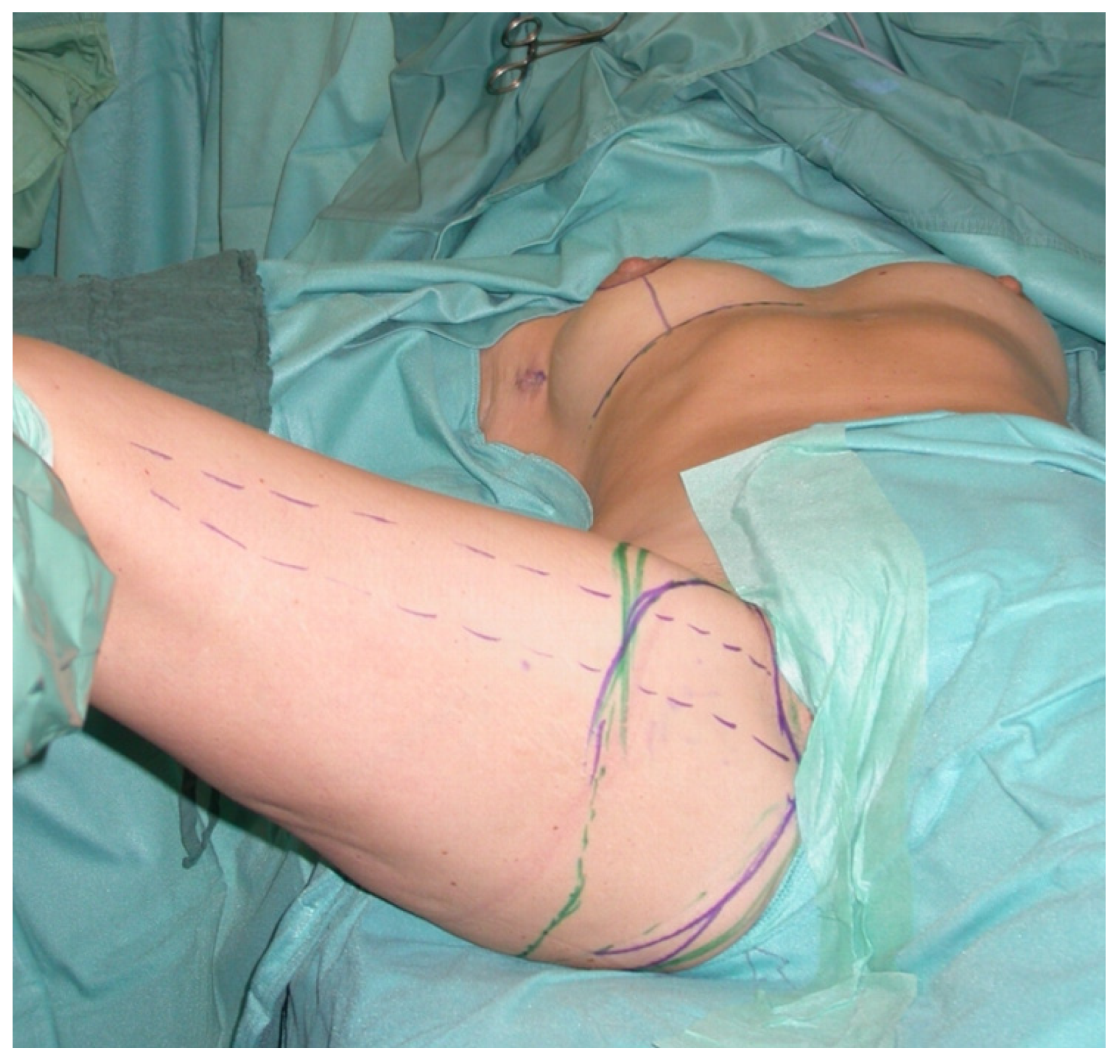
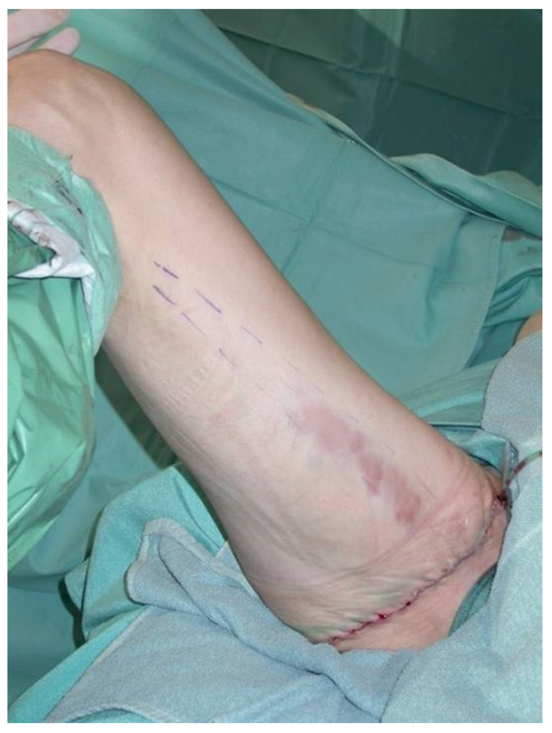
Shaping techniques as described by other authors [30] as demonstrated in Figure 9 and Figure 10 were used to increase the flap projection and improve the overall shape of the breast after reconstruction. We dismissed these time consuming shaping techniques today since they do not seem to remain stable over time and flap inset can be more difficult using them in some instances due to the short pedicle. Therefore, we most often place the skin island in the lower pole and the muscle in the upper and lateral quadrant of the breast. The gracilis is then anchored to the pectoralis muscle using one or two strong resorbable sutures.
A noteworthy drawback of the TMG flap is the darker and sometimes mismatching colour of the skin island from the medial upper thigh which sometimes can include pubic hair. This can be an issue in secondary breast reconstructions in fair-skinned individuals and may need laser treatment postoperatively for hair removal and skin lightening/bleaching treatments [31].
In cases where larger flaps with skin islands greater than 10 × 30 cm and more than the average 250 to 300 g are needed for breast reconstruction an extended version of the TMG flap can be raised [19]. In few cases (n = 29) two TMG flaps were utilized for unilateral breast reconstruction by the senior authors when one flap did not offer enough volume [19]. Other authors described hybrid breast reconstructions using TMG flaps and a silicone implant instead when more volume and projection was needed [32].
In a study from 2018, the senior authors were able to demonstrate that the TMG flap offers a reliable scaffold for postoperative fat transfer if indicated. Here overall 83 out of 139 patients (59%) received postoperative Lipofilling to achieve better symmetry [33]. A similar percentage (54%; 163/300) of patients received lipofilling in the present study collective. In a study including 408 DIEP flaps and 56 LAP flaps, Opsomer et al. stated the need for later lipofilling to be 39% in LAP and 64% in their DIEP flaps [34]. The exact numbers regarding the indication of secondary surgeries including lipofilling are highly variable and depend on multiple factors though such as patient satisfaction, demand, and the individual local health care provider. A recent study demonstrated that increased age and overweight are no contraindications to breast reconstructions with the TMG flap and did not cause any increase of complications [35].
Compared to other popular and most commonly used alternatives to the DIEP flap for autologous breast reconstruction such as the lumbar artery perforator (LAP) flap, the superior gluteal artery perforator (SGAP) flap or the fasciocutaneous infragluteal free flap (FCI) flap, the TMG flap offers a comparable size and a similar complication profile, the mean pedicle length is significantly shorter though [36,37,38]. An overview of the common used free flaps and their detailed characteristics is shown in Table 5 [21,23,34,39,40].
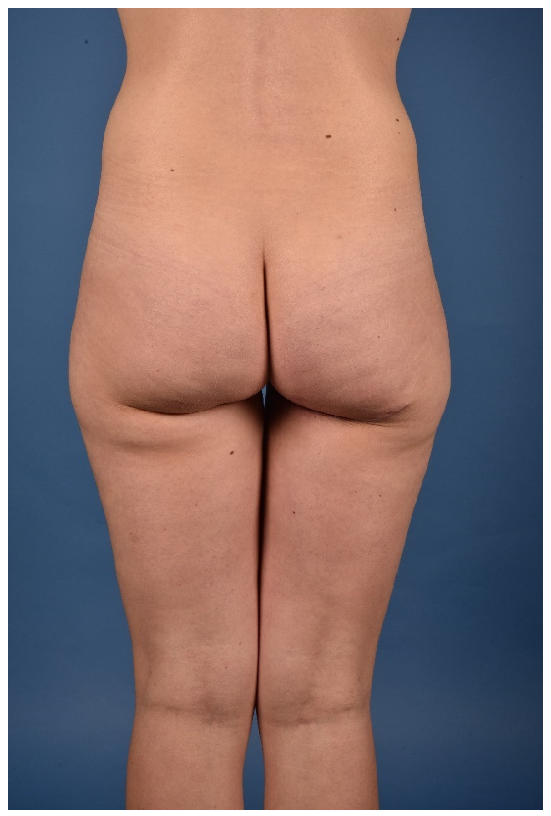
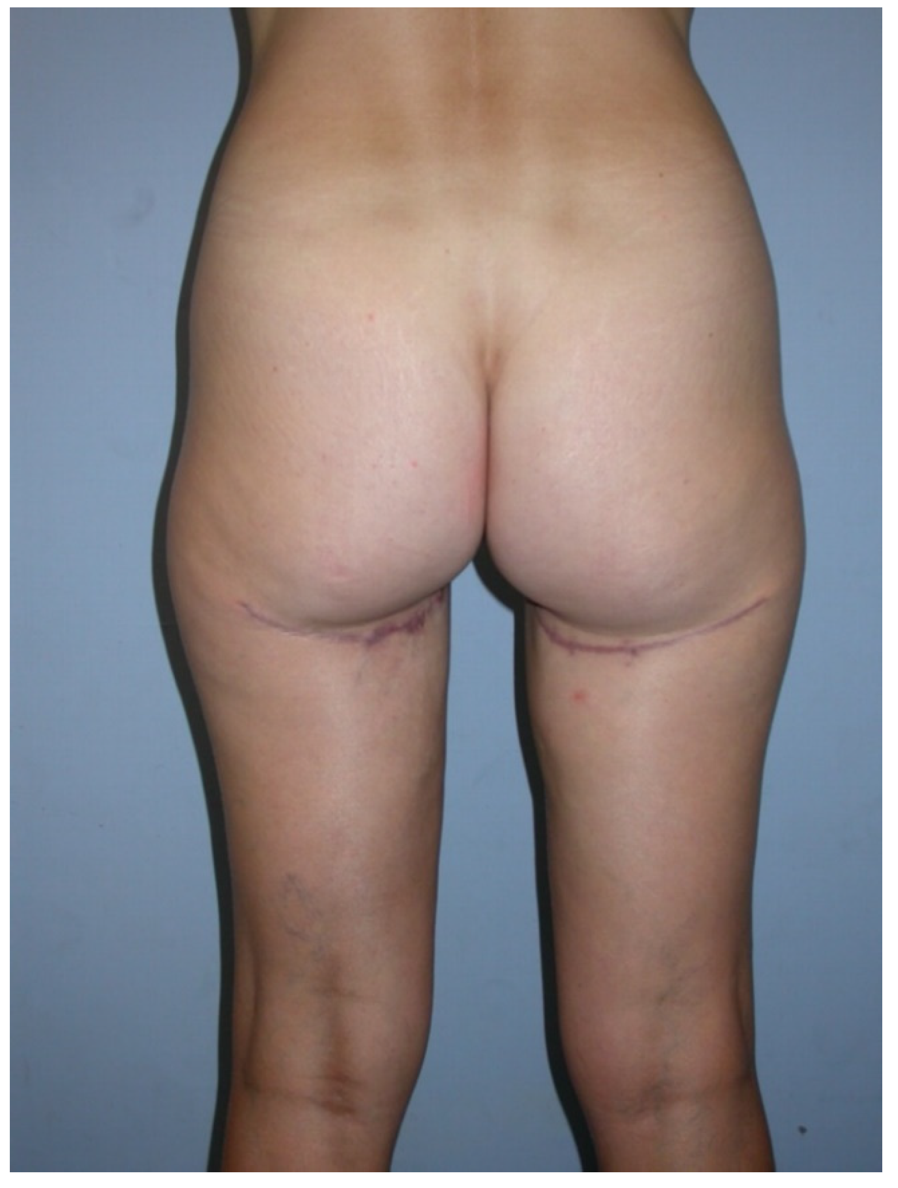
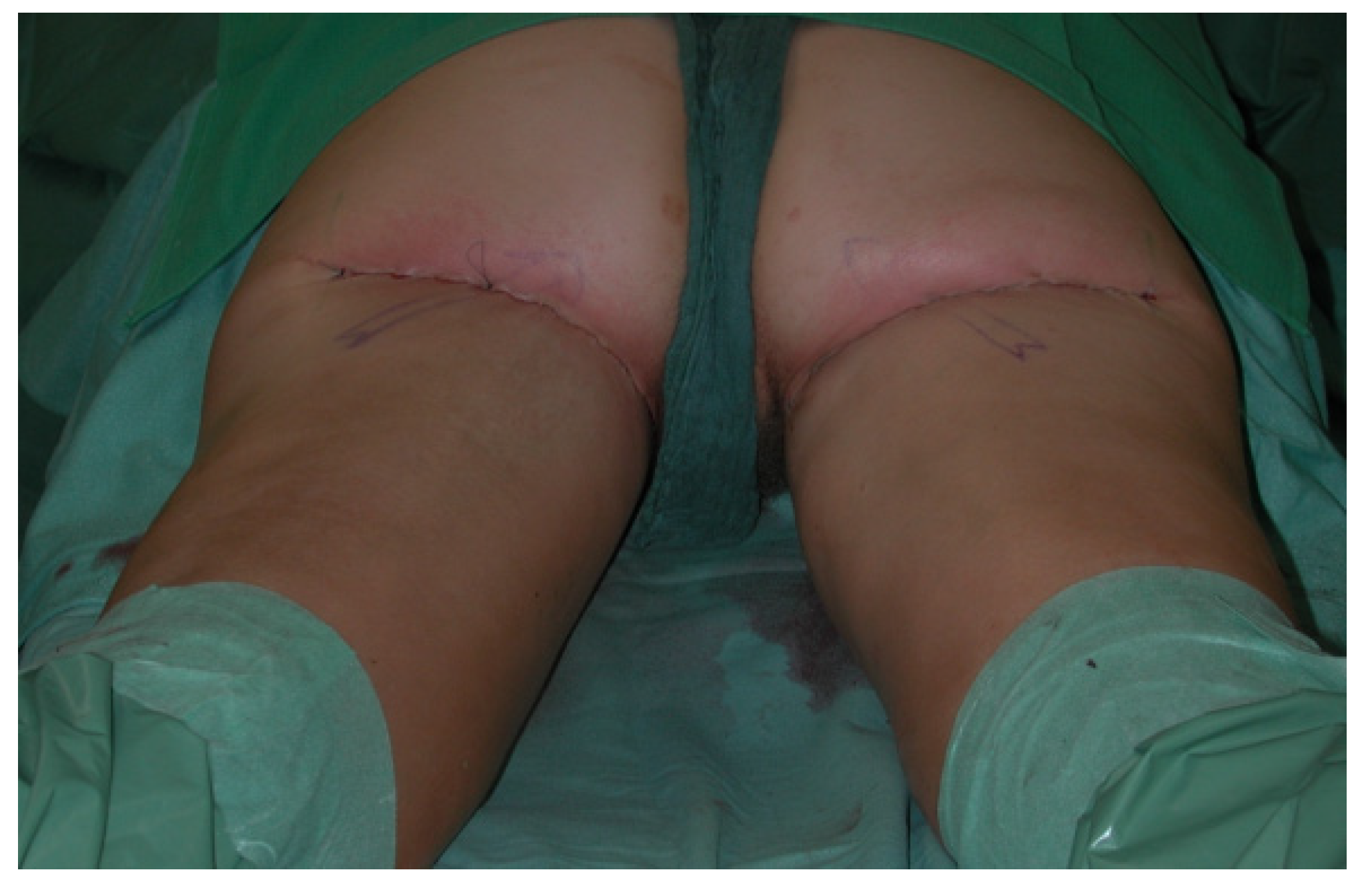
Other observed donor site complications include a widening and caudal migration of the scar which can easily be corrected with scar revision and suture fixation to the deep fascia or the periost of the ischial tuberosity. A scar revision of the donor site with refixation of the gluteal fold was performed in 3/300 (1%) patients, as demonstrated in Figure 11, Figure 12, Figure 13, Figure 14 and Figure 15.
A complication sometimes observed after FCI flap harvest is post-operative cluneal nerve pain which was entirely absent in our series of 300 TMG flap donor sites [40]. This complication was observed in the early beginnings of the TMG flap raises by the senior authors but can be avoided when the soft tissue part of the flap posterior to the gracilis muscle is in a plane strictly superficial to the deep fascia. In the last three decades the TMG flap has been used for all types of breast reconstruction. It was used for uni- and bilateral primary, secondary and tertiary reconstructive procedures of the breast [6,11,41]. As described earlier, in rare instances where a larger unilateral breast reconstruction is anticipated two TMG flaps can be used when one flap is anastomosed to the mammary while the other the thoracodorsal artery and vein [42].
In massive weight loss patients who had a failed breast augmentation with implants or recurrent capsular fibrosis and ptosis the TMG flap represents a unique source for a bilateral autologous tertiary breast reconstruction while simultaneously addressing the excess skin in a similar fashion as a thigh lift procedure [4].
An important limitation of this study and the present literature in general is the fact that no patient reported outcomes (PROMs) after breast reconstructions with the TMG flap have been evaluated and published yet. More studies focussing on PROMs are needed for a meaningful and wholesome comparison of different free flaps used for breast reconstruction using the Breast-Q or similar questionnaires [43].
In summary, we have learned many lessons during the last 30 years of breast reconstructions using the TMG flap. Itemized key points are listed in Table 6 for a better overview. Safe and reliable uni-, and bilateral breast reconstructions with a low risk for donor site complications can be offered to patients using the TMG flap today. This flap rightly represents one of the most popular and widely offered alternatives to the DIEP flap. We hope that reconstructive surgeons worldwide will maintain this tradition and will implement the TMG flap into their armamentarium for breast reconstructions in the future, thereby offering a high and up to date standard of care and at the same time further improving the availability for breast reconstruction for all patients regardless of their body shape or body mass index and donor site characteristic.

Author Contributions

Conceptualization, L.W. and T.S.; methodology, K.S.; software, K.B.; validation, M.M., K.S. and L.W.; formal analysis, G.W.; investigation, L.W.; resources, K.B.; data curation, K.B., E.R.; writing—original draft preparation, L.W.; writing—review and editing, K.S., E.R.; visualization, M.M.; supervision, G.W.; project administration, T.S.; funding acquisition, T.S. All authors have read and agreed to the published version of the manuscript.

Funding

This research received no external funding.

Institutional Review Board Statement

The study was conducted according to the guidelines of the Declaration of Helsinki, and approved by the Institutional Ethics Review Board of the Marienhospital Stuttgart.

Informed Consent Statement

Informed consent was obtained from all subjects involved in the study.

Conflicts of Interest

None of the authors has a financial interest in any of the products, devices, or drugs mentioned in this manuscript.

References

  1. Yousif, N.J. The Transverse Gracilis Musculocutaneous Flap. Ann. Plast. Surg. 1993, 31, 382. [Google Scholar] [CrossRef]
  2. Core, G.B.; Weimar, R.; Meland, N.B. The Turbo Gracilis Myocutaneous Flap. J. Reconstr. Microsurg. 1992, 8, 267–275. [Google Scholar] [CrossRef] [PubMed]
  3. Giordano, P.A.; Abbes, M.; Pequignot, J. Gracilis blood supply: Anatomical and clinical reevaluation. Br. J. Plast. Surg. 1990, 43, 266–272. [Google Scholar] [CrossRef]
  4. Schoeller, T.; Meirer, R.; Otto-Schoeller, A.; Wechselberger, G.; Piza-Katzer, H. Medial thigh lift free flap for autologous breast augmentation after bariatric surgery. Obes. Surg. 2002, 12, 831–834. [Google Scholar] [CrossRef]
  5. Wechselberger, G.; Schoeller, T. The transverse myocutaneous gracilis free flap: A valuable tissue source in autologous breast reconstruction. Plast. Reconstr. Surg. 2004, 114, 69–73. [Google Scholar] [CrossRef]
  6. Wechselberger, G.; Schoeller, T.; Bauer, T.; Schwabegger, A.; Ninkovic, M.; Rainer, C.; Ninkovic, M. Surgical technique and clinical application of the transverse gracilismyocutaneous free flap. Br. J. Plast. Surg. 2001, 54, 423–427. [Google Scholar] [CrossRef]
  7. Arnez, Z.M.; Pogorelec, D.; Planinsek, F.; Ahcan, U. Breast reconstruction by the free transverse gracilis (TUG) flap. Br. J. Plast. Surg. 2004, 57, 20–26. [Google Scholar] [CrossRef]
  8. Bodin, F.; Dissaux, C.; Dupret-Bories, A.; Schohn, T.; Fiquet, C.; Bruant-Rodier, C. The transverse musculo-cutaneous gracilis flap for breast reconstruction: How to avoid complications. Microsurgery 2016, 36, 42–48. [Google Scholar] [CrossRef]
  9. Fansa, H.; Schirmer, S.; Warnecke, I.C.; Cervelli, A.; Frerichs, O. The Transverse Myocutaneous Gracilis Muscle Flap: A Fast and Reliable Method for Breast Reconstruction. Plast. Reconstr. Surg. 2008, 122, 1326–1333. [Google Scholar] [CrossRef]
  10. Vega, S.J.; Sandeen, S.N.; Bossert, R.P.; Perrone, A.; Ortiz, L.; Herrera, H. Gracilis Myocutaneous Free Flap in Autologous Breast Reconstruction. Plast. Reconstr. Surg. 2009, 124, 1400–1409. [Google Scholar] [CrossRef]
  11. Wechselberger, G.; Traintinger, H.; Larcher, L.; Russe, E.; Edelbauer, M.; Ensat, F. Clinical Applications of the Transverse Musculocutaneous Gracilis Flap for Secondary Breast Reconstruction after Simple Mastectomy. Plast. Reconstr. Surg. 2016, 137, 19–28. [Google Scholar] [CrossRef]
  12. Haddock, N.T.; Greaney, P.; Otterburn, D.; Levine, S.; Allen, R.J. Predicting perforator location on preoperative imaging for the profunda artery perforator flap. Microsurgery 2012, 32, 507–511. [Google Scholar] [CrossRef]
  13. Saad, A.; Sadeghi, A.; Allen, R.J. The Anatomic Basis of the Profunda Femoris Artery Perforator Flap: A New Option for Autologous Breast Reconstruction—A Cadaveric and Computer Tomography Angiogram Study. J. Reconstr. Microsurg. 2012, 28, 381–386. [Google Scholar] [CrossRef]
  14. Neaman, K.C.; Armstrong, S.D.; Baca, M.E.; Albert, M.; Vander Woude, D.L.; Renucci, J.D. Outcomes of traditional cosmetic abdominoplasty in a community setting: A retrospective analysis of 1008 patients. Plast. Reconstr. Surg. 2013, 131, 403e–410e. [Google Scholar] [CrossRef] [Green Version]
  15. Wechselberger, G.; Schwaiger, K. Transverse Upper Gracilis Flap in Breast Reconstruction. In Breast Reconstruction: Modern and Promising Surgical Techniques; Mayer, H.F., Ed.; Springer International Publishing: Cham, Germany, 2020; pp. 253–260. [Google Scholar]
  16. Hallock, G.G. Further experience with the medial circumflex femoral (GRACILIS) perforator free flap. J. Reconstr. Microsurg. 2004, 20, 115–122. [Google Scholar]
  17. Huemer, G.M. TMG or TUG-Who is the winner? A PLEA for a uniform denomination. Microsurgery 2013, 33, 331–332. [Google Scholar] [CrossRef]
  18. Holm, C.; Mayr, M.; Höfter, E.; Ninkovic, M. Perfusion Zones of the DIEP Flap Revisited: A Clinical Study. Plast. Reconstr. Surg. 2006, 117, 37–43. [Google Scholar] [CrossRef] [Green Version]
  19. Wong, C.; Mojallal, A.; Bailey, S.H.; Trussler, A.; Saint-Cyr, M. The extended transverse musculocutaneous gracilis flap: Vascular anatomy and clinical implications. Ann. Plast. Surg. 2011, 67, 170–177. [Google Scholar] [CrossRef]
  20. Pignatti, M.; Pinto, V.; Giorgini, F.A.; Lozano Miralles, M.E.; D’Arpa, S.; Cipriani, R.; De Santis, G. Different Hydraulic Constructs to Optimize the Venous Drainage of DIEP Flaps in Breast Reconstruction: Decisional Algorithm and Review of the Literature. J. Reconstr. Microsurg. 2021, 37, 216–226. [Google Scholar]
  21. Blondeel, P.N. One hundred free DIEP flap breast reconstructions: A personal experience. Br. J. Plast. Surg. 1999, 52, 104–111. [Google Scholar] [CrossRef]
  22. Hunter, J.E.; Lardi, A.M.; Dower, D.R.; Farhadi, J. Evolution from the TUG to PAP flap for breast reconstruction: Comparison and refinements of technique. J. Plast. Reconstr. Aesthet. Surg. 2015, 68, 960–965. [Google Scholar] [CrossRef]
  23. Haddock, N.T.; Teotia, S.S. Consecutive 265 Profunda Artery Perforator Flaps: Refinements, Satisfaction, and Functional Outcomes. Plast. Reconstr. Surg. Glob Open 2020, 8, e2682. [Google Scholar] [CrossRef]
  24. Yim, J.H.; Yun, J.; Lee, T.J.; Kim, E.K.; Cho, J.; Eom, J.S. Outcomes of Take-Back Operations in Breast Reconstruction with Free Lower Abdominal Flaps. Arch. Plast. Surg. 2015, 42, 741–745. [Google Scholar] [CrossRef] [Green Version]
  25. Weitgasser, L.; Schwaiger, K.; Medved, F.; Hamler, F.; Wechselberger, G.; Schoeller, T. Bilateral Simultaneous Breast Reconstruction with DIEP- and TMG Flaps: Head to Head Comparison, Risk and Complication Analysis. J. Clin. Med. 2020, 9, 2031. [Google Scholar] [CrossRef]
  26. Allen, R.J., Jr.; Lee, Z.-H.; Mayo, J.L.; Levine, J.; Ahn, C.; Allen, R.J., Sr. The Profunda Artery Perforator Flap Experience for Breast Reconstruction. Plast. Reconstr. Surg. 2016, 138, 968–975. [Google Scholar] [CrossRef]
  27. Espinosa-De-Los-Monteros, A.; Frias-Frias, R.; Alvarez-Tostado-Rivera, A.; Caralampio-Castro, A.; Llanes, S.; Saldivar, A. Postoperative Abdominal Bulge and Hernia Rates in Patients Undergoing Abdominally Based Autologous Breast Reconstruction: Systematic Review and Meta-Analysis. Ann. Plast. Surg. 2021, 86, 476–484. [Google Scholar] [CrossRef]
  28. Marsh, D.; Patel, N.G.; Rozen, W.M.; Chowdhry, M.; Sharma, H.; Ramakrishnan, V.V. Three routine free flaps per day in a single operating theatre: Principles of a process mapping approach to improving surgical efficiency. Gland Surg. 2015, 5, 107–114. [Google Scholar] [CrossRef]
  29. Dayan, E.; Smith, M.L.; Sultan, M.; Samson, W.; Dayan, J.H. The Diagonal Upper Gracilis (DUG) Flap: A Safe and Improved Alternative to the TUG Flap. Plast. Reconstr. Surg. 2013, 132, 33–34. [Google Scholar] [CrossRef]
  30. Nickl, S.; Nedomansky, J.; Radtke, C.; Haslik, W.; Schroegendorfer, K.F. Optimization of breast reconstruction results using TMG flap in 30 cases: Evaluation of several refinements addressing flap design, shaping techniques, and reduction of donor site morbidity. Microsurgery 2018, 38, 489–497. [Google Scholar] [CrossRef]
  31. Schoeller, T.; Huemer, G.M.; Wechselberger, G. The transverse musculocutaneous gracilis flap for breast reconstruction: Guidelines for flap and patient selection. Plast. Reconstr. Surg. 2008, 122, 29–38. [Google Scholar] [CrossRef]
  32. Bach, A.D.; Morgenstern, I.H.; Horch, R.E. Secondary Hybrid Reconstruction Concept with Silicone Implants After Autologous Breast—Is It Safe and Reasonable? Med. Sci. Monit. 2020, 26, e921329. [Google Scholar] [CrossRef]
  33. Russe, E.; Kholosy, H.; Weitgasser, L.; Brandstetter, M.; Traintinger, H.; Neureiter, J.; Wechselberger, G.; Schoeller, T. Autologous fat grafting for enhancement of breast reconstruction with a transverse myocutaneous gracilis flap: A cohort study. J. Plast. Reconstr. Aesthet. Surg. 2018, 71, 1557–1562. [Google Scholar] [CrossRef]
  34. Opsomer, D.; Vyncke, T.; Depypere, B.; Stillaert, F.; Blondeel, P.; Van Landuyt, K. Lumbar Flap versus the Gold Standard: Comparison to the DIEP Flap. Plast. Reconstr. Surg. 2020, 145, 706e–714e. [Google Scholar] [CrossRef]
  35. Schwaiger, K.; Weitgasser, L.; Mahrhofer, M.; Bachleitner, K.; Abed, S.; Wimbauer, J.; Russe, E.; Scholler, T.; Wechselberger, G. Age and Overweight Are Not Contraindications for a Breast Reconstruction with a TMG-Flap-A Risk and Complication Analysis of a Retrospective Double Center Study Including 300 Patients. J. Clin. Med. 2021, 10, 926. [Google Scholar] [CrossRef]
  36. Guerra, A.B.; Metzinger, S.E.; Bidros, R.S.; Gill, P.S.; Dupin, C.L.; Allen, R.J. Breast reconstruction with gluteal artery perforator (GAP) flaps: A critical analysis of 142 cases. Ann. Plast. Surg. 2004, 52, 118–125. [Google Scholar] [CrossRef] [PubMed]
  37. Myers, P.L.; Nelson, J.A.; Allen, R.J., Jr. Alternative flaps in autologous breast reconstruction. Gland Surg. 2021, 10, 444–459. [Google Scholar] [CrossRef] [PubMed]
  38. Struckmann, V.; Peek, A.; Wingenbach, O.; Harhaus, L.; Kneser, U.; Holle, G. The free fasciocutaneous infragluteal (FCI) flap: Outcome and patient satisfaction after 142 breast reconstructions. J. Plast. Reconstr. Aesthet. Surg. 2016, 69, 461–469. [Google Scholar] [CrossRef] [PubMed]
  39. Papp, C.; Windhofer, C.; Michlits, W. Autologous breast augmentation with the deepithelialized fasciocutaneous infragluteal free flap: A 10-year experience. Ann. Plast. Surg. 2011, 66, 587–592. [Google Scholar] [CrossRef]
  40. Zoccali, G.; Mughal, M.; Giwa, L.; Roblin, P.; Farhadi, J. Breast reconstruction with Superior Gluteal Artery Perforator free flap: 8 years of experience. J. Plast. Reconstr. Aesthet. Surg. 2019, 72, 1623–1631. [Google Scholar] [CrossRef]
  41. Pülzl, P.; Huemer, G.M.; Schoeller, T. Transverse musculocutaneous gracilis flap for treatment of capsular contracture in tertiary breast reconstruction. Ann. Plast. Surg. 2015, 74, 167–172. [Google Scholar] [CrossRef]
  42. Werdin, F.; Haug, D.M.; Amr, A.; Schoeller, T. Double transverse myocutaneous gracilis free flaps for unilateral breast reconstruction. Microsurgery 2016, 36, 539–545. [Google Scholar] [CrossRef] [PubMed]
  43. Pusic, A.L.; Chen, C.M.; Cano, S.; Klassen, A.; McCarthy, C.; Collins, E.D.; Cordeiro, P.G. Measuring Quality of Life in Cosmetic and Reconstructive Breast Surgery: A Systematic Review of Patient-Reported Outcomes Instruments. Plast. Reconstr. Surg. 2007, 120, 823–837. [Google Scholar] [CrossRef] [PubMed]
Figure 1. Intraoperative image at the beginning of the operation.
Jcm 10 03629 g001
Figure 2. Intraoperative image after unilateral skin sparing mastectomy.
Jcm 10 03629 g002
Figure 3. Intraoperative image after unilateral TMG flap raise.
Jcm 10 03629 g003
Figure 4. Intraoperative image after unilateral donor site closure.
Jcm 10 03629 g004
Figure 5. Preoperative image of a BRCA patient.
Jcm 10 03629 g005
Figure 6. Postoperative image after two-staged bilateral autologous breast reconstruction with free TMG flaps.
Jcm 10 03629 g006
Figure 7. Postoperative donor site image.
Jcm 10 03629 g007
Figure 8. Transverse myocutaneous gracilis (TMG) flap publication trends between 2004 and today.
Jcm 10 03629 g008
Figure 9. TMG flap before shaping (blue arrows indicate folding of the flap).
Jcm 10 03629 g009
Figure 10. TMG flap after folding to generate an implant like flap shape.
Jcm 10 03629 g010
Figure 11. Post-operative caudal scar migration.
Jcm 10 03629 g011
Figure 12. Deepithelialising of the scar for refixation.
Jcm 10 03629 g012
Figure 13. Underminig and anchoring to the ischial tuberosity or deep fascia using resorbable sutures.
Jcm 10 03629 g013
Figure 14. Skin closure after scar revision.
Jcm 10 03629 g014
Figure 15. Post-operative result after scar revision with refixation of the gluteal fold.
Jcm 10 03629 g015
Table 1. Classification of Complications (modified from Neaman et al. [14]).
Major complications1. Hematoma or flap insufficiency requiring surgical intervention
2. Seroma requiring aspiration or surgery
3. Wound healing problems (also flap or fat necrosis) requiring surgery
4. Cellulitis requiring iv antibiotics
5. Deep Venous Thrombosis/Pulmonary Embolism
Minor complications1. Hematoma without treatment (+erythrocyte substitution with no other treatment necessary)
2. Seroma without treatment
3. Delayed wound healing
4. Cellulitis (also fat necrosis) treated with oral antibiotics without hospitalization
Table 2. Detailed patient characteristics.
CharacteristicNumber %
Cases included300 100
Age, years
   Mean 48.0
   SD 10.6
Follow-Up, months
   Mean 21.4
   SD 20.9
Lipofilling165 55.0
Preoperative radiotherapy116 38.6
Type of reconstruction
   Primary58 19.3
   Secondary124 41.3
   Tertiary118 39.4
Body Mass Index
   Mean 23.0
   SD 3.1
Table 3. Flap characteristics.
Flap CharacteristicMeanRangen%
Flap weight, gram320155–600
Skin island diameter, centimeter
   width97–13
   length3125–36
Venous coupler, millimeter2.51.5–3.5
Pedicle length, millimeter7043–110
Anastomosis
   Internal mammary (Artery /Vein) 27692.0
   Thoracodorsal (Artery/Vein) 248.0
Table 4. Complications overview; donor site, recipient site.
Complicationsn%
Overall14749.0
   Major8327.0
   Minor7023.0
Donor site complications
   Cellulitis103.3
   Hematoseroma217.0
   Wound healing disturbance289.3
   Scar revision31.0
Recipient site complications
   Cellulitis165.3
   Hematoseroma4414.7
   Wound healing disturbance165.3
   Fat necrosis155.0
Flap take-back4214.0
Flap loss196.3
Table 5. Overview of the common used free flaps for breast reconstruction and their properties.
FlapWeight (Mean, gram) Pedicle Length (Mean, millimeter) Source (PubMed)
TMG320 g70 mm Weitgasser et al., 2021
DIEP550 g150 mmBlondeel et al., 1999 [38]
PAP403 g112 mmHaddock et al., 2020 [23]
SGAP400 g85 mmZoccali et al., 2019 [39]
FCI 310 g150 mm Papp et al., 2011 [40]
LAP499 g40 mmOpsomer et al., 2020 [37]
Table 6. Overview of key lessons learned in the last 30 years of TMG flap breast reconstruction.
Flap characteristics320 g (155–600 g range)Skin island diameter: 9 cm (7–13 cm range) in width and 31 cm (25 to 36 cm range) in lengthPedicle length: 70 mm (43 mm to 110 mm range)
Indications for surgeryQualified for primary, secondary and tertiary breast reconstructions Can be used relatively independent of BMI and body shape
AnatomyConstant and reliable, no Computer Tomography (CT) Angio necessary for planningSupercharging with the saphenous vein is possible but is rarely necessaryFlap is raised without repositioning
Flap shapingDo not offer long term form stability and can make flap inset more difficult Are time consumingSkin island is usually placed in the lower breast pole and the muscle in the upper pole
Donor siteLymphatic complications are uncommonDonor site morbidity is comparable to other flapsDehiscence and wound break down is easy to manage, most often conservativelyNegative pressure wound therapy (NPWT) and skin grafting is rarely necessary
Color missmatchCan be an issue in secondary reconstructions where local breast skin is replacedLaser treatment and skin lightening or bleaching procedures can be offered
Widening of the donor site scarNot uncommon, especially in larger flaps and higher tenson on wound closure Can easily be corrected by anchoring the revised scar to the ischial tuberosity or deep fascia
Cluneal nerve painOccurs rarely and can be avoided through a dissection in a plane superficially to the deep fascia posterior to the gracilis
Large breast reconstructionsExternded TMG flap can be usedTwo flaps can be used for anastoming to the mammary and thoracodorsal vessels Lipofilling procedures are a powerful tool for volume adjustment
Publisher’s Note: MDPI stays neutral with regard to jurisdictional claims in published maps and institutional affiliations.

