Abstract
Ankle osteoarthritis affects 1% of the population and, unlike gonarthrosis or coxarthrosis, is secondary to previous trauma in more than 75% of cases. Another peculiarity of this disease is that it affects a younger and active population, with socio-occupational implications. Mechanical factors, such as incongruity, instability, malalignment, and impacts, which increase stress on isolated areas of the ankle cartilage, have been clearly associated with the development of osteoarthritis. However, we cannot ignore the importance of pro-inflammatory mediators present from the moment of fracture as triggers of the cascade that eventually causes chondrocyte cell death, ultimately responsible for ankle osteoarthritis.
1. Introduction
Ankle osteoarthritis is a chronic disease affecting approximately 1% of the population, with an estimated prevalence of 30 cases per 100,000 inhabitants, and corresponds to between 2% and 4% of all patients with osteoarthritis (OA) [1,2]. Cadaver, radiology, and clinical studies indicate that ankle osteoarthritis is less common than knee and hip OA. This is reflected in clinical practice as symptomatic knee osteoarthritis is 8 to 9 times more prevalent than ankle osteoarthritis, and approximately 24 times more total knee work is performed than arthrodesis and arthroplasty combined [3,4]. While its clinical impact on patients has not been considered particularly relevant, ankle OA is very debilitating in advanced stages and can have similar repercussions on quality of life as severe hip osteoarthritis, advanced kidney failure, or congestive heart failure [4,5].
There is scant scientific literature available regarding the aetiology of ankle OA; however, there is an apparent consensus that, unlike hip and knee OA, primary ankle osteoarthritis is not the most common aetiology [1,6,7]; only 7% to 9% of cases are classed as idiopathic osteoarthritis and 13% are secondary to other causes (rheumatoid arthritis, haemochromatosis, haemophilia, and osteonecrosis). Therefore, the main aetiology, representing 75% to 80% of all cases, is a traumatic event, with fractures around the ankle (malleolus, distal tibia, talus, and so on) (Figure 1) responsible for 62%, and a further 16% are the result of unresolved chronic ligament injuries, particularly those affecting the lateral collateral ligament (which some authors call “ligamentary ankle osteoarthritis”) [7,8]. Given its predominantly post-traumatic aetiology, patients with ankle OA tend to be younger (18–44 years) than those with other degenerative joint diseases in the lower limbs. They also suffer a more rapid loss of function, with progression to advanced stages of ankle OA occurring 10 to 20 years of onset, although clinical studies show that the initial degenerative changes secondary to an ankle fracture develop within 12 to 18 months of the traumatic injury [7,9].
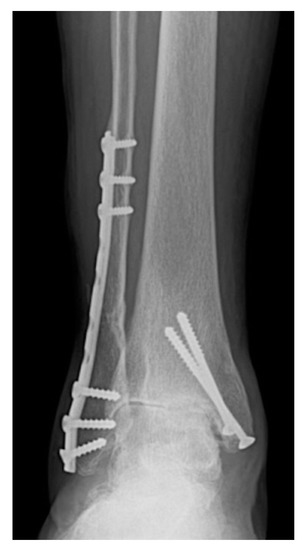
Figure 1.
Advanced symmetrical ankle OA secondary to ankle fracture dislocation surgically treated 8 years previously.
The present work sets out to describe the main causes of the onset of ankle osteoarthritis.
2. Pathophysiology: Peculiarities of Tibiotalar Cartilage
There is a lack of literature covering experimental work around ankle OA; however, some studies have furthered our knowledge of the pathogenesis of joint deterioration [9,10]. It is noteworthy that, despite being a frequently injured joint (fractures, sprains, and so on), clinically relevant ankle OA is less common than in other weight-bearing joints, which can undoubtedly be explained by the anatomical, biochemical, and biomolecular peculiarities of ankle cartilage [9]. Ankle cartilage receives the greatest force per unit area of all hyaline cartilage in the human body (500 N/350 mm2 compared with the same force per 1100 mm2 or 1120 mm2 in the hip or knee, respectively). Thus, ankle cartilage can be subject to three times greater force, yet, as mentioned previously, radiological, and clinical osteoarthritis is less common than in the knee or hip. Furthermore, the load distribution in the ankle differs from in other joints, such as the knee, which means the compressive forces are distributed over a greater area. Ankle cartilage (1–1.62 mm) is thus thinner than knee cartilage (1.69–2.55 mm) [10,11,12]. Biologically, ankle cartilage is believed to have a greater capacity for self-repair than knee cartilage [13]. It is stiffer and less permeable because it produces more water and proteoglycan [9]; in addition, the extracellular matrix improves its load-bearing capacity and reduces its susceptibility to mechanical damage. What is more, chondrocytes in the ankle are metabolically more active than in the knee and exhibit a greater response to anabolic factors influencing cartilage synthesis such as levels of osteogenic protein-1 and C-propeptide of type II collagen. They are also less sensitive to catabolic mediators such as fibronectin and interleukin-1 beta, which inhibit collagen synthesis [10,11,12,13]. Finally, the concentration of matrix metalloproteinases (MMPs), the enzymes responsible for protein degradation in the extracellular matrix, is known to vary between different joints; for instance, MMP-8 levels are significant in knee cartilage, but undetectable in the ankle [13]. Given all these reasons, ankle cartilage is less prone to joint degeneration than that of the knee or hip, but it is highly susceptible to lesions when there is an asymmetric distribution of stresses and forces, as in the case of joint fractures, injuries caused by impacts, or misalignment of the weight-bearing axis [9,14]. Such factors may explain the high correlation between ankle OA and a history of a traumatic event.
3. Predisposing Factors for Ankle Osteoarthritis
As mentioned above, most cases of ankle osteoarthritis are secondary to fractures or chronic, unresolved instabilities. Here, we discuss the various factors involved in the development of ankle OA.
3.1. Microscopic Cartilage Damage
Circumstances that overload ankle cartilage, either a one-off event (acute impact) or repetitive stress (cumulative contact stress), are known to be a potential cause of clinical osteoarthritis. In 2013, Buckwalter et al. stated that a chronic increase in the mechanical stress on cartilage also causes post-traumatic OA [15]. The proposed mechanism involves an increase in oxidative stress following the release of mitochondria from chondrocytes that are subject to greater contact stress from reactive oxygen species (ROS); mediators of cell damage (chondrocyte death) and extracellular matrix degeneration. Fibronectin fragments released from joint cartilage subjected to excessive loads also promote matrix degradation; inhibition of molecular pathways initiated by these fragments prevents this effect. Additionally, injured chondrocytes release alarmins that activate chondroprogenitor cells, which in turn propagate and migrate to areas of damaged cartilage [13,15]. These cells also release chemokines and cytokines that may contribute to inflammation, which causes progressive cartilage loss. In 2011, Tochigi et al. published an interesting article based on the analysis of cell death in seven ankle specimens collected from amputated patients [16]. They reported that chondrocyte apoptosis occurs mainly in the region of the fracture line, but even more noteworthy was how apoptosis spread spontaneously from the fracture line to apparently healthy areas in the 48 h after the trauma, suggesting that intra-articular mediators of cell damage are released from the injured chondrocytes, and they also affect the healthy chondrocytes.
3.2. Macroscopic Cartilage Damage
The direct relationship between osteochondral ankle lesions and future development of ankle OA is controversial; nevertheless, anterolateral talar, posteromedial tibial, and medial malleolar osteochondral injuries have the worst prognosis [5]. Several authors have reported the presence of occult or undiagnosed osteochondral lesions following ankle fractures [17,18], lesions that may explain why a long-term study found that up to 50% of patients with a fractured ankle had suboptimal functional outcomes in terms of persistent pain and a decline in activities of daily living [19]. A recent study observed a high rate of talar osteochondral lesions, both during the acute phase and after 12 months’ evolution (45% and 47%, respectively), among patients who had suffered an ankle fracture, particularly those caused by rotational mechanisms [20]. Consequently, the authors emphasized the need to document the lesions in the acute phase, where arthroscopy would play an essential role in diagnosis and treatment, as indicated in several other studies [17,18,20,21].
3.3. Joint Incongruence
It is evident that the presence of an intra-articular step-off favors the development of post-traumatic osteoarthritis in any joint, hence the importance of achieving anatomical reduction of the fracture [17]. This has been demonstrated by McKinley et al. [22], who created an intra-articular step-off in the distal tibia of 10 ankle specimens and observed a substantial increase of up to 300% in the peak contact stress, which could be decisive in the onset of post-traumatic osteoarthritis. Incongruence is closely related to instability; in fact, numerous studies have shown how instability in association with incongruence promotes cell damage to ankle cartilage. McKinley et al. also suggested that instability coupled with incongruence increases the contact stress per surface area by up to 60% in ankle cartilage compared with incongruence alone [23]. In the clinical set, several studies, including a review from 2018 by Verhage et al. [24], argue that postoperative step-off is the most important factor predicting post-traumatic osteoarthritis.
3.4. Instability
Instability itself increases the peak joint contact stress, resulting in cartilage deterioration. Authors such as Harrington have shown through radiological and arthroscopic follow-up that ankle instability inextricably leads to osteoarthritis [8]. However, apparent instability is not the only cause of joint damage. Tochigi et al. showed that so-called microinstability following a sprain, intra-articular fracture, or even ligament reconstruction surgery can create isolated peaks of stress in areas of cartilage that are known to lead to joint damage [14]. In clinical practice, many patients with osteoarthritis do not complain of manifest instability and already have advanced OA, so microinstability may play a key causative role in such cases. The term “ligamentary post-traumatic ankle osteoarthritis” (Figure 2) was coined by Valderrabano et al. [25] during an epidemiological study of the main characteristics of this type of ankle OA. It affected approximately 13% of their series, with less clinical impact and a longer mean latency time (34.3 years) than that observed for post-traumatic OA following a fracture. The authors showed, as did Hashimoto and Inokuchi [26], that most instabilities affect the lateral ligament complex, thus increasing stress on the medial joint surface owing to increased pronation and external rotation and producing varus ankle OA.
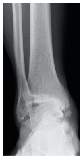
Figure 2.
”Ligamentary ankle OA”, advanced asymmetrical ankle OA secondary to chronic unsolved lateral instability.
3.5. Malalignment
Tibiotalar osteoarthritis is asymmetrical in almost 70% of cases [27]. In most patients, the deformity underlying the asymmetry is located close to the ankle joint, whether in the supramalleolar region or in the inferior peritalar (subtalar or calcaneal) complex—or both. A history of ankle fracture is very common, which ends up causing uneven load distribution owing to mal union, which, as previously mentioned, is very poorly tolerated by the talar cartilage [28]. Although the origin of most cases of asymmetric ankle arthropathy is close (supra or inframaleolar), it should not be forgotten that it must be explored at the proximal level because deformities at the level of the proximal tibia, knee, or distal femur could be the causes of an asymmetric load distribution on the ankle, which, in turn, results in asymmetric ankle osteoarthritis. In such cases, the problem originally manifesting in the ankle must be resolved surgically away from the ankle [29,30].
3.6. Proinflammatory Mediators in Synovial Fluid
Adams et al. reported an interesting finding about the presence of an ambient proinflammatory mediator and promoter of extracellular matrix degradation in the synovial fluid obtained perioperatively from patients undergoing osteosynthesis for an ankle fracture [13]. Interleukins IL-6 and IL-8 and matrix metalloproteinases MMP-2, 3, 9, and 10 were notable and may contribute to the development of post-traumatic OA. The same authors also reported how these proinflammatory factors were still elevated, compared with the healthy contralateral ankle, at 6 months post-surgery, highlighting that joint lavage at the time of fracture may act as a prophylactic [31]. In addition, Furman et al. [32] assure in their work that ankle joint fracture increases local inflammation, as indicated by increased synovitis, increased macrophage infiltration into synovial tissue, and increased synovial fluid concentrations of biomarkers on inflammation. These findings have opened a huge field of research on therapies based on inactivation of proinflammatory substances and cytokines in the treatment of osteoarthritis.
3.7. Ageing
Old age is a well-known risk factor for developing osteoarthritis [33,34]. On the other hand, molecular biology studies have shown that the presence of senescent cells in arthritic cartilage supports the idea that the cytokines produced by these cells play a part in the cartilage degeneration cascade that leads to osteoarthritis [35]. The waning capacity of joint cartilage to repair itself as we age is also a relevant factor [36]. Given these reasons, and as pointed out by Valderrabano et al. [2] and especially in the group with primary OA, patients are relatively older (58 years) at the time of diagnosis than those diagnosed with post-traumatic osteoarthritis.
4. Conclusions
Osteoarthritis of the ankle is secondary to a periarticular fracture in 75% of cases and chronic, unresolved ligament instability in around 13%. Mechanical factors, such as incongruence, instability, malalignment, and impacts, that increase the stress on isolated areas of ankle cartilage, have been clearly associated with the onset of osteoarthritis. However, we cannot ignore the importance of the proinflammatory mediators present from the moment of fracture as triggers of the cascade that eventually causes the cell damage responsible for osteoarthritis.
Author Contributions
Conceptualization, M.H.-P. and D.G.-M.; methodology, M.H.-P.; writing—original draft preparation, M.H.-P.; writing—review and editing, S.T., M.V.-M. and V.V.; supervision, M.H.-P., A.L.G.-S., and V.V.; project administration, M.H.-P. and S.T. All authors have read and agreed to the published version of the manuscript.
Funding
This research received no external funding.
Institutional Review Board Statement
Not applicable.
Informed Consent Statement
Not applicable.
Data Availability Statement
Not applicable.
Conflicts of Interest
The authors declare no conflict of interest.
References
- Goldberg, A.J.; MacGregor, A.; Dawson, J.; Singh, D.; Cullen, N.; Sharp, R.J.; Cooke, P.H. The demand incidence of symptomatic ankle osteoarthritis presenting to foot & ankle surgeons in the United Kingdom. Foot 2012, 22, 163–166. [Google Scholar] [CrossRef]
- Valderrabano, V.; Horisberger, M.; Russell, I.; Dougall, H.; Hintermann, B. Etiology of ankle osteoarthritis. Clin. Orthop. Relat. Res. 2009, 467, 1800–1806. [Google Scholar] [CrossRef] [PubMed] [Green Version]
- Thomas, R.H.; Daniels, T.R. Ankle arthritis. J. Bone Joint Surg. Am. 2003, 85, 923–936. [Google Scholar] [CrossRef]
- Glazebrook, M.; Daniels, T.; Younger, A.; Foote, C.J.; Penner, M.; Wing, K.; Lau, J.; Leighton, R.; Dunbar, M. Comparison of health-related quality of life between patients with end-stage ankle and hip arthrosis. J. Bone Joint Surg. Am. 2008, 90, 499–505. [Google Scholar] [CrossRef] [PubMed]
- Stufkens, S.A.; Knupp, M.; Horisberger, M.; Lampert, C.; Hintermann, B. Cartilage lesions and the development of osteoarthritis after internal fixation of ankle fractures: A prospective study. J. Bone Joint Surg. Am. 2010, 92, 279–286. [Google Scholar] [CrossRef] [Green Version]
- Horisberger, M.; Valderrabano, V.; Hintermann, B. Posttraumatic ankle osteoarthritis after ankle-related fractures. J. Orthop. Trauma 2009, 23, 60–67. [Google Scholar] [CrossRef]
- Saltzman, C.L.; Salamon, M.L.; Blanchard, G.M.; Huff, T.; Hayes, A.; Buckwalter, J.A.; Amendola, A. Epidemiology of ankle arthritis: Report of a consecutive series of 639 patients from a tertiary orthopaedic center. Iowa Orthop. J. 2005, 25, 44–46. [Google Scholar]
- Harrington, K.D. Degenerative arthritis of the ankle secondary to long-standing lateral ligament instability. J. Bone Joint Surg. Am. 1979, 61, 354–361. [Google Scholar] [CrossRef]
- Lindsjö, U. Operative treatment of ankle fractures. Acta Orthop. Scand Suppl. 1981, 189, 1–131. [Google Scholar] [CrossRef] [Green Version]
- Kraeutler, M.J.; Kaenkumchorn, T.; Pascual-Garrido, C.; Wimmer, M.A.; Chubinskaya, S. Peculiarities in Ankle Cartilage. Cartilage 2017, 8, 12–18. [Google Scholar] [CrossRef] [Green Version]
- Kuettner, K.E.; Cole, A.A. Cartilage degeneration in different human joints. Osteoarthr. Cartil. 2005, 13, 93–103. [Google Scholar] [CrossRef] [PubMed] [Green Version]
- Treppo, S.; Koepp, H.; Quan, E.C.; Cole, A.A.; Kuettner, K.E.; Grodzinsky, A.J. Comparison of biomechanical and biochemical properties of cartilage from human knee and ankle pairs. J. Orthop. Res. 2000, 18, 739–748. [Google Scholar] [CrossRef] [PubMed]
- Adams, S.B.; Setton, L.A.; Bell, R.D.; Easley, M.E.; Huebner, J.L.; Stabler, T.; Kraus, V.B.; Leimer, E.M.; Olson, S.A.; Nettles, D.L. Inflammatory Cytokines and Matrix Metalloproteinases in the Synovial Fluid After Intra-articular Ankle Fracture. Foot Ankle Int. 2015, 36, 1264–1271. [Google Scholar] [CrossRef] [PubMed]
- Tochigi, Y.; Rudert, M.J.; McKinley, T.O.; Pedersen, D.R.; Brown, T.D. Correlation of dynamic cartilage contact stress aberrations with severity of instability in ankle incongruity. J. Orthop. Res. 2008, 26, 1186–1193. [Google Scholar] [CrossRef] [Green Version]
- Buckwalter, J.A.; Anderson, D.D.; Brown, T.D.; Tochigi, Y.; Martin, J.A. The Roles of Mechanical Stresses in the Pathogenesis of Osteoarthritis: Implications for Treatment of Joint Injuries. Cartilage 2013, 4, 286–294. [Google Scholar] [CrossRef] [PubMed]
- Tochigi, Y.; Buckwalter, J.A.; Martin, J.A.; Hillis, S.L.; Zhang, P.; Vaseenon, T.; Lehman, A.D.; Brown, T.D. Distribution and progression of chondrocyte damage in a whole-organ model of human ankle intra-articular fracture. J. Bone Joint Surg. Am. 2011, 93, 533–539. [Google Scholar] [CrossRef] [PubMed] [Green Version]
- Day, G.A.; Swanson, C.E.; Hulcombe, B.G. Operative treatment of ankle fractures: A minimum ten-year follow-up. Foot Ankle Int. 2001, 22, 102–106. [Google Scholar] [CrossRef]
- Hintermann, B.; Regazzoni, P.; Lampert, C.; Stutz, G.; Gächter, A. Arthroscopic findings in acute fractures of the ankle. J. Bone Joint Surg. Br. 2000, 82, 345–351. [Google Scholar] [CrossRef]
- Nilsson, G.M.; Jonsson, K.; Ekdahl, C.S.; Eneroth, M. Unsatisfactory outcome following surgical intervention of ankle fractures. Foot Ankle Surg. 2005, 11, 11–16. [Google Scholar] [CrossRef]
- Martijn, H.A.; Lambers, K.T.A.; Dahmen, J.; Stufkens, S.A.S.; Kerkhoffs, G.M.M.J. High incidence of (osteo)chondral lesions in ankle fractures. Knee Surg. Sports Traumatol. Arthrosc. 2021, 29, 1523–1534. [Google Scholar] [CrossRef]
- Toth, M.J.; Yoon, R.S.; Liporace, F.A.; Koval, K.J. What’s new in ankle fractures. Injury 2017, 48, 2035–2041. [Google Scholar] [CrossRef] [PubMed]
- McKinley, T.O.; Rudert, M.J.; Tochigi, Y.; Pedersen, D.R.; Koos, D.C.; Baer, T.E.; Brown, T.D. Incongruity-dependent changes of contact stress rates in human cadaveric ankles. J. Orthop. Trauma 2006, 20, 732–738. [Google Scholar] [CrossRef] [PubMed]
- McKinley, T.O.; Tochigi, Y.; Rudert, M.J.; Brown, T.D. The effect of incongruity and instability on contact stress directional gradients in human cadaveric ankles. Osteoarthr. Cartil. 2008, 16, 1363–1369. [Google Scholar] [CrossRef] [Green Version]
- Verhage, S.M.; Hoogendoorn, J.M.; Krijnen, P.; Schipper, I.B. When and how to operate the posterior malleolus fragment in trimalleolar fractures: A systematic literature review. Arch. Orthop. Trauma Surg. 2018, 138, 1213–1222. [Google Scholar] [CrossRef]
- Valderrabano, V.; Hintermann, B.; Horisberger, M.; Fung, T.S. Ligamentous posttraumatic ankle osteoarthritis. Am. J. Sports Med. 2006, 34, 612–620. [Google Scholar] [CrossRef] [PubMed]
- Hashimoto, T.; Inokuchi, S. A kinematic study of ankle joint instability due to rupture of the lateral ligaments. Foot Ankle Int. 1997, 18, 729–734. [Google Scholar] [CrossRef]
- Yi, Y.; Lee, W. Peri-talar re-alignment osteotomy for joint preservation in asymmetrical ankle osteoarthritis. EFORT Open Rev. 2017, 2, 324–331. [Google Scholar] [CrossRef]
- Nwankwo, E.C., Jr.; Labaran, L.A.; Athas, V.; Olson, S.; Adams, S.B. Pathogenesis of Posttraumatic Osteoarthritis of the Ankle. Orthop. Clin. N. Am. 2019, 50, 529–537. [Google Scholar] [CrossRef]
- Xie, K.; Jiang, X.; Han, X.; Ai, S.; Qu, X.; Yan, M. Association between Knee Malalignment and Ankle Degeneration in Patients with End-Stage Knee Osteoarthritis. J. Arthroplast. 2018, 33, 3694–3698.e1. [Google Scholar] [CrossRef]
- Suero, E.M.; Sabbagh, Y.; Westphal, R.; Hawi, N.; Citak, M.; Wahl, F.M.; Krettek, C.; Liodakis, E. Effect of medial opening wedge high tibial osteotomy on intraarticular knee and ankle contact pressures. J. Orthop. Res. 2015, 33, 598–604. [Google Scholar] [CrossRef]
- Adams, S.B.; Leimer, E.M.; Setton, L.A.; Bell, R.D.; Easley, M.E.; Huebner, J.L.; Stabler, T.V.; Kraus, V.B.; Olson, S.A.; Nettles, D.L. Inflammatory Microenvironment Persists After Bone Healing in Intra-articular Ankle Fractures. Foot Ankle Int. 2017, 38, 479–484. [Google Scholar] [CrossRef]
- Furman, B.D.; Kimmerling, K.A.; Zura, R.D.; Reilly, R.M.; Zlowodzki, M.P.; Huebner, J.L.; Kraus, V.B.; Guilak, F.; Olson, S.A. Articular ankle fracture results in increased synovitis, synovial macrophage infiltration, and synovial fluid concentrations of inflammatory cytokines and chemokines. Arthritis Rheumatol. 2015, 67, 1234–1239. [Google Scholar] [CrossRef] [PubMed] [Green Version]
- Gelber, A.C.; Hochberg, M.C.; Mead, L.A.; Wang, N.Y.; Wigley, F.M.; Klag, M.J. Joint injury in young adults and risk for subsequent knee and hip osteoarthritis. Ann. Intern. Med. 2000, 133, 321–328. [Google Scholar] [CrossRef]
- Loeser, R.F. Aging and osteoarthritis. Curr. Opin. Rheumatol. 2011, 23, 492–496. [Google Scholar] [CrossRef] [PubMed] [Green Version]
- Jeon, O.H.; David, N.; Campisi, J.; Elisseeff, J.H. Senescent cells and osteoarthritis: A painful connection. J. Clin. Investig. 2018, 128, 1229–1237. [Google Scholar] [CrossRef] [PubMed]
- Martin, J.A.; Buckwalter, J.A. The role of chondrocyte senescence in the pathogenesis of osteoarthritis and in limiting cartilage repair. J. Bone Joint Surg. Am. 2003, 85-A, 106–110. [Google Scholar] [CrossRef]

Publisher’s Note: MDPI stays neutral with regard to jurisdictional claims in published maps and institutional affiliations. |
© 2021 by the authors. Licensee MDPI, Basel, Switzerland. This article is an open access article distributed under the terms and conditions of the Creative Commons Attribution (CC BY) license (https://creativecommons.org/licenses/by/4.0/).