
Recent Advances and Clinical Application of Color Scanning Laser Ophthalmoscope


Abstract
1. Introduction
1.1. History of SLOs
1.2. Recent Advances of Color SLO
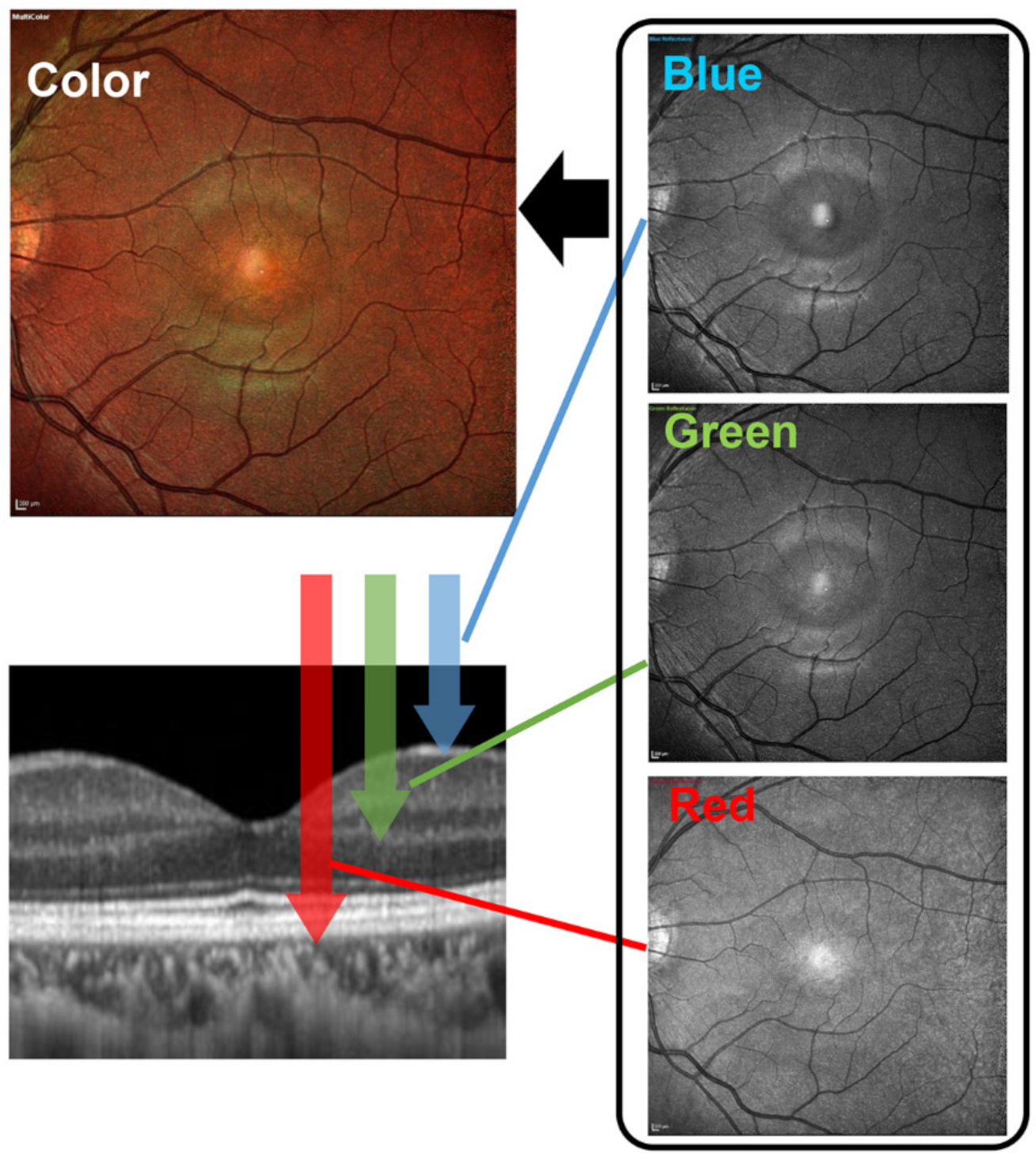
1.3. Characteristics of New SLO Devices
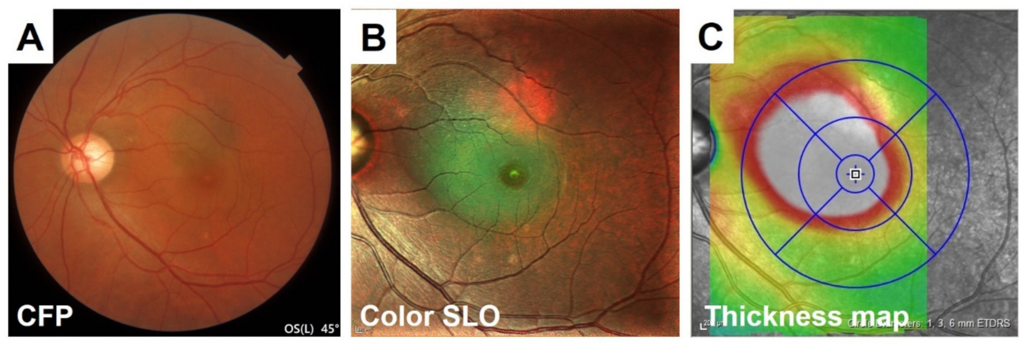
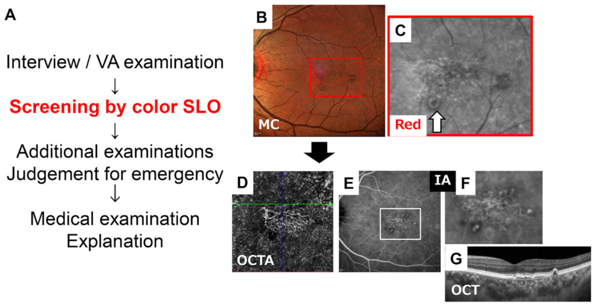
2. Recent Clinical Studies Using Color SLOs and the Effects on Clinical Practice
3. Representative Cases
3.1. Retinal Nerve Fiber Layer (RNFL) Defects
3.2. Epiretinal Membrane (ERM)
4. Retinal Capillary Microaneurysms
4.1. Retinal Vein Occlusion (RVO)
4.2. Central Serous Chorioretinopathy (CSC)
4.3. Age-Related Macular Degeneration and Other Acquired Macular Conditions
5. Conclusions
Author Contributions
Funding
Institutional Review Board Statement
Informed Consent Statement
Data Availability Statement
Acknowledgments
Conflicts of Interest
References
- Hyman, B.N. The Eye as a Target Organ: An Updated Classification of Hypertensive Retinopathy. J. Clin. Hypertens. 2000, 2, 194–197. [Google Scholar]
- Wilkinson, C.P.; Ferris, F.L., III; Klein, R.E.; Lee, P.P.; Agardh, C.D.; Davis, M.; Dills, D.; Kampik, A.; Pararajasegaram, R.; Verdaguer, J.T.; et al. Proposed international clinical diabetic retinopathy and diabetic macular edema disease severity scales. Ophthalmology 2003, 110, 1677–1682. [Google Scholar] [CrossRef]
- Webb, R.H.; Hughes, G.W.; Pomerantzeff, O. Flying spot TV ophthalmoscope. Appl. Opt. 1980, 19, 2991–2997. [Google Scholar] [CrossRef]
- Gabel, V.P.; Birngruber, R.; Nasemann, J. Das Scanning-Laser-Ophthalmoscope und seine Anwendung als Fluoreszenzangiogrpahie-Gerät [The scanning laser ophthalmoscope and its use as a fluorescein angiography instrument]. Fortschr. Ophthalmol. 1988, 85, 569–573. [Google Scholar] [PubMed]
- Scheider, A.; Schroedel, C. High resolution indocyanine green angiography with a scanning laser ophthalmoscope. Am. J. Ophthalmol. 1989, 108, 458–459. [Google Scholar] [CrossRef]
- Wolf, S. Recent developments in scanning laser ophthalmoscopy. Dev. Ophthalmol. 1997, 29, 1–7. [Google Scholar]
- Tanaka, T.; Muraoka, K.; Shimizu, K. Fluorescein fundus angiography with scanning laser ophthalmoscope. Visibility of leukocytes and platelets in perifoveal capillaries. Ophthalmology. 1991, 98, 1824–1829. [Google Scholar] [CrossRef]
- Wolf, S.; Arend, O.; Toonen, H.; Bertram, B.; Jung, F.; Reim, M. Retinal capillary blood flow measurement with a scanning laser ophthalmoscope. Preliminary results. Ophthalmology 1991, 98, 996–1000. [Google Scholar] [CrossRef]
- Timberlake, G.T.; Mainster, M.A.; Webb, R.H.; Hughes, G.W.; Trempe, C.L. Retinal localization of scotomata by scanning laser ophthalmoscopy. Investig. Ophthalmol. Vis. Sci. 1982, 22, 91–97. [Google Scholar]
- Sunness, J.S.; Schuchard, R.A.; Shen, N.; Rubin, G.S.; Dagnelie, G.; Haselwood, D.M. Landmark-driven fundus perimetry using the scanning laser ophthalmoscope. Investig. Ophthalmol. Vis. Sci. 1995, 36, 1863–1874. [Google Scholar]
- Hassenstein, A.; Meyer, C.H. Clinical use and research applications of Heidelberg retinal angiography and spectral-domain optical coherence tomography—A review. Clin. Exp. Ophthalmol. 2009, 37, 130–143. [Google Scholar] [CrossRef]
- Desmettre, T.; Devoisselle, J.M.; Mordon, S. Fluorescence properties and metabolic features of indocyanine green (ICG) as related to angiography. Surv. Ophthalmol. 2000, 45, 15–27. [Google Scholar] [CrossRef]
- Tan, A.C.; Fleckenstein, M.; Schmitz-Valckenberg, S.; Holz, F.G. Clinical Application of Multicolor Imaging Technology. Ophthalmologica 2016, 236, 8–18. [Google Scholar] [CrossRef] [PubMed]
- Nagiel, A.; Lalane, R.A.; Sadda, S.R.; Schwartz, S.D. Ultra-Widefield Fundus Imaging: A Review of Clinical Applications and Future Trends. Retina 2016, 36, 660–678. [Google Scholar] [CrossRef]
- Olvera-Barrios, A.; Heeren, T.F.; Balaskas, K.; Chambers, R.; Bolter, L.; Tufail, A.; Egan, C.E.; Anderson, J. Comparison of true-colour wide-field confocal scanner imaging with standard fundus photography for diabetic retinopathy screening. Br. J. Ophthalmol. 2020, 104, 1579–1584. [Google Scholar] [CrossRef] [PubMed]
- Olvera-Barrios, A.; Heeren, T.F.; Balaskas, K.; Chambers, R.; Bolter, L.; Egan, C.; Tufail, A.; Anderson, J. Diagnostic accuracy of diabetic retinopathy grading by an artificial intelligence-enabled algorithm compared with a human standard for wide-field true-colour confocal scanning and standard digital retinal images. Br. J. Ophthalmol. 2021, 105, 265–270. [Google Scholar] [CrossRef]
- Terasaki, H.; Sonoda, S.; Shiihara, H.; Kakiuchi, N.; Funatsu, R.; Shirasawa, M.; Sakamoto, T. More Effective Screening for Epiretinal Membranes with Multicolor Scanning Laser Ophthalmoscope than with Color Fundus Photographs. Retina 2020, 40, 1412–1418. [Google Scholar] [CrossRef]
- Graham, K.W.; Chakravarthy, U.; Hogg, R.E.; Muldrew, K.A.; Young, I.S.; Kee, F. Identifying Features of Early and Late Age-Related Macular Degeneration: A Comparison of Multicolor Versus Traditional Color Fundus Photography. Retina 2018, 38, 1751–1758. [Google Scholar] [CrossRef] [PubMed]
- Tan, A.C.S.; Yanagi, Y.; Cheung, G.C.M. Comparison of Multicolor Imaging and Color Fundus Photography in the Detection of Pathological Findings in Eyes with Polypoidal Choroidal Vasculopathy. Retina 2020, 40, 1512–1519. [Google Scholar] [CrossRef]
- He, L.; Chen, C.; Yi, Z.; Wang, X.; Liu, J.; Zheng, H. Clinical Application of Multicolor Imaging in Central Serous Chorioretinopathy. Retina 2020, 40, 743–749. [Google Scholar] [CrossRef]
- Govindahari, V.; Fraser-Bell, S.; Ayachit, A.G.; Invernizzi, A.; Nair, U.; Nair, D.V.; Lupidi, M.; Singh, S.R.; Rajendran, A.; Zur, D.; et al. Multicolor imaging in macular telangiectasia-a comparison with fundus autofluorescence. Graefes. Arch. Clin. Exp. Ophthalmol. 2020, 258, 2379–2387. [Google Scholar] [CrossRef]
- Zhang, Z.; Li, M.; Sun, Y.; Wei, Y.; Zhang, S. Multicolor Scanning Laser Ophthalmoscopy Strengthens Surgeons′ Preoperative Decision-Making and Intraoperative Performance on Epiretinal Membrane. Transl. Vis. Sci. Technol. 2020, 9, 36. [Google Scholar] [CrossRef]
- Ajlan, R.S.; Barnard, L.R.; Mainster, M.A. NONCONFOCAL ULTRA-WIDEFIELD SCANNING LASER OPHTHALMOSCOPY: Polarization Artifacts and Diabetic Macular Edema. Retina 2020, 40, 1374–1378. [Google Scholar] [CrossRef]
- Pang, C.E.; Freund, K.B. Ghost maculopathy: An artifact on near-infrared reflectance and multicolor imaging masquerading as chorioretinal pathology. Am. J. Ophthalmol. 2014, 158, 171–178.e2. [Google Scholar] [CrossRef]
- Feng, H.L.; Sharma, S.; Stinnett, S.; Asrani, S.; Mruthyunjaya, P. Characterization of Artifacts Associated With Multicolor Confocal Scanning Laser Ophthalmoscopy. Ophthalmic Surg. Lasers Imaging Retina. 2017, 48, 810–815. [Google Scholar] [CrossRef] [PubMed]
- Tuulonen, A.; Lehtola, J.; Airaksinen, P.J. Nerve fiber layer defects with normal visual fields. Do normal optic disc and normal visual field indicate absence of glaucomatous abnormality? Ophthalmology 1993, 100, 587–598. [Google Scholar] [CrossRef]
- Leung, C.K.-S.; Medeiros, F.A.; Zangwill, L.M.; Sample, P.A.; Bowd, C.; Ng, D.; Cheung, C.Y.L.; Lam, D.S.C.; Weinreb, R.N. American Chinese glaucoma imaging study: A comparison of the optic disc and retinal nerve fiber layer in detecting glaucomatous damage. Investig. Ophthalmol. Vis. Sci. 2007, 48, 2644–2652. [Google Scholar] [CrossRef]
- Medeiros, F.A.; Vizzeri, G.; Zangwill, L.M.; Alencar, L.M.; Sample, P.A.; Weinreb, R.N. Comparison of retinal nerve fiber layer and optic disc imaging for diagnosing glaucoma in patients suspected of having the disease. Ophthalmology 2008, 115, 1340–1346. [Google Scholar] [CrossRef]
- Terasaki, H.; Sonoda, S.; Kakiuchi, N.; Shiihara, H.; Yamashita, T.; Sakamoto, T. Ability of MultiColor scanning laser ophthalmoscope to detect non-glaucomatous retinal nerve fiber layer defects in eyes with retinal diseases. BMC Ophthalmol. 2018, 18, 324. [Google Scholar] [CrossRef]
- Wiley, H.E.; Ferris, F.L. Nonproliferative Diabetic Retinopathy, Ryan's Retina, 5th ed.; Elsevier: Amsterdam, The Netherlands, 2013; pp. 940–968. [Google Scholar]
- Arrigo, A.; Teussink, M.; Aragona, E.; Bandello, F.; Battaglia Parodi, M. MultiColor imaging to detect different subtypes of retinal microaneurysms in diabetic retinopathy. Eye 2021, 35, 277–281. [Google Scholar] [CrossRef] [PubMed]
- Hirano, T.; Toriyama, Y.; Iesato, Y.; Imai, A.; Hirabayashi, K.; Nagaoka, T.; Takamura, Y.; Sugimoto, M.; Murata, T. Effect of leaking perifoveal microaneurysms on resolution of diabetic macular edema treated by combination therapy using anti-vascular endothelial growth factor and short pulse focal/grid laser photocoagulation. Jpn. J. Ophthalmol. 2017, 61, 51–60. [Google Scholar] [CrossRef] [PubMed]
- Cheung, C.M.G.; Lee, W.K.; Koizumi, H.; Dansingani, K.; Lai, T.Y.Y.; Freund, K.B. Pachychoroid disease. Eye 2019, 33, 14–33. [Google Scholar] [CrossRef]
- Casalino, G.; Arrigo, A.; Introini, U.; Scialdone, A.; Coppola, M.; Bandello, F.; Chakravarthy, U.; Parodi, M.B. Clinical Course of Treated Choroidal Neovascularization in Eyes with Pre-existing Geographic Atrophy: Case Series and Reappraisal of the Literature. Curr. Eye. Res. 2020, 1–7. [Google Scholar] [CrossRef] [PubMed]
- Casalino, G.; Arrigo, A.; Romano, F.; Munk, M.R.; Bandello, F.; Parodi, M.B. Acute macular neuroretinopathy: Pathogenetic insights from optical coherence tomography angiography. Br. J. Ophthalmol. 2019, 103, 410–414. [Google Scholar] [CrossRef] [PubMed]
- Grzybowski, A.; Brona, P.; Lim, G.; Ruamviboonsuk, P.; Tan, G.S.W.; Abramoff, M.; Ting, D.S.W. Artificial intelligence for diabetic retinopathy screening: A review. Eye 2020, 34, 451–460. [Google Scholar] [CrossRef]
- Moraru, A.D.; Costin, D.; Moraru, R.L.; Branisteanu, D.C. Artificial intelligence and deep learning in ophthalmology—present and future (Review). Exp. Ther. Med. 2020, 20, 3469–3473. [Google Scholar]
- Ting, D.S.W.; Pasquale, L.R.; Peng, L.; Campbell, J.P.; Lee, A.Y.; Raman, R.; Tan, G.S.W.; Schmetterer, L.; Keane, P.A.; Wong, T.Y. Artificial intelligence and deep learning in ophthalmology. Br. J. Ophthalmol. 2019, 103, 167–175. [Google Scholar] [CrossRef]
- Shiihara, H.; Sakamoto, T.; Terasaki, H.; Kakiuchi, N.; Shinohara, Y.; Tomita, M.; Sonoda, S. Running pattern of choroidal vessel in en face OCT images determined by machine learning-based quantitative method. Graefes. Arch. Clin. Exp. Ophthalmol. 2019, 257, 1879–1887. [Google Scholar] [CrossRef]
- Yamashita, T.; Asaoka, R.; Terasaki, H.; Murata, H.; Tanaka, M.; Nakao, K.; Sakamoto, T. Factors in Color Fundus Photographs That Can Be Used by Humans to Determine Sex of Individuals. Transl. Vis. Sci. Technol. 2020, 9, 4. [Google Scholar]
- Ometto, G.; Montesano, G.; Afgeh, S.S.; Lazaridis, G.; Liu, X.; Keane, P.A.; Crabb, D.P.; Denniston, A.K. Merging Information From Infrared and Autofluorescence Fundus Images for Monitoring of Chorioretinal Atrophic Lesions. Transl. Vis. Sci. Technol. 2020, 9, 38. [Google Scholar] [CrossRef]
- Cavichini, M.; An, C.; Bartsch, D.-U.G.; Jhingan, M.; Amador-Patarroyo, M.J.; Long, C.P.; Zhang, J.; Wang, Y.; Chan, A.X.; Madala, S.; et al. Artificial Intelligence for Automated Overlay of Fundus Camera and Scanning Laser Ophthalmoscope Images. Transl. Vis. Sci. Technol. 2020, 9, 56. [Google Scholar] [CrossRef] [PubMed]


Publisher’s Note: MDPI stays neutral with regard to jurisdictional claims in published maps and institutional affiliations. |
© 2021 by the authors. Licensee MDPI, Basel, Switzerland. This article is an open access article distributed under the terms and conditions of the Creative Commons Attribution (CC BY) license (http://creativecommons.org/licenses/by/4.0/).
Share and Cite
Terasaki, H.; Sonoda, S.; Tomita, M.; Sakamoto, T. Recent Advances and Clinical Application of Color Scanning Laser Ophthalmoscope. J. Clin. Med. 2021, 10, 718. https://doi.org/10.3390/jcm10040718
Terasaki H, Sonoda S, Tomita M, Sakamoto T. Recent Advances and Clinical Application of Color Scanning Laser Ophthalmoscope. Journal of Clinical Medicine. 2021; 10(4):718. https://doi.org/10.3390/jcm10040718
Chicago/Turabian StyleTerasaki, Hiroto, Shozo Sonoda, Masatoshi Tomita, and Taiji Sakamoto. 2021. "Recent Advances and Clinical Application of Color Scanning Laser Ophthalmoscope" Journal of Clinical Medicine 10, no. 4: 718. https://doi.org/10.3390/jcm10040718
APA StyleTerasaki, H., Sonoda, S., Tomita, M., & Sakamoto, T. (2021). Recent Advances and Clinical Application of Color Scanning Laser Ophthalmoscope. Journal of Clinical Medicine, 10(4), 718. https://doi.org/10.3390/jcm10040718