
Prognostic Value of Reduced Heart Rate Reserve during Exercise in Hypertrophic Cardiomyopathy

 , , , , , ,
  ,
,  , , ,
, , ,  , ,
, ,  , , , and add
Show full author list
, , , and add
Show full author list
Abstract
:1. Introduction
2. Materials and Methods
2.1. Patients Population
2.2. Resting and Stress Echocardiography
2.3. Heart Rate Reserve
2.4. Follow-Up Data
2.5. Statistical Analysis
3. Results
3.1. Patient Characteristics
3.2. SE Positivity Criteria
3.3. Outcome
4. Discussion
4.1. Comparison with Previous Studies
4.2. Clinical Implications
4.3. Pathophysiology of Blunted HRR in HCM
4.4. Study Limitations
5. Conclusions
Author Contributions
Funding
Institutional Review Board Statement
Informed Consent Statement
Data Availability Statement
Acknowledgments
Conflicts of Interest
References
- Maron, B.J.; Maron, M.S. Hypertrophic cardiomyopathy. Lancet 2013, 381, 242–255. [Google Scholar] [CrossRef]
- Elliott, P.M.; Anastasakis, A.; Borger, M.A.; Borggrefe, M.; Cecchi, F.; Charron, P.; Hagege, A.A.; Lafont, A.; Limongelli, G.; Mahrholdt, H.; et al. 2014 ESC Guidelines on diagnosis and management of hypertrophic cardiomyopathy: The Task Force for the Diagnosis and Management of Hypertrophic Cardiomyopathy of the European Society of Cardiology (ESC). Eur. Heart J. 2014, 35, 2733–2779. [Google Scholar] [PubMed]
- Maron, M.S.; Rowin, E.J.; Wessler, B.S.; Mooney, P.J.; Fatima, A.; Patel, P.; Koethe, B.C.; Romashko, M.; Link, M.S.; Maron, B.J. Enhanced American College of Cardiology/American Heart Association Strategy for Prevention of Sudden Cardiac Death in High-Risk Patients with Hypertrophic Cardiomyopathy. JAMA Cardiol. 2019, 4, 644–657. [Google Scholar] [CrossRef] [Green Version]
- Maron, M.S.; Olivotto, I.; Betocchi, S.; Casey, S.A.; Lesser, J.R.; Losi, M.A.; Cecchi, F.; Maron, B.J. Effect of left ventricular outflow tract obstruction on clinical outcome in hypertrophic cardiomyopathy. N. Engl. J. Med. 2003, 348, 295–303. [Google Scholar] [CrossRef] [PubMed]
- Peteiro, J.; Bouzas-Mosquera, A.; Fernandez, X.; Monserrat, L.; Pazos, P.; Estevez-Loureiro, R.; Castro-Beiras, A. Prognostic value of exercise echocardiography in patients with hypertrophic cardiomyopathy. J. Am. Soc. Echocardiogr. 2012, 25, 182–189. [Google Scholar] [CrossRef]
- Magri, D.; Agostoni, P.; Sinagra, G.; Re, F.; Correale, M.; Limongelli, G.; Zachara, E.; Mastromarino, V.; Santolamazza, C.; Casenghi, M.; et al. Clinical and prognostic impact of chronotropic incompetence in patients with hypertrophic cardiomyopathy. Int. J. Cardiol. 2018, 271, 125–131. [Google Scholar] [CrossRef]
- Lazzeroni, E.; Picano, E.; Morozzi, L.; Maurizio, A.R.; Palma, G.; Ceriati, R.; Iori, E.; Barilli, A.; for the Echo Persantine Italian Cooperative (EPIC) Study group; Subproject Hypertrophic Cardiomyopathy. Dipyridamole-induced ischemia as a prognostic marker of future adverse cardiac events in adult hypertrophic cardiomyopathy. Circulation 1997, 96, 4268–4272. [Google Scholar] [CrossRef]
- Cortigiani, L.; Rigo, F.; Gherardi, S.; Galderisi, M.; Sicari, R.; Picano, E.; on behalf of the Echo Persantine Italian Cooperative (EPIC) EPIC—flow reserve (FR) study group. Prognostic implications of coronary flow reserve in left anterior descending coronary artery in hypertrophic cardiomyopathy. Am. J. Cardiol. 2008, 102, 926–932. [Google Scholar] [CrossRef]
- Ciampi, Q.; Olivotto, I.; Gardini, C.; Mori, F.; Peteiro, J.; Monserrat, L.; Fernandez, X.; Cortigiani, L.; Rigo, F.; Lopes, L.R.; et al. Prognostic Role of Stress Echocardiography in Hypertrophic Cardiomyopathy. The International Stress Echo registry. Int. J. Cardiol. 2016, 219, 331–338. [Google Scholar] [CrossRef]
- Picano, E.; Ciampi, Q.; Citro, R.; D’Andrea, A.; Scali, M.C.; Cortigiani, L.; Olivotto, I.; Mori, F.; Galderisi, M.; Costantino, M.F.; et al. Stress echo 2020: The international Stress Echo study in ischemic and non-ischemic heart disease. Cardiovasc. Ultrasound 2017, 15, 3. [Google Scholar] [CrossRef] [Green Version]
- Lang, R.M.; Badano, L.P.; Mor-Avi, V.; Afilalo, J.; Armstrong, A.; Ernande, L.; Flachskampf, F.A.; Foster, E.; Goldstein, S.A.; Kuznetsova, T.; et al. Recommendations for cardiac chamber quantification by echocardiography in adults: An update from the American Society of Echocardiography and the European Association of Cardiovascular Imaging. Eur. Heart J. Cardiovasc. Imaging 2015, 16, 233–270. [Google Scholar] [CrossRef]
- Sicari, R.; Nihoyannopoulos, P.; Evangelista, A.; Kasprzak, J.; Lancellotti, P.; Poldermans, D.; Voigt, J.U.; Zamorano, J.L.; on behalf of the European Association of Echocardiography. Stress echocardiography expert consensus statement. European Association of Echocardiography (EAE) (a registered branch of the ESC). Eur. J. Echocardiogr. 2008, 9, 415–437. [Google Scholar] [CrossRef] [Green Version]
- Pellikka, P.A.; Arruda-Olson, A.; Chaudhry, F.A.; Chen, M.H.; Marshall, J.E.; Porter, T.R.; Sawada, S.G. Guidelines for Performance, Interpretation, and Application of Stress Echocardiography in Ischemic Heart Disease: From the American Society of Echocardiography. J. Am. Soc. Echocardiogr. 2020, 33, 1–41.e8. [Google Scholar] [CrossRef] [Green Version]
- Lancellotti, P.; Pellikka, P.A.; Budts, W.; Chaudhry, F.A.; Donal, E.; Dulgheru, R.; Edvardsen, T.; Garbi, M.; Ha, J.W.; Kane, G.C.; et al. Recommendations for the clinical use of stress echocardiography in non-ischemic heart disease: Joint document of the European Association of Cardiovascular imaging and the American Society of Echocardiography. Eur. Heart J. Cardiov. Imaging 2016, 190, 1191–1229. [Google Scholar] [CrossRef] [PubMed] [Green Version]
- Ciampi, Q.; Picano, E.; Paterni, M.; Daros, C.B.; Simova, I.; de Castro, E.; Silva Pretto, J.L.; Scali, M.C.; Gaibazzi, N.; Severino, S.; et al. Quality control of regional wall motion analysis in Stress Echo 2020. Int. J. Cardiol. 2017, 249, 479–485. [Google Scholar] [CrossRef] [PubMed]
- Cortigiani, L.; Carpeggiani, C.; Landi, P.; Raciti, M.; Bovenzi, F.; Picano, E. Usefulness of blunted heart rate reserve as an imaging-independent prognostic predictor during dipyridamole-echocardiography test. Am. J. Cardiol. 2019, 124, 972–977. [Google Scholar] [CrossRef] [PubMed]
- Lauer, M.S.; Francis, G.S.; Okin, P.M.; Pashkow, F.J.; Snader, C.E.; Marwick, T.H. Impaired chronotropic response to exercise stress testing as a predictor of mortality. JAMA 1999, 281, 524–529. [Google Scholar] [CrossRef]
- Lauer, M.; Blackstone, E.; Young, J.; Topol, E. Cause of death in clinical research: Time for reassessment? J. Am. Coll. Cardiol. 1999, 34, 618–620. [Google Scholar] [CrossRef] [Green Version]
- Brubaker, P.H.; Kitzman, D.W. Chronotropic incompetence. Causes, consequences and management. Circulation 2011, 123, 1010–1020. [Google Scholar] [CrossRef] [Green Version]
- Fukuda, K.; Kanazawa, H.; Aizawa, Y.; Ardell, J.L.; Shivkumar, K. Cardiac innervation and sudden cardiac death. Circ. Res. 2015, 116, 2005–2019. [Google Scholar] [CrossRef] [Green Version]
- Efthimiadis, G.K.; Giannakoulas, G.; Parcharidou, D.G.; Pagourelias, E.D.; Kouidi, E.J.; Spanos, G.; Kamperidis, V.; Gavrielides, S.; Karvounis, H.; Styliadis, I.; et al. Chronotropic incompetence and its relation to exercise intolerance in hypertrophic cardiomyopathy. Int. J. Cardiol. 2011, 153, 179–184. [Google Scholar] [CrossRef] [PubMed]
- Luo, H.C.; Dimaano, V.L.; Kembro, J.M.; Hilser, A.; Hurtado-de-Mendoza, D.; Pozios, I.; Tomas, M.S.; Yalcin, H.; Dolores-Cerna, K.; Mormontoy, W.; et al. Exercise heart rates in patients with hypertrophic cardiomyopathy. Am. J. Cardiol. 2015, 115, 1144–1150. [Google Scholar] [CrossRef] [Green Version]
- Sen-Chowdhry, S.; Jacoby, D.; Moon, J.C.; McKenna, W.J. Update on hypertrophic cardiomyopathy and a guide to the guidelines. Nat. Rev. Cardiol. 2016, 13, 651–675. [Google Scholar] [CrossRef] [PubMed]
- Smith, E.D.; Tome, J.; Mcgrath, R.; Kumar, S.; Concannon, M.; Day, S.M.; Saberi, S.; Helms, A.S. Exercise hemodynamics in hypertrophic cardiomyopathy identify risk of incident heart failure but not ventricular arrhythmias or sudden cardiac death. Int. J. Cardiol. 2019, 274, 226–231. [Google Scholar] [CrossRef] [PubMed]
- Terai, H.; Shimizu, M.; Ino, H.; Yamaguchi, M.; Uchiyama, K.; Oe, K.; Nakajima, K.; Taki, J.; Kawano, M.; Mabuchi, H.; et al. Cardiac sympathetic nerve activity in patients with hypertrophic cardiomyopathy with malignant ventricular tachyarrhythmias. J. Nucl. Cardiol. 2003, 10, 304–310. [Google Scholar] [CrossRef]
- Omodani, H.; Kinugawa, T.; Ogino, K.; Furuse, Y.; Yamaguchi, M.; Mori, M.; Endo, A.; Kato, M.; Kato, T.; Osaki, S.; et al. Augmented exercise plasma noradrenaline with impaired chronotropic responsiveness in patients with hypertrophic cardiomyopathy. Clin. Exp. Pharmacol. Physiol. 1998, 25, 1018–1023. [Google Scholar] [CrossRef]
- Schäfers, M.; Dutka, D.; Rhodes, C.G.; Lammertsma, A.A.; Hermansens, F.; Schober, O.; Camici, P.G. Myocardial presynaptic and postsynaptic autonomic dysfunction in hypertrophic cardiomyopathy. Circ. Res. 1998, 82, 57–62. [Google Scholar] [CrossRef] [Green Version]
- Brush, J.E., Jr.; Eisenhofer, G.; Garty, M.; Stull, R.; Maron, B.J.; Cannon, R.O., 3rd; Panza, J.A.; Epstein, S.E.; Goldstein, D.S. Cardiac norepinephrine kinetics in hypertrophic cardiomyopathy. Circulation 1989, 79, 836–844. [Google Scholar] [CrossRef] [Green Version]
- Isobe, S.; Izawa, H.; Iwase, M.; Nanasato, M.; Nonokawa, M.; Ando, A.; Ohshima, S.; Nagata, K.; Kato, K.; Nishizawa, T.; et al. Cardiac 123I-MIBG reflects left ventricular functional reserve in patients with nonobstructive hypertrophic cardiomyopathy. J. Nucl. Med. 2005, 46, 909–916. [Google Scholar]
- Pace, L.; Betocchi, S.; Losi, M.A.; Della Morte, A.M.; Ciampi, Q.; Nugnez, R.; Chiariello, M.; Salvatore, M. Sympathetic nervous function in patients with hypertrophic cardiomyopathy assessed by [123I]-MIBG: Relationship with left ventricular perfusion and function. Q. J. Nucl. Med. Mol. Imaging 2004, 48, 20–25. [Google Scholar] [PubMed]
- Maron, B.J.; Rowin, E.J.; Casey, S.A.; Haas, T.S.; Chan, R.H.; Udelson, J.E.; Garberich, R.F.; Lesser, J.R.; Appelbaum, E.; Manning, W.J.; et al. Risk stratification and outcome of patients with hypertrophic cardiomyopathy ≥60 years of age. Circulation 2013, 127, 585–593. [Google Scholar] [CrossRef] [PubMed] [Green Version]


| Variable | Overall Population (n = 917) | Group 1 HRR: >2.13 (n = 224) | Group 2 HRR: 1.88–2.13 (n = 227) | Group 3 HRR: 1.62–1.87 (n = 237) | Group 4 HRR: ≤1.61 (n = 229) | p |
|---|---|---|---|---|---|---|
| Age (years) | 49 ± 15 | 43 ± 15 | 48 ± 15 * | 50 ± 16 * | 53 ± 14 *^ | <0.001 |
| Male gender, n (%) | 516 (56.3%) | 123 (54.9%) | 126 (55.5%) | 140 (59.1%) | 127 (55.5%) | 0.792 |
| BSA (m2) | 1.89 ± 0.21 | 1.86 ± 0.19 | 1.85 ± 0.21 | 1.91 ± 0.23 | 1.95 ± 0.20 *^ | 0.001 |
| NYHA functional class | 1.5 ± 0.6 | 1.3 ± 0.5 | 1.4 ± 0.6 * | 1.6 ± 0.6 * | 1.8 ± 0.7 *^§ | <0.001 |
| LV end-diastolic diameter (mm) | 52.7 ± 18.3 | 47.6 ± 9.4 | 52.3 ± 18.9 | 53.4 ± 17.9 | 57.5 ± 23.3* | 0.002 |
| LV end-systolic diameter (mm) | 27.0 ± 7.3 | 27.2 ± 6.6 | 26.7 ± 6.9 | 26.2 ± 7.1 | 27.8 ± 8.4 | 0.430 |
| Maximal wall thickness (mm) | 20.9 ± 5.4 | 20.2 ± 5.4 | 21.2 ± 5.3 | 20.7 ± 5.4 | 21.4 ± 5.4 | 0.126 |
| ≥Moderate MR, n (%) | 95/672 (14.1%) | 19/203 (9.4%) | 25/179 (14.0%) | 27/172 (15.7%) | 24/118 * (20.3%) | 0.048 |
| Beta-blockers, n (%) | 383 (41.8%) | 81 (36.2%) | 104 (45.8%) | 98 (41.4%) | 100 (43.7%) | 0.189 |
| Calcium-channel blockers n (%) | 91 (9.9%) | 10 (4.5%) | 16 (7%) | 30 (12.7%) | 35 (15.3%) | <0.001 |
| Diuretics, n (%) | 110 (12.3%) | 13 (5.8%) | 23 (10.1%) | 33 (13.9%) * | 44 (19.2%) *^ | <0.001 |
| ESC risk score SCD | 5.5 ± 7.4 | 5.8 ± 6.6 | 6.1 ± 8.5 | 4.4 ± 6.6 | 5.5 ± 7.6 | 0.269 |
| Overall Population (n = 917) | Group 1 HRR: >2.13 (n = 224) | Group 2 HRR: 1.88–2.13 (n = 227) | Group 3 HRR: 1.62–1.87 (n = 237) | Group 4 HRR: ≤1.61 (n = 229) | p | |
|---|---|---|---|---|---|---|
| HR at rest (b/m) | 70.9 ± 13.6 | 63.1 ± 8.8 | 68.9 ± 11.1 * | 73.6 ± 13.2 *^ | 77.6 ± 15.7 *^§ | <0.001 |
| HR at peak (b/m) | 132.3 ± 27.3 | 152.8 ± 20.0 | 138.1 ± 22.7 * | 129.1 ± 22.7 *^ | 110.0 ± 24.7 *^§ | <0.001 |
| HRR | 1.90 ± 0.40 | 2.44 ± 0.26 | 2.00 ± 0.08 * | 1.76 ± 0.07 *^ | 1.42 ± 0.15 *^§ | <0.001 |
| SBP at rest (mmHg) | 124.9 ± 16.5 | 122.6 ± 15.1 | 123.2 ± 17.3 | 126.0 ± 15.9 | 127.9 ± 17.2 *^ | 0.001 |
| SBP at peak (mmHg) | 161.5 ± 28.8 | 166.3 ± 28.5 | 161.0 ± 30.1 | 162.5 ± 27.5 | 156.1 ± 28.2 * | 0.002 |
| WMSI at rest | 1.01 ± 0.07 | 1.01 ± 0.09 | 1.01 ± 0.06 | 1.01 ± 0.06 | 1.00 ± 0.01 | 0.293 |
| WMSI at peak | 1.01 ± 0.09 | 1.02 ± 0.10 | 1.02 ± 0.10 | 1.01 ± 0.09 | 1.00 ± 0.01 | 0.146 |
| WMSI | 0.01 ± 0.05 | 0.01 ± 0.05 | 0.01 ± 0.05 | 0.01 ± 0.07 | 0.00 ± 0.01 | 0.157 |
| RWMA, n (%) | 22 (2.4%) | 10 (4.5%) | 5 (2.2%) | 5 (2.1%) | 2 (0.9%) | 0.091 |
| LV EDV at rest (mL, n = 660) | 90.3 ± 37.6 | 85.1 ± 29.8 | 86.6 ± 33.7 | 71.3 ± 41.4 | 101.3 ± 44.7 | 0.001 |
| LV EDV at peak (mL, n = 284) | 60.1 ± 27.8 | 68.9±27.8 | 68.7 ± 26.4 | 68.0 ± 31.6 | 65.5 ± 25.8 | 0.914 |
| LV ESV at rest, (mL, n = 428) | 26.9 ± 7.2 | 27.3 ± 11.8 | 28.0 ± 14.1 | 28.3 ± 16.4 | 29.4 ± 14.7 | 0.561 |
| LV ESV at peak, (mL, n = 311) | 20.1 ± 11.8 | 20.8±12.6 | 20.0 ± 11.4 | 19.6 ± 11.3 | 19.4 ± 11.7 | 0.749 |
| LV EF at rest (%) | 68.2 ± 9.1 | 68.2 ± 8.9 | 68.0 ± 9.6 | 68.2 ± 8.5 | 68.2 ± 9.3 | 0.804 |
| LV EF at peak (%) | 72.5 ± 11.0 | 72.3 ± 13.1 | 72.2 ± 9.8 | 72.4 ± 10.5 | 73.9 ± 8.4 | 0.804 |
| LVOTO at rest, n (%) | 150 (16.4%) | 14 (6.3%) | 29 (12.8%) | 39 (16.5%) * | 68 (29.7%) *^§ | <0.001 |
| LVOTO at peak, n (%) | 281 (30.7%) | 52 (23.7%) | 60 (27.1%) | 76 (32.3%) | 93 (42.1%) *^ | <0.001 |
| Mets | 7.5 ± 3.2 | 9.4 ± 3.1 | 7.8 ± 2.6 * | 7.1±3.0*^ | 5.3 ± 2.4 *^§ | <0.001 |
| Univariate Analysis | Multivariate Analysis | |||
|---|---|---|---|---|
| OR (95% CI) | p Value | OR (95% CI) | p Value | |
| Age | 1.029 (1.019–1.040) | <0.001 | 1.021 (1.009–1.033) | 0.001 |
| Gender (male) | 0.957 (0.708–1.293) | 0.775 | ||
| B-blocker therapy | 1.109 (0.820–1.501) | 0.501 | ||
| NYHA functional class ≥ 2 | 3.653 (2.118–6.301) | <0.001 | ||
| Calcium channel blocker therapy | 2.450 (1.475–4.069) | 0.001 | ||
| Type of exercise | 1.460 (0.739–2.885) | 0.276 | ||
| Mets | 0.729 (0.671–0.770) | <0.001 | 0.761 (0.708–0.817) | <0.001 |
| Heart Rate at rest | 1.050 (1.038–1.062) | <0.001 | 1.027 (1.018–1.036) | <0.001 |
| LV ejection fraction | 1.000 (0.983–1.048) | 0.969 | ||
| Maximal wall thickness | 1.022 (0.994–1.050) | 0.123 | ||
| LVOTO at rest | 3.121 (2.166–4.498) | <0.001 | 1.504 (1.043–2.170) | 0.029 |
| RWMA | 3.398 (0.778–14.452) | 0.101 | ||
| Univariate Analysis | Multivariate Analysis | |||
|---|---|---|---|---|
| HR (95% CI) | p Value | HR (95% CI) | p Value | |
| Age (years) | 1.059 (1.041–1.077) | <0.001 | 1.064 (1.043–1.085) | <0.001 |
| Gender (male) | 1.060 (0.698–1.609) | 0.785 | ||
| NYHA functional class ≥2 | 2.130 (1.177–3.854) | 0.012 | ||
| B-blockers therapy | 1.264 (0.807–1.978) | 0.306 | ||
| Exercise hypotension | 1.457 (0.956–2.222) | 0.080 | ||
| ESC risk score | 0.970 (0.932–1.010) | 0.140 | ||
| Maximal wall thickness | 1.042 (1.008–1.078) | 0.015 | 1.081 (1.037–1.128) | <0.001 |
| LVOTO at rest | 1.492 (0.897–2.480) | 0.123 | ||
| LVOTO at peak | 1.086 (0.701–1.682) | 0.711 | ||
| RWMA | 2.956 (1.366–6.398) | 0.006 | 3.279 (1.441–7.461) | 0.005 |
| Mets | 0.901 (0.836–0.970) | 0.006 | ||
| HRR > 2.13 | 1 | |||
| HRR: 1.88–2.13 | 1.742 (0.860–3.532) | 0.123 | ||
| HRR: 1.62–1.87 | 2.311 (1.187–4.502) | 0.014 | ||
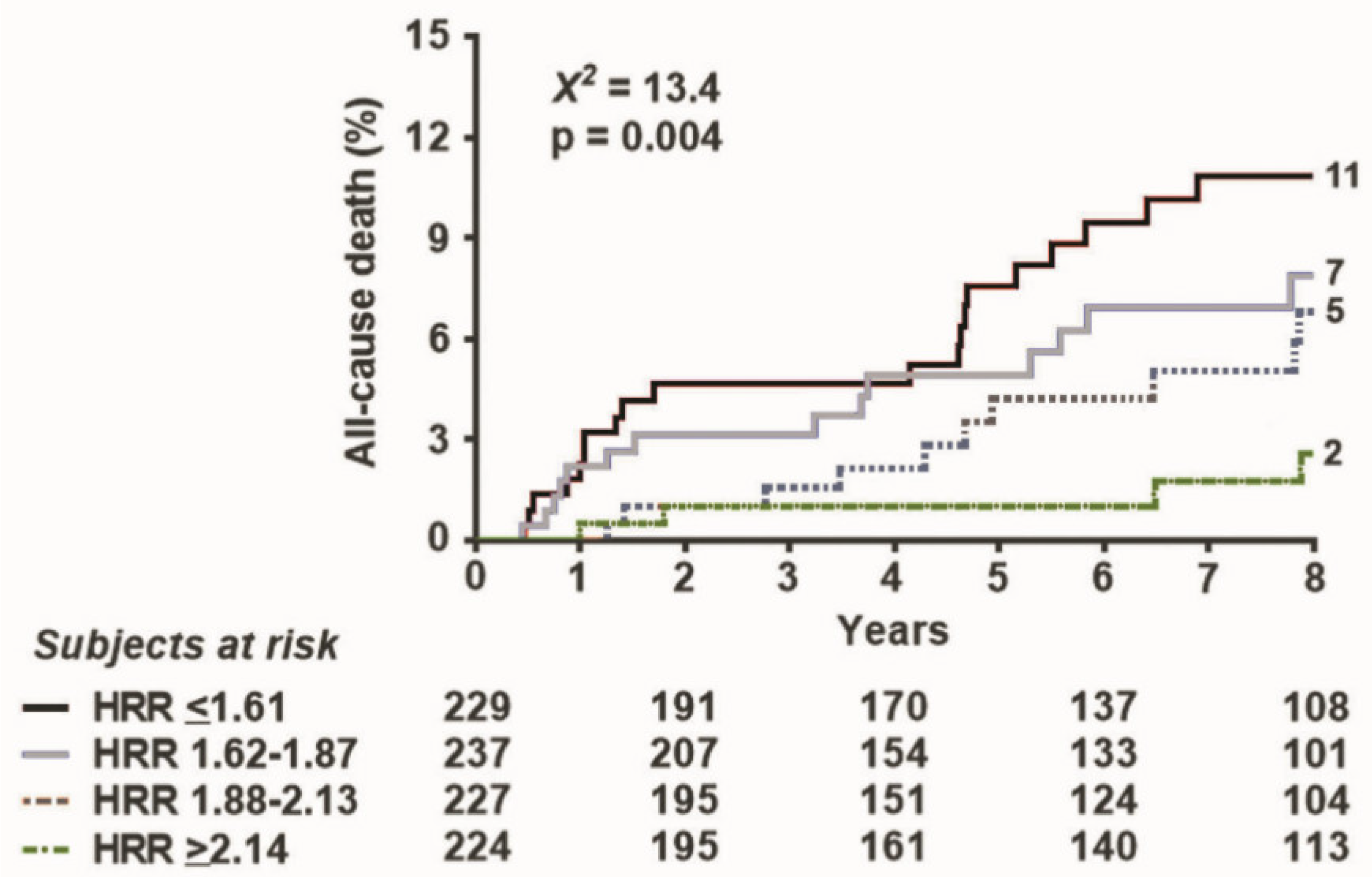
| HRR ≤ 1.61 | 3.100 (1.620–5.930) | 0.001 | 0.025 | |
Publisher’s Note: MDPI stays neutral with regard to jurisdictional claims in published maps and institutional affiliations. |
© 2021 by the authors. Licensee MDPI, Basel, Switzerland. This article is an open access article distributed under the terms and conditions of the Creative Commons Attribution (CC BY) license (http://creativecommons.org/licenses/by/4.0/).
Share and Cite
Ciampi, Q.; Olivotto, I.; Peteiro, J.; D’Alfonso, M.G.; Mori, F.; Tassetti, L.; Milazzo, A.; Monserrat, L.; Fernandez, X.; Pálinkás, A.; et al. Prognostic Value of Reduced Heart Rate Reserve during Exercise in Hypertrophic Cardiomyopathy. J. Clin. Med. 2021, 10, 1347. https://doi.org/10.3390/jcm10071347
Ciampi Q, Olivotto I, Peteiro J, D’Alfonso MG, Mori F, Tassetti L, Milazzo A, Monserrat L, Fernandez X, Pálinkás A, et al. Prognostic Value of Reduced Heart Rate Reserve during Exercise in Hypertrophic Cardiomyopathy. Journal of Clinical Medicine. 2021; 10(7):1347. https://doi.org/10.3390/jcm10071347
Chicago/Turabian StyleCiampi, Quirino, Iacopo Olivotto, Jesus Peteiro, Maria Grazia D’Alfonso, Fabio Mori, Luigi Tassetti, Alessandra Milazzo, Lorenzo Monserrat, Xusto Fernandez, Attila Pálinkás, and et al. 2021. "Prognostic Value of Reduced Heart Rate Reserve during Exercise in Hypertrophic Cardiomyopathy" Journal of Clinical Medicine 10, no. 7: 1347. https://doi.org/10.3390/jcm10071347
APA StyleCiampi, Q., Olivotto, I., Peteiro, J., D’Alfonso, M. G., Mori, F., Tassetti, L., Milazzo, A., Monserrat, L., Fernandez, X., Pálinkás, A., Pálinkás, E. D., Sepp, R., Re, F., Cortigiani, L., Tesic, M., Djordjevic-Dikic, A., Beleslin, B., Losi, M., Canciello, G., ... the Stress Echo 2020 Study Group on behalf of the Italian Society of Echocardiography and Cardiovascular Imaging (SIECVI). (2021). Prognostic Value of Reduced Heart Rate Reserve during Exercise in Hypertrophic Cardiomyopathy. Journal of Clinical Medicine, 10(7), 1347. https://doi.org/10.3390/jcm10071347