Article

Prenatal Diagnosis Using Chromosomal Microarray Analysis in High-Risk Pregnancies

1
Department of Biomedical Engineering, National Yang Ming Chiao Tung University, Taipei 112, Taiwan
2
Department of Obstetrics and Gynecology, Taipei City Hospital, Women and Children Campus, Taipei 100, Taiwan
3
School of Medicine for International Students, I-Shou University, Kaohsiung 840, Taiwan
4
Department of Bioscience Technology, Chung Yuan Christian University, Taoyuan 320, Taiwan
5
Union Clinical Laboratory, Taipei 106, Taiwan
6
Department of Life Sciences and Institute of Genome Sciences, National Yang Ming Chiao Tung University, Taipei 112, Taiwan
7
Department of Obstetrics and Gynecology, Ultrasound Center of Taiwan IVF Group, Ton-Yen General Hospital, Zhubei 302, Taiwan
*
Author to whom correspondence should be addressed.
J. Clin. Med. 2022, 11(13), 3624; https://doi.org/10.3390/jcm11133624
Submission received: 13 May 2022 / Revised: 16 June 2022 / Accepted: 21 June 2022 / Published: 23 June 2022
(This article belongs to the Section Obstetrics & Gynecology)

Abstract

:
Background: To assess the value of chromosomal microarray analysis (CMA) during the prenatal diagnosis of high-risk pregnancies. Methods: Between January 2016 and November 2021, we included 178 chorionic villi and 859 amniocentesis samples from consecutive cases at a multiple tertiary hospital. Each of these high-risk singleton pregnancies had at least one of the following indications: (1) advanced maternal age (AMA; ≥35 years; 546, 52.7%); (2) fetal structural abnormality on ultrasound (197, 19.0%); (3) high-risk first- or second-trimester Down syndrome screen (189, 18.2%), including increased nuchal translucency (≥3.5 mm; 90, 8.7%); or (4) previous pregnancy, child, or family history (105, 10.1%) affected by chromosomal abnormality or genetic disorder. Both G-banding karyotype analysis and CMA were performed. DNA was extracted directly and examined with oligonucleotide array-based comparative genomic hybridization. Results: Aneuploidies were detected by both G-banding karyotyping and CMA in 42/1037 (4.05%) cases. Among the 979 cases with normal karyotypes, 110 (10.6%) cases had copy number variants (CNVs) in CMA, including 30 (2.9%) cases with reported pathogenic and likely pathogenic CNVs ≥ 400 kb, 37 (3.6%) with nonreported VOUS, benign, or likely benign CNVs ≥ 400 kb, and 43 (4.1%) with nonreported CNVs < 400 kb. Of the 58 (5.6%) cases with aneuploidy rearrangements, 42 (4.1%) were diagnosed by both G-banding karyotyping and CMA; four inversions, six balanced translocations, and six low mosaic rates were not detected with CMA. Conclusions: CMA is an effective first step for the prenatal diagnosis of high-risk pregnancies with fetal structural anomalies found in ultrasonography or upon positive findings.

1. Background

Prenatal cytogenetic testing is currently offered most often to couples at high risk of having a child with a chromosomal abnormality. Cytogenetic fetal karyotyping can detect aneuploidy and large chromosomal rearrangements of up to 5–10 megabases (Mb), and was previously the “gold standard” prenatal genetic testing. Chromosomal microarray analysis (CMA) is a cytogenetic molecular technique that has a high detection rate for microscopic and submicroscopic chromosomal aberrations in patients with neurodevelopmental disorders (10–14%) [1]. CMA is used to assess microdeletions and microduplications called copy number variants (CNVs), which can be as small as 50–100 kb. Thus, CMA provides a ~100-fold higher resolution than conventional karyotyping, depending on the probe spacing and CMA platform used. The ability to examine the genome at this high resolution has resulted in the discovery of widespread CNVs in the human genome, including both polymorphic variations in healthy individuals and novel pathogenic copy number imbalances [2], and this has had a major impact on genetic diagnosis over the past decade [3]. Furthermore, CMA also provides additional clinically useful information in approximately 5% (range: 2.3–8.3%) of cases [4,5].
CMA enables the detection of smaller pathogenic chromosomal variants that are undetectable using standard cytogenetic analyses, and it can be highly customized and is amenable to high throughput. However, a potential drawback of CMA is that it does not allow for the detection of balanced chromosomal rearrangements, triploidy, and some instances of mosaicism. The biggest challenge that limits CMA is the detection of chromosomal variants of uncertain significance (VOUS). The difference in the reported prevalence of only pathogenic CNVs and the prevalence of both VOUS and pathogenic CNVs is likely to increase with the use of high-resolution, genome-wide array platforms, which have been applied more often in recent studies. However, unlike single nucleotide polymorphism (SNP) arrays, oligo-based CMA cannot identify balanced translocations, inversions, uniparental disomy, or polyploidy [3].
A pregnancy can be suspected of having a high risk of a chromosomal abnormality after screening due to a family history of chromosomal abnormalities or the detection of a structural anomaly in a prenatal ultrasound. CMA has now become the first-tier technique for the genetic follow-up of fetal structural anomalies identified by ultrasonography [6]. Recent studies have reported pathogenic CNVs at a rate of 0–15.0% among fetuses with increased nuchal translucency (NT; ≥2.5 to 3.5 mm, corresponding to the 95th to 99th percentile in the general population) or cystic hygroma [7,8]. The incidence of chromosomal anomalies in prenatal examinations is reported to be as high as 18–22% for all cases of congenital heart disease (CHD); the most common variants are trisomy 21 and 18, along with 22q11 microdeletion [9,10].
In this study, we report a multicenter, prospective cohort study of 1037 high-risk pregnancies that underwent prenatal diagnosis using G-banding karyotyping along with CMA. These analyses were carried out for suspected high-risk pregnancies or after observing a structural anomaly in an ultrasound during prenatal diagnosis.

2. Materials and Methods

2.1. Patients and Indications

We prospectively recruited 1037 high-risk singleton pregnancies after invasive diagnostic testing between January 2016 and December 2021. The inclusion criteria were high-risk singleton pregnancies with at least one of the following indications: (1) advanced maternal age (AMA; ≥35 years; 546, 52.7%); (2) fetal structural abnormality detected by ultrasound (197, 19.0%); (3) high risk on first- or second-trimester Down syndrome screening (189, 18.2%), including only increased nuchal translucency (≥3.5 mm; 90, 8.7%); or (4) previous pregnancy or live birth affected by or a family history of a chromosomal abnormality or another genetic disorder (105, 10.1%).
Both karyotype analysis and CMA were performed for all patients. The cohort comprised identified results from 1037 consecutive prenatal specimens; 929 cases underwent amniotic fluid (AF) sampling and 108 cases underwent chorionic villus sampling (CVS). AF studies were performed using direct (uncultured) specimens for 921 cases and cultured specimens for 8 cases. CVS studies were performed using uncultured specimens for 105 cases and cultured specimens for three cases. The subjects provided informed written consent for CMA when a positive indication was being considered. The Institutional Review Board approved this study (TCHIRB-10602101).

2.2. Prenatal Samples

Oral pretest counseling and written information was provided by a specialist in fetal medicine to inform the participants about the chance and implications of detecting aberrations with unknown or uncertain clinical relevance (variants of uncertain significance: VOUS) and incidental findings. Cases with high-risk indications underwent prenatal diagnosis using G-banding karyotyping along with CMA. If CMA analysis revealed an aberration, the parents’ blood samples were collected and analyzed to determine whether the fetal CNVs were inherited or de novo. CNVs of clinical significance or unknown significance were confirmed with fluorescence in situ hybridization (FISH) or quantitative real-time PCR (q-PCR), if necessary.
Maternal cell contamination is a possibility when studying uncultured amniotic fluid or CVS cells. Both the amniocytes and villi were dissected under a microscope to remove the maternal cells and decidua prior to DNA extraction.

2.3. DNA Preparation and Chromosomal Microarray Analysis

Chorionic villus sampling (CVS) and amniocentesis were performed for G-banding karyotype analysis and CMA. The material was immediately extracted from the uncultured villi (10–20 mg) and amniocentesis (5–10 mL) samples using Norgen’s Blood Genomic DNA Isolation Mini Kit (Norgen Biotek Corp., Thorold, ON, Canada).
For CMA, all samples were tested on targeted arrays using the CytoOneArray® platform (versions 1.0 to 4.0) depending on the date of specimen receipt, as the design of these arrays changed over time (Phalanx Biotech Group Inc., Hsinchu, Taiwan) [11]. The targeted design of CytoOneArray® can analyze more than 500 DNA regions associated with known genetic disorders, especially in Asian populations (https://www.phalanx.com.tw/reproductive-medicine/prenatal-testing/cyto-390/?lang=en (accessed on 1 March 2022)). Sample and reference genomic DNA (50 ng) were labeled with Cy5 (reference) or Cy3 (specimen) using a low-input DNA amplification and labeling kit. All experimental procedures were performed according to the manufacturer’s protocol. Scanning and image acquisition were carried out using an Agilent microarray scanner (G2565C/G2600D), and microarray image files were quantified using GenePix Pro 6 (Axon Instruments, Union City, CA, USA). Data analysis was performed using R version 3.4.1 (R Foundation for Statistical Computing, Vienna, Austria) and human genome version GRCh37 (hg19).

2.4. Interpretation and Reporting of CNVs
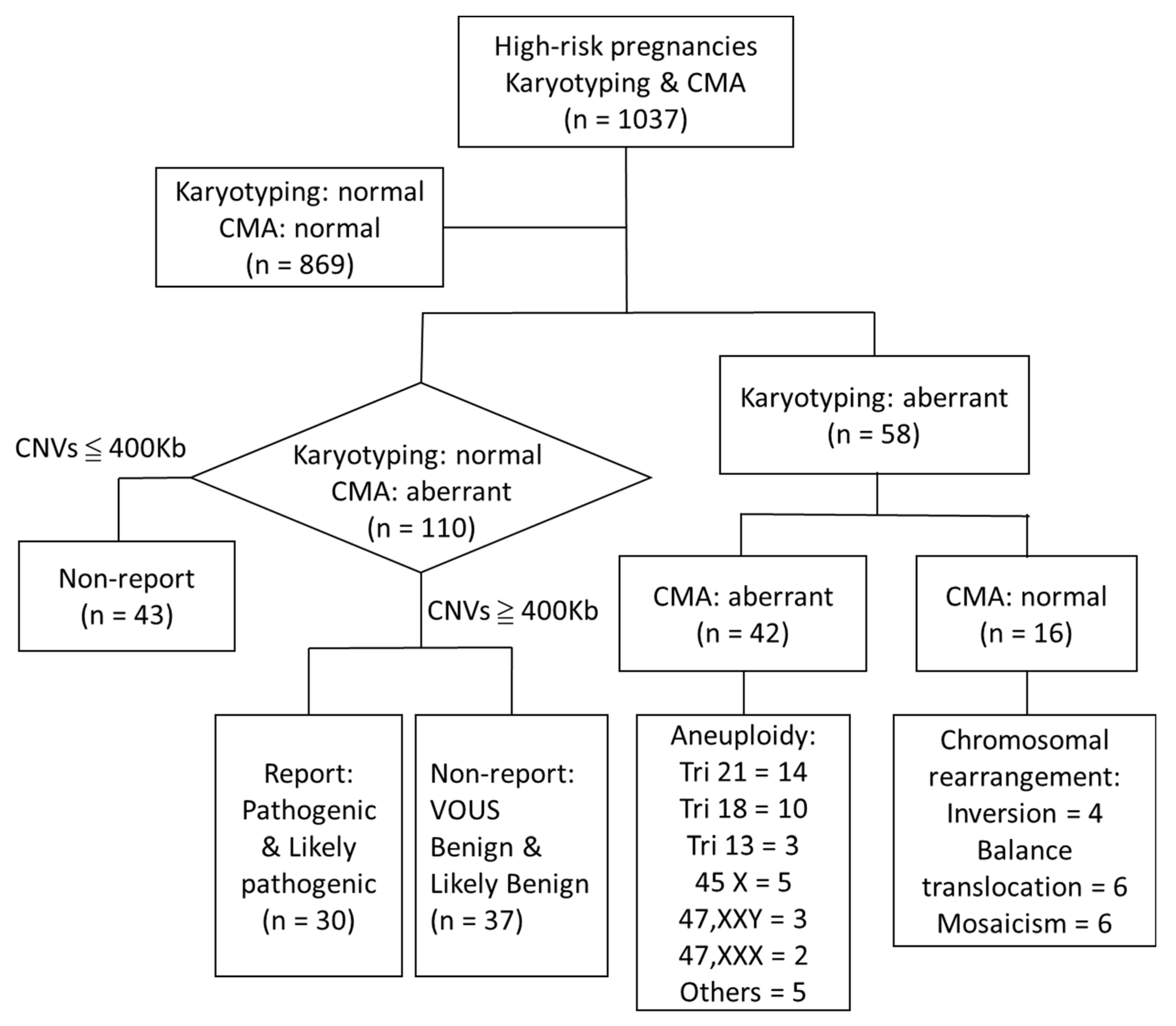
The detected CNVs were classified as (1) pathogenic, (2) likely pathogenic, (3) of uncertain significance, (4) likely benign, or (5) benign, along with standard definitions for each term, in accordance with the guidelines from the American College of Medical Genetics (ACMG) and the International Standard Cytogenomic Array (ISCA). Copy number gains or losses detected with CMA were evaluated systematically for clinical significance via comparison with public CNV databases and with our inhouse database. If aberrant CNVs were detected in fetal DNA, the parental DNA was further analyzed to determine the inheritance of variants in the family to provide information about the significance of VOUS. For clinical arrays, CNV gains or losses of at least ≥400 kb are reported, and CNVs smaller than these size limits for clinical significance are not reported [1,2,6,12,13].
The findings were interpreted according to whether the CNVs were described as pathogenic or benign in the scientific literature, general genome databases, integrated databases, and curated databases. Genomic structural variations were annotated using ClinVar (https://www.ncbi.nlm.nih.gov/clinvar/ (accessed on 1 March 2022)) and ClinGen (https://clinicalgenome.org/ (accessed on 1 March 2022)). Disease-causing genes, their functions, and their inheritance patterns were confirmed using the Online Mendelian Inheritance in Man (OMIM, http://www.omim.org/ (accessed on 1 March 2022)) Database of Genomic Variants (DGV http://dgt.tcag.ca/sgv/app/home (accessed on 1 March 2022)). The Database of Chromosomal Imbalance and Phenotype in Humans using Ensemble Resources (DECIPHER, https://decipher.sanger.ac.uk/ (accessed on 1 March 2022)) was used as a reference for known microdeletion and microduplication syndromes. In addition, the SFARI GENE database (https://gene.sfari.org/autdb/Welcome.do (accessed on 1 March 2022)) was employed to provide genetic information on evaluated CNVs that are associated with autism spectrum disorders (ASD), developmental delay (DD), and intellectual disability (ID) [2]. Additionally, a subgroup of the pathogenic CNVs was labeled as susceptibility variants, according to Rosenfeld et al. [14].

3. Results

The total number of cases enrolled with indications for invasive prenatal testing by CMA and conventional cytogenetics are presented in Figure 1. For all cases with findings of pathologic/unknown penetrance or VOUS, parental a-CGH analysis was performed to determine whether the findings were inherited or de novo. Furthermore, all fetuses with duplication or deletion findings were evaluated with detailed ultrasound scans to confirm the structural information related to the chromosomal abnormalities. Both the G-banding karyotyping and CMA results were normal for 869 of the 1037 cases. We excluded 43 cases with a normal karyotype and CNVs < 400 kb. Of the 110 cases with a normal karyotype and CNVs ≥ 400 kb, pathogenic variants and likely pathogenic variants were reported for 30 cases, and 37 cases had nonreported VOUS, benign, or likely benign variants. G-banding karyotype rearrangements were detected in 58 cases (58/1037, 5.6%), including 42 (4.1%) cases of aneuploidy, of which 14, 10, 3, 5, 3, and 2 cases were trisomy 21, 18, 13, monosomy X, 47,XXY, and 47,XXX, respectively. G-banding indicated six balanced-translocations, four inversions, and six mosaic aberrant karyotypes among cases with normal CMA results. Therefore, in our cohort, CMA produced an additional diagnostic yield of 2.9% (30/1037) for CNVs ≥ 400 kb, including reported pathogenic or likely pathogenic variants, 3.6% (37/1037) for nonreported variants, including VOUS, benign, and likely benign variants, and 4.1% (43/1037) for CNVs < 400 kb, corresponding to a total additional diagnostic yield of 10.6% (110/1037).
As shown in Table 1, among the 546 cases (52.7%; 546/1037) cases with AMA only (the major indication for prenatal testing in this cohort), 4% (22/546) of them showed chromosomal abnormality with CMA. However, only 1.1% (6/546) showed pathogenic/likely pathogenic CNVs. This indicated that AMA only is less relevant than other indications in Table 1. Among the 197 (19.0%, 197/1037) cases with abnormal ultrasound findings, reported CNVs ≥ 400 kb were detected in 11 cases (1.06%, 11/1037). The major anomalies observed on ultrasound included hydrocephalus, congenital fetal heart anomalies, megacystis, pyelectasis, gastroschisis, omphalocele, and the pentalogy of Cantrell. Reported CNVs ≥ 400 kb were detected in 13 (1.25%, 13/1037) of the 189 cases (18.2%, 189/1037) with high-risk Down syndrome screening results (≥1:270). All cases of trisomy 21, 18, and 13 diagnosed by G-banding and CMA were classified as high risk in Down syndrome screening (≥1:270). Thirteen of thirty (43.3%) cases with reported CNVs ≥ 400 kb had high-risk Down syndrome screening results (≥1:270). Reported and nonreported CNVs ≥ 400 kb were detected for one and five cases among the 10.1% (105/1037) of pregnancies with a family history or previous pregnancy with genetic abnormalities, respectively.
As shown in Table 2, pathogenic and likely pathogenic CNVs ≥ 400 kb were reported for 30 cases. Most of these cases presented multiple high-risk indications during chromosomal screening (NT ≥ 3.5 mm, 9 cases) or ultrasound scan anomalies (10 cases); 25 variants were de novo and five cases were pathogenic penetrance inheritance. The outcomes for these 30 cases were 20 terminations of pregnancy (TOP), 7 healthy births, 2 live births with signs of mental retardation, and 1 intrauterine fetal death at 32 weeks of gestation.

4. Discussion

Due to the incrementally higher diagnostic yield of CMA for chromosomal abnormalities compared to traditional G-banding karyotyping, numerous studies have used CMA to detect microdeletions and microduplications in high-risk pregnancies. Prenatal CMA is now more commonly employed in cases where fetal anomalies have been detected by ultrasound [15]. For isolated fetal anomalies, prenatal CMA has been shown to provide additional diagnostic yield over conventional karyotyping, and has thus been recommended in such situations. Metanalyses of CMA results in cases with isolated ultrasound defects indicate a pathogenic variant rate of ~5% [16]. Prenatal CMA identified clinically significant genomic alterations in 9.1% of cases with one or more abnormal ultrasound finding, and the majority of these variants were below the resolution of karyotyping, with the greatest yield observed for cardiac and renal anomalies [17,18]. Thus, it is likely that CMA will replace karyotyping in high-risk pregnancies [15]. Based on the available evidence, the American College of Obstetricians and Gynecologists and the Society of Maternal–Fetal Medicine recommend CMA should be performed instead of karyotyping in pregnancies with anomalous fetuses undergoing invasive testing [4,19].
Most clinical laboratories performing aCGH in postnatal studies report clinically significant imbalances in the range of 50–100 kb. The reporting size range is usually larger in prenatal studies, and it may vary according to the indication for testing [3]. A number of guidelines or recommendations have been developed in several countries. The practice guidelines of the Canadian College of Medical Geneticists–Society of Obstetricians and Gynecologists of Canada (CCMG-SOGC) reported that CNVs overlap completely with an established dosage-sensitive region. The Royal College of Pathologists, the British Society for Genetic Medicine, and the Royal College of Obstetricians and Gynecologists (RCP-BSGM-RCOG) also recommend the use of CMA in high-risk pregnancies [20,21]. VOUS above the size of the CMA cutoffs are only reported if there is significant supporting evidence that deletion or duplication of the region may be pathogenic. Secondary findings associated with a medically actionable disorder with childhood onset must be reported, whereas variants associated with adult-onset conditions are not reported unless requested by the parents or if disclosure could prevent serious harm to family members. In this study, the pathogenic dosage-sensitive region cutoffs used for the reported CNVs are in agreement with both the Canadian and British practice guidelines.

4.1. Overview of CMA Platforms and Interpretation of Prenatal Examinations

Clinical arrays are typically designed to detect imbalances of 20–50 kb in targeted regions (e.g., within known Mendelian genes) and imbalances of 100–250 kb in nontargeted (backbone) regions of the genome [1]. This study used the CytoOneArray® cytogenomic microarray. This CMA (~33,000 probes of 60 nt) is based on a minimum consecutive probe number of 15 in pathogenic/likely pathogenic CNV hotspots, rather than spreading the probes uniformly within pathogenic/likely pathogenic gene regions. The target regions are selected based on pathogenic/likely pathogenic CNV frequency data in CNV databases to implement CMA designs with added targeted coverage of known disease-associated genes and regions (e.g., OMIM morbid genes). The nontargeted regions are designed as five consecutive probes at a distance of 10–30 kb per 1 Mb [22]. Prenatal array platforms conforming to the European and international consensus have also been established for a lower limit threshold of 400 kb across the genome [21]. The literature indicates that the platform, size filter cutoffs, and target regions of cytogenomic microarrays affect the detection of CNVs in prenatal diagnosis. The findings of this study underscore the significant benefits of the CMA platform, as well as the recommended size filter cutoffs and target regions used in data analysis, for the detection of CNVs in a cohort of prenatal cases. An overview of the recent literature on CMA platforms and the interpretation of prenatal examinations is shown in Table 3. We used the ACMG standards and guidelines for the interpretation and reporting of prenatal constitutional CNVs. Parental studies provide some of the most useful evidence on the clinical significance of the observed CNVs. The interpretation of a VOUS can be aided by information about whether the variant was inherited from a healthy parent or occurred de novo in the proband. Population studies suggest that >99% of all benign CNVs are inherited, and the vast majority of inherited CNVs are much smaller than 500 kb [1]. Most CMA platforms—including Affymetrix, Agilent, Illumina, and the Phalanx CytoOneArray—have a resolution of 100 kb, and most follow the 400 kb interpretation cutoff recommended by the ACMG. Depending on the patient population (Table 3), the variable detection rate ranges from 2.15 to 39.79%, and reported pathogenic and likely pathogenic CNVs vary from 0.38% to 8.27%. In accordance with the guidelines, the CMA analysis in this study achieved a 2.89% (30/1037) higher detection rate for reported pathogenic and likely pathogenic CNVs ≥ 400 kb and a 3.57% (37/1037) higher rate for nonreported CNVs. When including the 42 cases of aneuploidy and 43 cases with CNVs < 400 kb, the overall detection rate was 14.7% (152/1037).

4.2. Detection of Uniparental Disomy by CGH and SNP Platforms

Most occurrences of uniparental disomy (UPD) in chromosomes do not result in phenotypic anomalies. Maternal UPD involving chromosomes 2, 7, 14, and 15 and paternal UPD involving chromosomes 6, 11, 15, and 20 are associated with phenotypic growth, neurodevelopmental, and behavioral abnormalities [27,34]. UPD in maternal chromosome 7 is associated with a phenotype similar to Russell–Silver syndrome with intrauterine growth restriction [35]. As shown in the pathogenic and likely pathogenic CNVs in Table 2, case 9 exhibited a de novo 11.45 Mb gene deletion at the 7q21.11q21.3 loci related to Russell–Silver syndrome; both parents had a normal chromosome 7. In actuality, aCGH cannot determine the UPD condition in a patient; however, case 7 shows that a loss of copy has the potential to result in an isodisomy-related phenotype. Unfortunately, the methylation analysis is not available and we lack data to rule out the disorder. As this imprinting disorder is characterized by pre- and postnatal growth retardation; a triangular face; and facial, limb or truncal asymmetry, the parents decided to terminate the pregnancy. Another case exhibited a maternally inherited 3.805 Mb 7q21.12q21.2 duplication classified as a VOUS. Because the risk of heterodisomy or isodisomy is very low in a case with the gain of imprinting loci, the parents decided to continue the pregnancy, and a healthy baby was born (Supplementary Table S1). Neither of these cases met the criteria for UPD.

4.3. Does CMA Provide a Higher Detection YIELD Than Karyotyping?

In this study (Figure 1), all 42 cases with aneuploidy, including trisomy 21 (n = 14), 18 (n = 10), and 13 (n = 3); monosomy X (n = 5); 47,XXY (n = 3); 47,XXX (n = 2); and other variants (n = 5), were diagnosed with both prenatal G-banding karyotyping and CMA. However, six balanced translocations, four inversions, and six low-percentage mosaicisms were not detected with CMA. The aCGH used in this article can also detect the mosaic level above 60%. The overall detection rate of CMA was 14.65% (152/1037). Despite the aneuploidies, the incremental yield of 10.6% (110/1037) included 67 cases with CNVs ≥ 400 kb (10.6%; 67/1037), including cases with reported pathogenic or likely pathogenic variants (2.9% (30/1037)) and cases with nonreported VOUS, benign, or likely benign variants (3.6% (37/1037)). Moreover, cases with nonreported CNVs < 400 kb were also detected by CMA (4.1% (43/1037)). According to our review, CGH + SNP almost dominates the current prenatal testing platform. CGH + SNP can provide more clinical information on genetic findings, such as smaller CNV, UPD or tetraploidy, etc., and those findings provide valuable information for the consideration of possible clinical outcomes. However, for some findings, further specific tests are necessary to confirm the diagnosis; for instance, a methylation test is still necessary to rule out whether the loss of imprinting expressed in the fetus results in UPD disorder; thus, it still a difficult and urgent condition in prenatal diagnosis. We recognize that the CGH platform may not provide the information as the SNP array does. In order to make up for this shortcoming, our strategy is to conduct the karyotyping and aCGH side by side, and the results show that targeted aCGH can indeed help discover most of the significant CNVs to improve the diagnosis rate.

4.4. Does CMA Have a Higher Detection Yield for Fetuses with Increased NT and cFTS?

RCP/RCOG/BSGM recommendations indicate that CMA should be performed if nuchal translucency (NT) ≥ 3.5 mm occurs when the crown–rump length measures between 45–84 mm [21]. A systematic review and metanalysis of 17 studies reported that genomic microarrays provided a 5.0% incremental yield for detecting CNVs and aberrations, including those involving 22q11.2, in fetuses with isolated increased NT and normal karyotypes [36]. Lund et al. [37] reported a detection rate of 12.8% in a prospective clinical series using high-resolution prenatal CMA on uncultured CVS. The detection rate of clinically important chromosomal anomalies was significantly higher in pregnancies with NT ≥ 3.5 mm. In pregnancies with NT ≥ 4.5 mm, the detection rate was as high as 26.5% [38]. In this study, CMA detected CNVs ≥ 400 kb in 33.3% (10/30) of cases with NT ≥ 3.5 mm. In cFTS risk assessment, when combining ultrasound soft markers (abnormal doppler flow of the ductus venosus or tricuspid and an absent nasal bone) and serum in the evaluation, the major indication for high-risk screening of CMA was revealed to be 43.3% (13/30), which is a significantly higher detection rate (Table 2). This means that NT and cFTS in CMA are more effective than other single soft markers.

4.5. Relationship between CMA and Ultrasound Scan Abnormalities

Congenital anomalies are highly correlated with chromosomal abnormalities, and vary depending on the number and type of scan anomalies. De Wit reported that pathogenic CNVs can be detected in 5.6% of fetuses with isolated anomalies and 9.1% of fetuses with multiple anomalies [16]. In this study (Table 1), the major indications in abnormal ultrasounds were omphalocele, micrognathia, radial aplasia, megacystis, pyelectasis, ambiguous genitalia, echogenic bowel, VACTERL, and heart defects; one or more of these features was detected among 36.7% (11/30) of fetuses with reported pathogenic and likely pathogenic CNVs ≥ 400 kb. Thus, CMA obviously increases the detection yield in prenatal diagnosis.
CMA is recommended for all types of CHD in cases with prenatally diagnosed fetal cardiovascular malformations [38,39,40]. Combined CMA and next-generation sequencing detected pathogenic chromosomal anomalies in 21 of 115 (18.3%) fetuses with CHDs [10]. As shown in Table 2, CMA detected two cases with reported pathogenic and likely CNVs ≥ 400 kb associated with pathogenic congenital heart abnormalities. Case 14 had a double outlet of the ventricle (10q23 deletion syndrome; MIM: 612242) and case 16 had tetralogy of Fallot (16p11.2 deletion syndrome; MIM: 611913); both of these cardiovascular malformations are associated with aberrations in CMA, which suggests that CMA may enable more detailed analysis for the detection of CHD.

4.6. CNVs and Advanced Maternal Age

The aneuploidy abnormality rate increases with AMA. CMA has been reported to increase the prenatal diagnostic rate by about 1.4–1.7% for pregnancies in women with AMA [4,26]. In this study (Table 1), AMA was the major indication for CMA, and 0.55% (3/546) of the reported CNVs ≥ 400 kb were lower than those reported in the literature. However, in the nonreported CNVs ≥ 400 kb, 2.56% (14/546) were more common than those reported in the AMA group.

4.7. Prenatal Clinical Counseling for Inherited CNVs or CNVs with Variable Penetrance and Expressivity

Although the generalization between the size and significance of CNVs holds true as a general rule, very large CNVs can be benign in nature, and very small CNVs can be clinically significant [2]. To estimate the penetrance for recurrent pathogenic CNVs, the background risk for congenital anomalies/developmental delay/intellectual disability was assumed to be ~5%. RCOP/BSGM/RCOG recommendations for the use of CMA in pregnancy susceptibility includes distal 1q21.1 deletions and duplications, 15q13.3 deletions, distal and proximal 16p11.2 deletions, and 17q12 deletions. Detailed scans looking for associated anomalies in a clinical context should be considered when reporting these variations, including 22q11.2 duplication, proximal 1q21.1 deletion, and 17q12 duplication [21]. To compare the frequency of CNVs of variable penetrance in low-risk and high-risk prenatal samples, the CNVs were categorized based on clinical penetrance as: (i) high (>40%), (ii) moderate (10–40%), and (iii) low (<10%). High-penetrance CNVs play a major role in the overall heritability of developmental, intellectual, and structural anomalies. Low-penetrance CNVs do not seem to contribute to these anomalies [41].
Table 2 shows the cases with pathogenic and likely pathogenic CNVs ≥ 400 kb. Firstly, two cases of de novo proximal 16p 11.2 deletion CNVs involving the recurrent BP4 and BP5 breakpoint (BP) regions, including the morbid gene TBX6 (MIM: 611913), were detected in this study; the deletion of this region may be associated with developmental delay, cognitive impairment, language delay, autism spectrum disorder, delayed language development, or minor dysmorphic facial features. In both of these cases, the parents decided to TOP due to the associated ultrasound anomalies and a high penetrance estimate of 46.8%. Case 18 presented with 16p 11.2 duplication with a moderate penetrance estimate of 27.2%; the parents decided to continue the pregnancy, and a live baby was born. Secondly, case 19 had maternally inherited 16p13.11 duplication affecting the morbid gene MYH11 (MIM: 160745); cFTS risk evaluation revealed tricuspid regurgitation and the absence of a nasal bone, and intrauterine fetal death occurred at 32 weeks gestation. Thirdly, both case 20 (de novo) and case 21 (maternally inherited) involved 22q11.1q11.21 duplication affecting the morbid genes TBX1 (MIM: 602054) and CECR2 (MIM: 115470); furthermore, the duplication region partially overlapped with a critical region of cat eye syndrome (CES). CES is characterized by large phenotypic variability, ranging from near normal to severe malformations, as reflected by varied neurodevelopmental outcomes [42]. In general, CES is typically associated with a supernumerary bisatellited marker chromosome (inv dup 22pter-22q11.2), resulting in four copies of this region, but aCGH cannot well discriminate the difference between three and four copies; thus, we further checked the karyotype data of both cases. Case 20 showed a mosaic karyotype with a supernumerary marker chromosome (47,XY+ mar de novo [42]/46,XY [29]) and pyelectasis was found with an ultrasound. Therefore, the fetus was diagnosed as having CES, and, finally, the parents decided to TOP. However, the gain of CNV in case 21 was maternally inherited and the karyotype was normal. Some publications report that the interstitial duplication of 22q11.2 is associated with typical CES [43]. Based on those reports, the risk of pathogenicity cannot be ruled out, and it was considered to be a likely pathogenic case. The parents decided to continue the pregnancy, and a healthy baby was born; after three years following, no significant clinical issues were found. Fourthly, 22q11.21 duplication syndrome (MIM: 608363) was noted in two cases. Case 25 had increased NT (4.4 mm) and the variant was de novo; thus, the parents decided to TOP. Case 26 was maternally inherited; thus, the parents decided to continue the pregnancy, and a healthy baby was born [14]. Lastly, Xq28 duplication syndrome (MIM: 300815) affecting the morbid gene RB39B, associated with intellectual developmental disorder, was noted in case 25. The ultrasound scan revealed ambiguous genitalia, and the variant was de novo (MIM 300815); thus, the parents decided to TOP. In case 30, the fetal ultrasound scan revealed an echogenic bowel; in this case, the variant was paternally inherited, and a healthy baby was born.
When a CNV is found in a parent or another relevant family member, numerous caveats should be considered. Many genes may include variations related to secondary or incidental findings associated with adult-onset conditions or carrier status. It is recommended that solicited CNV pathogenic findings should only be reported when the identified variant may inform present or future management of the pregnancy or family; nonactionable findings should not be reported. Clinics and laboratories should report female carriers of X-linked recessive mutations associated with childhood-onset disorders, since there may be significant risk to the family if affected males are conceived [21]. In this study, one case with a maternally inherited CNV reported as Xp21.1 deletion was diagnosed as pathogenic Duchene muscular dystrophy (MIM: 310200), which affects the female carriers of X-linked recessive mutations associated with childhood-onset disorders. There may be significant risk to the family if affected males are conceived. As the fetus was female, we disclosed the heterozygous recessive findings. After thorough counseling, the parents decided to carry on with the pregnancy, and a healthy female was born.
The Supplementary Table S1 shows all 37 cases with nonreported CNVs < 400 kb detected by CMA and interpreted as VOUS, benign, or likely benign for all types of inheritance. There is evidence to refute the significance of the duplication of the Xp22.31 (STS) region. Duplications of this region are common in the general population (~0.32–0.41%). Recent studies on carriers of this duplication, identified through large cohort studies of the general population, showed that carriers perform similarly in neurocognitive tests to noncarrier controls [44]. However, recessive X-linked ichthyosis (MIM: 308100) is carried in females and only phenotypically manifests in males. Firstly, eight cases of Xp22.31 duplication were detected in this study: one de novo deletion associated with pathogenic X-linked mental retardation and seven benign inherited variants; all parents decided to continue the pregnancies and healthy babies were born. During counseling to take family histories, most male family members reported ichthyosis, but the females were healthy. Secondly, the prevalence of Y-chromosome deletion or microdeletion is estimated to be one in 2000–3000 in males. The frequency of Yq microdeletions in males with azoospermia and oligozoospermia is about 5–15% [45]. This study detected eight cases with Yq11.223q11.23 variants, all paternally inherited (four deletions and four duplications) ranging in size from 0.415 to 2.246 Mb; we did not report all of these variants. Moreover, three and four cases of common CNVs ≥ 400 kb associated with 15q11.2 deletion and 15q13.3 duplication were detected, respectively. ClinGen queries of the Database of Genomic Variants (DGV) gold-standard GRCh37 dataset indicated no relationships have been reported between the gene(s) included in this region and human disease. Given the high population frequency, this region has been classified as “dosage sensitivity unlikely”.
Finally, providing information on incidental prenatal findings may have profound consequences, as the parents can opt to terminate the pregnancy. Many CNVs are associated with phenotypes that have reduced penetrance and/or variable expressivity. Some cases may inherit CNVs from the parents, but their penetrance varies dramatically, and ultrasound may show severe multiple fetal anomalies. Some women find this information to be “toxic” and to cause considerable amounts of anxiety during pregnancy and beyond, and even during their baby’s childhood [46]. The identification of a CNV allows for a more precise understanding of the medical and neurocognitive implications of the anomaly, which is important when making decisions about the pregnancy and in planning care for the child [18]. In this study, the chromosomal microarray technologies standard was followed up with the ACMG update guideline in order to provide good quality in the clinical application of the diagnostic evaluation of constitutional disorders [47]. Studies using pre- and post-tests have described the potential limitations of CMA as a clinical test, especially with regard to the detection of VOUS, balanced translocations, and low-level mosaicism. The reporting of VOUS to families in a prenatal setting should be discussed in the context of VOUS detected by CMA.
In conclusion, this study demonstrates the clear value of CMA for the evaluation of cFTS and fetal structural anomalies. The variable clinical implications of inherited CNVs also depend on the mechanisms of inheritance that influence the expression of a trait. Genetic counseling and the evaluation of the risk of recurrence of the genetic abnormality are important for families. However, larger prospective cohort studies with greater focus on additional information are needed.

5. Conclusions

Prenatal CMA is recommended for high-risk pregnancies.

Supplementary Materials

The following supporting information can be downloaded at: https://www.mdpi.com/article/10.3390/jcm11133624/s1, Table S1. Overview of the cases with inherited benign, likely benign, or susceptibility copy number variants non-report after chromosome microarray (≥400 kb) in this study.

Author Contributions

This study was designed by C.-H.H., who was the major contributor in writing the manuscript. C.-H.H., J.-S.C., Y.-M.S., Y.-C.W. and Y.-J.C. analyzed and interpreted the fetal copy number variants in the chromosomal microarray analysis-associated disease data. C.-H.C. conducted the statistical analyses. W.-C.C. critically reviewed the manuscript. All authors have read and agreed to the published version of the manuscript.

Funding

This research received no external funding.

Institutional Review Board Statement

This study was conducted in accordance with the principles of the Declaration of Helsinki. Ethical approval for this study was obtained from the Institutional Review Board of Taipei City Hospital (Reg. No. TCHIRB-10602101) for studies involving humans.

Informed Consent Statement

All participants were informed of the purpose of the study and provided informed written consent before participation.

Data Availability Statement

The data are not publicly available as they contain information that could compromise the privacy of the research participants, but encoded data are available from the corresponding author upon reasonable request.

Acknowledgments

We would like to acknowledge the study participants for providing the necessary personal information and Taipei City Hospital for allowing us to access the patients’ charts. Genetic Counselor Li Wei Kou supported the clinical laboratory interpretation of information.

Conflicts of Interest

The authors declare no conflict of interest.

Abbreviations

cFTS: combined first-trimester screening; CMA: chromosomal microarray analysis; CNV: copy number variants; VOUS: variant of unknown significance.

References

  1. Miller, D.T.; Adam, M.P.; Aradhya, S.; Biesecker, L.G.; Brothman, A.R.; Carter, N.P.; Church, D.M.; Crolla, J.A.; Eichler, E.E.; Epstein, C.J.; et al. Consensus statement: Chromosomal microarray is a first-tier clinical diagnostic test for individuals with developmental disabilities or congenital anomalies. Am. J. Hum. Genet. 2010, 86, 749–764. [Google Scholar] [CrossRef] [PubMed]
  2. Kearney, H.M.; Thorland, E.C.; Brown, K.K.; Quintero-Rivera, F.; South, S.T. American College of Medical Genetics standards and guidelines for interpretation and reporting of postnatal constitutional copy number variants. Genet. Med. 2011, 13, 680–685. [Google Scholar] [CrossRef] [PubMed] [Green Version]
  3. Levy, B.; Wapner, R. Prenatal diagnosis by chromosomal microarray analysis. Fertil. Steril. 2018, 109, 201–212. [Google Scholar] [CrossRef] [PubMed] [Green Version]
  4. Wapner, R.J.; Martin, C.L.; Levy, B.; Ballif, B.C.; Eng, C.M.; Zachary, J.M.; Savage, M.; Platt, L.D.; Saltzman, D.; Grobman, W.A. Chromosomal microarray versus karyotyping for prenatal diagnosis. N. Engl. J. Med. 2012, 367, 2175–2184. [Google Scholar] [CrossRef] [Green Version]
  5. Zhu, X.; Chen, M.; Wang, H.; Guo, Y.; Chau, M.H.K.; Yan, H.; Cao, Y.; Kwok, Y.K.Y.; Chen, J.; Hui, A.S.Y.; et al. Clinical utility of expanded non-invasive prenatal screening and chromosomal microarray analysis in high-risk pregnancy. Ultrasound Obstet Gynecol. 2021, 57, 459–465. [Google Scholar] [CrossRef]
  6. South, S.T.; Lee, C.; Lamb, A.N.; Higgins, A.W.; Kearney, H.M. ACMG standards and Guidelines for constitutional cytogenomic microarray analysis, including postnatal and prenatal applications: Revision 2013. Genet. Med. 2013, 15, 901–909. [Google Scholar] [CrossRef]
  7. Vogel, I.; Petersen, O.B.; Christensen, R.; Hyett, J.; Lou, S.; Vestergaard, E.M. Chromosomal microarray as primary diagnostic genomic tool for pregnancies at increased risk within a population-based combined first-trimester screening program. Ultrasound Obstet. Gynecol. 2018, 51, 480–486. [Google Scholar] [CrossRef] [Green Version]
  8. Zhang, Z.; Hu, T.; Wang, J.M.; Li, Q.Q.; Wang, H.; Liu, S.L. Prenatal Diagnostic Value of Chromosomal Microarray in Fetuses with Nuchal Translucency Greater than 2.5 mm. BioMed Res. Int. 2019, 2019, 6504159. [Google Scholar] [CrossRef] [Green Version]
  9. Mademont-Soler, I.; Morales, C.; Soler, A.; Mart´ınez-Crespo, J.M.; Shen, Y.; Margarit, E.; Clusellas, N.; Obo’n, M.; Wu, B.L.; Sa´nchez, A. Prenatal diagnosis of chromosomal abnormalities in fetuses with abnormal cardiac ultrasound findings: Evaluation of chromosomal microarray-based analysis. Ultrasound Obstet. Gynecol. 2013, 41, 375–382. [Google Scholar] [CrossRef]
  10. Zhu, X.; Li, J.; Ru, T.; Wang, Y.; Xu, Y.; Yang, Y.; Wu, X.; Cram, D.S.; Hu, Y. Identification of copy number variations associated with congenital heart disease by chromosomal microarray analysis and next-generation sequencing. Prenat. Diagn. 2016, 36, 321–327. [Google Scholar] [CrossRef]
  11. Charlotte, B.A.; Behar, D.M.; Herrero, S.G.; Rubio, C. Overcoming Challenges in Reproductive Health Applications by Deploying More Sensitive and Accurate Molecular Technologies. EMJ Repro. Health 2019, 5 (Suppl. S1), 2–12. [Google Scholar]
  12. Richards, S.; Aziz, N.; Bale, S.; Bick, D.; Das, S.; Gastier-Foster, J.; Grody, W.W.; Hegde MLyon, E.; Spector, E.; Voelkerding, K.; et al. Standards and guidelines for the interpretation of sequence variants: A joint consensus recommendation of the American College of Medical Genetics and Genomics and the Association for Molecular Pathology. Genet. Med. 2015, 17, 405–423. [Google Scholar] [CrossRef] [PubMed] [Green Version]
  13. RooneyRiggs, E.; Andersen, E.F.; Cherry, A.M.; Kantarci, S.; Kearney, H.; Patel, A.; Raca, G.; Ritter, D.I.; South, S.T.; Thorland, E.C.; et al. Technical standards for the interpretation and reporting of 451 constitutional copy-number variants: A joint consensus recommendation of the American College of Medical 452 Genetics and Genomics (ACMG) and the Clinical Genome Resource (ClinGen). Genet. Med. 2020, 22, 245–257. [Google Scholar]
  14. Rosenfeld, J.A.; Coe, B.P.; Eichler, E.E.; Cuckle, H.; Shaffer, L.G. Estimates of penetrance for recurrent pathogenic copy-number variations. Genet. Med. 2013, 15, 478–481. [Google Scholar] [CrossRef]
  15. Hillman, S.; McMullan, D.J.; Maher, E.R.; Kilby, M.D. The use of chromosomal microarray in prenatal diagnosis. Obstet. Gynaecol. 2013, 15, 80–84. [Google Scholar] [CrossRef]
  16. De Wit, M.C.; Srebniak, M.I.; Govaerts, L.C.P.; Van Opstal, D.; Galjaard, R.J.H.; Go, A.T.J.I. Additional value of prenatal genomic array testing in fetuses with isolated structural ultrasound abnormalities and a normal karyotype: A systematic review of the literature. Ultrasound Obstet. Gynecol. 2014, 43, 139–146. [Google Scholar] [CrossRef]
  17. Shaffer, L.G.; Rosenfeld, J.A.; Dabell, M.P.; Coppinger, J.; Bandholz, A.M.; Ellison, J.W.; Ravnan, J.B.; Torchia, B.S.; Ballif, B.C.; Fisher, A.J. Detection rates of clinically significant genomic alterations by microarray analysis for specific anomalies detected by ultrasound. Prenat. Diagn. 2012, 32, 986–995. [Google Scholar] [CrossRef] [Green Version]
  18. Donnelly, J.C.; Platt, L.P.; Rebarber, A.; Zachary, J.; Grobman, W.A.; Wapner, R.J. Association of Copy Number Variants with Specific Ultrasonographically Detected Fetal Anomalies. Obstet. Gynecol. 2014, 124, 83–90. [Google Scholar] [CrossRef]
  19. American College of Obstetricians and Gynecologists Committee on Genetics. Committee opinion no. 682: Microarray and Next-Generation Sequencing Technology: The use of Advanced Genetic Diagnostic Tools in Obstetrics and Gynecology. Obstet. Gynecol. 2016, 128, e262–e268. [Google Scholar] [CrossRef] [PubMed]
  20. Armour, C.M.; Dougan, S.D.; Brock, J.A.; Chari, R.; Chodirker, B.N.; DeBie, I.; Evans, J.A.; Gibson, W.T.; Kolomietz, E.; Nelson, T.N.; et al. Practice guideline: Joint CCMG-SOGC recommendations for the use of chromosomal microarray analysis for prenatal diagnosis and assessment of fetal loss in Canada. J. Med. Genet. 2018, 55, 215–221. [Google Scholar] [CrossRef]
  21. Gardiner, C.; Wellesley, D.; Kilby, M.D.; Kerr, B. Recommendations for the Use of Chromosome Microarray in Pregnancy. 2015, Volume 2015, pp. 1–17. Available online: https://www.rcpath.org/resourceLibrary/recommendations-for-the-use-of-chromosome-microarray-inpregnancy.html (accessed on 1 March 2022).
  22. Coe, B.P.; Witherspoon, K.; A Rosenfeld, J.; Van Bon, B.W.M.; Silfhout, A.T.V.-V.; Bosco, P.; Friend, K.L.; Baker, C.; Buono, S.; Vissers, L.; et al. Refining analyses of copy number variation identifies specific genes associated with developmental delay. Nat. Genet. 2014, 46, 1063–1071. [Google Scholar] [CrossRef] [PubMed]
  23. Oneda, B.; Baldinger, R.; Reissmann, R.; Reshetnikova, I.; Krejci, P.; Masood, R.; Ochsenbein-Kölble, N.; Bartholdi, D.; Steindl, K.; Morotti, D. High-resolution chromosomal microarrays in prenatal diagnosis significantly increase diagnostic power. Prenat. Diagn. 2014, 34, 525–533. [Google Scholar] [CrossRef] [PubMed]
  24. Egloff, M.; Hervé, B.; Quibel, T.; Jaillard, S.; Le Bouar, G.; Uguen, K.; Saliou, A.-H.; Valduga, M.; Perdriolle, E.; Coutton, C. Diagnostic yield of chromosomal microarray analysis in fetuses with isolated increased nuchal translucency: A French multicenter study. Ultrasound Obstet. Gynecol. 2018, 52, 715–721. [Google Scholar] [CrossRef] [PubMed] [Green Version]
  25. Sagi-Dain, L.; Maya, I.; Reches, A.; Frumkin, A.; Grinshpun-Cohen, J.; Segel, R.; Manor, E.; Khayat, M.; Tenne, T.; Banne, E.; et al. Chromosomal Microarray Analysis Results from Pregnancies with Various Ultrasonographic Anomalies. Obstet. Gynecol. 2018, 132, 1368–1375. [Google Scholar] [CrossRef]
  26. Shi, Y.; Ma, J.; Xue, Y.; Wang, J.; Yu, B.; Wang, T. The assessment of combined karyotype analysis and chromosomal microarray in pregnant women of advanced maternal age: A multicenter study. Ann. Transl. Med. 2019, 7, 318. [Google Scholar] [CrossRef]
  27. Wang, J.C.; Radcliff, J.; Coe, S.J.; Mahon, L.W. Effects of platforms, size filter cutoffs, and targeted regions of cytogenomic microarray on detection of copy number variants and uniparental disomy in prenatal diagnosis: Results from 5026 pregnancies. Prenat. Diagn. 2019, 39, 137–156. [Google Scholar] [CrossRef] [Green Version]
  28. Lin, Y.U.; Jong, Y.J.; Huang, P.C.; Tsai, C. Detection of copy number variants with chromosomal microarray in 10 377 pregnancies at a single laboratory. Acta Obstet. Gynecol. Scand. 2020, 99, 775–782. [Google Scholar] [CrossRef]
  29. Xia, M.J.; Yang, X.; Fu, J.; Teng, Z.; Lv, Y.; Yu, L. Application of chromosome microarray analysis in prenatal diagnosis. BMC Pregnancy Childbirth 2020, 20, 696. [Google Scholar] [CrossRef]
  30. Hu, T.; Tian, T.; Zhang, Z.; Wang, J.M.; Hu, R.; Xiao, L.; Zhu, H.M.; Lai, Y.; Wang, H.; Liu, S.L. Prenatal chromosomal microarray analysis in 2466 fetuses with ultrasonographic soft markers: A prospective cohort study. Am. J. Obstet. Gynecol. 2021, 224, e1–e516. [Google Scholar] [CrossRef]
  31. Hu, Z.M.; Li, L.L.; Zhang, H.; Zhang, H.G.; Liu, R.Z.; Yu, Y. Clinical Application of Chromosomal Microarray Analysis in Pregnant Women with Advanced Maternal Age and Fetuses with Ultrasonographic Soft Markers. Med. Sci. Monit. 2021, 27, e929074-e1. [Google Scholar] [CrossRef]
  32. Stern, S.; Hacohen, N.; Meiner, V.; Yagel, S.; Zenvirt, S.; Shkedi-Rafid, S.; Macarov, M.; Valsky, D.V.; Porat, S.; Yanai, N.; et al. Universal chromosomal microarray analysis reveals high proportion of copy number variants in low risk pregnancies. Ultrasound Obstet. Gynecol. 2021, 57, 813–820. [Google Scholar] [CrossRef] [PubMed]
  33. Wu, X.Q.; Li, Y.; Lin, N.; Xie, X.R.; Su, L.; Cai, M.Y.; Lin, Y.; Wang, L.S.; Wang, M.Y.; Xu, L.P.; et al. Chromosomal microarray analysis for pregnancies with abnormal maternal serum screening who undergo invasive prenatal testing. J. Cell Mol. Med. 2021, 25, 6271–6279. [Google Scholar] [CrossRef] [PubMed]
  34. Shaffer, L.G.; Agan, N.; Goldberg, J.D.; Ledbetter, D.H.; Longshore, J.W.; Cassidy, S.B. American College of Medical Genetics statement of diagnostic testing for uniparental disomy. Genet. Med. 2001, 3, 206–211. [Google Scholar] [CrossRef] [PubMed] [Green Version]
  35. Gaudio, D.D.; Shinawi, M.; Astbury, C.; Tayeh, M.K.; Deak, K.L.; Raca, G.; ACMG Laboratory Quality Assurance Committee. Diagnostic testing for uniparental disomy: A points to consider statement from the American College of Medical Genetics and Genomics (ACMG). Genet. Med. 2020, 22, 1133–1141. [Google Scholar] [CrossRef] [PubMed] [Green Version]
  36. Grande, M.; Jansen, F.A.R.; Blumenfeld, Y.J.; Fisher, A.; Odibo, A.O.; Haak, M.C.; Borrell, A. Genomic microarray in fetuses with increased nuchal translucency and normal karyotype: A systematic review and meta-analysis. Ultrasound Obstet. Gynecol. 2015, 46, 650–658. [Google Scholar] [CrossRef]
  37. Lund, I.C.B.; Christensen, R.; Petersen, O.B.; Vogel, I.; Vestergaard, E.M. Chromosomal microarray in fetuses with increased nuchal translucency. Ultrasound Obstet. Gynecol. 2015, 45, 95–100. [Google Scholar] [CrossRef]
  38. Jansen, F.A.R.; Blumenffld, Y.J.; Fisher, A.; Cobben, J.M.; Odibo, A.O.; Borrell, A.; Haak, M.C. Array comparative genomic hybridization and fetal congenital heart defects: A systematic review and meta-analysis. Ultrasound Obstet. Gynecol. 2015, 45, 27–35. [Google Scholar] [CrossRef] [Green Version]
  39. Landstrom, A.P.; Kim, J.J.; Gelb, B.D.; Helm, B.M.; Kannankeril, P.J.; Semsarian, C.; Sturm, A.C.; Tristani-Firouzi, M.; Ware, S.M. Genetic Testing for Heritable Cardiovascular Diseases in Pediatric Patients. Circ. Genom. Precis. Med. 2021, 14, e000086. [Google Scholar] [CrossRef]
  40. Zaidi, S.; Brueckner, M. Genetics and Genomics of Congenital Heart Disease. Circ. Res. 2017, 120, 923–940. [Google Scholar] [CrossRef]
  41. Maya, I.; Sharony, R.; Yacobson, R.; Kahana, S.; Yeshaya, J.; Tenne, T.; Agmon-Fishman, T.; Cohen-Vig, L.; Goldberg, Y.; Berger, R.; et al. When genotype is not predictive of phenotype: Implications for genetic counseling based on 21,594 chromosomal microarray analysis examinations. Genet. Med. 2018, 20, 128–131. [Google Scholar] [CrossRef] [Green Version]
  42. Rosias, P.R.; Sijstermans, J.M.; Theunissen, P.M.; Pulles-Heintzberger, C.F.; De Die-Smulders, C.E.; Engelen, J.J.; Van Der Meer, S.B. Phenotypic variability of the cat eye syndrome. Case report and review of the literature. Genet. Couns. 2001, 12, 273–282. [Google Scholar] [PubMed]
  43. Meins, M.; Burfeind, P.; Motsch, S.; Trappe, R.; Burtmus, D.; Langer, S.; Speicher, M.R.; Mühlendyck, H.; Bartels, I.; Zoll, B. Partial trisomy of chromosome 22 resulting from an interstitial duplication of 22q11.2 in a child with typical cat eye syndrome. J. Med. Genet. 2003, 40, e62. Available online: http://www.jmedgenet.com/cgi/content/full/40/5/e62 (accessed on 1 March 2022). [CrossRef] [PubMed] [Green Version]
  44. Crawford, K.; Bracher-Smith, M.; Owen, D.; Kendall, K.M.; Rees, E.; Pardiñas Einon, M.; Escott-Price, V.; Walters, J.T.R.; O’Donovan, M.C.; Owen, M.J.; et al. Medical consequences of pathogenic CNVs in adults: Analysis of the UK Biobank. J. Med. Genet. 2019, 56, 131–138. [Google Scholar] [CrossRef] [PubMed] [Green Version]
  45. Fan, Y.; Silber, S.J. Y Chromosome Infertility 1993–2022; University of Washington: Seattle, WA, USA, 2002; ISSN 2372-0697. [Google Scholar]
  46. Bernhardt, B.A.; Soucier, D.; Hanson, K.; Savage, M.S.; Jackson, L.; Wapner, R.J. Women’s experiences receiving abnormal prenatal chromosomal microarray testing results. Genet. Med. 2013, 15, 139–145. [Google Scholar] [CrossRef] [PubMed] [Green Version]
  47. Shao, L.; Akkari, Y.; Cooley, L.D.; Miller, D.T.; Seifert, B.A.; Wolff, D.J.; Mikhail, F.M.; ACMG Laboratory Quality Assurance Committee. Chromosomal microarray analysis, including constitutional and neoplastic disease applications, 2021 revision: A technical standard of the American College of Medical Genetics and Genomics (ACMG). Genet. Med. 2021, 23, 1818–1829. [Google Scholar] [CrossRef]
Figure 1. Flowchart of patient G-banding karyotype and chromosomal microarray analysis. CMA: chromosomal microarray (CMA); CNVs: copy number variants; VOUS: variant of uncertain significance.
Jcm 11 03624 g001
Table 1. Major indications for prenatal chromosomal microarray analysis (CMA) for 1037 pregnancies with aberrant findings.
IndicationNoT21T18T13Reported
CNVs ≥ 400 kb
Nonreported
CNVs ≥ 400 kb
Advanced maternal age only546 (52.7%)113314
Abnormal ultrasound finding197 (19.0%)241112
High risk on Down syndrome screening (≥1:270)189 (18.2%)13103139
Increased NT (≥3.5 mm) only9065213
FTS soft markers: DV(+), TR(+), NB(−)5675114
Previous pregnancy, child, or familial risk105 (10.1%) 15
T21: trisomy 21; T18: trisomy 18; T13: trisomy 13; CNVs: copy number variants; NT: nuchal translucency; FTS: first-trimester screening; DV(+): ductus venosus feverse; TR(+): tricuspid regurgitation; NB(−): absent nasal bone.
Table 2. Overview of the pathologic, likely pathologic, and susceptibility copy number variants reported after chromosome microarray analysis of pregnancies in this study.
CaseAgeNT (mm)Indication/Ultrasound FindingCMA [hg19] ResultsSize (Mb)InheritanceCandidate OMIM GenesDisordersInterpretationOutcome
1342.5TR(+), High risk1q21.1q21.2 (146,627,038–147,384,032) × 10.757De novo6124741q21.1 deletion syndromePathogenicTOP
2353.5NT(+), High risk2q37.2q37.3 (236,330,093–243,040,324) × 16.710De novo6004302q37 deletion syndromePathogenicTOP
3363.2Omphalocele, High risk3p26.33p26.2 (2,146,782–3,771,742) × 31.625Paternal6072803pter-p25 duplicationVOUSTOP
4212.2CM(+), TR(+), NB(−),High risk3q22.1q25.32 (130,521,560–157,015,801) × 126.494De novo220200Syndromic intellectual disability
Dandy Walker Syndrome
PathogenicTOP
5385.1NT(+), DV(+), High risk, AMA3q27.2q29 (184,799,629–197,803,820) × 3 9p24.3p22.3 (271,257–14,680,180) × 1 9p22.3p13.1 (14,844,795–38,663,271) × 313.004
14.409
23.818
De novo611936/602424/604935/612900/158170/608980/156540/6016733q29 duplication syndrome
9p24.3p22 deletion
9p22.3p13.1 duplication
9p duplication & deletion
LP
Pathogenic
TOP
6272.8High risk4q34.3q35.1 (180,742,112–183,532,267) × 3
4q35.1q35.2 (183,532,267–190,957,460) × 1
2.790
7.425
De novo610083/5189004q34.3q35.1 duplication
4q35.1q35.2 deletion
LPTOP
7254Micrognathia, Low set ear, NT(+)5q32 (145,755,389–150,297,954) × 12.575De novoN/ATreacher Collins syndromePathogenicTOP
8362.8TR(+), High Risk, AMA6q22.1q22.31 (115,853,923–119,245,348) × 33.391De novo605942/604714/612647/612659/610463/618865/172405/610098/1201106q22.1q22.31 duplicationVOUSLB
(MR)
9372AMA7q21.11q21.3 (84,600,949–96,051,291) × 111.45De novo600028/604149Split-hand/foot malformation 1
Silver–Russell syndrome
Myoclonus-Dystonia
PathogenicTOP
10362.6High risk, AMA8q23.1 (106,336,068–106,715,982) × 10.380 603693congenital diaphragmatic herniaLPBH
11355.1NT(+), AMA9p24.3p22.2 (204,193–16,626,507) × 116.422De novo1581709p24.3p22.2 deletion syndromePathogenicTOP
12315.7Radial aplasia, NT(+), High risk9q21.2 (80,191,465–80,601,045) × 30.41De novo600998Radial aplasiaVOUSTOP
13361.9High risk, AMA10q22.3 (79,617,635–81,707,527) × 32.090De novo602412/614258/607159/178642/178630/61863910q22.3 duplicationVOUSBH
14321.4Double outlet of right ventricle, TR(+)10q23.1 (86,767,729–86,984,308) × 10.217De novoN/A10q23 deletion syndromeVOUSTOP
15354.5NT(+), High risk, AMA14q32.31q32.33 (101,758,166–106,852,173) × 15.094De novo614062/605799/61473014q32.31q32.33 deletion syndromePathogenicTOP
16271.9TOF of Heart, NB(−), High risk16p11.2 (29,653,115–30,198,522) × 10.545De novo611913Proximal 16p11.2 deletion syndromePathogenicTOP
17314.3R/O: VACTERL, NT(+), High risk16p11.2 (29,653,115–30,198,581) × 10.545De novo611913Proximal 16p11.2 deletion syndromePathogenicTOP
18321.5Megacystis16p11.2 (29,698,283–30,198,582) × 30.500De novo614671Proximal 16p11.2 duplication syndromePathogenicBH
19372.3TR(+), NB(−), High risk16p13.11 (15,131,575–16,288,874) × 31.157Maternal609449/160745/60323416p13.1 duplicationLPIUFD
20401.7Pyelectasis22q11.1q11.21 (17,444,646–18,106,018) × 30.661De novo115470Cat eye syndrome
(47,XY+ mar de novo [42]/46,XY [29])
PathogenicTOP
21372AMA22q11.1q11.21 (17,444,646–17,993,089) × 30.548Maternal11547022q11.1q11.21 duplication
(46,XY)
LPBH
22319.4NT(+), NB(−), TR(+), High risk22q11.21 (19,035,231–21,449,413) × 12.414De novo188400DiGeorge SyndromePathogenicTOF
23424.3NT(+), TR(+), High risk, AMA22q11.21 (19,006,943–21,461,068) × 12.454De novo188400DiGeorge syndromePathogenicTOP
24422.5TR(+), High risk, AMA22q11.21 (19,006,943–21,461,005) × 1 17q12 (34,823,708–36,247,940) × 32.454
1.426
De novo614526
188400
DiGeorge syndrome
17q12 duplication syndrome
PathogenicTOP
25334.4NT(+)22q11.21 (18,104,691–21,461,005) × 33.356De novo60836322q11.2 duplication syndromePathogenicTOP
26352.1PHx; Genetic Hx22q11.21 (19,006,943–21,461,005) × 32.454Maternal60836322q11.2 duplication syndromePathogenicBH
(Health)
27402.3AMAXp22.31 (6,460,120–8,101,239) × 11.641De novo308100X-linked mental retardation
Ichthyosis, X-linked (XLI)
PathogenicTOP
28311.4NB(−), Mental retardationXq22.1q22.2 (100,907,854–102,659,284) × 11.751De novo300319/300969Xq22.1q22.2 deletion
X-linked mental retardation
LPLB
(MR)
29292.1Ambigious genitalXq28 (154,130,347–154,527,746) × 30.397De novo300815Xq28 duplication syndromePathogenicTOP
30312.5Echogenic bowelXq28 (154,161,678–154,650,677) × 3
7q36.2 (153,923,581–154,024,097) × 1
0.351
0.087
Paternal
Maternal
300815
612956
Xq28 duplication syndrome
7q36.2 deletion syndrome
Pathogenic
VOUS
BH
NT: nuchal translucency; NB(−): absent nasal bone; TR(+): tricuspid regurgitation; DV(+): ductus venosus reverse; CMA: chromosome microarray; OMIN: Online Mendelian Inheritance in Man; VOUS: variants of uncertain significance; TOP: termination of pregnancy; LP: likely pathogenic; LB: live born; BH: born healthy.
Table 3. Overview of current CMA platforms and interpretation of prenatal examinations in the literature.
AuthorPatient PopulationCases NoCMA PlatformChip DesignCMA ResolutionInterpretation Cut OffDetection RateP/LP CNVs
Oneda et al. (2014) [23]High risk #464Affymetrix cytogenetics Whole
Genome 2.7 M array/Cytoscan HD Array
CGH + SNP20–100 Kb20–100 Kb17/464 (3.70%)15/464 (3.23%)
Zhu et al. (2016) [5]Heart anomaly115Affymetrix CytoScan 750 KCGH + SNP100 KbN/A21/115 (18.3%)13/115 (11.3%)
Egloff et al. (2018) [24]High risk #599Agilent PreCytoNEMCGH + SNP60 &180 KbN/A53/599 (8.85%)16/599 (2.67%)
Sagi-Dain et al. (2018) [25]Ultrasound anomaly5750Affymetrix CytoScan 750 K array
Infinium OmniExpress-24 v1.2 BeadChip
BlueGnome Cytochip ISCA 8 × 60 K format
Agilent CGH + SNP (4 × 180 K)
CGH + SNP
SNP
CGH
CGH + SNP
100 Kb1 M (loss)/2 M (gain)272/5750 (4.73%)157/5750 (2.73%)
Vogel et al. (2018) [7]cFTS high risk575Agilent CytoGenomicsCGH + SNP180 KN/A51/575 (8.87%)15/575 (2.61%)
Shi et al. (2019) [26]High risk, AMA703Affymetrix CytoScan 750 KCGH + SNP100 KbN/A48/703 (6.83%)10/703 (1.42%)
Wang et al. (2019) [27]High risk #5026Affymetrix Human SNP Array 6.0
Affymetrix CytoScan HD
SNP
CGH + SNP
Target: 20 kb (loss)/100 kb (gain)
Nontarget: 50 kb (loss)/200 kb (gain)
400 K562/5026 (11.2%)19/5026 (0.38%)
Lin et al. (2020) [28]General population10,377Thermo-Fisher CytoScan750 KCGH + SNP100 Kb200 K223/10,377 (2.15%)126/10,377 (1.21%)
Xia et al. (2020) [29]Ultrasound anomaly477Affymetrix CytoScan 750 KCGH + SNP50 Kb (loss)/100 Kb (gain)100 K (loss)/200 K (gain)71/447 (15.88%)17/447 (3.80%)
Hu et al. (2021) [30]Ultrasound anomaly2466Thermo-Fisher CytoScan750 KCGH + SNP100 Kb400 K107/2466 (4.34%)64/2466 (2.59%)
Hu et al. (2021) [31]AMA, soft marker1521Affymetrix CytoScan 750 KCGH + SNP100 Kb400 K330/1527 (21.61%)37/1520 (2.42%)
Stern et al. (2021) [32]Ultrasound low risk6431Affymetrix CytoScan 750 KCGH + SNP100 KbN/A319/6431 (4.96%)27/6431 (0.42%)
Wu et al. (2021) [33]Serum screening high risk713Affymetrix CytoScan 750 KCGH + SNP100 Kb400 K82/713 (11.5%)59/713 (8.27%)
Zhu et al. (2021) [10]High risk #774Affymetrix CytoScan 750 K
Iliumina HumanCytoSNP-12
Agilent CGH 8 × 60 K (customized)
CGH + SNP
SNP
CGH + SNP
100 Kb400 K308/774 (39.79%)17/774 (2.20%)
Present studyHigh risk #1037Phalanx CytoOneCGHTarget: 50–100 K; Non-target: 1 Mb400 K153/1037 (14.75%)30/1037 (2.89%)
High risk #: includes trisomy, CNVs; high risk with AMA, structural abnormalities on ultrasound, screening high risk, family history.
Publisher’s Note: MDPI stays neutral with regard to jurisdictional claims in published maps and institutional affiliations.

