Review

Anti-Inflammatory and/or Anti-Fibrotic Treatment of MPO-ANCA-Positive Interstitial Lung Disease: A Short Review

1
Department of Respiratory Medicine, Saitama Red Cross Hospital, 1-5 Shintoshin, Chuo-ku, Saitama 330-8553, Japan
2
Department of Respiratory Medicine, Tokyo Jikei University Hospital, 3-25-8 Nishi-shinbashi, Minato-ku, Tokyo 105-8461, Japan
3
Department of Internal Medicine, Japanese Red Cross Kochi Hospital, 1-4-63-11 Hadaminamimachi, Kochi 780-8562, Japan
4
Department of Respiratory Medicine, Kanagawa Cardiovascular and Respiratory Center, 6-16-1 Tomioka-higashi, Kanazawa-ku, Yokohama 236-0051, Japan
5
Department of Respiratory Medicine, Okinawa Chubu Hospital, Okinawa, 81 Miyazato, Uruma 904-2293, Japan
6
Department of Radiology, The Jikei University Daisan Hospital, 4-11-1 Izumihoncho Komae-shi, Tokyo 201-8601, Japan
7
Department of Pathology, Kanagawa Cardiovascular and Respiratory Center, 6-16-1 Tomioka-higashi, Kanazawa-ku, Yokohama 236-0051, Japan
*
Author to whom correspondence should be addressed.
J. Clin. Med. 2022, 11(13), 3835; https://doi.org/10.3390/jcm11133835
Submission received: 16 May 2022 / Revised: 27 June 2022 / Accepted: 30 June 2022 / Published: 1 July 2022
(This article belongs to the Section Pulmonology)

Abstract

:
The presence of a lung lesion is common in microscopic polyangiitis (MPA), and interstitial lung disease (ILD) can lead to a poor prognosis. Although myeloperoxidase antineutrophil cytoplasmic antibodies (MPO-ANCA) are often present in patients with MPA, patients with ILD and MPO-ANCA positivity but without other manifestations of systemic vasculitis have also been reported. Therefore, the possible association between MPO-ANCA, MPA, and idiopathic ILD remains unclear. This problematic matter has influenced the treatment strategy of MPO-ANCA-positive ILD patients without systemic vasculitis. Clinicians should undertake treatment with careful consideration of the four major causes of death in MPO-ANCA-positive ILD: acute exacerbation of ILD, progressive lung fibrosis, infectious comorbidities, and diffuse alveolar hemorrhage. Further, clinicians need to carefully judge whether inflammation or fibrosis is the dominant condition with reference to the patient’s clinical domain and radiopathological lung features. Recently, anti-fibrotic agents such as nintedanib and pirfenidone were shown to be effective in treating various etiologies associated with ILD and have thus led to the widening of treatment options. In this review, the clinical characteristics, radiopathology, prognosis, and therapeutic options in patients with MPO-ANCA-positive ILD are summarized using limited information from previous studies.

1. Introduction

Antineutrophil cytoplasmic antibodies (ANCA) are generally detected in multisystemic diseases, namely ANCA-associated vasculitides such as microscopic polyangiitis (MPA), granulomatosis with polyangiitis (GPA), and eosinophilic granulomatosis with polyangiitis (EGPA) [1,2,3]. The presence of a lung lesion is a common and important clinical feature in ANCA-associated vasculitides [2,4]. However, interstitial lung disease (ILD) is rarely seen in patients with GPA and EGPA [1,2,3,4], but the presence of ILD is a common and important clinical feature in MPA [1,3,4]. In addition, MPO-ANCA have been frequently identified in patients with ILD without multisystemic disease. A recent report showed that ILD was diagnosed before (52%) or simultaneously (39%) with ANCA-associated vasculitides [5]. Therefore, its association with ILD has often been discussed [6,7,8] because ILD can lead to a poor prognosis in patients with MPA [5,9,10,11]. In addition, patients with pulmonary fibrosis and ANCA positivity but without other manifestations of systemic vasculitis have also been reported, which was sometimes diagnosed as “pulmonary-limited type of ANCA-associated vasculitis” [8,12]. Furthermore, ANCA-positive conversion has been described in patients with an initial diagnosis of idiopathic ILD, with manifestations of systemic vasculitis occurring in some patients [13,14]. However, it is unclear whether ILD observed in the presence of only MPO-ANCA differs from idiopathic ILD and ILD with overt MPA [14]. Needless to say, this problematic matter has influenced the treatment strategy of MPO-ANCA-positive ILD patients without systemic vasculitis as a form of idiopathic ILD. Therefore, the aim of the present review was to assess the characteristics of MPO-ANCA-positive ILD using limited information from previous studies in this field and then focus on the future treatment of MPO-ANCA-positive ILD.

2. Prevalence and Clinical Manifestations

The prevalence of MPO-ANCA positivity ranges from 1.7% to 22.2% in patients with idiopathic ILD [8,13,15,16]. From the viewpoint of MPA, the prevalence of ILD ranges from 2.7% to 47.4% in patients with MPA [8,17,18,19,20,21,22,23,24,25]. ILD is more frequently associated with MPO-ANCA positivity in Japanese patients than in Western patients [5,26]. MPO-ANCA-positive ILD is usually observed in patients older than 65 years old as idiopathic pulmonary fibrosis (IPF) [17].
Conversion to MPO-ANCA positivity occurs in patients initially diagnosed as having idiopathic ILD at a reported rate of prevalence of 3.3% to 5.7% [13,15]. Moreover, a quarter of the patients with MPO-ANCA positivity at the diagnosis of idiopathic ILD or with conversion to MPO-ANCA positivity during follow-up developed MPA [8,13,15]. To put it differently, clinicians should be careful to note that MPA develops at a certain incidence rate in MPO-ANCA-positive ILD patients [8,10,13,15].

3. Morphological Domain

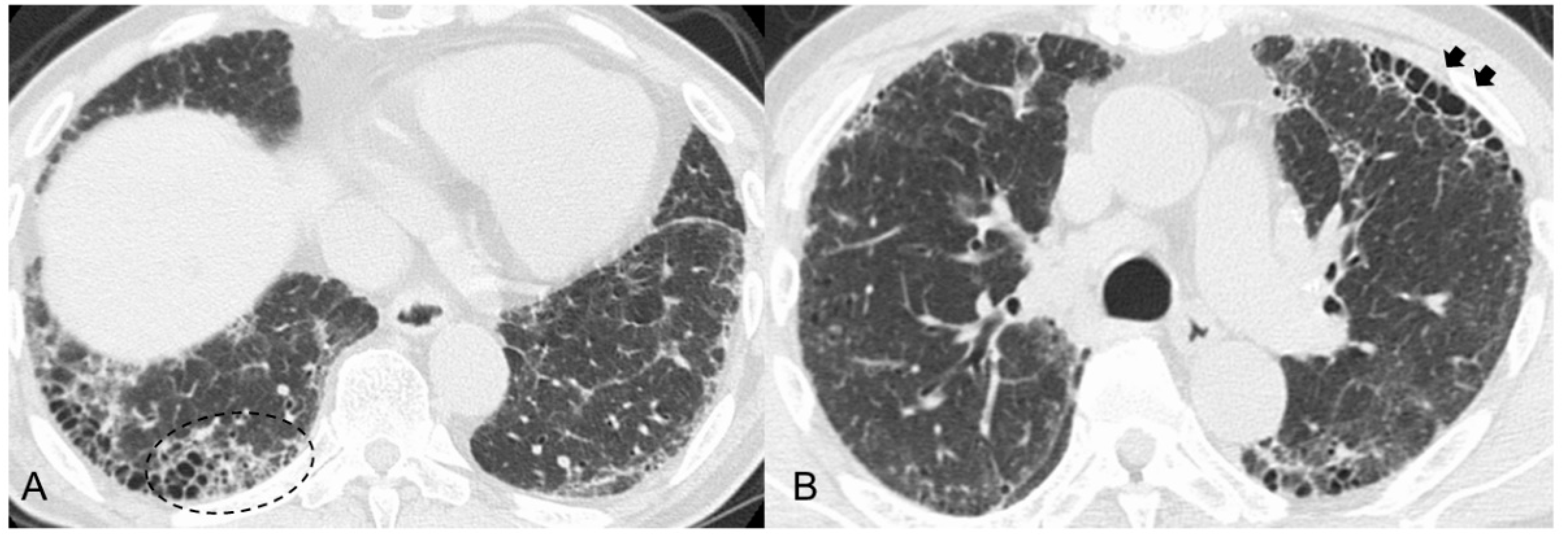
3.1. Radiological Findings

In previous reports, radiological analysis, especially by high-resolution computed tomography (HRCT), was used for the classification of patterns [27,28] in patients with MPO-ANCA-positive ILD. In patients with MPA, the most frequently occurring pattern was usual interstitial pneumonia (UIP) (50–78%), followed by nonspecific interstitial pneumonia (NSIP) (7–58%) and others (13–31%) [14,29,30,31,32]. MPO-ANCA-positive ILD without systemic vasculitis (i.e., idiopathic ILD) also showed a similar tendency (UIP: 12.9–53.9%, non-UIP: 13.6–58.1%) [31,32]. Honeycombing was seen in about 30% of the MPO-ANCA-positive ILD patients regardless of whether they had MPA-ILD or idiopathic ILD [30,31,32].

3.2. Pathological Findings

In studies that specifically described the characteristics of patients with ILD associated with MPA and MPO-ANCA-positive idiopathic ILD, UIP was the most frequent pathological pattern (46–100%), and marked dense fibrosis and areas of honeycombing were common in these specimens [17,20,21,33,34,35]. In addition, a high incidence of findings such as lymphoid hyperplasia, organizing pneumonia, pleuritis, and bronchiolitis is a characteristic observed in ILD associated with MPA and MPO-ANCA-positive idiopathic ILD [18,33,34].

3.3. Differences between MPO-ANCA-Positive ILD and UIP/IPF

For the reason stated above in regard to radiopathological findings, similar to UIP/IPF, MPO-ANCA-positive ILD most commonly showed a UIP pattern and honeycombing as rheumatoid arthritis (RA)-ILD [9,17,36], whereas ILD in patients with systemic sclerosis and myositis is predominantly associated with a non-UIP pattern [9,17,37]. A recent study reported that the single nucleotide polymorphism (SNP) rs35705950T was more strongly associated with UIP than with non-UIP [38]. The SNP rs35705950 (G/T) in the promotor region of MUC5B, which encodes mucin 5B, is a strong genetic factor for UIP/IPF and RA-UIP [39,40]. The MUC5B promoter variant rs35705950 might also be a strong risk factor for MPA-ILD [41]. Therefore, these findings suggest at least a partially shared genetic susceptibility between MPO-ANCA-positive ILD and UIP/IPF.
However, some previous studies reported that MPO-ANCA-positive ILD (including MPA-ILD) and UIP/IPF showed different radiopathological features, despite having the same basic UIP pattern [30,31,33,34,35]. Radiologically, increased attenuation around areas of honeycombing and traction bronchiectasis (Figure 1A) was more frequently seen in MPO-ANCA-positive ILD than in UIP/IPF [30,31,34]. We reported that an anterior upper lobe honeycomb-like lesion, which represents a concentration of cystic air spaces within the anterior aspect of the upper lobes (Figure 1B), might be found with higher frequency in MPO-ANCA-positive ILD and RA-ILD than in other etiologies associated with ILD [31,42]. Pathologically, MPO-ANCA-positive ILD even with a UIP pattern showed more prominent inflammatory cell infiltration and cellular bronchiolitis, unlike UIP/IPF (Figure 2A–E) [8,14,17,33,34,35]. In other words, these pathological features correspond well to the presence of a radiological feature such as increased attenuation around areas of honeycombing and traction bronchiectasis [30,31,34]. Therefore, these features may be associated with a better response to anti-inflammatory therapies in MPO-ANCA-positive ILD patients, and clinicians should be careful to note the development of MPA in patients having these features [31,34] (Table 1).

4. Prognosis and Causes of Death

Studies of previous cohorts of patients with MPA-ILD reported median survival times of 4.1–11 years and 5-year survival rates of 46–60%, although long-term outcomes are less well reported [10,31,32,34,43]. In MPO-ANCA-positive ILD without systemic vasculitis as a form of idiopathic ILD, whether there is a difference in survival time and prognosis between patients with MPA-ILD and those with idiopathic ILD is currently controversial [31,32]. As a caveat, there is a high probability that patients with MPO-ANCA-positive ILD even if showing a UIP pattern that had longer survival than those with UIP/IPF [10,13,34]. The poor prognostic factors reported included older age, higher ANCA titers, higher levels of inflammatory markers (i.e., erythrocyte sedimentation rate [ESR] and C-reactive protein [CRP]), lower forced vital capacity, and anterior upper lobe honeycomb-like lesions [10,13,14,16,31,32,34,44].
The major causes of death in patients with MPO-ANCA-positive ILD were acute exacerbation (AE) of ILD (10–25.5%), chronic progressive fibrosing ILD (PF-ILD) (7.8–19%), infectious disease (19.2–20%), and diffuse alveolar hemorrhage (DAH) (4.3–30%) [10,11,31]. As a cautionary point, it is possible that patients with MPO-ANCA-positive idiopathic ILD may present with DAH as the initial manifestation of MPA development, which is a serious condition, as DAH can be life threatening [10,13,15,31]. Taken together, these findings suggest that clinicians should create a decision-making strategy for the treatment of MPO-ANCA-positive ILD with an awareness of the presence of these four major causes of death (i.e., AE of ILD, PF-ILD, infectious disease, and DAH).

5. Therapeutic Assessment of MPO-ANCA-Positive ILD

5.1. ILD Patients with MPA

When ILD associated with MPA is diagnosed, treatment should be based on organ involvements of vasculitis [8]. Treatment of patients with MPA should include induction therapy using highly potent anti-inflammatory agents (e.g., corticosteroid, cyclophosphamide, rituximab, mycophenolate, and azathioprine) [9,14,17,32,44]. However, no studies have shown whether therapy to treat MPA itself is also effective on ILD. Unfortunately, ILD often independently leads to progressive lung fibrosis [45].

5.2. MPO-ANCA-Positive ILD Patients without Systemic Vasculitis as Idiopathic ILD

Initially, when MPO-ANCA-positive ILD without systemic vasculitis is considered, clinicians should determine whether the patient has a UIP pattern on HRCT. In patients with NSIP or an organizing pneumonia pattern, anti-inflammatory agents (mainly as corticosteroids) should be started to treat these conditions as idiopathic ILD [14,17].
Second, the therapeutic assessment of MPO-ANCA-positive ILD with a UIP pattern is most difficult and controversial due to the lack of evidence [14]. This also includes progressive pulmonary fibrosis (i.e., PF-ILD) with MPA despite anti-inflammatory therapies directed at treating MPA itself. Two main factors are important when considering the decision to start or switch treatment, whether inflammatory change or fibrotic change in ILD is the dominant factor [46].
One factor favoring inflammatory change is the presence of MPA-like disease (mock MPA), as indicated by recurrent unexplained fever and/or higher CRP/ESR and an upward trend in the MPO-ANCA titer. Moreover, the second factor is increased attenuation around fibrotic change seen on HRCT and prominent inflammatory cell infiltration in pathological lung lesions [1,2,3,47,48]. Regarding the former (i.e., mock MPA), if patients with MPO-ANCA-positive idiopathic ILD develop MPA, they should be treated with anti-inflammatory therapy because untreated MPA (particularly that with DAH) is normally progressive and fatal [13,31]. In terms of the latter as a radiopathological finding, anti-inflammatory therapy can be an effective course of treatment even if a UIP pattern is present, as shown in Figure 3 and Figure 4. Therefore, if these conditions are present, anti-inflammatory therapy should be considered.
In contrast, the factors favoring fibrotic change are nonincreasing levels of CRP or ESR, a decrease or leveling off of the MPO-ANCA titer, expansion of fibrotic change as reticulation with traction bronchiectasis and honeycombing on HRCT without increasing attenuation (i.e., ground-glass opacity [GGO] and consolidation), and pathologically patchy fibrosis as a UIP pattern and/or fibroblastic foci in a lung specimen without prominent inflammatory cells [1,2,3,30,31,33,34,47,48,49,50]. As a caveat, GGO, which is a finding typically associated with inflammation or infiltration, does not always represent reversible lung disease, but rather in some cases, it may represent microscopic fibrosis [51]. Therefore, lung biopsy may be useful to assess whether inflammatory or fibrotic change is dominant [36,49,52]. Importantly, recent reports have shown that all etiologies associated with ILD can lead to a poor prognosis, particularly in cases in which a component of UIP is found to a certain degree [53,54,55]. In other words, some cases represent the natural evolution of the inflammatory change of ILD into fibrotic change [56]. Adegunsoye et al. noted that honeycombing represents a progressive fibrotic ILD phenotype regardless of underlying conditions such as connective tissue disease, idiopathic ILD, or fibrotic hypersensitive pneumonia [57]. In addition, AE of ILD can affect all patients with ILD but apparently occurs more frequently in patients with an underlying UIP pattern, which is a most harmful event and can be a major cause of death [58,59]. Therefore, the presence and/or expansion of fibrotic change particularly in the UIP pattern appearing as reticulation with traction bronchiectasis and honeycombing during follow-up has an important prognostic implication in all etiologies associated with ILD [36,40,53]. If we determine that fibrotic change is dominant, we consider starting an anti-fibrotic therapy such as nintedanib or pirfenidone, which has been approved for the treatment of IPF [60,61,62]. The INBUILD study showed the effectiveness of nintedanib on PF-ILD other than IPF, and nintedanib might have the potential to suppress the AE of ILD [63]. Post hoc analysis of the INBUILD study suggested a treatment benefit of nintedanib across all subgroups of patients with PF-ILD, including autoimmune ILD, although the trial was not powered to provide evidence to address this specific question [64]. In the RELIEF study, although the quality of evidence was rated as low, pirfenidone also significantly suppressed the deterioration of forced vital capacity in patients with progressive fibrotic ILD due to four diagnoses: connective tissue disease-associated ILDs, fibrotic NSIP, fibrotic hypersensitivity pneumonitis, and asbestos-induced lung fibrosis [65]. Taken together, expanding fibrotic change in patients with a UIP pattern can lead to the high risk of PF-ILD and AE of ILD, which means a poorer prognosis even in those with MPO-ANCA-positive ILD. Therefore, starting anti-fibrotic therapy such as nintedanib should be considered in such a case (Figure 5, same patient as in Figure 2).
Additionally, considering therapeutic assessment from a different perspective, anti-inflammatory therapy could be harmful in some cases. The importance of separating IPF from ILD associated with specific systemic conditions such as those related to connective tissue diseases has been emphasized [33,66]. It is recommended that IPF patients should not be treated with anti-inflammatory therapies [66,67]. A recent report also showed anti-inflammatory therapy as indicated by steroid use to have a higher risk of developing PF-ILD in connective tissue disease-associated ILD [68]. In RA-ILD, prednisolone doses higher than 10 mg are associated with a higher risk of infectious comorbidity [69]. As also with MPA-ILD in elderly patients, the administration of high-dose corticosteroid therapy is a significant risk factor for severe infection, which is a main cause of death [70]. In general, corticosteroids had been considered to result in a higher risk of chronic pulmonary infection, such as nontuberculous mycobacteria and pulmonary aspergillosis [71,72]. Because the presence of emphysema may also be a risk factor for chronic pulmonary infection in ILD patients [73,74], when considering the initiation or strength of anti-inflammatory therapy, clinicians should pay particular attention to those patients having pulmonary emphysema with ILD to some extent. Moreover, Kawamura et al. reported that corticosteroid use was associated with an increased risk of AE of ILD even in a dose-dependent manner at low doses [75]. Although we cannot say for certain, incomplete anti-inflammatory therapy may cause harmful events such as infectious disease and AE of ILD that can lead to a poor prognosis. Taken together, when it is difficult to select between anti-inflammatory and anti-fibrotic therapy, anti-fibrotic therapy should be given priority in the patients at high risk for infection and AE of ILD such as initiation or long-term administration of moderate- to high-dose corticosteroid, concomitant to a certain extent with pulmonary emphysema (Figure 6).
A prospective study should clarify the natural history of MPA-ILD and the role of ANCA positivity in patients with idiopathic ILD and offer a better therapeutic approach to treating the different groups of patients with this condition [76].

6. Conclusions

MPO-ANCA-positive ILD is not uncommon in any case of MPA-ILD or idiopathic ILD. Because some patients with MPO-ANCA positivity at the diagnosis of idiopathic ILD or with MPO-ANCA-positive conversion during follow-up develop MPA, clinicians should pay particular attention to the presence of DAH because this complication can be fatal. In patients with MPA-ILD, MPA itself should first be stabilized with anti-inflammatory therapies. When MPO-ANCA-positive ILD with UIP pattern including PF-ILD with MPA is present despite anti-inflammatory therapies, clinicians need to carefully judge whether inflammation due to fibrosis is the dominant condition. When it is difficult to select between anti-inflammatory and anti-fibrotic therapy, anti-fibrotic therapy should be given priority in the patients at high risk for infection and AE of ILD. Because MPO-ANCA-positive ILD can result in the development of systemic vasculitides and/or a poor prognosis, the monitoring of patients over time is important. Multidisciplinary discussion among the pulmonologist, rheumatologist, radiologist, and pathologist can be useful to correctly approach the therapeutic assessment of patients with MPO-ANCA positivity.

Author Contributions

Conceptualization, H.Y., Y.T. and T.K.; methodology, T.B.; writing—original draft preparation, H.Y.; writing—review and editing, Y.T., T.B., T.K., T.F., T.T. and K.K.; supervision, K.K.; project administration, H.Y. All authors have read and agreed to the published version of the manuscript.

Funding

This research received no external funding.

Institutional Review Board Statement

Not applicable.

Informed Consent Statement

Not applicable.

Data Availability Statement

Not applicable.

Conflicts of Interest

H.Y., Y.T., T.K. and T.B. received honoraria from Nippon Boehringer Ingelheim Co., Ltd. T.F., T.T. and K.K. have no conflict of interest to declare.

References

  1. Xiao, H.; Heeringa, P.; Hu, P.; Liu, Z.; Zhao, M.; Aratani, Y.; Maeda, N.; Falk, R.J.; Jennette, J.C. Antineutrophil cytoplasmic autoantibodies specific for myeloperoxidase cause glomerulonephritis and vasculitis in mice. J. Clin. Investig. 2002, 110, 955–963. [Google Scholar] [CrossRef]
  2. Jennette, J.C.; Falk, R.J.; Bacon, P.A.; Basu, N.; Cid, M.C.; Ferrario, F.; Flores-Suarez, L.F.; Gross, W.L.; Guillevin, L.; Hagen, E.C.; et al. 2012 revised International Chapel Hill Consensus Conference Nomenclature of Vasculitides. Arthritis Rheumatol. 2013, 65, 1–11. [Google Scholar] [CrossRef]
  3. Kallenberg, C.G. The diagnosis and classification of microscopic polyangiitis. J. Autoimmun. 2014, 48–49, 90–93. [Google Scholar] [CrossRef]
  4. Suppiah, R.; Robson, J.C.; Grayson, P.C.; Ponte, C.; Craven, A.; Khalid, S.; Judge, A.; Hutchings, A.; Merkel, P.A.; Luqmani, R.A.; et al. 2022 American College of Rheumatology/European Alliance of Associations for Rheumatology classification criteria for microscopic polyangiitis. Ann. Rheum. Dis. 2022, 81, 321–326. [Google Scholar] [CrossRef]
  5. Maillet, T.; Goletto, T.; Beltramo, G.; Dupuy, H.; Jouneau, S.; Borie, R.; Crestani, B.; Cottin, V.; Blockmans, D.; Lazaro, E.; et al. Usual interstitial pneumonia in ANCA-associated vasculitis: A poor prognostic factor. J. Autoimmun. 2020, 106, 102338. [Google Scholar] [CrossRef]
  6. Katsumata, Y.; Kawaguchi, Y.; Yamanaka, H. Interstitial lung disease with ANCA-associated vasculitis. Clin. Med. Insights Circ. Respir. Pulm. Med. 2015, 9 (Suppl. 1), 51–56. [Google Scholar] [CrossRef]
  7. Kishaba, T. Current perspective of progressive-fibrosing interstitial lung disease. Respir. Investig. 2022. [Google Scholar] [CrossRef] [PubMed]
  8. Bando, M.; Homma, S.; Harigai, M. MPO-ANCA positive interstitial pneumonia: Current knowledge and future perspectives. Sarcoidosis Vasc. Diffus. Lung Dis. 2022, 38, e2021045. [Google Scholar]
  9. Kwon, M.; Lee, A.S.; Mira-Avendano, I.; Rojas, C.A.; Grage, R.; Abril, A. Interstitial lung disease in antineutrophil cytoplasmic antibody-associated vasculitis patients: Comparison with idiopathic pulmonary fibrosis. J. Clin. Rheumatol. 2021, 27, 324–330. [Google Scholar] [CrossRef]
  10. Hozumi, H.; Kono, M.; Hasegawa, H.; Yasui, H.; Suzuki, Y.; Karayama, M.; Furuhashi, K.; Enomoto, N.; Fujisawa, T.; Inui, N.; et al. Clinical significance of interstitial lung disease and its acute exacerbation in microscopic polyangiitis. Chest 2021, 159, 2334–2345. [Google Scholar] [CrossRef]
  11. Matsuda, S.; Kotani, T.; Suzuka, T.; Kiboshi, T.; Fukui, K.; Wakama, M.; Ishida, T.; Fujiki, Y.; Shiba, H.; Nagai, K.; et al. Evaluation of poor prognostic factors of respiratory related death in microscopic polyangiitis complicated by interstitial lung disease. Sci. Rep. 2021, 11, 1490. [Google Scholar] [CrossRef] [PubMed]
  12. Ozaki, S.; Atsumi, T.; Hayashi, T.; Ishizu, A.; Kobayashi, S.; Kumagai, S.; Kurihara, Y.; Kurokawa, M.S.; Makino, H.; Nagafuchi, H.; et al. Severity-based treatment for Japanese patients with MPO-ANCA-associated vasculitis: The JMAAV study. Mod. Rheumatol. 2012, 22, 394–404. [Google Scholar] [CrossRef] [PubMed]
  13. Kagiyama, N.; Takayanagi, N.; Kanauchi, T.; Ishiguro, T.; Yanagisawa, T.; Sugita, Y. Antineutrophil cytoplasmic antibody-positive conversion and microscopic polyangiitis development in patients with idiopathic pulmonary fibrosis. BMJ Open Respir. Res. 2015, 2, e000058. [Google Scholar] [CrossRef] [PubMed]
  14. Kadura, S.; Raghu, G. Antineutrophil cytoplasmic antibody-associated interstitial lung disease: A review. Eur. Respir. Rev. 2021, 30, 210123. [Google Scholar] [CrossRef]
  15. Hozumi, H.; Oyama, Y.; Yasui, H.; Suzuki, Y.; Kono, M.; Karayama, M.; Furuhashi, K.; Enomoto, N.; Fujisawa, T.; Inui, N.; et al. Clinical significance of myeloperoxidase-anti-neutrophil cytoplasmic antibody in idiopathic interstitial pneumonias. PLoS ONE 2018, 13, e0199659. [Google Scholar] [CrossRef] [PubMed]
  16. Liu, G.Y.; Ventura, I.B.; Achtar-Zadeh, N.; Elicker, B.M.; Jones, K.D.; Wolters, P.J.; Collard, H.R.; Adegunsoye, A.; Strek, M.E.; Ley, B. Prevalence and clinical significance of antineutrophil cytoplasmic antibodies in North American patients with idiopathic pulmonary fibrosis. Chest 2019, 156, 715–723. [Google Scholar] [CrossRef] [PubMed]
  17. Alba, M.A.; Flores-Suárez, L.F.; Henderson, A.G.; Xiao, H.; Hu, P.; Nachman, P.H.; Falk, R.J.; Charles Jennette, J. Interstital lung disease in ANCA vasculitis. Autoimmun. Rev. 2017, 16, 722–729. [Google Scholar] [CrossRef] [PubMed]
  18. Homma, S.; Suzuki, A.; Sato, K. Pulmonary involvement in ANCA-associated vasculitis from the view of the pulmonologist. Clin. Exp. Nephrol. 2013, 17, 667–671. [Google Scholar] [CrossRef] [PubMed]
  19. Sada, K.E.; Yamamura, M.; Harigai, M.; Fujii, T.; Dobashi, H.; Takasaki, Y.; Ito, S.; Yamada, H.; Wada, T.; Hirahashi, J.; et al. Classification and characteristics of Japanese patients with antineutrophil cytoplasmic antibody-associated vasculitis in a nationwide, prospective, inception cohort study. Arthritis Res. Ther. 2014, 16, R101. [Google Scholar] [CrossRef] [PubMed] [Green Version]
  20. Tzelepis, G.E.; Kokosi, M.; Tzioufas, A.; Toya, S.P.; Boki, K.A.; Zormpala, A.; Moutsopoulos, H.M. Prevalence and outcome of pulmonary fibrosis in microscopic polyangiitis. Eur. Respir. J. 2010, 36, 116–121. [Google Scholar] [CrossRef] [PubMed] [Green Version]
  21. Arulkumaran, N.; Periselneris, N.; Gaskin, G.; Strickland, N.; Ind, P.W.; Pusey, C.D.; Salama, A.D. Interstitial lung disease and ANCA-associated vasculitis: A retrospective observational cohort study. Rheumatology 2011, 50, 2035–2043. [Google Scholar] [CrossRef] [PubMed] [Green Version]
  22. Mohammad, A.J.; Mortensen, K.H.; Babar, J.; Smith, R.; Jones, R.B.; Nakagomi, D.; Sivasothy, P.; Jayne, D.R.W. Pulmonary involvement in antineutrophil cytoplasmic antibodies (ANCA)-associated vasculitis: The influence of ANCA subtype. J. Rheumatol. 2017, 44, 1458–1467. [Google Scholar] [CrossRef] [PubMed]
  23. Schirmer, J.H.; Wright, M.N.; Vonthein, R.; Herrmann, K.; Nölle, B.; Both, M.; Henes, F.O.; Arlt, A.; Gross, W.L.; Schinke, S.; et al. Clinical presentation and long-term outcome of 144 patients with microscopic polyangiitis in a monocentric German cohort. Rheumatology 2016, 55, 71–79. [Google Scholar] [CrossRef] [PubMed] [Green Version]
  24. Furuta, S.; Chaudhry, A.N.; Hamano, Y.; Fujimoto, S.; Nagafuchi, H.; Makino, H.; Matsuo, S.; Ozaki, S.; Endo, T.; Muso, E.; et al. Comparison of phenotype and outcome in microscopic polyangiitis between Europe and Japan. J. Rheumatol. 2014, 41, 325–333. [Google Scholar] [CrossRef] [PubMed]
  25. Fernandez Casares, M.; Gonzalez, A.; Fielli, M.; Caputo, F.; Bottinelli, Y.; Zamboni, M. Microscopic polyangiitis associated with pulmonary fibrosis. Clin. Rheumatol. 2015, 34, 1273–1277. [Google Scholar] [CrossRef]
  26. Katsuyama, T.; Sada, K.E.; Makino, H. Current concept and epidemiology of systemic vasculitides. Allergol. Int. 2014, 63, 505–513. [Google Scholar] [CrossRef] [Green Version]
  27. Lynch, D.A.; Sverzellati, N.; Travis, W.D.; Brown, K.K.; Colby, T.V.; Galvin, J.R.; Goldin, J.G.; Hansell, D.M.; Inoue, Y.; Johkoh, T.; et al. Diagnostic criteria for idiopathic pulmonary fibrosis: A Fleischner Society White Paper. Lancet Respir. Med. 2018, 6, 138–153. [Google Scholar] [CrossRef]
  28. Raghu, G.; Remy-Jardin, M.; Myers, J.L.; Richeldi, L.; Ryerson, C.J.; Lederer, D.J.; Behr, J.; Cottin, V.; Danoff, S.K.; Morell, F.; et al. Diagnosis of Idiopathic Pulmonary Fibrosis. An Official ATS/ERS/JRS/ALAT Clinical Practice Guideline. Am. J. Respir. Crit. Care Med. 2018, 198, e44–e68. [Google Scholar] [CrossRef]
  29. Palmucci, S.; Inì, C.; Cosentino, S.; Fanzone, L.; Di Pietro, S.; Di Mari, A.; Galioto, F.; Tiralongo, F.; Vignigni, G.; Toscano, S.; et al. Pulmonary vasculitides: A radiological review emphasizing parenchymal HRCT features. Diagnostics 2021, 11, 2318. [Google Scholar] [CrossRef]
  30. Suzuki, A.; Sakamoto, S.; Kurosaki, A.; Kurihara, Y.; Satoh, K.; Usui, Y.; Nanki, T.; Arimura, Y.; Makino, H.; Okada, Y.; et al. Chest high-resolution CT findings of microscopic polyangiitis: A Japanese first nationwide prospective cohort study. AJR Am. J. Roentgenol. 2019, 213, 104–114. [Google Scholar] [CrossRef]
  31. Yamakawa, H.; Sato, S.; Nakamura, T.; Nishizawa, T.; Kawabe, R.; Oba, T.; Horikoshi, M.; Akasaka, K.; Amano, M.; Kuwano, K.; et al. Prognostic value of radiological findings indeterminate for UIP pattern and anterior upper lobe honeycomb-like lesion in chronic fibrosing interstitial lung disease associated with MPO-ANCA. BMC Pulm. Med. 2021, 21, 346. [Google Scholar] [CrossRef] [PubMed]
  32. Sun, X.; Peng, M.; Zhang, T.; Li, Z.; Song, L.; Li, M.; Shi, J. Clinical features and long-term outcomes of interstitial lung disease with anti-neutrophil cytoplasmic antibody. BMC Pulm. Med. 2021, 21, 88. [Google Scholar] [CrossRef] [PubMed]
  33. Tanaka, T.; Otani, K.; Egashira, R.; Kashima, Y.; Taniguchi, H.; Kondoh, Y.; Kataoka, K.; Shiraki, A.; Kitasato, Y.; Leslie, K.O.; et al. Interstitial pneumonia associated with MPO-ANCA: Clinicopathological features of nine patients. Respir. Med. 2012, 106, 1765–1770. [Google Scholar] [CrossRef] [PubMed] [Green Version]
  34. Hosoda, C.; Baba, T.; Hagiwara, E.; Ito, H.; Matsuo, N.; Kitamura, H.; Iwasawa, T.; Okudela, K.; Takemura, T.; Ogura, T. Clinical features of usual interstitial pneumonia with anti-neutrophil cytoplasmic antibody in comparison with idiopathic pulmonary fibrosis. Respirology 2016, 21, 920–926. [Google Scholar] [CrossRef]
  35. Baqir, M.; Yi, E.E.; Colby, T.V.; Cox, C.W.; Ryu, J.H.; Specks, U. Radiologic and pathologic characteristics of myeloperoxidase-antineutrophil cytoplasmic antibody-associated interstitial lung disease: A retrospective analysis. Sarcoidosis Vasc. Diffus. Lung Dis. 2019, 36, 195–201. [Google Scholar] [CrossRef] [Green Version]
  36. Yamakawa, H.; Ogura, T.; Kameda, H.; Kishaba, T.; Iwasawa, T.; Takemura, T.; Kuwano, K. Decision-making strategy for the treatment of rheumatoid arthritis-associated interstitial lung disease (RA-ILD). J. Clin. Med. 2021, 10, 3806. [Google Scholar] [CrossRef]
  37. Juge, P.A.; Lee, J.S.; Ebstein, E.; Furukawa, H.; Dobrinskikh, E.; Gazal, S.; Kannengiesser, C.; Ottaviani, S.; Oka, S.; Tohma, S.; et al. MUC5B promoter variant and rheumatoid arthritis with interstitial lung disease. N. Engl. J. Med. 2018, 379, 2209–2219. [Google Scholar] [CrossRef]
  38. Putman, R.K.; Gudmundsson, G.; Araki, T.; Nishino, M.; Sigurdsson, S.; Gudmundsson, E.F.; Eiríksdottír, G.; Aspelund, T.; Ross, J.C.; San José Estépar, R.; et al. The MUC5B promoter polymorphism is associated with specific interstitial lung abnormality subtypes. Eur. Respir. J. 2017, 50, 1700537. [Google Scholar] [CrossRef] [Green Version]
  39. Seibold, M.A.; Wise, A.L.; Speer, M.C.; Steele, M.P.; Brown, K.K.; Loyd, J.E.; Fingerlin, T.E.; Zhang, W.; Gudmundsson, G.; Groshong, S.D.; et al. A common MUC5B promoter polymorphism and pulmonary fibrosis. N. Engl. J. Med. 2011, 364, 1503–1512. [Google Scholar] [CrossRef] [Green Version]
  40. Akiyama, M.; Kaneko, Y. Pathogenesis, clinical features, and treatment strategy for rheumatoid arthritis-associated interstitial lung disease. Autoimmun. Rev. 2022, 21, 103056. [Google Scholar] [CrossRef]
  41. Namba, N.; Kawasaki, A.; Sada, K.E.; Hirano, F.; Kobayashi, S.; Yamada, H.; Furukawa, H.; Shimada, K.; Hashimoto, A.; Matsui, T.; et al. Association of MUC5B promoter polymorphism with interstitial lung disease in myeloperoxidase-antineutrophil cytoplasmic antibody-associated vasculitis. Ann. Rheum. Dis. 2019, 78, 1144–1146. [Google Scholar] [CrossRef] [PubMed]
  42. Yamakawa, H.; Ogura, T.; Sato, S.; Nishizawa, T.; Kawabe, R.; Oba, T.; Kato, A.; Horikoshi, M.; Akasaka, K.; Amano, M.; et al. The potential utility of anterior upper lobe honeycomb-like lesion in interstitial lung disease associated with connective tissue disease. Respir. Med. 2020, 172, 106125. [Google Scholar] [CrossRef] [PubMed]
  43. Hirayama, K.; Kobayashi, M.; Usui, J.; Arimura, Y.; Sugiyama, H.; Nitta, K.; Muso, E.; Wada, T.; Matsuo, S.; Yamagata, K. Pulmonary involvements of anti-neutrophil cytoplasmic autoantibody-associated renal vasculitis in Japan. Nephrol. Dial. Transplant. 2015, 30 (Suppl. 1), i83–i93. [Google Scholar] [CrossRef] [PubMed] [Green Version]
  44. Foulon, G.; Delaval, P.; Valeyre, D.; Wallaert, B.; Debray, M.P.; Brauner, M.; Nicaise, P.; Cadranel, J.; Cottin, V.; Tazi, A.; et al. ANCA-associated lung fibrosis: Analysis of 17 patients. Respir. Med. 2008, 102, 1392–1398. [Google Scholar] [CrossRef]
  45. Sebastiani, M.; Luppi, F.; Sambataro, G.; Castillo Villegas, D.; Cerri, S.; Tomietto, P.; Cassone, G.; Bocchino, M.; Atienza-Mateo, B.; Cameli, P.; et al. Interstitial lung disease and anti-myeloperoxidase antibodies: Not a simple association. J. Clin. Med. 2021, 10, 2548. [Google Scholar] [CrossRef]
  46. Wijsenbeek, M.; Cottin, V. Spectrum of fibrotic lung diseases. N. Engl. J. Med. 2020, 383, 958–968. [Google Scholar] [CrossRef]
  47. Terrier, B.; Saadoun, D.; Sène, D.; Ghillani, P.; Amoura, Z.; Deray, G.; Fautrel, B.; Piette, J.C.; Cacoub, P. Antimyeloperoxidase antibodies are a useful marker of disease activity in antineutrophil cytoplasmic antibody-associated vasculitides. Ann. Rheum. Dis. 2009, 68, 1564–1571. [Google Scholar] [CrossRef]
  48. Kondoh, Y.; Makino, S.; Ogura, T.; Suda, T.; Tomioka, H.; Amano, H.; Anraku, M.; Enomoto, N.; Fujii, T.; Fujisawa, T.; et al. 2020 guide for the diagnosis and treatment of interstitial lung disease associated with connective tissue disease. Respir. Investig. 2021, 59, 709–740. [Google Scholar] [CrossRef]
  49. Ravaglia, C.; Wells, A.U.; Tomassetti, S.; Dubini, A.; Cavazza, A.; Piciucchi, S.; Sverzellati, N.; Gurioli, C.; Gurioli, C.; Costabel, U.; et al. Transbronchial lung cryobiopsy in diffuse parenchymal lung disease: Comparison between biopsy from 1 segment and biopsy from 2 segments-diagnostic yield and complications. Respiration 2017, 93, 285–292. [Google Scholar] [CrossRef]
  50. Enomoto, N.; Suda, T.; Kato, M.; Kaida, Y.; Nakamura, Y.; Imokawa, S.; Ida, M.; Chida, K. Quantitative analysis of fibroblastic foci in usual interstitial pneumonia. Chest 2006, 130, 22–29. [Google Scholar] [CrossRef]
  51. Elicker, B.M.; Kallianos, K.G.; Henry, T.S. The role of high-resolution computed tomography in the follow-up of diffuse lung disease. Eur. Respir. Rev. 2017, 26, 170008. [Google Scholar] [CrossRef] [PubMed]
  52. Bulpa, P.A.; Dive, A.M.; Mertens, L.; Delos, M.A.; Jamart, J.; Evrard, P.A.; Gonzalez, M.R.; Installé, E.J. Combined bronchoalveolar lavage and transbronchial lung biopsy: Safety and yield in ventilated patients. Eur. Respir. J. 2003, 21, 489–494. [Google Scholar] [CrossRef] [PubMed]
  53. Makino, S. Progressive fibrosing interstitial lung diseases: A new concept and indication of nintedanib. Mod. Rheumatol. 2021, 31, 13–19. [Google Scholar] [CrossRef] [PubMed]
  54. Yamakawa, H.; Kitamura, H.; Takemura, T.; Ikeda, S.; Sekine, A.; Baba, T.; Iwasawa, T.; Hagiwara, E.; Sato, S.; Ogura, T. Prognostic factors and disease behaviour of pathologically proven fibrotic non-specific interstitial pneumonia. Respirology 2018, 23, 1032–1040. [Google Scholar] [CrossRef] [Green Version]
  55. Gibson, C.D.; Kugler, M.C.; Deshwal, H.; Munger, J.S.; Condos, R. Advances in targeted therapy for progressive fibrosing interstitial lung disease. Lung 2020, 198, 597–608. [Google Scholar] [CrossRef] [PubMed]
  56. Silva, C.I.; Müller, N.L.; Hansell, D.M.; Lee, K.S.; Nicholson, A.G.; Wells, A.U. Nonspecific interstitial pneumonia and idiopathic pulmonary fibrosis: Changes in pattern and distribution of disease over time. Radiology 2008, 247, 251–259. [Google Scholar] [CrossRef]
  57. Adegunsoye, A.; Oldham, J.M.; Bellam, S.K.; Montner, S.; Churpek, M.M.; Noth, I.; Vij, R.; Strek, M.E.; Chung, J.H. Computed tomography honeycombing identifies a progressive fibrotic phenotype with increased mortality across diverse interstitial lung diseases. Ann. Am. Thorac. Soc. 2019, 16, 580–588. [Google Scholar] [CrossRef]
  58. Leuschner, G.; Behr, J. Acute exacerbation in interstitial lung disease. Front. Med. 2017, 4, 176. [Google Scholar] [CrossRef] [Green Version]
  59. Suzuki, A.; Kondoh, Y.; Brown, K.K.; Johkoh, T.; Kataoka, K.; Fukuoka, J.; Kimura, T.; Matsuda, T.; Yokoyama, T.; Fukihara, J.; et al. Acute exacerbations of fibrotic interstitial lung diseases. Respirology 2020, 25, 525–534. [Google Scholar] [CrossRef]
  60. Richeldi, L.; du Bois, R.M.; Raghu, G.; Azuma, A.; Brown, K.K.; Costabel, U.; Cottin, V.; Flaherty, K.R.; Hansell, D.M.; Inoue, Y.; et al. Efficacy and safety of nintedanib in idiopathic pulmonary fibrosis. N. Engl. J. Med. 2014, 370, 2071–2082. [Google Scholar] [CrossRef] [Green Version]
  61. Fletcher, S.V.; Jones, M.G.; Renzoni, E.A.; Parfrey, H.; Hoyles, R.K.; Spinks, K.; Kokosi, M.; Kwok, A.; Warburton, C.; Titmuss, V.; et al. Safety and tolerability of nintedanib for the treatment of idiopathic pulmonary fibrosis in routine UK clinical practice. ERJ Open Res. 2018, 4, 00049–02018. [Google Scholar] [CrossRef] [PubMed]
  62. King, T.E., Jr.; Bradford, W.Z.; Castro-Bernardini, S.; Fagan, E.A.; Glaspole, I.; Glassberg, M.K.; Gorina, E.; Hopkins, P.M.; Kardatzke, D.; Lancaster, L.; et al. A phase 3 trial of pirfenidone in patients with idiopathic pulmonary fibrosis. N. Engl. J. Med. 2014, 370, 2083–2092. [Google Scholar] [CrossRef] [PubMed] [Green Version]
  63. Flaherty, K.R.; Wells, A.U.; Cottin, V.; Devaraj, A.; Walsh, S.L.F.; Inoue, Y.; Richeldi, L.; Kolb, M.; Tetzlaff, K.; Stowasser, S.; et al. Nintedanib in progressive fibrosing interstitial lung diseases. N. Engl. J. Med. 2019, 381, 1718–1727. [Google Scholar] [CrossRef] [PubMed] [Green Version]
  64. Wells, A.U.; Flaherty, K.R.; Brown, K.K.; Inoue, Y.; Devaraj, A.; Richeldi, L.; Moua, T.; Crestani, B.; Wuyts, W.A.; Stowasser, S.; et al. Nintedanib in patients with progressive fibrosing interstitial lung diseases-subgroup analyses by interstitial lung disease diagnosis in the INBUILD trial: A randomised, double-blind, placebo-controlled, parallel-group trial. Lancet Respir. Med. 2020, 8, 453–460. [Google Scholar] [CrossRef]
  65. Behr, J.; Prasse, A.; Kreuter, M.; Johow, J.; Rabe, K.F.; Bonella, F.; Bonnet, R.; Grohe, C.; Held, M.; Wilkens, H.; et al. Pirfenidone in patients with progressive fibrotic interstitial lung diseases other than idiopathic pulmonary fibrosis (RELIEF): A double-blind, randomised, placebo-controlled, phase 2b trial. Lancet Respir. Med. 2021, 9, 476–486. [Google Scholar] [CrossRef]
  66. Raghu, G.; Collard, H.R.; Egan, J.J.; Martinez, F.J.; Behr, J.; Brown, K.K.; Colby, T.V.; Cordier, J.F.; Flaherty, K.R.; Lasky, J.A.; et al. An official ATS/ERS/JRS/ALAT statement: Idiopathic pulmonary fibrosis: Evidence-based guidelines for diagnosis and management. Am. J. Respir. Crit. Care Med. 2011, 183, 788–824. [Google Scholar] [CrossRef] [Green Version]
  67. Naccache, J.M.; Jouneau, S.; Didier, M.; Borie, R.; Cachanado, M.; Bourdin, A.; Reynaud-Gaubert, M.; Bonniaud, P.; Israël-Biet, D.; Prévot, G.; et al. Cyclophosphamide added to glucocorticoids in acute exacerbation of idiopathic pulmonary fibrosis (EXAFIP): A randomised, double-blind, placebo-controlled, phase 3 trial. Lancet Respir. Med. 2022, 10, 26–34. [Google Scholar] [CrossRef]
  68. Chiu, Y.H.; Spierings, J.; de Jong, P.A.; Hoesein, F.M.; Grutters, J.C.; van Laar, J.M.; Voortman, M. Predictors for progressive fibrosis in patients with connective tissue disease associated interstitial lung diseases. Respir. Med. 2021, 187, 106579. [Google Scholar] [CrossRef]
  69. Zamora-Legoff, J.A.; Krause, M.L.; Crowson, C.S.; Ryu, J.H.; Matteson, E.L. Risk of serious infection in patients with rheumatoid arthritis-associated interstitial lung disease. Clin. Rheumatol. 2016, 35, 2585–2589. [Google Scholar] [CrossRef]
  70. Yamaguchi, K.; Yamaguchi, A.; Itai, M.; Onuki, Y.; Shin, Y.; Uno, S.; Hanazato, C.; Taguchi, K.; Umetsu, K.; Aikawa, M.; et al. Interstitial lung disease with myeloperoxidase-antineutrophil cytoplasmic antibody-associated vasculitis in elderly patients. Rheumatol. Int. 2021, 41, 1641–1650. [Google Scholar] [CrossRef]
  71. Denning, D.W.; Cadranel, J.; Beigelman-Aubry, C.; Ader, F.; Chakrabarti, A.; Blot, S.; Ullmann, A.J.; Dimopoulos, G.; Lange, C. Chronic pulmonary aspergillosis: Rationale and clinical guidelines for diagnosis and management. Eur. Respir. J. 2016, 47, 45–68. [Google Scholar] [CrossRef] [PubMed]
  72. Yamakawa, H.; Takayanagi, N.; Miyahara, Y.; Ishiguro, T.; Kanauchi, T.; Hoshi, T.; Yanagisawa, T.; Sugita, Y. Prognostic factors and radiographic outcomes of nontuberculous mycobacterial lung disease in rheumatoid arthritis. J. Rheumatol. 2013, 40, 1307–1315. [Google Scholar] [CrossRef] [PubMed]
  73. Kurosaki, F.; Bando, M.; Nakayama, M.; Mato, N.; Nakaya, T.; Yamasawa, H.; Yoshimoto, T.; Fukushima, N.; Sugiyama, Y. Clinical features of pulmonary aspergillosis associated with interstitial pneumonia. Intern. Med. 2014, 53, 1299–1306. [Google Scholar] [CrossRef] [PubMed] [Green Version]
  74. Odashima, K.; Kagiyama, N.; Kanauchi, T.; Ishiguro, T.; Takayanagi, N. Incidence and etiology of chronic pulmonary infections in patients with idiopathic pulmonary fibrosis. PLoS ONE 2020, 15, e0230746. [Google Scholar] [CrossRef] [PubMed]
  75. Kawamura, K.; Ichikado, K.; Ichiyasu, H.; Anan, K.; Yasuda, Y.; Suga, M.; Sakagami, T. Acute exacerbation of chronic fibrosing interstitial pneumonia in patients receiving antifibrotic agents: Incidence and risk factors from real-world experience. BMC Pulm. Med. 2019, 19, 113. [Google Scholar] [CrossRef]
  76. Sebastiani, M.; Manfredi, A.; Vacchi, C.; Cassone, G.; Faverio, P.; Cavazza, A.; Sverzellati, N.; Salvarani, C.; Luppi, F. Epidemiology and management of interstitial lung disease in ANCA-associated vasculitis. Clin. Exp. Rheumatol. 2020, 38 (Suppl. 124), 221–231. [Google Scholar]
Figure 1. (A) High-resolution computed tomography (HRCT) scan shows increased attenuation around honeycombing and traction bronchiectasis (dashed circle). (B) HRCT shows anterior upper lobe honeycomb-like lesion as a concentration of cystic air spaces within the anterior aspect of the upper lobes (black arrows).
Jcm 11 03835 g001
Figure 2. Transbronchial lung cryobiopsy in a case of MPO-ANCA-positive ILD. (A) Hematoxylin and eosin staining shows that the lesion is characterized by peribronchial dense fibrosis of alveoli with architectural destruction as a framework of usual interstitial pneumonia (UIP) pattern along with inflammatory cells in the interstitial tissue (×1) (Br, bronchus). (B) Elastica van Gieson staining shows partial loss of elastic lamina of a bronchiole as bronchiolitis and cystic air spaces filled with mucus (arrow) (×1). (C,D) A high-power magnification view of the closed square in (A) shows dilated bronchioles with subepithelial fibroblastic foci (arrows) and moderate infiltration of lymphocytes and plasma cells in the fibrosis ((C): ×2.5; Alcian blue staining, (D): ×2.5; hematoxylin and eosin staining). (E) A magnified view of the dashed square in (A) shows a dilated bronchiole revealing shedding of epithelial cells, subepithelial fibrosis, and infiltration of lymphocytes and plasma cells, which defines bronchiolitis (×2.5; hematoxylin and eosin staining).
Jcm 11 03835 g002
Figure 3. High-resolution computed tomographic images of a patient with a stable course of MPA-ILD with UIP pattern for less than 3 years. The patient received anti-inflammatory therapies, after which wall thickening in the areas of honeycombing had improved, and the fibrotic lesion did not extend without requiring administration of an anti-fibrotic agent.
Jcm 11 03835 g003
Figure 4. Pathological findings of the same patient with MPA-ILD shown in Figure 3. The transbronchial lung cryobiopsy specimens revealed the presence of dense fibrosis with elastosis apposed to a bronchus, located at the perilobular area (arrow), and an area of normal alveoli can be also seen, which makes up the framework of the UIP pattern ((A): ×1; hematoxylin and eosin staining; Br, bronchus. (B): ×1; Elastica van Gieson staining).
Jcm 11 03835 g004
Figure 5. Radiologic course of the same patient with MPO-ANCA-positive ILD shown in Figure 2. (A) At the time of transbronchial lung cryobiopsy, high-resolution computed tomographic (HRCT) imaging showed subpleural reticulations with traction bronchiectasis as the framework of UIP and ground-glass opacities (GGOs) to some extent in the bilateral lung field. (B) At six months after anti-inflammatory treatments, the findings regarding GGOs were improved. (C) At six more months later, HRCT showed a moderate increase in disease extent of the reticulations and traction bronchiectasis as indications of progressive fibrosis, for which therapy with the anti-fibrotic agent nintedanib was initiated.
Jcm 11 03835 g005
Figure 6. Proposed protocol for the treatment of MPO-ANCA-positive ILD. MPA, microscopic polyangiitis; MPO-ANCA, myeloperoxidase antineutrophil cytoplasmic antibody; ILD, interstitial lung disease; CRP, C-reactive protein; ESR, erythrocyte sedimentation rate; PF-ILD, progressive fibrosing ILD; HRCT, high-resolution computed tomography; UIP, usual interstitial pneumonia.
Jcm 11 03835 g006
Table 1. Characteristics and HRCT/pathological findings in MPO-ANCA-positive ILD and IPF.
MPO-ANCA-Positive ILD/MPA-ILDIPF
AgeCommonly older than 65 years old (patients younger than 50 years old are rare)
SmokingThe majority of patients have a history of past cigarette smoking
SexMale (45.2–66.7%) ≒ Female (33.3–54.7%)Male (>70%) > Female
HRCT findingsFramework: Subpleural and basal predominant distribution is often heterogeneous
Increased attenuation around honeycombing and traction bronchiectasis (19–39%)
Pathological findingsFramework: Dense fibrosis with architectural distortion, predominant subpleural and/or paraseptal distribution of fibrosis
More prominent inflammatory cell infiltration and cellular bronchiolitis compared with IPF
Publisher’s Note: MDPI stays neutral with regard to jurisdictional claims in published maps and institutional affiliations.

