Review

Gastric Cancer Screening in Japan: A Narrative Review

1
Division of Gastroenterology and Nephrology, Faculty of Medicine, Tottori University, 36-1 Nishicho, Yonago 683-8504, Japan
2
Sanin Rosai Hospital, 1-8-1 Kaikeshinden, Yonago 683-8605, Japan
*
Author to whom correspondence should be addressed.
J. Clin. Med. 2022, 11(15), 4337; https://doi.org/10.3390/jcm11154337
Submission received: 27 June 2022 / Revised: 22 July 2022 / Accepted: 24 July 2022 / Published: 26 July 2022

Abstract

:
Gastric cancer is the second leading cause of cancer incidence in Japan, although gastric cancer mortality has decreased over the past few decades. This decrease is attributed to a decline in the prevalence of H. pylori infection. Radiographic examination has long been performed as the only method of gastric screening with evidence of reduction in mortality in the past. The revised 2014 Japanese Guidelines for Gastric Cancer Screening approved gastric endoscopy for use in population-based screening, together with radiography. While endoscopic gastric cancer screening has begun, there are some problems associated with its implementation, including endoscopic capacity, equal access, and cost-effectiveness. As H. pylori infection and atrophic gastritis are well-known risk factors for gastric cancer, a different screening method might be considered, depending on its association with the individual’s background and gastric cancer risk. In this review, we summarize the current status and problems of gastric cancer screening in Japan. We also introduce and discuss the results of gastric cancer screening using H. pylori infection status in Hoki-cho, Tottori prefecture. Further, we review risk stratification as a system for improving gastric cancer screening in the future.

1. Introduction

Gastric cancer is the fifth most common cancer and the fourth leading cause of cancer-related deaths worldwide [1]. Helicobacter pylori (H. pylori) infection is considered the main cause of gastric cancer [2,3]. In Japan, the adjusted incidence and mortality rates of gastric cancer have decreased over the past few decades [4]. This decrease is mainly attributed to the reduction in H. pylori infection rates and the preventative effects of the H. pylori eradication therapy [5,6,7,8,9,10]. Despite this reduction, the number of gastric cancer cases ranks second and the number of deaths caused by gastric cancer ranks third in Japan [11], making it a critical public health problem.
In Japan, radiographic examination has been conducted since the 1960s as a secondary preventive measure for gastric cancer [12]. The revised 2014 Japanese Guidelines for Gastric Cancer Screening approved gastric endoscopy for use in population-based screening, together with radiography [13]. Currently, the government of Japan recommends either radiography or gastroscopic examination for gastric cancer screening [14]. However, there are some barriers, such as participation rate, endoscopic capacity, equal access, and cost-effectiveness [15,16,17,18].
Over 99% of gastric cancers in Japan are predisposed by a current or past H. pylori infection [19,20]. Furthermore, the background of gastric cancer risk has changed compared to the past due to the rapid decrease in the infection rate of H. pylori [5,6,7,8,9,10]. It has become necessary for efficient gastric cancer screening to classify patients as H. pylori-infected [8,21,22].
In recent years, image-enhanced endoscopy (IEE) [23], as well as artificial intelligence (AI), have been introduced in endoscopic diagnostics [24,25,26]. In this review, the present status and problems of gastric cancer screening in Japan are summarized. We present the results of gastric cancer screening using H. pylori infection status in Hoki-cho, Tottori prefecture. Further, we introduce risk stratification as a system for improving gastric cancer screening in the future.

2. Gastric Cancer in Japan

2.1. Epidemiology of Gastric Cancer

In Japan, gastric cancer accounted for almost half of all cancer deaths in the 1960s, but the proportion continues to decline. According to the 2021 cancer statistics forecast of the National Cancer Center Cancer-Information Service, “Cancer Registration and Statistics”, gastric cancer ranked third in the number of deaths after lung cancer and colorectal cancer. The total number of cancer deaths was 11.1% (42,000 people) [11]. The number of gastric cancer deaths has remained at 50,000 per year for the past few decades, and since 2011, it has been declining. However, more than 40,000 people lose their lives to stomach cancer every year. Gastric cancer has the second highest incidence rate at 12.9% (130,500 people), following colorectal cancer. As for the annual transition of gastric cancer, the age-standardized incidence and mortality are steadily decreasing, the number of cases is increasing, and the number of deaths tends to plateau due to an increase in the incidence and deaths caused by gastric cancer in the elderly population.

2.2. H. pylori and Gastric Cancer

The International Agency for Research on Cancer designated H. pylori as a clear gastric cancer carcinogenic factor (group 1) in 1994 [27] and recommended prevention by eradication in 2014 [28]. The presence of H. pylori infection is determined by histologic examination, the rapid urease test, serum antibody test, stool antigen test, or 13C-urea breath test. The effectiveness of the eradication treatment on gastric cancer prevention has been shown in a randomized controlled trial [29], and this primary preventive effect of the eradication of gastric cancer has been reported in recent meta-analyses [30,31,32]. Eradication of H. pylori reduces the risk of gastric cancer and mortality [33,34,35,36], but the risk still remains in the second decade after eradication [37]. Moreover, the pathogenicity and carcinogenicity of H. pylori depend on its strain. The East Asian type of H. pylori, which is popular in Japan, is more carcinogenic than the European-type H. pylori [38,39]. In addition, the presence of H. pylori with a positive babA2 gene may contribute to an increased risk of GC, especially in the Asian population [39,40]. In Japan, the eradication treatment for gastric and duodenal ulcers was covered by the National Health Insurance in 2000, and H. pylori-infected gastritis was added as an indication in 2013 [41]. According to recent reports in Japan, the risk of cumulative incidence of gastric cancer was 17.0% in men and 7.7% in women in the H. pylori-infected population, and <1% in the non-infected population [42]. More than 99% of gastric cancers in Japan are associated with H. pylori-infection gastritis [19,20]. Histopathological diagnosis of gastric cancer is performed according to the Japanese Classification of Gastric Carcinoma and the Vienna classification system [43,44]. Although gastric cancer that is not associated with H. pylori infection is extremely rare, gastric is cancer associated with autoimmune gastritis, gastric cancer due to CDH1 gene mutation, fundic gland-type cancer, signet ring cell carcinoma, and cardia cancer are known [45]. Cardia cancer is often discovered at an advanced stage; thus, particular attention should be paid to it [46]. Moreover, the main risk factors of cardia cancer, which include gastroesophageal reflux disease and obesity, are different from those of gastric cancer associated with H. pylori [47].
As mentioned above, in Japan, the age-standardized incidence and mortality rate of gastric cancer has decreased over the past few decades due to a decrease in the incidence of H. pylori infection [4,5,6,7,8,9,10,11,12,13,14,15]. H. pylori infection rates in the 1960s, 1970s, and 1980s or later were 30%, 20%, and <10%, respectively [7]. A meta-analysis of the Japanese population shows that H. pylori infection rate is high in patients born in the 1940s; however, the infection rate decreased in patients who were born later, in the 1950s [9]. Although the morbidity rate of gastric cancer has continued to decrease due to the reduced H. pylori infection rates and the preventative effect of the H. pylori eradication therapy, the prevalence of H. pylori eradication has increased remarkably in recent years [8]. In the midst of dynamic changes in the incidence of H. pylori infection, it is considered to be important to pay attention to the high-risk groups in gastric cancer screening.

3. Gastric Cancer Screening Methods Used in Japan

3.1. Current Status and Problems of Upper Gastrointestinal Series

Annual radiographic screening for everyone >40 years of age in Japan was implemented in the 1960s as a secondary preventive measure for gastric cancer [12,14]. Gastric cancer screening using radiographic examination has proven to reduce mortality. It has an excellent mass-processing ability, and good accuracy, and is safe and cost-effective [48,49]. Furthermore, in recent case-control studies in Japan and South Korea, the effect of radiographic screening on mortality reduction was limited [50,51]. The Japan Society of Gastroenterological Cancer Screening formulated a revised version of the new gastric radiography guidelines (2011) [52]. The ability to view lesions by gastric radiographic examination has been greatly improved with the use of high-concentration, low-viscosity barium preparations and the advent of digital X-ray devices. Consequently, the rate of early detection of gastric cancer has exceeded 70% [53]. In addition, gastric cancer screening has been performed using imaging and AI to detect H. pylori-infected gastritis and gastric mucosal atrophy [54]. However, due to aging and immobilization of patients, radiation exposure, and lack of reading physicians and aging facilities, the rate of participation has been sluggish. Although endoscopic examinations have been approved by the revised 2014 Japanese Guidelines for Gastric Cancer Screening [13], it is impossible to replace all conventional radiography with endoscopic examinations due to problems relating to the capacity of endoscopy, budget, and access to examinees [14,15]. In population-based gastric cancer screening, it will be necessary to continue to utilize radiographic examinations with high processing capacity as a safety net.

3.2. Current Status and Problems of Upper Gastrointestinal Endoscopy

Radiographic examination is a screening method limited to Japan, but there is a growing international interest in endoscopic screening [55]. In Korea, in response to the results of domestic research, gastric cancer screening has been limited to endoscopic examinations [55,56].
In 2013, a case-control study was conducted in Japan and Korea. The research conducted in Japan involved a study on the population of Goto Islands in Nagasaki Prefecture [57] and a study on the population of Tottori Prefecture and Niigata City [58]. Although the sample size is small in the Nagasaki study, the mortality rate of gastric cancer was significantly decreased by 79% in participants of endoscopic screening (odds ratio [OR]: 0.206, 95% confidence interval [CI]: 0.044–0.965) [57]. In 2013, a case-control study that was conducted in Niigata City and four cities in Tottori Prefecture reported that the mortality rate was significantly lower by approximately 30% in people who underwent endoscopy 36 months before the date of gastric cancer diagnosis (OR: 0.695, 95% CI: 0.489–0.986) [58]. The studies that were conducted in Korea were large-scale research based on national databases. When the gastroscopic examination was performed even once in the past, the effect of reducing the gastric cancer mortality rate was confirmed to be 47% in individuals aged 40–74 years old (OR: 0.53, 95% CI: 0.51–0.56) [56]. Based on these results, a gastroscopy was recommended as a population-based screening method according to the revised 2014 Japanese Guidelines for Gastric Cancer Screening [13]. At the same time, it has changed from once a year for individuals aged >40 to once every 2 years for individuals aged >50 years, reflecting the recent decline in gastric cancer mortality by age group. In 2015, a study of Tottori Prefecture showed that endoscopic screening reduced the gastric cancer mortality rate by 67% compared with radiographic screening [50]. Zhang et al. conducted a meta-analysis that included 342,013 individuals in the six-cohorts and four-case-control studies that were previously published. This analysis demonstrated that endoscopic examination showed a 40% reduction in gastric cancer mortality rate (relative risk: 0.60, 95% CI: 0.49–0.73) [59].
According to reports from the area where endoscopic examinations were introduced, the gastric cancer detection rate was 0.05–0.32% for gastric X-ray examination and 0.30–0.87% for gastroscopic examinations [8,60]. Further, the gastric cancer detection rate of endoscopy was reported to have been approximately three times higher than that of X-ray examination. In Japanese studies, the proportion of early-stage cancer was approximately 70% in the radiographic screening group and >80% in the endoscopic screening group. Similarly, Hosokawa et al. previously reported that the detection rate of early cancer was higher in the endoscopic screening group than in the radiographic screening group [61]. However, the effectiveness of gastric cancer screening should be evaluated by the mortality reduction, and not by the detection rate.
Endoscopy can diagnose early-stage cancers that can be treated by endoscopic surgical dissection. Endoscopic surgical dissection has been performed for approximately half of early-stage cancers detected by endoscopic screening [62]. It seems to contribute to the maintenance of the quality of life after treatment. Moreover, recent development and widespread use of IEE and magnifying endoscopy have improved the endoscopic diagnosis of gastric cancer [23]. IEE is useful for diagnosing gastric cancer after eradication, which is usually difficult to detect [63]. In a recent study, we showed that photodynamic endoscopic diagnosis—based on the fluorescence of photosensitizers that accumulate in tumors—may be useful in the diagnosis of early gastric cancer regardless of the endoscopist’s experience and is useful for tumor detection; however, its usefulness has not been established because no prospective studies evaluating its usefulness have been performed [64].
As the participation rate in gastric cancer screening has decreased, its impact on mortality reduction has become limited. Although the participation rate in radiographic screening for gastric cancer has sunk below 10% [65], it is possible to improve the participation rate by introducing endoscopic screening as a method of gastric cancer screening. Notably, the participation rate is approximately 25% in municipalities that have already undergone endoscopic screening [66,67]. Thus, endoscopy is now the first choice for gastrointestinal tract examination instead of X-ray examination.

4. Risk Stratification for Gastric Cancer Screening

4.1. Risk Factors for Gastric Cancer

Risk factors for gastric cancer include H. pylori infection and accompanying gastric mucosal atrophy, smoking, and hereditary diseases, such as Lynch syndrome and familial adenomatous coli [23]. In addition, diet, lifestyle preferences, and Epstein-Barr virus infection have been reported as possible risk factors. Recently, it has been reported that approximately one-fifth of diffuse-type gastric cancers in Japan were attributable to the combination of alcohol intake and defective ALDH2 allele or CDH1 variants [68]. The most important method of obtaining information about these risk factors before endoscopic screening is a medical questionnaire. In addition, during the endoscopic examination, individuals can be stratified by gastric cancer risk based on H. pylori infection status and relevant findings suggestive of gastric cancer risk, as described in the endoscopy-based Kyoto classification of gastritis [69,70,71]. Endoscopic findings related to the risk of gastric cancer include moderate-to-severe gastric atrophy, enlarged gastric folds, nodular gastritis, xanthoma [72,73], and map-like redness [70]. As a result of examining the accuracy of H. pylori infection diagnosis by the “Kyoto classification of gastritis”, the sensitivity and specificity of detecting uninfected, existing infection, and current infection were 88.3% and 92.9%, 78.8% and 90.0%, and 67.1% and 91.4%, respectively. Moreover, risk classification by endoscopic examination was confirmed to have very high accuracy. However, to avoid false-negative results, an H. pylori antibody test was recommended [74].

4.2. Tests Used for Risk Stratification

According to the 2019 Basic Survey on National Life, 54.2% of men and 45.1% of women aged 40–69 years had undergone gastric cancer screening [75], approaching the target value of 50% of the 3rd Basic Plan for Cancer Countermeasures in Japan. However, in recent years, the number of H. pylori-negative people has increased, and the gastric cancer-adjusted mortality rate has naturally decreased [5,6,7,8,9,10,11]; following this, there has been a problem with cost-effectiveness in the strategy of simply increasing the participation rate. In the future, it may be necessary to stratify individuals according to gastric cancer risk by determining risk factors—such as a history of H. pylori infection and gastric mucosal atrophy—and reflect them in the selection of endoscopy and the determination of the screening interval.
The “ABC method”, a combined assay for serum anti-H. pylori IgG antibody and serum pepsinogen (PG) levels, is generally used in Japan as a gastric cancer risk classification system [76]. Itoh et al. reported a strong correlation between the ABC classification system and radiological findings in relation to the risk of gastric cancer [77]. However, the revised 2014 Japanese Guidelines for Gastric Cancer Screening do not recommend this method due to insufficient scientific evidence regarding its effectiveness in gastric cancer screening [13]. The risk of gastric cancer can be stratified based on factors, such as the presence of H. pylori infection and the extent and severity of gastric atrophy. The serum anti-H. pylori IgG antibody titer can predict an individual’s H. pylori infection status, whereas its titers vary greatly depending on the test kit used.Serum PG levels reflect the status of gastric mucosal inflammation and serve as a marker for atrophic gastritis. Individuals with PG I levels of ≤70 ng/ml and PG I/II ratio of <3 are classified as PG test positive, and people with a history of H. pylori eradication, treatment of proton pump inhibitors, previous gastric resection and impairment of renal function are excluded to ensure correct stratification. This method classifies individuals into the following four groups according to their serological status: (1) group A, anti-H. pylori IgG antibody (−)PG (−); (2) group B, anti-H. pylori IgG antibody (+)/PG (−); (3) group C, anti-H. pylori IgG antibody (+)/PG (+); and (4) group D, anti-H. pylori IgG antibody (−)/PG (+), which also included those with autoimmune gastritis (type A gastritis) [76]. Notably, a meta-analysis conducted by Terasawa et al. demonstrated that groups A, B, and C + D were significantly different in their respective gastric cancer risk [78]; thus, this stratification is expected to serve as a mass screening system for this disease.
As the development of gastric cancer in patients not infected with H. pylori is extremely rare in Japan, it may be expected that the H. pylori-uninfected population could be excluded from the mass screening system for gastric cancer. However, group A included patients with a high risk of developing gastric cancer and could not be regarded as truly H. pylori-negative [79,80]. The presence of H. pylori-infected individuals in group A is a crucial problem because the individuals are wrongly considered to have an extremely low risk for gastric cancer, similar to healthy, H. pylori-uninfected individuals. The endoscopic grade of atrophy is an accurate predictive marker for gastric cancer [81,82]. To exclude individuals who are truly H. pylori-negative, an endoscopic evaluation of the gastric mucosa should be performed [83,84]. It is inefficient to perform endoscopy in all patients as this is expensive and requires high manpower of endoscopists.
According to a report by the Kanazawa City Medical Association [84], gastric cancer may develop at an annual rate of 0.31% in a state with advanced atrophy (O-3) classified by Kimura and Takemoto [85], and it is possible to stratify the risk of gastric cancer using endoscopic diagnosis. Therefore, endoscopic diagnosis of atrophy may be more effective than the ABC classification system for predicting the risk of gastric cancer.
Several cost-effectiveness analyses demonstrated that endoscopic surveillance is a cost-effective method to reduce gastric cancer mortality. A comprehensive systematic review showed that endoscopic screening is cost-effective in high-incidence countries, and that targeted endoscopic screening of high-risk populations is also generally cost-effective in low-intermediate incidence countries [86]. Recently, Kowada et al. demonstrated that biennial endoscopy for patients with mild-to-moderate gastric mucosal atrophy and annual endoscopy for patients with severe gastric mucosal atrophy were the most cost-effective measures after H. pylori eradication [87].

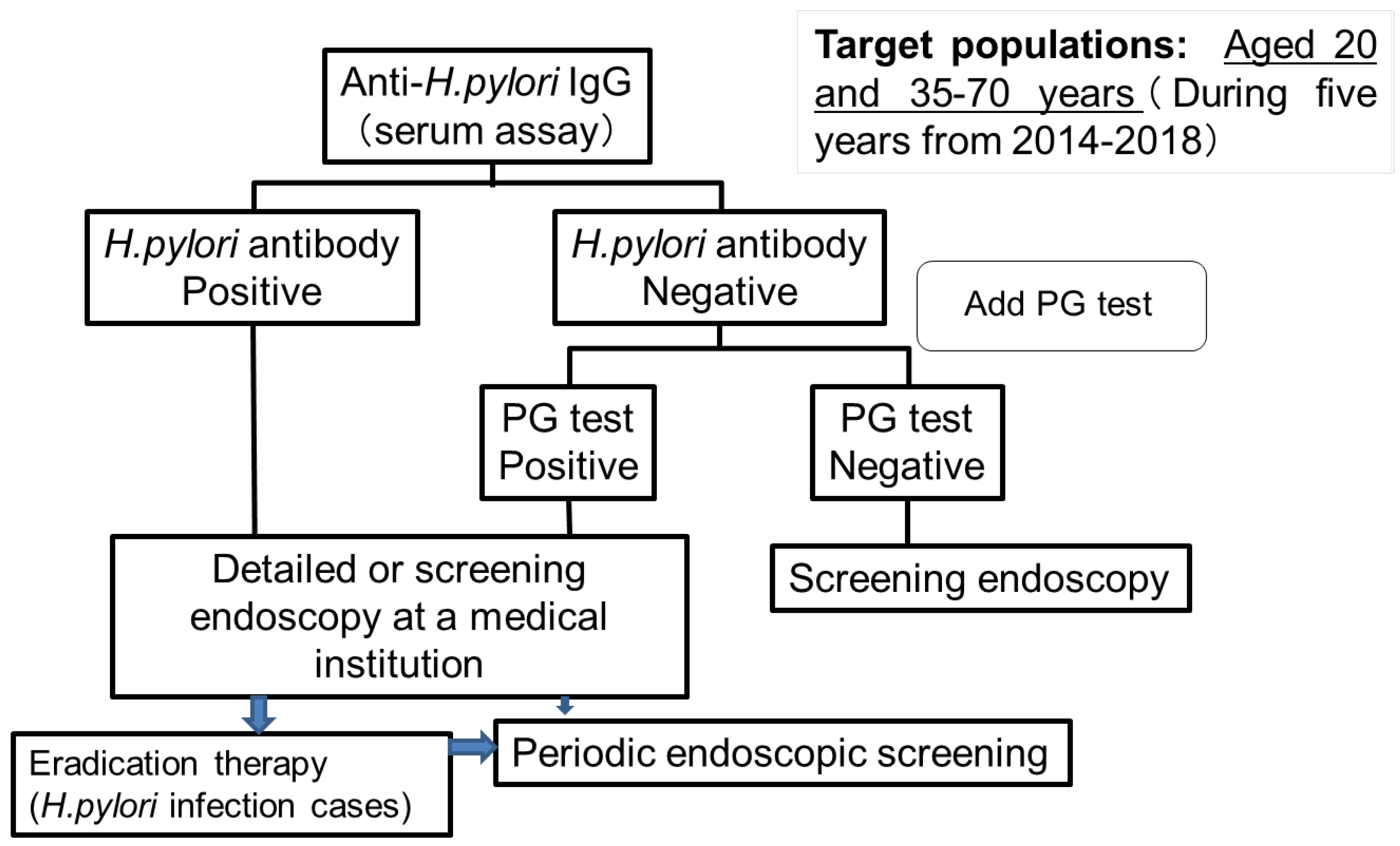
4.3. Gastric Cancer Screening Tests Performed at Hoki-cho, Tottori Prefecture

Since 2000, patients in Tottori Prefecture were able to select between endoscopic and radiographic examinations. The rate of gastric cancer screening by endoscopic or radiographic examination in Hoki-cho, Tottori Prefecture has remained around 20%, which is not sufficient, as the national target is 50%. With the aim of accelerating endoscopic screening and eradication therapy for H. pylori infection, Hoki-cho in Tottori Prefecture has implemented a risk evaluation system for gastric cancer for 5 years since 2014 by testing the serum for H. pylori antibodies [88]. Target populations included individuals aged 20 and 35–70 years in each year, and who underwent at least one examination through the evaluation system during this period (Figure 1).
In cases with negative results for H. pylori diagnosis, we incorporated the serum PG method. During the 5 years from 2014 to 2018, there were a total of 6191 target individuals, of whom 2464 were screened (participation rate: 39.8%). The total number of H. pylori-positive cases was 753 (30.6%), and that of cases negative for H. pylori antibody and positive for the PG method was 58 (2.4%). The frequency of H. pylori positivity was 9.2% in individuals aged 20 years and <40% in individuals aged 60–70 years. This gradually increased with advancing age (Figure 2). The rate was highest (38.4%) among patients aged 60–70 years of age.
Consequently, during the 5-year study period, 71.3% of the examinees underwent a detailed endoscopic examination (Table 1), and two patients with early gastric cancer were detected. Eradication therapy was implemented in 97.6% of cases that had a positive result for H. pylori infection after undergoing a detailed endoscopic examination. On the other hand, only 33.7% and 22.8% of individuals with positive screening results in 2014 and 2015, respectively, had received a periodic endoscopic screening at least once during the three years after the following year. Therefore, it is important to increase the participation rate of this project and the rate of detailed endoscopic examinations to further increase in the detection of the risk of gastric cancer and implement periodic endoscopic screening.
The rate of population-based gastric cancer screening in Hoki-cho was 20.6% in 2013; however, after the introduction of the H. pylori infection screening, it increased to 26.2% in 2015, 22.8% in 2016, 23.2% in 2017, and 24.3% in 2018. In 2018, 657 (63.4%) of the 1036 patients had opted for endoscopic examination (26.1% in 2013, 35.3% in 2014, 52.9% in 2015, 50.7% in 2016, and 57.0% in 2017), contributing to the steady increase in the use of endoscopy (Table 2).
This implies that screening using the H. pylori antibody test is useful for improving the rate of participation and efficient gastric cancer endoscopy. In the future, it will be necessary to verify the effect of reducing gastric cancer mortality by combining H. pylori antibody testing and endoscopic examination and to implement the optimal screening interval for each H. pylori-infected and uninfected person. In addition, it is important to improve the true rate of participation by recommending endoscopic examination to those who require it.

5. Future Directions for Gastric Cancer Screening

5.1. Optimal Age and Intervals for Screening

According to Japan’s national screening program, the recommended age for gastric cancer screening was changed to >50 years due to a decrease in the incidence of gastric cancer in 40-year-olds [13]. Similarly, the British Society of Gastroenterology guidelines suggested endoscopy screening be considered in individuals aged >50 years with multiple risk factors for gastric adenocarcinoma (male, smokers, and pernicious anemia) [89]. In Korea, gastric cancer screening is conducted for populations aged 40–74 years [55]. A study in Japan based on nationwide data showed that the endoscopic screening program would be cost-effective when implemented for populations aged 50–75 years [90]. A nationwide study in Singapore revealed that gastric cancer screening was cost-effective when used among Chinese men aged 50–70 years [91].
A different screening interval might be defined and considered depending on its relationship to the individual’s background and gastric cancer risk. The incidence of gastric cancer differs according to individual risks and is mainly defined by H. pylori infection status and atrophic gastritis. In Korea, an interval of 2 years is recommended [92]. The British Society of Gastroenterology recommends that endoscopic follow-up should be performed every 3 years for individuals with severe chronic atrophic gastritis or intestinal metaplasia, and within one-year intervals for low-grade intraepithelial neoplasia—similar to the management of epithelial precancerous conditions and lesions in the stomach (MAPS II) guideline [93]. In Japan, high-grade intraepithelial neoplasia should be treated clinically. The national program in Japan recommends repeated gastric cancer screening every 2–3 years [14]. However, high-quality prospective research is required to determine the optimal follow-up interval for endoscopic screening in Japan. If individuals with a low risk of gastric cancer could be identified and adopted in the screening programs, their screening interval could be expanded. Hamashima et al. introduced infection atrophy diagnosis using endoscopy and serological testing or risk stratification and conducted a nationwide prospective study to set the interval between risk-specific screenings [17]. It is expected that the results of this research will reduce the burden on patients by appropriately classifying the risk of gastric cancer and extending the interval between screenings for low-risk patients. The research also aims to establish a system that enables the target population to access endoscopic screening fairly by effectively utilizing limited medical resources.

5.2. AI as a New Screening Method

In gastric cancer screening, both radiographic and endoscopic examinations may be eluded by gastric cancer [56,94,95]. In population-based screening, the specialist is required to carry out a double check, the labor is intensive, and the evaluation of the accuracy is difficult. Recently, diagnosis of H. pylori infection and detection of gastric cancer using AI have been reported. The sensitivity and specificity of endoscopic H. pylori infection diagnosis were 81.9% and 83.4% using AI, 79.0% and 83.2% by an average endoscopist, and 85.2% and 89.3% by an endoscopic specialist, respectively [96]. On the other hand, when AI detection was conducted in three groups. That is, H. pylori-positive, H. pylori-negative, and eradicated H. pylori, the rate of correct diagnosis decreased to 77% [97]; hence, there is room for further improvement in diagnosis using AI, including that of cases following H. pylori eradication. AI has a high sensitivity for gastric cancer, but its positive predictive value is low [24,25,26]. However, this has rapidly improved [98]. In addition to its accuracy, AI diagnostic imaging is expected to reduce the burden of double-checking and effectively extract patients who need follow-up endoscopy [98]. It is expected that intervention of gastric cancer screening using AI may reduce gastric cancer deaths more efficiently than the conventional methods of screening.

6. Conclusions

While endoscopic gastric cancer screening has been initiated nationwide in Japan, the incidence of H. pylori infection has decreased and the number of cases following H. pylori eradication has increased. Moreover, the importance of ABC classification reflecting H. pylori infection status and gastric atrophy before endoscopic screening is being increasingly recognized. Considering its cost-effectiveness, spreading the use of endoscopic screening is desirable to establish a new medical examination provision system that conducts examinations at appropriate screening intervals, according to the individual’s background and risks.

Author Contributions

Writing—original draft preparation, K.Y.; writing—review and editing, M.S. and H.I.; supervision, M.S., H.K., K.K. and H.I. All authors have read and agreed to the published version of the manuscript.

Funding

This research received no external funding.

Institutional Review Board Statement

This study was approved by the institutional ethics committee of complies with “The Treaty of Helsinki”. The study was conducted in accordance with the Declaration of Helsinki and approved by the Institutional Ethics Committee of Tottori University (1511A080).

Informed Consent Statement

Informed consent was obtained from all subjects involved in the study.

Data Availability Statement

Data will be available from the corresponding author upon reasonable request.

Acknowledgments

The study of H. pylori antibody project was supported by Hoki-cho, Tottori prefecture.

Conflicts of Interest

The authors declare no conflict of interest.

References

  1. Sung, H.; Ferlay, J.; Siegel, R.L.; Laversanne, M.; Soerjomataram, I.; Jemal, A.; Bray, F. Global cancer statistics 2020: GLOBOCAN estimates of incidence and mortality worldwide for 36 cancers in 185 countries. CA Cancer J. Clin. 2021, 71, 209–249. [Google Scholar] [CrossRef] [PubMed]
  2. Schistosomes, liver flukes and Helicobacter pylori. International Agency for Research on Cancer monographs on the evaluation of carcinogenesis risks to humans. IARC Monogr. Eval. Carcinog. Risks Hum. 1994, 61, 208–220.
  3. Uemura, N.; Okamoto, S.; Yamamoto, S.; Matsumura, N.; Yamaguchi, S.; Yamakido, M.; Taniyama, K.; Sasaki, N.; Schlemper, R.J. Helicobacter pylori infection and the development of gastric cancer. N. Engl. J. Med. 2001, 345, 784–789. [Google Scholar] [CrossRef] [PubMed]
  4. Katanoda, K.; Hori, M.; Saito, E.; Shibata, A.; Ito, Y.; Minami, T.; Ikeda, S.; Suzuki, T.; Matsuda, T. Updated trends in cancer in Japan: Incidence in 1985–2015 and mortality in 1958–2018—A sign of decrease in cancer incidence. J. Epidemiol. 2021, 31, 426–450. [Google Scholar] [CrossRef]
  5. Kobayashi, T.; Kikuchi, S.; Lin, Y.; Yagyu, K.; Obata, Y.; Ogihara, A.; Hasegawa, A.; Miki, K.; Kaneko, E.; Mizukoshi, H.; et al. Trends in the incidence of gastric cancer in Japan and their associations with Helicobacter pylori infection and gastric mucosal atrophy. Gastric Cancer 2004, 7, 233–239. [Google Scholar] [CrossRef] [Green Version]
  6. Ueda, J.; Gosho, M.; Inui, Y.; Matsuda, T.; Sakakibara, M.; Mabe, K.; Nakajima, S.; Shimoyama, T.; Yasuda, M.; Kawai, T.; et al. Prevalence of Helicobacter pylori infection by birth year and geographic area in Japan. Helicobacter 2014, 19, 105–110. [Google Scholar] [CrossRef]
  7. Kamada, T.; Haruma, K.; Ito, M.; Inoue, K.; Manabe, N.; Matsumoto, H.; Kusunoki, H.; Hata, J.; Yoshihara, M.; Sumii, K.; et al. Time trends in Helicobacter pylori infection and atrophic gastritis over 40 years in Japan. Helicobacter 2015, 20, 192–198. [Google Scholar] [CrossRef]
  8. Sugano, K. Screening of gastric cancer in Asia. Best Pract. Res. Clin. Gastroenterol. 2015, 29, 895–905. [Google Scholar] [CrossRef]
  9. Wang, C.; Nishiyama, T.; Kikuchi, S.; Inoue, M.; Sawada, N.; Tsugane, S.; Lin, Y. Changing trends in the prevalence of H. pylori infection in Japan (1908–2003): A systematic review and meta-regression analysis of 170,752 individuals. Sci. Rep. 2017, 7, 15491. [Google Scholar] [CrossRef]
  10. Lin, Y.; Kawai, S.; Sasakabe, T.; Nagata, C.; Naito, M.; Tanaka, K.; Sugawara, Y.; Mizoue, T.; Sawada, N.; Matsuo, K. Effects of Helicobacter pylori eradication on gastric cancer incidence in the Japanese population: A systematic evidence review. Jpn. J. Clin. Oncol. 2021, 51, 1158–1170. [Google Scholar] [CrossRef]
  11. National Cancer Center. Center for Cancer Control and Information Services. 2021. Available online: https://ganjoho.jp/public/index.html (accessed on 1 January 2022).
  12. Oshima, A. A critical review of cancer screening programs in Japan. Int. J. Technol. Assess. Health Care 1994, 10, 346–358. [Google Scholar] [CrossRef]
  13. Hamashima, C. Systematic review group and guideline development group for gastric cancer screening guidelines. Update version of the Japanese Guidelines for Gastric Cancer Screening. Jpn. J. Clin. Oncol. 2018, 48, 673–683. [Google Scholar] [CrossRef] [Green Version]
  14. Hamashima, C. Cancer screening guidelines and policy making: 15 years of experience in cancer screening guideline development in Japan. Jpn. J. Clin. Oncol. 2018, 48, 278–286. [Google Scholar] [CrossRef]
  15. Hamashima, C.; Goto, R. Potential capacity of endoscopic screening for gastric cancer in Japan. Cancer Sci. 2017, 108, 101–107. [Google Scholar] [CrossRef] [Green Version]
  16. Hamashima, C. Overdiagnosis of gastric cancer by endoscopic screening. World J. Gastrointest. Endosc. 2017, 9, 55–60. [Google Scholar] [CrossRef]
  17. Hamashima, C.; Yoshimura, K.; Fukao, A. A study protocol for expanding the screening interval of endoscopic screening for gastric cancer based on individual risks: Prospective cohort study of gastric cancer screening. Ann. Transl. Med. 2020, 8, 1604. [Google Scholar] [CrossRef]
  18. Mabe, K.; Inoue, K.; Kamada, T.; Kato, K.; Kato, M.; Haruma, K. Endoscopic screening for gastric cancer in Japan: Current status and future perspectives. Dig. Endosc. 2022, 34, 412–419. [Google Scholar] [CrossRef]
  19. Matsuo, T.; Ito, M.; Takata, S.; Tanaka, S.; Yoshihara, M.; Chayama, K. Low prevalence of Helicobacter pylori-negative gastric cancer among Japanese. Helicobacter 2011, 16, 415–419. [Google Scholar] [CrossRef]
  20. Ono, S.; Kato, M.; Suzuki, M.; Ishigaki, S.; Takahashi, M.; Haneda, M.; Mabe, K.; Shimizu, Y. Frequency of Helicobacter pylori-negative gastric cancer and gastric mucosal atrophy in a Japanese endoscopic submucosal dissection series including histological, endoscopic and serological atrophy. Digestion 2012, 86, 59–65. [Google Scholar] [CrossRef]
  21. Mizota, Y.; Yamamoto, S. How long should we continue gastric cancer screening? From an epidemiological point of view. Gastric Cancer 2019, 22, 456–462. [Google Scholar] [CrossRef] [Green Version]
  22. Kishikawa, H. The clinical benefits, limitations, and perspectives of the ABC method. Intern. Med. 2020, 59, 1471–1472. [Google Scholar] [CrossRef] [Green Version]
  23. Yao, K.; Uedo, T.; Kamada, T.; Hirasawa, T.; Nagahama, T.; Yoshinaga, S.; Oka, M.; Inoue, K.; Mabe, K.; Yao, T.; et al. Guidelines for endoscopic diagnosis of early gastric cancer. Dig. Endosc. 2020, 32, 663–698. [Google Scholar] [CrossRef]
  24. Hirasawa, T.; Aoyama, K.; Tanimoto, T.; Ishihara, S.; Shichijo, S.; Ozawa, T.; Ohnishi, T.; Fujishiro, M.; Matsuo, K.; Fujisaki, J.; et al. Application of artificial intelligence using a convolutional neural network for detecting gastric cancer in endoscopic images. Gastric Cancer 2018, 21, 653–660. [Google Scholar] [CrossRef] [Green Version]
  25. Ishioka, M.; Hirasawa, T.; Tada, T. Detecting gastric cancer from video images using convolutional neural networks. Dig. Endosc. 2019, 31, e34–e35. [Google Scholar] [CrossRef] [Green Version]
  26. Ikenoyama, Y.; Hirasawa, T.; Ishioka, M.; Namikawa, K.; Yoshimizu, S.; Horiuchi, Y.; Ishiyama, A.; Yoshio, T.; Tsuchida, T.; Takeuchi, Y.; et al. Detecting early gastric cancer: Comparison between the diagnostic ability of convolutional neural networks and endoscopists. Dig. Endosc. 2021, 33, 141–150. [Google Scholar] [CrossRef]
  27. Park, J.Y.; Greenberg, E.R.; Parsonnnet, J.; Wild, C.P.; Forman, D.; Herrero, R. Summary of IARC Working Group Meeting on Helicobacter pylori Eradication as a Strategy for Preventing Gastric Cancer; IARC Working Group Report 8; International Agency for Research on Cancer: Lyon, France, 2014; pp. 1–4. [Google Scholar]
  28. Greenberg, E.R.; Park, J.Y. Effectiveness of Helicobacter pylori eradication. In Helicobacter pylori Eradication as a Strategy for Preventing Gastric Cancer; IARC Working Group Report 8; International Agency for Research on Cancer: Lyon, France, 2014; pp. 64–71. [Google Scholar]
  29. Li, W.Q.; Ma, J.L.; Zhang, L.; Brown, L.M.; Li, J.Y.; Shen, L.; Pan, K.F.; Liu, W.D.; Hu, Y.; Han, Z.X.; et al. Effects of Helicobacter pylori treatment on gastric cancer incidence and mortality in subgroups. J. Natl. Cancer Inst. 2014, 106, dju116. [Google Scholar] [CrossRef] [Green Version]
  30. Lee, Y.C.; Chiang, T.H.; Chou, C.K.; Tu, Y.K.; Liao, W.C.; Wu, M.S.; Graham, D.Y. Association between Helicobacter pylori eradication and gastric cancer incidence: A systematic review and meta-analysis. Gastroenterology 2016, 150, 1113–1124.e5. [Google Scholar] [CrossRef] [Green Version]
  31. Sugano, K. Effect of helicobacter pylori eradication on the incidence of gastric cancer: A systematic review and meta-analysis. Gastric Cancer 2019, 22, 435–445. [Google Scholar] [CrossRef] [Green Version]
  32. Ford, A.C.; Yuan, Y.; Moayyedi, P. Helicobacter pylori eradication therapy to prevent gastric cancer: Systematic review and metaanalysis. Gut 2020, 69, 2113–2121. [Google Scholar] [CrossRef]
  33. Fukase, K.; Kato, M.; Kikuchi, S.; Inoue, K.; Uemura, N.; Okamoto, S.; Terao, S.; Amagai, K.; Hayashi, S.; Asaka, M.; et al. Effect of eradication of Helicobacter pylori on incidence of metachronous gastric carcinoma after endoscopic resection of early gastric cancer: An openlabel, randomised controlled trial. Lancet 2008, 372, 392–397. [Google Scholar] [CrossRef] [Green Version]
  34. Li, W.Q.; Zhang, J.Y.; Ma, J.L.; Li, Z.X.; Zhang, L.; Zhang, Y.; Guo, Y.; Zhou, T.; Li, J.Y.; Shen, L.; et al. Effects of helicobacter pylori treatment and vitamin and garlic supplementation on gastric cancer incidence and mortality: Follow-up of a randomized intervention trial. BMJ 2019, 366, l5016. [Google Scholar] [CrossRef] [PubMed] [Green Version]
  35. Choi, I.J.; Kim, C.G.; Lee, J.Y.; Kim, Y.I.; Kook, M.C.; Park, B.; Joo, J. Family History of Gastric Cancer and Helicobacter pylori Treatment. N. Engl. J. Med. 2020, 382, 427–436. [Google Scholar] [CrossRef] [PubMed]
  36. Chiang, T.; Chang, W.; Chen, S.L.; Yen, A.M.; Fann, J.C.; Chiu, S.Y.; Chen, Y.R.; Chuang, S.L.; Shieh, C.F.; Liu, C.Y.; et al. Mass eradication of Helicobacter pylori to reduce gastric cancer incidence and mortality: A long-term cohort study on Matsu Islands. Gut 2021, 70, 243–250. [Google Scholar] [CrossRef] [PubMed]
  37. Take, S.; Mizuno, M.; Ishiki, K.; Kusumoto, C.; Imada, T.; Hamada, F.; Yoshida, T.; Yokota, K.; Mitsuhashi, T.; Okada, H. Risk of gastric cancer in the second decade of follow-up after helicobacter pylori eradication. J. Gastroenterol. 2020, 55, 281–288. [Google Scholar] [CrossRef] [Green Version]
  38. Satomi, S.; Yamakawa, A.; Matsunaga, S.; Masaki, R.; Inagaki, T.; Okuda, T.; Suto, H.; Ito, Y.; Yamazaki, Y.; Kuriyama, M.; et al. Relationship between the diversity of the cagA gene of Helicobacter pylori and gastric cancer in Okinawa, Japan. J. Gastroenterol. 2006, 41, 668–673. [Google Scholar] [CrossRef]
  39. Yamaoka, Y.; Kato, M.; Asaka, M. Geographic differences in gastric cancer incidence can be explained by differences between Helicobacter pylori strains. Intern. Med. 2008, 47, 1077–1083. [Google Scholar] [CrossRef] [Green Version]
  40. Kpoghomou, M.A.; Wang, J.; Wang, T.; Jin, G. Association of Helicobacter pylori babA2 gene and gastric cancer risk: A meta-analysis. BMC Cancer 2020, 20, 465. [Google Scholar] [CrossRef]
  41. Asaka, M.; Mabe, K. Strategies for eliminating death from gastric cancer in Japan. Proc. Jpn. Acad. Ser. B Phys. Biol. Sci. 2014, 90, 251–258. [Google Scholar] [CrossRef] [Green Version]
  42. Kawai, S.; Wang, C.; Lin, Y.; Sasakabe, T.; Okuda, M.; Kikuchi, S. Lifetime incidence risk for gastric cancer in the Helicobacter pylori-infected and uninfected population in Japan: A Monte Carlo simulation study. Int. J. Cancer 2022, 150, 18–27. [Google Scholar] [CrossRef]
  43. Japanese Gastric Cancer Association. Japanese classification of gastric carcinoma-3rd English edition. Gastric Cancer 2011, 14, 101–112. [Google Scholar] [CrossRef] [Green Version]
  44. Schlemper, R.J.; Riddell, R.H.; Kato, Y.; Borchard, F.; Cooper, H.S.; Dawsey, S.M.; Dixon, M.F.; Fenoglio-Preiser, C.M.; Fléjou, J.F.; Geboes, K.; et al. The Vienna classification of gastrointestinal epithelial neoplasia. Gut 2000, 47, 251–255. [Google Scholar] [CrossRef] [Green Version]
  45. Yamamoto, Y.; Fujisaki, J.; Omae, M.; Hirasawa, T.; Igarashi, M. Helicobacter pylori-negative gastric cancer: Characteristics and endoscopic findings. Dig. Endosc. 2015, 27, 551–561. [Google Scholar] [CrossRef]
  46. Imamura, Y.; Watanabe, M.; Oki, E.; Morita, M.; Baba, H. Esophagogastric junction adenocarcinoma shares characteristics with gastric adenocarcinoma: Literature review and retrospective multicenter cohort study. Ann. Gastroenterol. Surg. 2020, 5, 46–59. [Google Scholar] [CrossRef]
  47. Crew, K.D.; Neugut, A.I. Epidemiology of gastric cancer. World J. Gastroenterol. 2006, 12, 354–362. [Google Scholar] [CrossRef]
  48. Ohshima, A.; Hirata, N.; Ubukata, T.; Umeda, K.; Fujimoto, I. Evaluation of a mass screening program for stomach with a case control study design. Int. J. Cancer 1986, 38, 829–833. [Google Scholar] [CrossRef]
  49. Fukao, A.; Tsubono, Y.; Tsuji, I.; Hisamichi, S.; Sugahara, N.; Takano, A. The evaluation of screening for gastric cancer in Miyagi Prefecture, Japan: A population-based case-control study. Int. J. Cancer 1995, 60, 45–48. [Google Scholar] [CrossRef]
  50. Hamashima, C.; Shabana, M.; Okada, K.; Okamoto, M.; Osaki, Y. Mortality reduction from gastric cancer by endoscopic and radiographic screening. Cancer Sci. 2015, 106, 1744–1749. [Google Scholar] [CrossRef] [Green Version]
  51. Jun, J.K.; Choi, K.S.; Lee, H.Y.; Suh, M.; Park, B.; Song, S.H.; Jung, K.W.; Lee, C.W.; Choi, I.J.; Park, E.C.; et al. Effectiveness of the Korean National Cancer Screening Program in Reducing Gastric Cancer Mortality. Gastroenterology 2017, 152, 1319–1328.e7. [Google Scholar] [CrossRef]
  52. The Japanese Society of Gastrointestinal Cancer Screening. New Guidelines of Radiography for Gastric Cancer Screening. 2011. Available online: https://www.jsgcs.or.jp (accessed on 14 December 2020).
  53. The Japanese Society of Gastrointestinal Cancer Screening. Annual Report of Gastrointestinal Cancer Screening 2014. Available online: https://www.jsgcs.or.jp (accessed on 14 December 2020).
  54. Togo, R.; Yamamichi, N.; Mabe, K.; Takahashi, Y.; Takeuchi, C.; Kato, M.; Sakamoto, N.; Ishihara, K.; Ogawa, T.; Haseyama, M. Detection of gastritis by a deep convolutional neural network from double-contrast upper gastrointestinal barium x-ray radiography. J. Gastroenterol. 2019, 54, 321–329. [Google Scholar] [CrossRef] [Green Version]
  55. Choi, K.S.; Jun, J.K.; Suh, M.; Park, B.; Noh, D.K.; Song, S.H.; Jung, K.W.; Lee, H.Y.; Choi, I.J.; Park, E.C. Effect of endoscopy screening on stage at gastric cancer diagnosis: Results of the National Cancer Screening Programme in Korea. Br. J. Cancer 2015, 112, 608–612. [Google Scholar] [CrossRef] [Green Version]
  56. Choi, K.S.; Jun, J.K.; Park, E.C.; Park, S.; Jung, K.W.; Han, M.A.; Choi, I.J.; Lee, H.Y. Performance of different gastric cancer screening methods in Korea: A population-based study. PLoS ONE 2012, 7, e50041. [Google Scholar] [CrossRef] [PubMed] [Green Version]
  57. Matsumoto, S.; Yoshida, Y. Efficacy of endoscopic screening in an isolated island: A case-control study. Indian J. Gastroenterol. 2014, 33, 46–49. [Google Scholar] [CrossRef]
  58. Hamashima, C.; Ogoshi, K.; Okamoto, M.; Shabana, M.; Kishimoto, T.; Fukao, A. A community-based, case-control study evaluating mortality reduction from gastric cancer by endoscopic screening in Japan. PLoS ONE 2013, 8, e79088. [Google Scholar] [CrossRef] [Green Version]
  59. Zhang, X.; Li, M.; Chen, S.; Hu, J.; Guo, Q.; Liu, R.; Zheng, H.; Jin, Z.; Yuan, Y.; Xi, Y.; et al. Endoscopic Screening in Asian Countries Is Associated With Reduced Gastric Cancer Mortality: A Meta-analysis and Systematic Review. Gastroenterology 2018, 155, 347–354.e9. [Google Scholar] [CrossRef] [PubMed] [Green Version]
  60. The Japanese Society of Gastrointestinal Cancer Screening. Quality Assurance Manual of Endoscopic Screening for Gastric Cancer in Japanese Communities. Jpn. J. Clin. Oncol. 2016, 46, 1053–1061. (In Japanese) [Google Scholar] [CrossRef] [PubMed] [Green Version]
  61. Hosokawa, O.; Shinbo, T.; Matsuda, K.; Miyanaga, T. Impact of opportunistic endoscopic screening on the decrease of mortality from gastric cancer. J. Gatsroenterol. Cancer Screen 2011, 49, 401–409. (In Japanese) [Google Scholar]
  62. Ogoshi, K.; Narisawa, R.; Kato, T.; Saito, S.; Funagoshi, K.; Kinameri, K. Evaluation of endoscopic screening for gastric cancer in Niigata City: The reduction of the mortality rate. J. Gatsroenterol. Cancer Screen 2009, 47, 531–541. (In Japanese) [Google Scholar]
  63. Shichijo, S.; Uedo, N.; Michida, T. Detection of Early Gastric Cancer after Helicobacter pylori Eradication. Digestion 2021, 2, 54–61. [Google Scholar] [CrossRef]
  64. Kurumi, H.; Kanda, T.; Ikebuchi, Y.; Yoshida, A.; Kawaguchi, K.; Yashima, K.; Isomoto, H. Current Status of Photodynamic Diagnosis for Gastric Tumors. Diagnostics 2021, 11, 1967. [Google Scholar] [CrossRef]
  65. Ministry of Health, Labour and Welfare. The Report of Health Promotion and Community Health 2013. Available online: http://www.e-stat.go.jp/SG1/estat/GL08020101.do?_-toGL08020101_&tstatCode=000001030884&requestSender=dsearch (accessed on 1 September 2015).
  66. Shabana, M.; Hamashima, C.; Nishida, M.; Miura, K.; Kishimoto, T. Current status and evaluation of endoscopic screening for gastric cancer. Jpn. J. Cancer Det. Diagn. 2010, 17, 229–235. (In Japanese) [Google Scholar]
  67. Kitagawa, S.; Miyagawa, K.; Iriguchi, Y. The report of gastroenterological screening in 2012. J. Gastroenterol. Cancer Screen 2015, 53, 60–86. (In Japanese) [Google Scholar]
  68. Suzuki, A.; Katoh, H.; Komura, D.; Kakiuchi, M.; Tagashira, A.; Yamamoto, S.; Tatsuno, K.; Ueda, H.; Nagae, G.; Fukuda, S.; et al. Defined lifestyle and germline factors predispose Asian populations to gastric cancer. Sci. Adv. 2020, 6, eaav9778. [Google Scholar] [CrossRef]
  69. Sugano, K.; Tack, J.; Kuipers, E.J.; Graham, D.Y.; El-Omar, E.M.; Miura, S.; Haruma, K.; Asaka, M.; Uemura, N.; Malfertheiner, P. Kyoto global consensus report on Helicobacter pylori gastritis. Gut 2015, 64, 1353–1367. [Google Scholar] [CrossRef] [Green Version]
  70. Haruma, K.; Kato, M.; Inoue, K.; Murakami, K.; Kamada, T. Kyoto Classification of Gastritis; Nihon Medical Center: Tokyo, Japan, 2017. [Google Scholar]
  71. Yoshii, S.; Mabe, K.; Watano, K.; Ohno, M.; Matsumoto, M.; Ono, S.; Kudo, T.; Nojima, M.; Kato, M.; Sakamoto, N. Validity of endoscopic features for the diagnosis of helicobacter pylori infection status based on the Kyoto classification of gastritis. Dig. Endosc. 2020, 32, 74–83. [Google Scholar] [CrossRef] [Green Version]
  72. Isomoto, H.; Mizuta, Y.; Inoue, K.; Matsuo, T.; Hayakawa, T.; Miyazaki, M.; Onita, K.; Takeshima, F.; Murase, K.; Shimokawa, I.; et al. A close relationship between Helicobacter pylori infection and gastric xanthoma. Scand J. Gastroenterol. 1999, 34, 346–352. [Google Scholar] [CrossRef]
  73. Sekikawa, A.; Fukui, H.; Sada, R.; Fukuhara, M.; Marui, S.; Tanke, G.; Endo, M.; Ohara, Y.; Matsuda, F.; Nakajima, J.; et al. Gastric atrophy and xanthelasma are markers for predicting the development of early gastric cancer. J. Gastroenterol. 2016, 51, 35–42. [Google Scholar] [CrossRef]
  74. Hirai, R.; Hirai, M.; Shimodate, Y.; Minami, M.; Ishikawa, S.; Kanadani, T.; Takezawa, R.; Doi, A.; Nishimura, N.; Mouri, H.; et al. Feasibility of endoscopic evaluation of Helicobacter pylori infection status by using the Kyoto classification of gastritis in the population-based gastric cancer screening program: A prospective cohort study. Health Sci. Rep. 2021, 4, e325. [Google Scholar] [CrossRef]
  75. Overview of the 2019 Basic Survey on National Life. Available online: https://www.mhlw.go.jp/toukei/saikin/hw/k-tyosa/k-tyosa19/index.html (accessed on 27 June 2022).
  76. Miki, K. Gastric cancer screening by combined assay for serum anti-Helicobacter pylori IgG antibody and serum pepsinogen levels “ABC method”. Proc. Jpn. Acad. Ser. B Phys. Biol. Sci. 2011, 87, 405–414. [Google Scholar] [CrossRef] [Green Version]
  77. Itoh, T.; Saito, M.; Marugami, N.; Hirai, T.; Marugami, A.; Takahama, J.; Tanaka, T.; Kichikawa, K. Correlation between the ABC classification and radiological findings for assessing gastric cancer risk. Jpn. J. Radiol. 2015, 33, 636–644. [Google Scholar] [CrossRef]
  78. Terasawa, T.; Nishida, H.; Kato, K.; Miyashiro, I.; Yoshikawa, T.; Takaku, R.; Hamashima, C. Prediction of gastric cancer development by serum pepsinogen test and Helicobacter pylori seropositivity in Eastern Asians: A systematic review and meta-analysis. PLoS ONE 2014, 9, e109783. [Google Scholar] [CrossRef]
  79. Kiso, M.; Yoshihara, M.; Ito, M.; Inoue, K.; Kato, K.; Nakajima, S.; Mabe, K.; Kobayashi, M.; Uemura, N.; Yada, T.; et al. Characteristics of gastric cancer in negative test of serum anti-Helicobacter pylori antibody and pepsinogen test: A multicenter study. Gastric Cancer 2017, 20, 764–771. [Google Scholar] [CrossRef] [Green Version]
  80. Kishino, T.; Oyama, T.; Tomori, A.; Takahashi, A.; Shinohara, T. Usefulness and limitations of a serum screening system to predict the risk of gastric cancer. Intern. Med. 2020, 59, 1473–1480. [Google Scholar] [CrossRef] [Green Version]
  81. Masuyama, H.; Yoshitake, N.; Sasai, T.; Nakamura, T.; Masuyama, A.; Zuiki, T.; Kurashina, K.; Mieda, M.; Sunada, K.; Yamamoto, H.; et al. Relationship between the degree of endoscopic atrophy of the gastric mucosa and carcinogenic risk. Digestion 2015, 91, 30–36. [Google Scholar] [CrossRef] [Green Version]
  82. Spence, A.D.; Cardwell, C.R.; McMenamin, U.C.; Hicks, B.M.; Johnston, B.T.; Murray, L.J.; Coleman, H.G. Adenocarcinoma risk in gastric atrophy and intestinal metaplasia: A systematic review. BMC Gastroenterol. 2017, 17, 157. [Google Scholar] [CrossRef] [Green Version]
  83. Kotachi, T.; Ito, M.; Yoshihara, M.; Boda, T.; Kiso, M.; Masuda, K.; Matsuo, T.; Tanaka, S.; Chayama, K. Serological evaluation of gastric cancer risk based on pepsinogen and helicobacter pylori antibody: Relationship to endoscopic findings. Digestion 2017, 95, 314–318. [Google Scholar] [CrossRef]
  84. Kaji, K.; Hashiba, A.; Uotani, C.; Yamaguchi, Y.; Ueno, T.; Ohno, K.; Takabatake, I.; Wakabayashi, T.; Doyama, H.; Ninomiya, I.; et al. Grading of atrophic gastritis is useful for risk stratification in endoscopic screening for gastric cancer. Am. J. Gastroenterol. 2019, 114, 71–79. [Google Scholar] [CrossRef]
  85. Kimura, K.; Takemoto, T. An endoscopic recognition for the atrophic border and its significance in chronic gastritis. Endoscopy 1969, 1, 87–97. [Google Scholar] [CrossRef]
  86. Canakis, A.; Pani, E.; Saumoy, M.; Shah, S.C. Decision model analyses of upper endoscopy for gastric cancer screening and preneoplasia surveillance: A systematic review. Therap. Adv. Gastroenterol. 2020, 13, 1756284820941662. [Google Scholar] [CrossRef] [PubMed]
  87. Kowada, A. Endoscopy Is Cost-effective for gastric cancer screening after successful Helicobacter pylori eradication. Dig. Dis. Sci. 2021, 66, 4220–4226. [Google Scholar] [CrossRef] [PubMed]
  88. Yashima, K.; Hasegawa, R.; Shabana, M.; Kawaguchi, G.; Isomoto, H. Mass screening considering Helicobacter pylori infection status for gastric cancer in Hoki-cho, Tottori prefecture. J. Gatsroenterol. Cancer Screen 2019, 57, 561–570. (In Japanese) [Google Scholar]
  89. Banks, M.; Graham, D.; Jansen, M.; Gotoda, T.; Coda, S.; di Pietro, M.; Uedo, N.; Bhandari, P.; Pritchard, D.M.; Kuipers, E.J.; et al. British Society of Gastroenterology guidelines on the diagnosis and management of patients at risk of gastric adenocarcinoma. Gut 2019, 68, 1545–1575. [Google Scholar] [CrossRef] [Green Version]
  90. Huang, H.L.; Leung, C.Y.; Saito, E.; Katanoda, K.; Hur, C.; Kong, C.Y.; Nomura, S.; Shibuya, K. Effect and cost-effectiveness of national gastric cancer screening in Japan: A microsimulation modeling study. BMC Med. 2020, 18, 257. [Google Scholar] [CrossRef]
  91. Dan, Y.Y.; So, J.B.; Yeoh, K.G. Endoscopic screening for gastric cancer. Clin. Gastroenterol. Hepatol. 2006, 4, 709–716. [Google Scholar] [CrossRef]
  92. Suh, Y.S.; Lee, J.; Woo, H.; Shin, D.; Kong, S.H.; Lee, H.J.; Shin, A.; Yang, H.K. National cancer screening program for gastric cancer in Korea: Nationwide treatment benefit and cost. Cancer 2020, 126, 1929–1939. [Google Scholar] [CrossRef]
  93. Pimentel-Nunes, P.; Libânio, D.; Marcos-Pinto, R.; Areia, M.; Leja, M.; Esposito, G.; Garrido, M.; Kikuste, I.; Megraud, F.; Matysiak-Budnik, T.; et al. Management of epithelial precancerous conditions and lesions in the stomach (MAPS II): European Society of Gastrointestinal Endoscopy (ESGE), European Helicobacter and Microbiota96. Study Group (EHMSG), European Society of Pathology (ESP), and Sociedade Portuguesa de Endoscopia Digestiva (SPED) guideline update 2019. Endoscopy 2019, 51, 365–388. [Google Scholar] [CrossRef] [Green Version]
  94. Hosokawa, O.; Tsuda, S.; Kidani, E.; Watanabe, K.; Tanigawa, Y.; Shirasaki, S.; Hayashi, H.; Hinoshita, T. Diagnosis of gastric cancer up to three years after negative upper gastrointestinal endoscopy. Endoscopy 1998, 30, 669–674. [Google Scholar] [CrossRef]
  95. Pimentaelo, A.R.; Monteirooares, M.; Libanio, D.; Dinisibeiro, M. Missing rate for gastric cancer during upper gastrointestinal endoscopy: A systematic review and meta-analysis. Eur. J. Gastroenterol. Hepatol. 2016, 28, 1041–1049. [Google Scholar] [CrossRef]
  96. Shichijo, S.; Nomura, S.; Aoyama, K.; Nishikawa, Y.; Miura, M.; Shinagawa, T.; Takiyama, H.; Tanimoto, T.; Ishihara, S.; Matsuo, K.; et al. Application of Convolutional Neural Networks in the Diagnosis of Helicobacter pylori Infection Based on Endoscopic Images. EBioMedicine 2017, 25, 106–111. [Google Scholar] [CrossRef] [Green Version]
  97. Shichijo, S.; Endo, Y.; Aoyama, K.; Takeuchi, Y.; Ozawa, T.; Takiyama, H.; Matsuo, K.; Fujishiro, M.; Ishihara, S.; Ishihara, R.; et al. Application of convolutional neural networks for evaluating Helicobacter pylori infection status on the basis of endoscopic images. Scand J. Gastroenterol. 2019, 54, 158–163. [Google Scholar] [CrossRef]
  98. Oura, H.; Matsumura, T.; Fujie, M.; Ishikawa, T.; Nagashima, A.; Shiratori, W.; Tokunaga, M.; Kaneko, T.; Imai, Y.; Oike, T.; et al. Development and evaluation of a double-check support system using artificial intelligence in endoscopic screening for gastric cancer. Gastric Cancer 2022, 25, 392–400. [Google Scholar] [CrossRef]
Figure 1. Flow chart of H. pylori antibody test project in Hoki-cho, Tottori prefecture. Individuals with PG I levels of ≤70 ng/mL and PG I/II ratio of <3 are classified as PG test positive, which is equal to gastric atrophy. PG, pepsinogen.
Jcm 11 04337 g001
Figure 2. The frequency of H. pylori positivity according to age (2014~2018).
Jcm 11 04337 g002
Table 1. Results of H. pylori antibody test project in Hoki-cho, Tottori Prefecture.
Year2014201520162017,
2018
Total
Examinees (n)9107763114672464
Cases requiring detailed endoscopy (n)323259109121811
Examination required rate (%)35.433.435.025.932.9
Cases undergone screeningendoscopy (n)2581816178578
Examination rate (%)79.969.956.064.571.3
Table 2. Annual trends in the rate of participation for population-based gastric cancer screening in Hoki-cho, Tottori Prefecture.
Year2013201420152016201720182019
Target population (n)4533453345334257425742574257
Examinees (n)934963118897098610361039
Participation rate (%)20.621.226.222.823.224.324.4
Proportion of endoscopy among gastric cancer screening tests (%)26.135.352.950.757.063.465.9
The data were obtained from “Cancer Screening Report in Tottori Prefecture”.
Publisher’s Note: MDPI stays neutral with regard to jurisdictional claims in published maps and institutional affiliations.

