Review

Probiotics in the Management of Mental and Gastrointestinal Post-COVID Symptomes

by
Igor Łoniewski
1,
Karolina Skonieczna-Żydecka
1,*,
Joanna Sołek-Pastuszka
2 and
Wojciech Marlicz
3
1
Department of Biochemical Science, Pomeranian Medical University in Szczecin, 71-460 Szczecin, Poland
2
Department of Anesthesiology and Intensive Therapy, Pomeranian Medical University, 70-204 Szczecin, Poland
3
Department of Gastroenterology, Pomeranian Medical University, 70-204 Szczecin, Poland
*
Author to whom correspondence should be addressed.
J. Clin. Med. 2022, 11(17), 5155; https://doi.org/10.3390/jcm11175155
Submission received: 31 July 2022 / Revised: 17 August 2022 / Accepted: 29 August 2022 / Published: 31 August 2022
(This article belongs to the Section Epidemiology & Public Health)

Abstract

:
Patients with “post-COVID” syndrome manifest with a variety of signs and symptoms that continue/develop after acute COVID-19. Among the most common are gastrointestinal (GI) and mental symptoms. The reason for symptom occurrence lies in the SARS-CoV-2 capability of binding to exact receptors, among other angiotensin converting enzyme 2 (ACE2) receptors in gastrointestinal lining and neuropilin-1 (NRP-1) in the nervous system, which leads to loss of gastrointestinal and blood-brain barriers integrity and function. The data are mounting that SARS-CoV-2 can trigger systemic inflammation and lead to disruption of gut-brain axis (GBA) and the development of disorders of gut brain interaction (DGBIs). Functional dyspepsia (FD) and irritable bowel syndrome (IBS) are the most common DGBIs syndromes. On the other hand, emotional disorders have also been demonstrated as DGBIs. Currently, there are no official recommendations or recommended procedures for the use of probiotics in patients with COVID-19. However, it can be assumed that many doctors, pharmacists, and patients will want to use a probiotic in the treatment of this disease. In such cases, strains with documented activity should be used. There is a constant need to plan and conduct new trials on the role of probiotics and verify their clinical efficacy for counteracting the negative consequences of COVID-19 pandemic. Quality control is another important but often neglected aspect in trials utilizing probiotics in various clinical entities. It determines the safety and efficacy of probiotics, which is of utmost importance in patients with post-acute COVID-19 syndrome.

Graphical Abstract

1. Introduction

After the first cases of SARS-CoV-2 virus contamination in Wuhan, Hubei Province, China, in December 2019 and the announcement of World Health Organization (WHO) declaring a global pandemic on 11 March 2020, COVID-19 infected over 589 million people around the world, caused over 6,400,000 deaths on 13 August 2022 [1]. Due to the genetic evolution of the virus, up to date five different variants of concern have been identified, namely, Alpha (B.1.1.7), Beta (B.1.351), Gamma (P.1), Delta (B.1.617.2), and Omicron (B.1.1.529) [2].
Respiratory presentation of COVID-19 is predominant; however, a set of other symptoms, including gastrointestinal ones along with hepatic malfunctions, might precede full symptomatic occurrence or co-occur with respiratory infection [3,4]. Most COVID-19 patients are oligosymptomatic with scarce or discreet symptoms. In selected individuals, SARS-CoV-2 damage may be intensified due to an inadequate immune response [5] leading to whole body failure [6]. Apart from acute COVID-19 symptoms, of relevance are adverse events associated with long COVID-19 disease. In fact, in persons who experienced mild symptoms of COVID-19, as well as survivors, post-acute COVID-19 syndrome (PACS) might occur [7] as a set of persistent symptoms and/or delayed or long-term complications. Therefore, apart from therapeutic protocols administered by physicians, there is a need to develop novel, efficacious, safe, and economically acceptable protocols to manage long term disease and treat related adverse events.
Probiotics are live microorganisms that are intended to have health benefits when consumed or applied to the human body with a proved efficacy in DGBIs. The popularity of using probiotics is growing and indications for their use are not limited to co-prescription with antibiotics only. Now more than ever, clinicians need clear guidelines and recommendations, including safety and efficacy for implementation of probiotics into clinical practice. Although there are no official recommendations on the use of probiotics in patients with COVID-19, we aimed to present the current knowledge and discuss clinical implications for the use of probiotics in post COVID-19 patients.

2. COVID-19 Pandemic and Microbiota

2.1. Environment

Common use of antiseptic and aseptic detergents and cleaning reagents in household, workplace, and public spaces can lead to a loss of microbial environmental diversity [8]. Commonly used detergents target the whole microbial environment and can facilitate horizontal antibiotic resistant genes among individuals [9]. According to “Hygiene Theory”, reducing the number of environmental microorganisms can diminish the risk of infection, however at the same time increase the risk of allergic disorders [9]. The current knowledge on the effect of cleaning detergents on the human microbiome is scarce and limited mostly to animal data. There is a constant need to expand our knowledge on the environmental effect and human health of the wide use of a variety of aseptic/prophylactic methods in the era of the COVID-19 pandemic [10]. The data are mounting that fermented food and probiotics are capable of diminishing the risk of environmental risk factors by limiting the microbial loss. Probiotics are aimed to maintain and support microbial biodiversity and limit the negative consequences of exposure to cleaning detergents [11]. However, this hypothesis has yet to be supported by the evidence-based medicine. Further studies in the field of environmental and evolutionary microbiology are needed in order to define recommendations for prebiotics and probiotics use to minimize microbiome alterations caused by environmental hygienization and implement their use into real life and practice.

2.2. Microbiota and COVID-19

The data are mounting that besides COVID-19 pulmonary infection, SARS-CoV-2 can also affect other organs, including the gastrointestinal tract. Gastrointestinal microbiota have been found to be altered in COVID-19 patients, who manifested with common GI symptoms. The presence of GI symptoms was associated with significantly increased risk of admittance to intensive care units (ICUs) and the rate of mortality in comparison to those without GI symptoms [12]. Individuals with comorbidities, such as diabetes mellitus (DM), hypertension, obesity, and cardiovascular diseases (CVD) and characterized by a low abundance of Bacteroides species were reported to have the highest COVID-19 mortality [13,14]. Hill et al. [15] in their pilot study of 15 patients with COVID-19 found persistent alterations in the fecal microbiome at a time of hospitalization, compared to control individuals, both of which were associated with fecal levels of SARS-CoV-2 and COVID-19 severity.
Microbiota alterations were reported as the risk factors for COVID-19 severity as assessed by univariate and multivariable logistic regression models. In contrast to mild COVID-19 patients, the gut microbiota of patients with moderate and severe symptoms have been found to be of: (a) lower Firmicutes/Bacteroidetes ratio; (b) higher abundance of Proteobacteria; and (c) lower abundance Roseburia and Lachnospira. It has also been demonstrated that the lower Shannon diversity index (odds ratio (OR) = 2.85, 95% CI = 1.09–7.41, p = 0.032) and higher (≥96.8 mg/L) C-reactive protein (OR = 3.45, 95% CI = 1.33–8.91, p = 0.011) were predictors of severe COVID-19 (a score of 6 or higher in the WHO Clinical Progression Scale). Others have also pointed out that hospitalized patients with moderate and severe COVID-19 disease had microbial signatures of gut dysbiosis and that the gut microbiota diversity has been pointed out as a prognostic biomarker of COVID-19 severity [16]. Moreover, Ward et al. [17] pointed that based on the composition of intestinal and oral microbiomes, scientists were able to predict, with more than 80% accuracy, the risk toward severe coronavirus disease and death.
Li et al. [18] using shotgun metagenomic sequencing and analyzing taxonomic indices found that the microbial diversity of COVID-19 patients was diminished as compared to controls. Streptococcus thermophilus, Bacteroides oleiciplenus, Fusobacterium ulcerans, and Prevotella bivia were found to be detected only in COVID-19 patients, whereas Enterococcus faecium, Candidate division TM7 single-cell isolate TM7c, Actinomyces graevenitzii, and Solobacterium moorei were found in healthy controls. In COVID-19 patients, nine metabolic pathways, among others D-glutamine and D-glutamate metabolism, thiamine metabolism, and pyruvate metabolism were significantly decreased, and several others, e.g., lysine degradation and arginine and proline metabolism increased as compared to healthy individuals. Overall, 15 species made up a set of microbiological markers of COVID-19. Moreover, the researchers found that some clinical indicators were associated with taxonomy. For instance, alanine transaminase (ALT), red blood cells (RBC), and hemoglobin levels correlated positively with Coprococcus catus. Other correlations were also found, including the asparagine transaminase (AST) and abundance of Streptococcus salivarius, Clostridium nexile counts correlated negatively with RBC, and hemoglobin but positively with neutrophils. The disease severity analysis among subgroups collectively confirmed a decreased number of species as compared to the healthy control, with two genera, namely, Roseburia and Megasphaer, negatively and Paraprevotella, Lachnospiraceae, and Erysipelotrichaceae positively linked with the severity of COVID-19. At the species level, six species including a butyrate producer Roseburia inulinivorans, were linked with the poor outcome in COVID-19 patients. On the other hand, Paraprevotella sp., Streptococcus thermophilus, Clostridium ramosum, and B. animalis showed the opposite trend in the association with the disease phenotype.
Of note, dysbiosis in patients with SARS-CoV-2 infection can be also caused by age, comorbidities, malnutrition, superinfections, antibiotics, and antivirals. Dysbiosis can affect intestinal, pulmonary, brain, and skin axes, which can be termed as “immunity dysregulation dysbiosis cycle” (IDDC) hypothesis in SARS-CoV-2 patients. Consequently, vicious circles of immune dysregulation, exaggerating, and leading to further microbiota alterations and dysbiosis followed by immune dysregulation may contribute to a severe prognosis and development of post-COVID-19 syndrome [19].

2.3. Post COVID-19 Syndrome

Post-acute COVID-19 syndrome (PACS) [7] as a phenotype of persistent symptoms and/or delayed or long-term complications beyond 4 weeks from the onset of symptoms [20], has been diagnosed in as much as 23.2% of COVID-19 patients. Approximately 10% of patients might have experienced health issues lasting up to a year long [21]. Commonly reported multisystem symptoms included: (i) fatigue, (ii) muscle weakness, and (iii) sleep difficulties [22]. The “loss of appetite”, nausea, vomiting, diarrhea, and abdominal pain make up a set of gastrointestinal symptoms [23]. These might occur as a consequence of SARS-CoV-2 ligation to the angiotensin-converting enzyme 2 (ACE2) receptors in intestinal epithelial cells, which might negatively affect normal gut function [24]. Importantly, nausea, abdominal pain, and diarrhea may last for 3 to 6 months after acute COVID-19 [25,26]. Multiple studies have shown that the taxonomy of gut microbiota may to at least some extent predict COVID-19 outcome [27,28,29]. Patients with an altered gut microbiota are more prone to develop severe forms of COVID-19 due to: (i) a higher risk of developing immunopathology linked to “leaky gut syndrome” [30], (ii) a higher risk of developing complications associated with the growth of opportunistic pathogens and the low counts of immunomodulatory beneficial bacteria [31]. Altered gut microbiota after the infection clearance [27,29] might be responsible for ongoing symptoms. For example, the overproduction of fermentation gas in the small intestine due to bacterial overgrowth might aggravate GI symptoms. [32]. On the other hand, beneficial microbes such as Faecalibacterium prausnitzii, Eubacterium rectale, and bifidobacteria were under-represented in symptomatic patients [27]. Interestingly, gut microbiota composition at hospital admission was linked to occurrence of post-acute COVID-19 symptoms (PACS). Patients without PACS showed a recovered gut microbiome profile at 6 months comparable to that of non-COVID-19 controls. A PACS-like microbiome were found to be abundant in Ruminococcus gnavus, Bacteroides vulgatus, and contained lower counts of Faecalibacterium prausnitzii. Persistent respiratory symptoms were associated with the presence of opportunistic gut pathogens, whereas neuropsychiatric symptoms were associated with nosocomial intestinal pathogens, such as Clostridium innocuum and Actinomyces naeslundii (all p < 0.05). Butyrate producers, such as B. pseudocatenulatum and Faecalibacterium prausnitzii, were inversely linked to PACS at 6 months. Further studies should formulate an opinion whether microbiota modulation can be somehow helpful in PACS [25].
Health care of patients with COVID-19 does not end with virus clearance ad PACS cases has been increasing along with new cases being discovered. Nutritional regimens to alleviate gastrointestinal symptoms might not only increase quality of life but also, to a lesser extent, important economic and social aspects, especially in the context of a long-term pandemic. [7].
Gastrointestinal dysbiosis after COVID-19 can occur, even in the absence of gastrointestinal symptoms [29]. In addition, antibiotic therapy, secondary bacterial infections, and enteral nutrition can also lead to dysbiosis [33,34]. Consequences of alterted microbiota can include inflammatory changes in the gastrointestinal tract, malnutrition [33], and viral and bacterial infections [29]. It is also possible that COVID-19 patients had an altered gut microbiota even before the disease [35]. In these patients, the COVID-19 may exacerbate dysbiosis leading to chronic diseases such as obesity and metabolic disorders [35]. It is important to consider that proper nutrition plays a significant role in restoring the microbiota after COVID-19 outbreeding, with prebiotics and probiotics playing a particular role [29,33]. By modulating the microbiota, we can expect to restore microbiome function and improve the immune response [36].

3. Probiotics and COVID-19

The modulation of the gut/lung microbiome with the implementation of probiotics is gaining new focus on the horizon [37], although the number of active or completed high level clinical trials is still low. Overall, these randomized controlled trials (RCTs) found that the ingestion of probiotics might favourably affect the outcome of COVID-19 with the most recently reported improvement regarding fatigue, anosmia and breathlessness, nausea, vomiting, and other gastrointestinal symptoms. On the other hand, probiotics should not be recommended to immunocompromised patients on corticosteroid therapy. Therefore, the use of prebiotics and postbiotics as well as next-generation probiotics or live immune-therapeutics may also represent a promising target of investigation in the fight against SARS-CoV-2 infection, as pointed out by Cunningham et al. [38].

3.1. Mechanism of Probiotics Action

Due to their multidirectional action, probiotics can influence the course of COVID-19 infection through various mechanisms (Figure 1). These include: (i) improving anti-viral responses, (ii) production of antimicrobial peptides, (iii) protection from secondary infections, and (iv) antinflammatory activity [39]. Probiotics may have antiviral activity and modulate the immune response in the event of viral infections [40,41], and also reduce tissue damage resulting from infection [42,43]. Their direct effect on SARS-CoV-2 virus is associated with the production of antiviral compounds (hydrogen peroxide, nitric oxide, bacteriocins, subtilisin), inactivation of virulence factors, blocking of virus receptors, binding of its particles, direct and indirect blocking of the renin-angiotensin system, inhibition janus kinase (JAK)/signal transducer and activator of transcription protein (STAT) signaling pathway, and inhibition of histone deacetylase [44]. The immunomodulatory effects of probiotics is mainly based on increasing the production of IL-12, which activates NK, Th1, and Th2 immune cells, and increasing the production of IL-10 [45] leading to increased production of Treg cells to control inflammation [46,47,48]. A therapeutic approach with prebiotics, probiotics, and synbiotics can modulate other key points in the severity of COVID-19 cases: (a) reduced ferritin synthesis and regulation of iron metabolism by polyphenols (natural iron chelators), helping to diminish inflammation and oxidative stress [49,50]; (b) reduced D-dimer level involved in COVID-19 coagulopathy [51]; (c) increase in immune efficacy of COVID-19 vaccine [52,53]; and (d) reduced occurrence of persistent post-COVID-19 symptoms such as dyspnoea, tiredness, and joint and chest pain. The detailed description of mechanisms of probiotic action is beyond the scope of this paper and has been discussed elsewhere [54].

3.2. Clinical Efficacy

Several pilot clinical studies aimed at assessing the efficacy of probiotics in COVID-19 have already been published. For example, d’Ettorre et al. [55] administered Streptococcus thermophilus DSM 32345, L. acidophilus DSM 32241, L. helveticus DSM 32242, Lacticaseibacillus paracasei DSM 32243, L. plantarum DSM 32244, LeviL. brevis DSM 27961, B. lactis DSM 32246, and B. lactis DSM 32247 for seven days to 70 hospitalized patients with COVID-19. A reduction in diarrhea and other symptoms severity was shown in patients given probiotics compared to the placebo group. Such therapy was demonstrated to diminish the risk of respiratory diseases by eight times. In China, 800 COVID-positive patients experiencing diarrhea were supplemented with probiotics to finally recover from the disease faster including a relief in abdominal distension, nausea, vomiting, and other gastrointestinal symptoms [56]. Gutierrez-Castrellon et al. [57] performed a RCT involving 150 symptomatic COVID-19 outpatients that were treated with L. plantarum KABP022, KABP023 and KAPB033, and Pediococcus acidilactici KABP021 for 30 days. Remission rate of 53% and 28% in probiotic and placebo groups, respectively, were demonstrated. Tang et al. [58] recruited 1132 COVID-19-positive individuals and administered them with L. rhamnosus GG for 28 days but the results of the primary and secondary outcomes have yet to be published, although authors concluded that such option was inexpensive and safe. Finally, good clinical evidence exists to underline that probiotics improve human immunity, via inhibiting gut colonization by pathogens which consequently decrease the disease severity [59]. However, more studies have to be carried out to supporting the use of probiotics either in the form of oral supplements or nasal sprays as mitigating the effects of COVID-19.

3.3. Probiotics and Secondary Infections in COVID-19

SARS-CoV-2 virus infection is associated with secondary bacterial infections [60,61], especially in predisposing states such as, e.g., chronic obstructive pulmonary disease [62]. It might result from downregulation of immune genes [63]. Bacteria responsible for secondary infections predominantly are: Escherichia coli, K. pneumoniae, P. aeruginosa, Methicillin-Resistant S. aureus (MRSA) [64]. The basic procedure in the prevention of secondary infections is appropriate antibiotic therapy, however, some probiotics may find potential application here, especially those that have good adhesion properties to the gastrointestinal mucosa and antagonistic properties against pathogens. L. plantarum 299v adheres to epithelial cells along the entire length of the gastrointestinal tract in both healthy persons [65,66,67,68,69] and ill patients [70]. The binding of mannose residues exposed to the epithelial surface is the best-known mechanism allowing L. plantarum 299v to adhere to the intestinal epithelium [71]. L. plantarum 299v binds to epithelial receptors and competitively (through competition for receptors) inhibits the adhesion of numerous bacteria (f.i.: Escherichia coli (ETEC/EPEC): Salmonella enterica serovar Enteritidis, Vibrio cholerae, Pseudomonas aeruginosa [72], Streptococcus pneumoniae [73], Streptococcus aureus [74], group A streptococci [75], staphylococci [76], and also Candida albicans [72,77] and Schistosoma mansoni [77]. Additionally, L. plantarum 299v increases the production of mucin by intestinal epithelial cells, which explains the antagonistic activity of this bacterium in relation to Escherichia coli [78,79].

3.4. Postbiotics

A panel of experts of International Scientific Association for Probiotics and Prebiotics (ISAPP) defined a postbiotic as a “preparation of inanimate microorganisms and/or their components that confers a health benefit on the host” [80]. Interest in postbiotics is still increasing due to their good stability, favourable safety profile, mechanism of action based on active molecules, beneficial effects on the intestinal microbiota, immune system, metabolism and signaling in the nervous system [80]. Few clinical trials and experimental studies regarding postbiotics and COVID-19 have been published.
A recent study reported that the metabolites of a L. plantarum, namely plantaricin BN, plantaricin D, plantaricin W, plantaricin JLA-9, blocked the protein S of SARS-CoV-2 [81]. Similar properties were presented with lactococcine G—another metabolite of L. plantarum [82].

3.5. Safety

Some probiotic strains can be responsible for bacteremia (e.g., Lactobacillus strains, rhamnosus, acidophilus, casei and GG; Bacillus subtilis; B. longum and B. breve), sepsis (B. infantis) and fungemia (i.e., S. boulardii and S. cerevisiae) in immunocompromised subjects, e.g., preterm infants.
Therefore, the high standard of quality control in production of probiotics is required (this problem is described in detail in Section 4) [83,84]. Other issues are: (i) contamination of probiotics with pathogenic bacteria, what could be especially harmful in vulnerable populations; (ii) wrong identification of strains; and (iii) loss of efficacy due to a decreased number of alive bacteria during storage period. Thus, authorities should be responsible for regulations concerning manufacturing of safe and effective probiotics [85,86,87]. Probiotics are Generally Recognised As Safe (GRAS) or Qualified Presumption of Safety (QPS), according to the Food and Drug Administration (FDA) and European Food Safety Authority (EFSA), respectively [88,89]. Despite many studies confirming the health benefits of probiotics, the health claims associated with the use of this product group have not yet been approved by the FDA and EFSA. The term prebiotics is also not officially recognised by the FDA and the European Union [87]. It is therefore necessary to establish a safe dosage and to resolve numerous complicated legislative issues [90]. It is also necessary to assess the benefit/risk ratio when using probiotics in immunocompromised patients, critically ill patients treated in an intensive care unit, and patients with a central vein catheter. Such situations are considered by some manufacturers to be a contraindication to the use of probiotics (e.g., S. boulardii CNCM I-745) [91]. The administration of probiotics via jejunostomy and the use of broad-spectrum antibiotics to which the probiotics used are resistant may also be risk factors [92]. Particularly noteworthy is the definition of the age categories in which the strains can be taken for specific indications. Of note, it is emphasized that the legislation or regulatory guidelines applied to prebiotics and probiotics vary significantly among countries, which in this time of the COVID-19 pandemic highlights the need to build globally uniform standards [93].

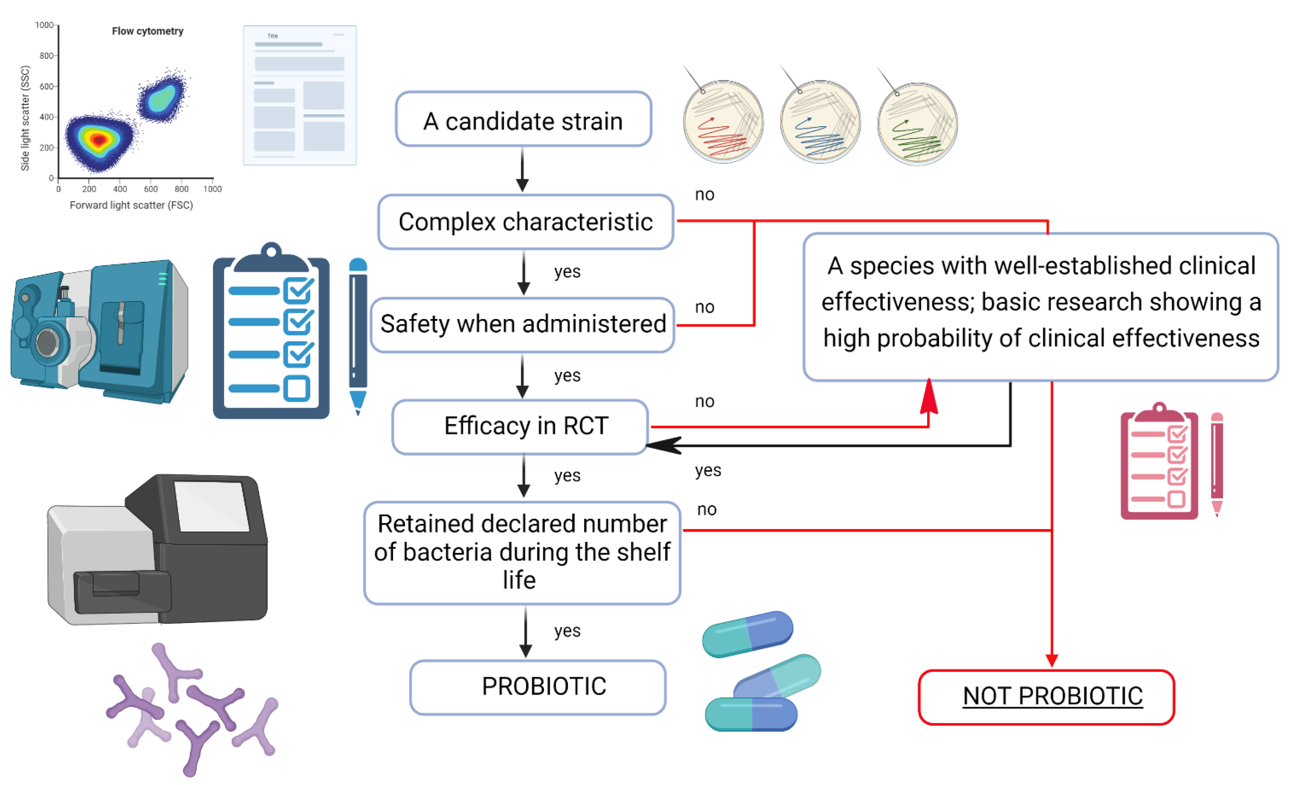
4. Probiotics—Quality Means Effectiveness

The legal regulations regarding the production of probiotics do not cover the specificity of this group of products and are the same as for food. In addition, the Food Safety Act practically excludes the possibility of informing consumers about the health effects of probiotics. Additional confusion is introduced by the division into probiotic drugs and dietary supplements. It often happens that the same strain, in the same quantity and produced by the same manufacturer, can be found both as a medicine and a dietary supplement. Therefore, medical practitioners are often confused and not eager to delve into these complex issues at a border of law and medicine, shifting the responsibility for the use of probiotics to patients or pharmacists. Probiotics must contain live bacteria, which are freeze-dried during production, i.e., dried at low temperature (this is the first critical moment for bacteria viability). Bacteria need to “animate” in the digestive tract and initiate the appropriate physiological and/or therapeutic effect. It is assumed that the minimum daily dose of probiotic bacteria should be approximately 1 × 10⁹ CFU (colony forming units) per day (deviations in the part of the logarithm are often not critical). Bacteria, after adhering to the epithelium of the gastrointestinal tract, transmit signals that “call” other microorganisms, which are to collectively create a biofilm that allows them to survive in the gastrointestinal tract. For this reason, it is more about the bacterial adhesion capacity than the dose of probiotic, which has the impact on the effectiveness of probiotics. The appropriate amount of probiotics in a given preparation should be kept at the end of the shelf-life. Other elements that ensure the viability of the bacteria throughout the shelf life of the product are the packaging that protects the probiotic from moisture (special barrier blisters are given out), transport under temperature-controlled conditions, and storage in a dry and cool place (some probiotics must be stored in the refrigerator). The most important is that probiotics must benefit the host’s health. That is, this effect should be documented by studies, preferably placebo-controlled clinical trials [94].
Probiotic strains must be well characterized, safe, and effective [94].
The most widely used technique for identifying strains is 16S ribosomal DNA sequencing. The sequence can be matched with available databases. The gold standard for strain identification, however, is whole genome sequencing (WGS), including any extrachromosomal elements. A fully sequenced genome allows for the recognition of microorganisms and facilitates the search for potential benefits and risk factors associated with the use of a given strain. One of the most important features of a safe probiotic is the lack of acquired antimicrobial resistance genes. Transmission of antibiotic resistance occurs through food, soil, and water reservoirs. That is why it is so important to properly select a probiotic strain, and ensure its verification and careful testing from every angle in order to prevent serious and irreversible consequences for the environment [94].
Assessing the viability of bacteria is one of the most important quality requirements for probiotics. Commonly used culturing methods are long-lasting, which do not ensure immediate correction of the production process and are burdened with the difficulties to culture various strains, which is especially important in the case of multi-strain products. The introduction of flow cytometry is very promising; however, it requires further validation studies [95].
The safety of probiotic use must be determined based on established scientific principles, including appropriate research material. EFSA has been keeping and updating a list of species considered safe for human consumption since 2007. One of the basic classifications of probiotic strains is QPS. Qualification for QPS is made on the basis of taxonomic identification and comprehensive scientific data on the safety of a given strain. QPS qualification is conducted at species level. It seems that since the activity of probiotics is strain-dependent, the safety of their use should also be determined for individual strains. Now, the only method to evaluate this at a strain level is to conduct in vitro toxicological studies and clinical trials. End-product-specific tests are particularly important, especially in the case of using probiotics in groups of seriously ill people [96,97]. The decision tree for the classification of a strain as a probiotic is shown in Figure 2.
Another problem concerns declaring the effectiveness of probiotics. The approach to this issue differs from country to country [98]. In the European Union, probiotics and dietary supplements are subject to food law (Regulation 178/2002/EC; Directive 2000/13/EU). All health claims made on probiotics must be approved by EFSA. EFSA has published a list of bacterial species with QPS. As already mentioned, EFSA has so far rejected all submitted health claims regarding probiotics. So, on the one hand, there is a stringent control of health claims, but on the other hand, there is little regulation of the manufacturing process and almost no post-marketing regulatory oversight. In the United States, most probiotic products are classified as food or dietary supplements. Dietary supplements must be manufactured in accordance with GMP guidelines, but are not covered by quality or efficacy testing. As in Europe, the packaging of dietary supplements cannot contain information regarding the indications for the use of probiotics in specific diseases, however, functional claims such as “supports healthy digestion” are allowed, with the reservation required by the FDA regarding the lack of medicinal properties of the product. These claims must be truthful, not mislead the consumer, and must be substantiated by scientific evidence. There is also a category of probiotics that has been developed to be ingested or administered enterally under medical supervision and which are intended for the dietary management of a specific disease or medical condition for which specific nutritional requirements exist. In the United States, these preparations are classified as medicinal foods. In Europe, manufacturers of dietary supplements cannot inform consumers about the effects of their products at all, unless they are accompanied by approved health claims. Therefore, the only way to obtain such information is to use professional studies or advice from doctors, pharmacists, nutritionists, and other health care system employees. Recommendations of scientific societies and other organizations setting standards of nutritional or dietary management are very important.
Taking into account the above considerations, below we discuss the evidence-based rationales of prescribing probiotics to post-acute COVID-19 patients, manifesting with gastrointestinal and neuropsychiatric symptoms. Despite lack of specific clinical trials among post COVID-19 patients with DGBIs, the available data already supports probiotic use in those patients.

5. Special Clinical Situations

5.1. COVID-19 and Central Nervous System

The body of evidence indicates that SARS-CoV-2 has the ability to invade the central nervous system (CNS) in either a direct way or—in most cases—indirectly, as a consequence of systemic hyper-inflammatory state. The viral agent was found to penetrate into brain parenchyma via at least a couple of routes [99]. It was hypothesized, however, that initially, peripheral nerves are invaded and then the infection spreads through synapses [100]. The olfactory nerve has been pointed as the most frequent route of infection [101] and seems a strong candidate as anosmia is a frequent COVID-19 symptom. Moreover, an angiotensin-converting enzyme 2 (ACE-2) receptors are co-expressed there with TMPRSS2 and neuropilins, among them NRP-1 invading CNS, all of which are necessary for SARS-CoV-2 infection [102,103]. Importantly, olfactory tract projections, for instance respiratory and cardiovascular control centers in medulla, have been found to contain SARS-CoV-2 [104], making the so-called transcribial route of neuroinfection. The other peripheral nerve found to play a role in SARS-CoV-2 infection is the vagus nerve, eventually reaching the brainstem [105]. Of note, the vagus nerve co-creates the gut brain axis, the dysfunction of which might explain neuropsychiatric symptoms that occur during COVID-19 [106]. SARS-CoV-2 was found in human faeces [107], as ACE receptors are expressed in intestinal epithelium. All together, gut originated viral particles might enter the enteric nervous system and finally reach the CNS via vagus nerve [108]. The other route of the virus neuroinvasion is of hematogenous type [109]. Viral transcytosis across vascular endothelial cells (expressing ACE-2 receptor) and the infected leukocytes mobilization towards the blood brain barrier make this one [110,111,112] the most probable and frequent one.
The first CNS-related symptoms might be due to hypoxia occurring along with respiratory distress. These alter the brain metabolism to a large extent [113] but do not serve as primary neurologic symptoms in COVID-19. Neuroinvasiveness and neurotropism are responsible for long COVID-19 symptoms. Short ones are nonspecific and include anosmia, lack of taste, headache, malaise, myalgias, and dizziness. Serious neurological disorders include acute disseminated encephalomyelitis (ADEM), such as demyelination, multiple sclerosis, optic neuritis, and encephalitis along with delirium and cerebrovascular events. Importantly, like in other pandemics in the past, psychiatric and cognitive sequelae do occur in COVID-19 survivors, despite the severity of the disease [114,115]. The mental well-being of each person has been altered by the pandemic-related stress to varying degrees, with children/adolescents, pregnant women, and the elderly being among the most affected [116]. It has been well established that extended exposure to overall stress contributes to alterations in biological stress-responsive systems (i.e., hypothalamic-pituitary-adrenal axis, autonomic nervous system, sympathetic-adreno-modulary system, immune system) and precipitate physical and mental disorders [117] or negatively affect ongoing conditions in people already affected. Behavioral restriction and social isolation also triggered unhealthy lifestyle choices which might additionally add on to poor neuropsychiatric state [118]. A large multinational survey, namely Collaborative Outcomes study on Health and Functioning during Infection Times (COH-FIT) [119,120] has been established in April 2020 to track the psychiatric state worldwide and to identify risk and protective factors towards poor outcomes. The very early results of the study, which up to the 1 January 2022 collected data on more than 166,000 respondents demonstrated worsening in stress, predominantly in women and the elderly. Similar observations were made for loneliness, anger, and obsessive-compulsive behaviors. These are in line with already published data stating that preventing mental health in long COVID-19 phenotypes might be of particular concern to health policy [121]. Indeed, the high prevalence of psychiatric disorders has become a catalyst for many unfavorable, social, health, and economic phenomena at the national and international level

5.2. COVID-19 and Gastrointestinal Tract

Up to date numerous retrospective and prospective studies as well as meta-analyses on the topic related to gastrointestinal manifestation of SARS-CoV-2 infection were published. Jin et al. [122] in their retrospective study reported at least one of common GI symptoms (nausea, vomiting or diarrhea) among 11.4% out of 651 patients due to SARS-CoV-2 infection at hospital admission in China. Most of the symptoms were self-limited; however, patients with gastrointestinal symptoms had more severe complications during treatment with a need of ICU admission [122] in comparison to those without gastrointestinal symptoms. In another multicenter, retrospective study and recruiting patients in China, investigators reported the presence of abdominal pain and melena in 21% of 230 COVID-19 patients [123]. Redd et al. in their cohort, multicenter retrospective study reported the presence of gastrointestinal symptoms (anorexia, diarrhea, nausea, and vomiting) in 61.3% out of 318 patients admitted to hospital due to SARS-CoV-2 infection [124]. Others also reported various frequency of GI symptoms among COVID-19 patients: (i) Cholaneril et al. [USA]—31.9% [125]; (ii) Zhang et al. [China]—39.6% [126]; and (iii) Cheung et al. [Hong Kong]—25.4% [127]. Li et al. in their meta-analysis, including 212 studies from 11 countries involving more than 280 000 individuals, reported that gastrointestinal (nausea, vomiting, abdominal pain) and respiratory symptoms (shortness of breath, chest pain) were associated with severe COVID-19, whereas pneumonia and end-organ failure were associated with mortality [128]. In another report, Li et al. reported no significant association between GI symptoms and COVID-19 mortality based on 20 studies with 58,423 COVID-19 patients with adjusted effect estimates analysis (pooled effect size, 0.93; 95% CI, 0.75 to 1.16; p = 0.535; random effects model) [129]. Cao et al. conducted a systematic review and meta-analysis of 31 studies with 46,959 individuals hospitalized in China and found the prevalence of diarrhea among 6.8% COVID-19 patients [130]. Another systematic review and meta-analysis of 47 papers and 10,890 patients revealed the presence of diarrhea in 7.7%, nausea and vomiting in 7.8%, and abdominal pain in 2.7% in patients hospitalized due to COVID-19 disease. Of importance, gastrointestinal symptoms were more prevalent in individuals hospitalized in countries other than China when comparison data analysis was conducted [131]. Transient liver function failure among COVID-19 individuals has also been observed and analyzed [4]. However, most acute gastrointestinal symptoms fade away, once the viral infection is cleared.
Nevertheless, SARS-CoV2 infection has already been implicated in persistence of chronic gastrointestinal symptoms, which may persist long after the COVID-19 disease [132]. SARS-CoV-2 has the ability to bind to ACE-2 receptors—found on the surface of many cells, including the gastrointestinal lining—and trigger their physiological role to processes conversion of angiotensin II (Ang II) to angiotensinI [133]. These two members of the RAAS family trigger opposing biological effects on target cells and stimulate: (i) the angiotensin 1 receptor (AT1R), and (ii) MasR, respectively. Activation of AT1R during SARS-CoV-2 infection has deleterious impacts, inducing an increase in reactive oxygen species (ROS) synthesis, vasoconstriction, fibrosis, and alterations of gut microbiota [134]. SARS-CoV-2, as capable of infecting gastrointestinal epithelial and endothelial cells, has been classified as an enteric pathogen. The interaction of the ACE2 receptor with the SARS-CoV-2 spike protein may trigger activation of the Nlrp3 inflammasome, which when hyperactivated further leads to cell death by pyroptosis [134]. Post-COVID-19 chronic gastrointestinal symptoms recall post-infectious irritable bowel syndrome (IBS), which is frequent after other viral and bacterial infections. According to ROME Foundation functional gastrointestinal disorders (FGIDs) or disorders of gut-brain interaction (DGBI) are disorders classified by GI symptoms related to any combination of: (i) motility disturbance, (ii) visceral hypersensitivity, (iii) altered mucosal and immune function; (iv) altered gut microbiota; and v) altered central nervous system (CNS) processing [135,136]. SARS-CoV-2 by altering mucosal, immune, and microbial function in the gastrointestinal tract impairs the gut barrier [136]. As we previously reported, an impaired gut barrier and microbial alterations may lead or be associated with gastrointestinal [54], mental [137], and metabolic [138] symptoms or disorders. Cooney et al. reported that gastrointestinal symptoms were common at 6 months among 43.8% individuals post COVID-19 disease [139]. Ours, yet unpublished data of a cohort of COVID-19 individuals in Poland, supports previous reports of a high percentage of patients with GI symptoms following COVID-19 recovery. Therefore, further research is essential to explore the nature of new post-COVID-19 symptoms as well as clinical courses and responses to various medical treatments.

5.3. Intensive Care of COVID-19 Patients—Evidence Based Medicine of a Single Center Experience

Here we share a brief report of a single center experience at the intensive care unit (ICU) at Pomeranian Medical University in Poland. ICU treatment significantly affected the COVID-19-related prognosis [3]. In one of the recent studies, Meconnen et al. reported a rate of 33% ICU transfer of hospitalized COVID-19 patients with a 33% mortality rate at ICU [140]. ICU treatment was associated with high mortality [141]. Variety of factors were reported to affect this outcome, including: (i) patients overall health condition at a time of hospitalization, (ii) age, (iii) comorbidities, and (iv) type of treatment. Most of the patients transferred to ICU were critically ill, with symptoms of acute respiratory failure due to viral pneumonia. Diagnostic x-ray and CT scans revealed matte glass appearance corresponding to massive pneumonitis affecting more than 90% of the lung area. Patients with corresponding lesions frequently required mechanical ventilation, deep analgo-sedation with muscle relaxant drugs administration and ECMO (Extra Corporeal Membrane Exygenation) along with other symptomatic pharmacological management. Despite the treatment, systemic hyper inflammatory state multiorgan failure affected the clinical picture. Targeted antibiotic therapy was frequently introduced to counteract secondary bacterial infections. Of recommended drugs, systemic steroids (dexamethason) were used in order to suppress systemic inflammation [142]. Of other ICU treatment regimens, anticoagulants in prophylactic (not therapeutic) doses were prescribed. Plasma of convalescents was not recommended to ICU critically ill patients. Similarly, the use of redemsivir was restricted only to patients within 72 h since COVID-19 diagnosis or as therapy continuation in patients transferring to ICU. (https://sccm.org/SurvivingSepsisCampaign/Guidelines/COVID-19, accessed on 30 July 2022).
Another problem reported among post-COVID ICU patients was significant undernutrition, which concerned 66% of patients (diagnosed based on Global Leadership Initiative on Malnutrition (GLIM) criteria). Despite intensive treatment, intensive catabolism was observed, which could have lasted for several months. Undernutrition was reported as an independent negative prognostic factor of ICU readmission, infection, and mortality. More than half of patients dismissed home after hospital and ICU treatment complained of poor health and incomplete recovery. Using the descriptive EQ5D-5L scale system, which comprises five dimensions: (i) mobility, (ii) self-care, (iii) usual activities, (iv) pain and discomfort, as well as (v) anxiety, complaints were more prevalent among women in their fifties [143]. Thromboembolic events were reported among other COVID-19-related disorders, including massive cerebral strokes. However, the full-scale epidemiologic data on thromboembolic events among COVID-19 patients are lacking. These thromboembolic events were reported also in young adults and children diagnosed with COVID-19 disease.

6. Specific Applications of Probiotics after COVID-19 Infection

6.1. Irritable Bowel Syndrome (IBS)

Disturbances in the interaction of the gut-brain axis play an important role in the multifactorial pathogenesis of the irritable bowel syndrome (IBS). The intestinal microbiota is an essential element of these interactions, and its disturbances directly affect other pathogenetic mechanisms of IBS. One of the first clinical trials assessing the effectiveness of probiotics in IBS, concerned the L. plantarum 299v strain in 40 patients with IBS. In all patients with IBS who took the probiotic for 4 weeks, a significant reduction in the frequency and intensity of abdominal pain was observed compared to the placebo group [144]. It should be noted that according to the current Rome IV Criteria, pain is a key symptom in IBS. In guidelines of Polish Gastroenterology Association [39], on the use of probiotic strains with a possible beneficial effect on the symptoms of IBS, experts—the authors of the monograph—suggested the use of individual strains or complex preparations tested for effectiveness in IBS rather than probiotics as a group. The recommended strains available in Poland include: L. plantarum 299V—1 × 10¹⁰ CFU 1 × daily, Saccharomyces boulardii CNCM I-745—5 × 10⁹ CFU 2 × daily, Bacillus coagulans GBI—15 × 107 3 × daily, and B. infantis 35624—108 CFU 1 × daily. It should be emphasized that the recommendations are based on the results of randomized clinical trials (RCTs) and not meta-analyzes. The authors of the British guidelines in 2021, based on the RCT meta-analysis, indicated that probiotics should be used in persons with IBS in the first place, for 12 weeks with diet modification, before starting pharmacological treatment [145]. Although the authors of the guidelines do not recommend individual strains, in the meta-analysis attached to the guidelines, among the groups of probiotic bacteria, lactobacilli showed the greatest effectiveness in IBS. Marlicz et al. further analysis of the effectiveness of individual strains of lactobacilli in IBS showed the greatest effectiveness of L. plantarum 299v, the strain for which the largest number of RCTs were conducted [146]. These observations were also confirmed by McFarland et al. in the latest meta-analysis on the effectiveness of individual probiotic strains in patients with IBS [147]. The effectiveness of L. plantarum 299v was also confirmed in a large study, carried out in 25 medical centers in Germany, for a period of 12 weeks, comprising 221 patients with IBS. Taking the probiotic had a significant, beneficial effect on all the analyzed symptoms of IBS and was associated with the reduction in abdominal pain, flatulence, and normalization of the bowel movement rhythm [148]. Probiotic strains that can be used in patients with post-acute COVID-19 IBS are shown in Table 1.

6.2. Psychobiotics

The COVID-19 pandemic had an impact on almost every individual worldwide [154]. This refers not only to the infectious aspect of this clinical entity but also to global healhtcare crisis, including mental health among patients and their caregivers. Lockdowns, huge economic and social instabilities—all rose along the pandemic. A number of meta-analyses confirmed the high incidence of anxiety, depression, and PTSD in COVID-19 patients and frontline healthcare workers [155,156]. Pharmacotherapies are effective for some but many side effects might occur from COVID-19 and its link with gut microbiota urged the scientific and medical community to focus on the gut microbiome modulation as a novel strategy to counteract COVID-19-related symptoms, including neuropsychiatric ailments. As evidenced, stress increases gut permeability thus inducing peripheral and neuronal inflammation. Empirical data exist to confirm an important role of the gut microbiota in mental health and well-being; however, few gaps need to be addressed, with the causal role of the microbiota in mental health as the priority [157]. Secondly, the role of fungi and phages should also be evaluated, as these microorganisms also make up the microbiota. Importantly, a few conflicting results in human trials with respect to probiotics use in alleviating depression and anxiety exist [158], probably due to strain specificity. In addition, probiotics might not work in the same way in all individuals due to host genetic suits, one’s diet, and the colonization potential of the probiotics. Hence, one size might not fit all regarding anxiety and depression. Large trials in a diverse population to define efficacy, duration, adverse effects, and dosage are needed. Future trials might also include genetic determinants, for instance, those referring to probiotic colonization and their efficacy. In the era of post COVID-19 pandemic, personalized medicine is getting more and more attraction among scientists and healthcare providers. In particular, testing dietary interventions among individuals and testing their microbiomes might opt out as an effective strategy to boost well-being and mental health in patients recovering from COVID-19 disease. Probiotics as non-expensive and safe agents can be viewed as dietary supplements effective among individuals with risk factors, especially in developing countries being at high risk of depression or other stress-related comorbidities [159].
Psychobiotics, i.e., probiotics that support mental health, are currently of great interest to researchers, doctors, and patients suffering from mental disorders, as well as people exposed to stress. The pioneers of research on psychobiotics were John F. Cryan and Timothy G. Dinan, who introduced this term into the medical dictionary [160]. Animal studies provided the first evidence for the role of probiotics in shaping emotional health. It has been proved that the administration of psychobiotics influences the composition of the intestinal microbiota and may change the behavior of the tested animals [161,162,163]. In addition, it has been proven that selected probiotic strains, belonging to the group of psychobiotics, affect the activity of the vagus nerve, and if administered long enough, they can change the expression of GABA receptors related to the pathogenesis of anxiety-depressive disorders [164]. Moreover, numerous experimental and clinical studies have shown that some probiotics are able to lower the concentration of cortisol in humans [165] and its counterpart—corticosterone in animals [166]. It follows that they are also indirectly involved in the regulation of the hypothalamic-pituitary-adrenal (HPA) axis [137]. Disturbances in the functioning of this axis are related to the so-called allostatic load [167]. The term “allostasis” was coined to conceptualize the physiological responses that enable adaptation to environmental demands arising from stressful situations [168]. Allostasis mediators include hormones, neurotransmitters, neurotrophic substances, markers of oxidative stress, and inflammation. Short-term activation of these mechanisms may be beneficial to the process of maintaining homeostasis of the organism; however, chronic is a harmful burden on the organism and has been defined as an allostatic load (AL). In turn, the negative effects of this burden on mental and physical health have been defined as allostatic overload. These changes lead to mental disorders and chronic somatic diseases, such as metabolic or cardiovascular disorders. One of the factors influencing the allostatic load may be changes in the gut microbiota [169]. The results of recent years’ research clearly indicate that the use of psychobiotics may bring health benefits to patients treated for mental disorders. One of the best-studied strains of bacteria from this group are L. heleveticus Rosell-52 and B. longum Rosell-175. It has been shown that feeding L. heleveticus Rosell 52 to animals exposed to stress reduces the adhesion of pathogens to intestinal epithelial cells, prevents their translocation, and reduces the synthesis of pro-inflammatory cytokines and may have a protective effect on the structures of the limbic system exposed to long-term stress [170]. Interestingly, the administration of L. heleveticus Rosell-52 and B. longum Rosell-175 to mothers of rats that are subjected to high stress may save the next generations from the negative consequences of this burden [171]. Especially in the era of the latest research on the effects of stress from generation to generation, this observation gives great hope for effective help not only for directly burdened individuals but also for their offspring. Clinical studies have shown that the administration of L. heleveticus Rosell-52 and B. longum Rosell-175 bacterial strains to healthy people reduces gastrointestinal discomfort caused by excessive stress [172]. It has been observed that the administration of these bacterial strains has a positive effect on the well-being of the respondents, reduces the intensity of anxiety, and reduces the excretion of cortisol. In March 2016, Canada’s Natural and Nonprescription Health Products Directorate issued the following recommendations for its use: (1) it helps to relieve general symptoms of anxiety; (2) alleviate gastrointestinal symptoms caused by stress; which (3) supports emotional balance. Below we discuss issues related to psychobiotic use in patients with (i) depression, (ii) anxiety, (iii) and stress, what is especially important for patients after post-acute COVID-19 infection.

6.2.1. Depression

In patients with depression, a different composition of the gut microbiota compared to healthy people was evidenced in multiple studies. The experimental animals, which have had stool transplanted from depressed patients, started to manifest depressive behavior after such a procedure. Although the mechanism of this relationship remains unknown [157], it is postulated that taxonomic changes observed in depressed patients are associated with pro-inflammatory effects, reduced production of short-chain fatty acids (SCFA), impaired intestinal barrier integrity, and disturbed metabolic pathways (inter alia tryptophan, GABA). However, only a few publications confirmed these via metagenomic and metabolomic analysis, evaluation of immunological parameters or markers of intestinal permeability. Future research requires a standardization process ranging from patient selection, biological material collection, sequencing (e.g., 16S, whole genome or “shotgun” shallow sequencing), and bioinformatics analysis. In clinical trials, the unequivocal effect of antidepressants on the microbiota is not observed, however, subsequent research projects require the involvement of a larger number of patients and mechanistic analyses [173,174]. It should be emphasized that the cause of depression is unknown, and the disease has a multifactorial etiology. Therefore, microbiota should not be treated as the Holy Grail that would answer all our questions regarding the etiology and treatment of this disease, but as an environmental element that participates in the etiopathogenesis of this disease and creates new possibilities for the monitoring and treatment of patients. Numerous studies and meta-analyses have shown the beneficial effect of probiotics on mood. Most of them were carried out in healthy people or with unconfirmed depression. A recently published systematic review comprised eight randomized clinical trials that investigated the relationship between the effectiveness of probiotics in depressed patients [175], and five of them also investigated the mechanism of action of probiotics. Other systematic reviews also describe studies of probiotics in patients with depression and other conditions, such as irritable bowel syndrome. Among the analyzed studies, four reported reductions in depression symptoms and two improved cognitive functions. Only one study in which probiotics were used as monotherapy did not confirm their clinical effectiveness. The composition of the gut microbiota was evaluated in one clinical trial and no changes were found under the influence of probiotics. It is however believed that probiotics, despite having no effect on the composition of the microbiota, affect the expression of bacterial genes and might induce anti-inflammatory responses within a cell. Two studies found a decrease in blood kynurenine. Additionally, an effect on the metabolism of tryptophan was observed after administration of the probiotic. Kynurenine may be of high importance in depressed patients due to its neurotoxic and neurodegenerative effects. Furthermore, it has recently been shown that the administration of L. helveticus R0052 and B. longum R0175 caused an increase in the concentration of Brain Derived Neurotrophic Factor (BDNF) in the blood, which may reduce the severity of depression symptoms [176]. Due to the scarcity of research, no clear conclusions can be drawn about the strains or their combinations, doses, and duration of their administration in depression; however, it is known that patients may benefit from the use of probiotics, especially in combination with antidepressants.

6.2.2. Anxiety

The anxiolytic properties of probiotics have been observed in preclinical and clinical studies in which the influence of this group of preparations on the symptoms of anxiety accompanying other diseases, such as IBS, was assessed. So far, only one study has been published in which the impact of probiotics on generalized anxiety disorder (DSM-V criteria) in patients treated with sertraline has been analyzed. After eight weeks of the study, there was an improvement in the results of the Hamilton Rating Scale for Anxiety (HAM-A) [177].

6.2.3. Stress

Stress-related comorbidities, such as depression and anxiety, were reported to be associated with gut microbiome alterations among COVID-19 frontline health care workers, persisting for at least half a year. During a half-year follow up, several microbes were linked to mental health status, including mainly Faecalibacterium spp. and [Eubacterium] eligens group spp. Both with anti-inflammatory effects. Of note, the prediction model indicated that low abundance of [Eubacterium] hallii group uncultured bacterium and high abundance of Bacteroides eggerthii at Day 0 (immediately after the two-month frontline work) were significant determinants of the reappearance of PTSD symptoms in FHWs [178]. Psychobiotic strains, which are useful in post-acute COVID-19 patients suffering from mental health problems, are included in Table 2.

7. Conclusions

COVID-19 pandemic had a significant impact on environmental, animal, and human microbiomes. Microbial alterations, frequently referred to as dysbiosis, played a role in the pathogenesis and prognosis of COVID-19 and associated diseases. Microbiota modulation by means of probiotics seems a very promising way of eliminating negative outcomes of SARS-CoV-2 infection. Particular probiotic strains have been tested and their efficacy documented in selected disorders, including IBS and neuropsychiatric disturbances. Currently, there are no official recommendations or recommended procedures for the use of probiotics in patients with COVID-19. However, it can be assumed that many doctors, pharmacists, and patients will want to use a probiotic in the treatment of this disease. In such cases, strains with documented activity should be used. There is a constant need to plan and conduct new RCTs on the role of probiotics and verify their clinical efficacy of counteracting negative consequences of COVID-19 pandemic. Quality control is another important but often neglected aspect in trials utilizing probiotics in various clinical entities. It determines the safety and efficacy of probiotics, which is of utmost importance in patients with post-acute COVID-19 syndrome.

Author Contributions

Conceptualization, I.Ł.; methodology, I.Ł.; software, K.S.-Ż.; validation, I.Ł.; formal analysis, I.Ł. and W.M.; investigation, I.Ł., K.S.-Ż., J.S.-P. and W.M.; data curation, I.Ł.; writing—original draft preparation, I.Ł.; writing—review and editing, all.; visualization, K.S.-Ż.; supervision, W.M.; project administration, K.S.-Ż.; funding acquisition, W.M. All authors have read and agreed to the published version of the manuscript.

Funding

This research received no external funding.

Institutional Review Board Statement

Not applicable.

Informed Consent Statement

Not applicable.

Data Availability Statement

Not applicable.

Conflicts of Interest

Karolina Skonieczna-Żydecka receives remuneration from Sanprobi, Szczecin, Poland—manufacturer of probiotics, and Igor Łoniewski and Wojciech Marlicz are shareholders of this company. However, the content of this study was not constrained by this fact. Moreover, our adherence to the Journal’s policies on the sharing of data and materials is unaffected. The other authors declare no conflicts of interest.

References

  1. Coronavirus COVID-19 (2019-NCoV). Available online: https://www.arcgis.com/apps/dashboards/bda7594740fd40299423467b48e9ecf6 (accessed on 13 August 2022).
  2. Cascella, M.; Rajnik, M.; Aleem, A.; Dulebohn, S.C.; Di Napoli, R. Features, Evaluation, and Treatment of Coronavirus (COVID-19). In StatPearls; StatPearls Publishing: Treasure Island, FL, USA, 2022. [Google Scholar]
  3. Kotfis, K.; Skonieczna-Żydecka, K. COVID-19: Gastrointestinal Symptoms and Potential Sources of SARS-CoV-2 Transmission. Anaesthesiol. Intensive Ther. 2020, 52, 171–172. [Google Scholar] [CrossRef] [PubMed]
  4. Kukla, M.; Skonieczna-Żydecka, K.; Kotfis, K.; Maciejewska, D.; Łoniewski, I.; Lara, L.F.; Pazgan-Simon, M.; Stachowska, E.; Kaczmarczyk, M.; Koulaouzidis, A.; et al. COVID-19, MERS and SARS with Concomitant Liver Injury—Systematic Review of the Existing Literature. J. Clin. Med. 2020, 9, 1420. [Google Scholar] [CrossRef]
  5. Böhmer, M.M.; Buchholz, U.; Corman, V.M.; Hoch, M.; Katz, K.; Marosevic, D.V.; Böhm, S.; Woudenberg, T.; Ackermann, N.; Konrad, R.; et al. Investigation of a COVID-19 Outbreak in Germany Resulting from a Single Travel-Associated Primary Case: A Case Series. Lancet Infect. Dis. 2020, 20, 920–928. [Google Scholar] [CrossRef]
  6. Mokhtari, T.; Hassani, F.; Ghaffari, N.; Ebrahimi, B.; Yarahmadi, A.; Hassanzadeh, G. COVID-19 and Multiorgan Failure: A Narrative Review on Potential Mechanisms. J. Mol. Histol. 2020, 51, 613–628. [Google Scholar] [CrossRef] [PubMed]
  7. Wang, Y.; Wu, G.; Zhao, L.; Wang, W. Nutritional Modulation of Gut Microbiota Alleviates Severe Gastrointestinal Symptoms in a Patient with Post-Acute COVID-19 Syndrome. mBio 2022, 13, e0380121. [Google Scholar] [CrossRef]
  8. Sanidad, K.Z.; Xiao, H.; Zhang, G. Triclosan, a Common Antimicrobial Ingredient, on Gut Microbiota and Gut Health. Gut Microbes 2018, 10, 434–437. [Google Scholar] [CrossRef]
  9. Jutkina, J.; Marathe, N.P.; Flach, C.-F.; Larsson, D.G.J. Antibiotics and Common Antibacterial Biocides Stimulate Horizontal Transfer of Resistance at Low Concentrations. Sci. Total Environ. 2018, 616–617, 172–178. [Google Scholar] [CrossRef]
  10. Ejtahed, H.-S.; Hasani-Ranjbar, S.; Siadat, S.D.; Larijani, B. The Most Important Challenges Ahead of Microbiome Pattern in the Post Era of the COVID-19 Pandemic. J. Diabetes Metab. Disord. 2020, 19, 2031–2033. [Google Scholar] [CrossRef]
  11. Zang, L.; Ma, Y.; Huang, W.; Ling, Y.; Sun, L.; Wang, X.; Zeng, A.; Dahlgren, R.A.; Wang, C.; Wang, H. Dietary Lactobacillus Plantarum ST-III Alleviates the Toxic Effects of Triclosan on Zebrafish (Danio Rerio) via Gut Microbiota Modulation. Fish Shellfish Immunol. 2019, 84, 1157–1169. [Google Scholar] [CrossRef]
  12. Woodruff, M.C.; Ramonell, R.P.; Cashman, K.S.; Nguyen, D.C.; Ley, A.M.; Kyu, S.; Saini, A.; Haddad, N.; Chen, W.; Howell, J.C.; et al. Critically Ill SARS-CoV-2 Patients Display Lupus-like Hallmarks of Extrafollicular B Cell Activation. MedRxiv 2020. [Google Scholar] [CrossRef]
  13. Cai, Q.; Chen, F.; Wang, T.; Luo, F.; Liu, X.; Wu, Q.; He, Q.; Wang, Z.; Liu, Y.; Liu, L.; et al. Obesity and COVID-19 Severity in a Designated Hospital in Shenzhen, China. Diabetes Care 2020, 43, 1392–1398. [Google Scholar] [CrossRef]
  14. Fang, L.; Karakiulakis, G.; Roth, M. Are Patients with Hypertension and Diabetes Mellitus at Increased Risk for COVID-19 Infection? Lancet Respir. Med. 2020, 8, e21. [Google Scholar] [CrossRef]
  15. Hill, M.A.; Mantzoros, C.; Sowers, J.R. Commentary: COVID-19 in Patients with Diabetes. Metabolism 2020, 107, 154217. [Google Scholar] [CrossRef]
  16. Moreira-Rosário, A.; Marques, C.; Pinheiro, H.; Araújo, J.R.; Ribeiro, P.; Rocha, R.; Mota, I.; Pestana, D.; Ribeiro, R.; Pereira, A.; et al. Gut Microbiota Diversity and C-Reactive Protein Are Predictors of Disease Severity in COVID-19 Patients. Front. Microbiol. 2021, 12, 1820. [Google Scholar] [CrossRef] [PubMed]
  17. Ward, D.; Bhattarai, S.; Rojas-Correa, M.; Purkayastha, A.; Holler, D.; Qu, M.D.; Mitchell, W.; Yang, J.; Fountain, S.; Zeamer, A.; et al. The Intestinal and Oral Microbiomes Are Robust Predictors of COVID-19 Severity the Main Predictor of COVID-19-Related Fatality [Preprint]. Univ. Mass. Med. Sch. Fac. Publ. 2021. [Google Scholar] [CrossRef]
  18. Li, S.; Yang, S.; Zhou, Y.; Disoma, C.; Dong, Z.; Du, A.; Zhang, Y.; Chen, Y.; Huang, W.; Chen, J.; et al. Microbiome Profiling Using Shotgun Metagenomic Sequencing Identified Unique Microorganisms in COVID-19 Patients With Altered Gut Microbiota. Front. Microbiol. 2021, 12, 2930. [Google Scholar] [CrossRef]
  19. Santacroce, L.; Inchingolo, F.; Topi, S.; Del Prete, R.; Di Cosola, M.; Charitos, I.A.; Montagnani, M. Potential Beneficial Role of Probiotics on the Outcome of COVID-19 Patients: An Evolving Perspective. Diabetes Metab. Syndr. 2021, 15, 295–301. [Google Scholar] [CrossRef] [PubMed]
  20. Nalbandian, A.; Sehgal, K.; Gupta, A.; Madhavan, M.V.; McGroder, C.; Stevens, J.S.; Cook, J.R.; Nordvig, A.S.; Shalev, D.; Sehrawat, T.S.; et al. Post-Acute COVID-19 Syndrome. Nat. Med. 2021, 27, 601–615. [Google Scholar] [CrossRef]
  21. Komaroff, A.L. The Tragedy of Long COVID. Available online: https://www.health.harvard.edu/blog/the-tragedy-of-the-post-covid-long-haulers-202010152479 (accessed on 25 April 2022).
  22. Huang, C.; Huang, L.; Wang, Y.; Li, X.; Ren, L.; Gu, X.; Kang, L.; Guo, L.; Liu, M.; Zhou, X.; et al. 6-Month Consequences of COVID-19 in Patients Discharged from Hospital: A Cohort Study. Lancet 2021, 397, 220–232. [Google Scholar] [CrossRef]
  23. Pan, L.; Mu, M.; Yang, P.; Sun, Y.; Wang, R.; Yan, J.; Li, P.; Hu, B.; Wang, J.; Hu, C.; et al. Clinical Characteristics of COVID-19 Patients With Digestive Symptoms in Hubei, China: A Descriptive, Cross-Sectional, Multicenter Study. Am. J. Gastroenterol. 2020, 115, 766–773. [Google Scholar] [CrossRef]
  24. Hashimoto, T.; Perlot, T.; Rehman, A.; Trichereau, J.; Ishiguro, H.; Paolino, M.; Sigl, V.; Hanada, T.; Hanada, R.; Lipinski, S.; et al. ACE2 Links Amino Acid Malnutrition to Microbial Ecology and Intestinal Inflammation. Nature 2012, 487, 477–481. [Google Scholar] [CrossRef] [PubMed]
  25. Liu, Q.; Mak, J.W.Y.; Su, Q.; Yeoh, Y.K.; Lui, G.C.-Y.; Ng, S.S.S.; Zhang, F.; Li, A.Y.L.; Lu, W.; Hui, D.S.-C.; et al. Gut Microbiota Dynamics in a Prospective Cohort of Patients with Post-Acute COVID-19 Syndrome. Gut 2022, 71, 544–552. [Google Scholar] [CrossRef] [PubMed]
  26. Weng, J.; Li, Y.; Li, J.; Shen, L.; Zhu, L.; Liang, Y.; Lin, X.; Jiao, N.; Cheng, S.; Huang, Y.; et al. Gastrointestinal Sequelae 90 Days after Discharge for COVID-19. Lancet Gastroenterol. Hepatol. 2021, 6, 344–346. [Google Scholar] [CrossRef]
  27. Yeoh, Y.K.; Zuo, T.; Lui, G.C.-Y.; Zhang, F.; Liu, Q.; Li, A.Y.; Chung, A.C.; Cheung, C.P.; Tso, E.Y.; Fung, K.S.; et al. Gut Microbiota Composition Reflects Disease Severity and Dysfunctional Immune Responses in Patients with COVID-19. Gut 2021, 70, 698–706. [Google Scholar] [CrossRef] [PubMed]
  28. Dhar, D.; Mohanty, A. Gut Microbiota and Covid-19- Possible Link and Implications. Virus Res. 2020, 285, 198018. [Google Scholar] [CrossRef]
  29. Zuo, T.; Zhang, F.; Lui, G.C.Y.; Yeoh, Y.K.; Li, A.Y.L.; Zhan, H.; Wan, Y.; Chung, A.C.K.; Cheung, C.P.; Chen, N.; et al. Alterations in Gut Microbiota of Patients With COVID-19 During Time of Hospitalization. Gastroenterology 2020, 159, 944–955.e8. [Google Scholar] [CrossRef]
  30. Cavaillon, J.-M. Exotoxins and Endotoxins: Inducers of Inflammatory Cytokines. Toxicon Off. J. Int. Soc. Toxinology 2018, 149, 45–53. [Google Scholar] [CrossRef] [PubMed]
  31. Wang, W.; Xu, Y.; Gao, R.; Lu, R.; Han, K.; Wu, G.; Tan, W. Detection of SARS-CoV-2 in Different Types of Clinical Specimens. JAMA 2020, 323, 1843–1844. [Google Scholar] [CrossRef]
  32. Small Intestinal Bacterial Overgrowth (SIBO)|OMICS International. Available online: https://www.omicsonline.org/open-access/small-intestinal-bacterial-overgrowth-sibo-2161-069X.1000225.php?aid=31428 (accessed on 25 April 2022).
  33. Zaher, S. Nutrition and the Gut Microbiome during Critical Illness: A New Insight of Nutritional Therapy. Saudi J. Gastroenterol. Off. J. Saudi Gastroenterol. Assoc. 2020, 26, 290–298. [Google Scholar] [CrossRef]
  34. Langford, B.J.; So, M.; Raybardhan, S.; Leung, V.; Westwood, D.; MacFadden, D.R.; Soucy, J.-P.R.; Daneman, N. Bacterial Co-Infection and Secondary Infection in Patients with COVID-19: A Living Rapid Review and Meta-Analysis. Clin. Microbiol. Infect. Off. Publ. Eur. Soc. Clin. Microbiol. Infect. Dis. 2020, 26, 1622–1629. [Google Scholar] [CrossRef]
  35. Alberca, R.W.; Oliveira, L.d.M.; Branco, A.C.C.C.; Pereira, N.Z.; Sato, M.N. Obesity as a Risk Factor for COVID-19: An Overview. Crit. Rev. Food Sci. Nutr. 2021, 61, 2262–2276. [Google Scholar] [CrossRef]
  36. Alberca, G.G.F.; Alberca, R.W. Nutrition and the Microbiota Post-COVID-19. Saudi J. Gastroenterol. Off. J. Saudi Gastroenterol. Assoc. 2021, 27, 111–112. [Google Scholar] [CrossRef]
  37. Xavier-Santos, D.; Padilha, M.; Fabiano, G.A.; Vinderola, G.; Gomes Cruz, A.; Sivieri, K.; Costa Antunes, A.E. Evidences and Perspectives of the Use of Probiotics, Prebiotics, Synbiotics, and Postbiotics as Adjuvants for Prevention and Treatment of COVID-19: A Bibliometric Analysis and Systematic Review. Trends Food Sci. Technol. 2022, 120, 174–192. [Google Scholar] [CrossRef]
  38. Cunningham, M.; Azcarate-Peril, M.A.; Barnard, A.; Benoit, V.; Grimaldi, R.; Guyonnet, D.; Holscher, H.D.; Hunter, K.; Manurung, S.; Obis, D.; et al. Shaping the Future of Probiotics and Prebiotics. Trends Microbiol. 2021, 29, 667–685. [Google Scholar] [CrossRef] [PubMed]
  39. Baindara, P.; Chakraborty, R.; Holliday, Z.M.; Mandal, S.M.; Schrum, A.G. Oral Probiotics in Coronavirus Disease 2019: Connecting the Gut-Lung Axis to Viral Pathogenesis, Inflammation, Secondary Infection and Clinical Trials. New Microbes New Infect. 2021, 40, 100837. [Google Scholar] [CrossRef]
  40. Kanauchi, O.; Andoh, A.; AbuBakar, S.; Yamamoto, N. Probiotics and Paraprobiotics in Viral Infection: Clinical Application and Effects on the Innate and Acquired Immune Systems. Curr. Pharm. Des. 2018, 24, 710–717. [Google Scholar] [CrossRef] [PubMed]
  41. Tiwari, S.K.; Dicks, L.M.T.; Popov, I.V.; Karaseva, A.; Ermakov, A.M.; Suvorov, A.; Tagg, J.R.; Weeks, R.; Chikindas, M.L. Probiotics at War Against Viruses: What Is Missing From the Picture? Front. Microbiol. 2020, 11, 1877. [Google Scholar] [CrossRef]
  42. Strauss, M.; Mičetić-Turk, D.; Pogačar, M.Š.; Fijan, S. Probiotics for the Prevention of Acute Respiratory-Tract Infections in Older People: Systematic Review. Healthcare 2021, 9, 690. [Google Scholar] [CrossRef] [PubMed]
  43. Tapiovaara, L.; Lehtoranta, L.; Poussa, T.; Mäkivuokko, H.; Korpela, R.; Pitkäranta, A. Absence of Adverse Events in Healthy Individuals Using Probiotics--Analysis of Six Randomised Studies by One Study Group. Benef. Microbes 2016, 7, 161–169. [Google Scholar] [CrossRef]
  44. Montazeri-Najafabady, N.; Kazemi, K.; Gholami, A. Recent Advances in Antiviral Effects of Probiotics: Potential Mechanism Study in Prevention and Treatment of SARS-CoV-2. Biologia 2022, 1–18. [Google Scholar] [CrossRef]
  45. Chiba, Y.; Shida, K.; Nagata, S.; Wada, M.; Bian, L.; Wang, C.; Shimizu, T.; Yamashiro, Y.; Kiyoshima-Shibata, J.; Nanno, M.; et al. Well-Controlled Proinflammatory Cytokine Responses of Peyer’s Patch Cells to Probiotic Lactobacillus Casei. Immunology 2010, 130, 352–362. [Google Scholar] [CrossRef] [PubMed]
  46. Chowdhury, A.H.; Cámara, M.; Verma, C.; Eremin, O.; Kulkarni, A.D.; Lobo, D.N. Modulation of T Regulatory and Dendritic Cell Phenotypes Following Ingestion of Bifidobacterium Longum, AHCC® and Azithromycin in Healthy Individuals. Nutrients 2019, 11, 2470. [Google Scholar] [CrossRef]
  47. Iemoli, E.; Trabattoni, D.; Parisotto, S.; Borgonovo, L.; Toscano, M.; Rizzardini, G.; Clerici, M.; Ricci, E.; Fusi, A.; De Vecchi, E.; et al. Probiotics Reduce Gut Microbial Translocation and Improve Adult Atopic Dermatitis. J. Clin. Gastroenterol. 2012, 46, S33–S40. [Google Scholar] [CrossRef]
  48. Dwivedi, M.; Kumar, P.; Laddha, N.C.; Kemp, E.H. Induction of Regulatory T Cells: A Role for Probiotics and Prebiotics to Suppress Autoimmunity. Autoimmun. Rev. 2016, 15, 379–392. [Google Scholar] [CrossRef] [PubMed]
  49. Speer, H.; D’Cunha, N.M.; Botek, M.; McKune, A.J.; Sergi, D.; Georgousopoulou, E.; Mellor, D.D.; Naumovski, N. The Effects of Dietary Polyphenols on Circulating Cardiovascular Disease Biomarkers and Iron Status: A Systematic Review. Nutr. Metab. Insights 2019, 12, 1178638819882739. [Google Scholar] [CrossRef]
  50. Ferlazzo, N.; Visalli, G.; Cirmi, S.; Lombardo, G.E.; Laganà, P.; Di Pietro, A.; Navarra, M. Natural Iron Chelators: Protective Role in A549 Cells of Flavonoids-Rich Extracts of Citrus Juices in Fe(3+)-Induced Oxidative Stress. Environ. Toxicol. Pharmacol. 2016, 43, 248–256. [Google Scholar] [CrossRef] [PubMed]
  51. Stiksrud, B.; Nowak, P.; Nwosu, F.C.; Kvale, D.; Thalme, A.; Sonnerborg, A.; Ueland, P.M.; Holm, K.; Birkeland, S.-E.; Dahm, A.E.A.; et al. Reduced Levels of D-Dimer and Changes in Gut Microbiota Composition After Probiotic Intervention in HIV-Infected Individuals on Stable ART. J. Acquir. Immune Defic. Syndr. 1999 2015, 70, 329–337. [Google Scholar] [CrossRef]
  52. Yeh, T.-L.; Shih, P.-C.; Liu, S.-J.; Lin, C.-H.; Liu, J.-M.; Lei, W.-T.; Lin, C.-Y. The Influence of Prebiotic or Probiotic Supplementation on Antibody Titers after Influenza Vaccination: A Systematic Review and Meta-Analysis of Randomized Controlled Trials. Drug Des. Devel. Ther. 2018, 12, 217–230. [Google Scholar] [CrossRef]
  53. Lei, W.-T.; Shih, P.-C.; Liu, S.-J.; Lin, C.-Y.; Yeh, T.-L. Effect of Probiotics and Prebiotics on Immune Response to Influenza Vaccination in Adults: A Systematic Review and Meta-Analysis of Randomized Controlled Trials. Nutrients 2017, 9, 1175. [Google Scholar] [CrossRef] [PubMed]
  54. Marlicz, W.; Yung, D.E.; Skonieczna-Żydecka, K.; Loniewski, I.; van Hemert, S.; Loniewska, B.; Koulaouzidis, A. From Clinical Uncertainties to Precision Medicine: The Emerging Role of the Gut Barrier and Microbiome in Small Bowel Functional Diseases. Expert Rev. Gastroenterol. Hepatol. 2017, 11, 961–978. [Google Scholar] [CrossRef]
  55. d’Ettorre, G.; Ceccarelli, G.; Marazzato, M.; Campagna, G.; Pinacchio, C.; Alessandri, F.; Ruberto, F.; Rossi, G.; Celani, L.; Scagnolari, C.; et al. Challenges in the Management of SARS-CoV-2 Infection: The Role of Oral Bacteriotherapy as Complementary Therapeutic Strategy to Avoid the Progression of COVID-19. Front. Med. 2020, 7, 389. [Google Scholar] [CrossRef] [PubMed]
  56. Ke, E.; Zhang, H. Clinical effects of probiotics in ordinary-type COVID-19 patients with diarrhea. World Chin. J. Dig. 2020, 28, 834–838. [Google Scholar] [CrossRef]
  57. Gutiérrez-Castrellón, P.; Gandara-Martí, T.; Abreu, Y.; Abreu, A.T.; Nieto-Rufino, C.D.; López-Orduña, E.; Jiménez-Escobar, I.; Jiménez-Gutiérrez, C.; López-Velazquez, G.; Espadaler-Mazo, J. Probiotic Improves Symptomatic and Viral Clearance in Covid19 Outpatients: A Randomized, Quadruple-Blinded, Placebo-Controlled Trial. Gut Microbes 2022, 14, 2018899. [Google Scholar] [CrossRef] [PubMed]
  58. Tang, H.; Bohannon, L.; Lew, M.; Jensen, D.; Jung, S.-H.; Zhao, A.; Sung, A.D.; Wischmeyer, P.E. Randomised, Double-Blind, Placebo-Controlled Trial of Probiotics To Eliminate COVID-19 Transmission in Exposed Household Contacts (PROTECT-EHC): A Clinical Trial Protocol. BMJ Open 2021, 11, e047069. [Google Scholar] [CrossRef]
  59. Olaimat, A.N.; Aolymat, I.; Al-Holy, M.; Ayyash, M.; Abu Ghoush, M.; Al-Nabulsi, A.A.; Osaili, T.; Apostolopoulos, V.; Liu, S.-Q.; Shah, N.P. The Potential Application of Probiotics and Prebiotics for the Prevention and Treatment of COVID-19. Npj Sci. Food 2020, 4, 17. [Google Scholar] [CrossRef]
  60. Zhou, F.; Yu, T.; Du, R.; Fan, G.; Liu, Y.; Liu, Z.; Xiang, J.; Wang, Y.; Song, B.; Gu, X.; et al. Clinical Course and Risk Factors for Mortality of Adult Inpatients with COVID-19 in Wuhan, China: A Retrospective Cohort Study. Lancet 2020, 395, 1054–1062. [Google Scholar] [CrossRef]
  61. Chen, N.; Zhou, M.; Dong, X.; Qu, J.; Gong, F.; Han, Y.; Qiu, Y.; Wang, J.; Liu, Y.; Wei, Y.; et al. Epidemiological and Clinical Characteristics of 99 Cases of 2019 Novel Coronavirus Pneumonia in Wuhan, China: A Descriptive Study. Lancet 2020, 395, 507–513. [Google Scholar] [CrossRef]
  62. Dong, X.; Cao, Y.-Y.; Lu, X.-X.; Zhang, J.-J.; Du, H.; Yan, Y.-Q.; Akdis, C.A.; Gao, Y.-D. Eleven Faces of Coronavirus Disease 2019. Allergy 2020, 75, 1699–1709. [Google Scholar] [CrossRef]
  63. Manna, S.; Baindara, P.; Mandal, S.M. Molecular Pathogenesis of Secondary Bacterial Infection Associated to Viral Infections Including SARS-CoV-2. J. Infect. Public Health 2020, 13, 1397–1404. [Google Scholar] [CrossRef]
  64. Tan, F.L.S.; Loo, W.L.; Tan, S.G.; Wong, C.Y.; Tan, Y.-M. Severe Acute Respiratory Syndrome in Surgical Patients: A Diagnostic Dilemma. ANZ J. Surg. 2005, 75, 21–26. [Google Scholar] [CrossRef] [Green Version]
  65. Johansson, M.-L.; Nobaek, S.; Berggren, A.; Nyman, M.; Björck, I.; Ahrné, S.; Jeppsson, B.; Molin, G. Survival of Lactobacillus Plantarum DSM 9843 (299v), and Effect on the Short-Chain Fatty Acid Content of Faeces after Ingestion of a Rose-Hip Drink with Fermented Oats. Int. J. Food Microbiol. 1998, 42, 29–38. [Google Scholar] [CrossRef]
  66. Johansson, M.L.; Molin, G.; Jeppsson, B.; Nobaek, S.; Ahrné, S.; Bengmark, S. Administration of Different Lactobacillus Strains in Fermented Oatmeal Soup: In Vivo Colonization of Human Intestinal Mucosa and Effect on the Indigenous Flora. Appl. Environ. Microbiol. 1993, 59, 15–20. [Google Scholar] [CrossRef] [PubMed]
  67. Mangell, P.; Thorlacius, H.; Syk, I.; Ahrné, S.; Molin, G.; Olsson, C.; Jeppsson, B. Lactobacillus Plantarum 299v Does Not Reduce Enteric Bacteria or Bacterial Translocation in Patients Undergoing Colon Resection. Dig. Dis. Sci. 2012, 57, 1915–1924. [Google Scholar] [CrossRef] [PubMed]
  68. Nobaek, S.; Johansson, M.-L.; Molin, G.; Ahrné, S.; Jeppsson, B. Alteration of Intestinal Microflora Is Associated With Reduction in Abdominal Bloating and Pain in Patients With Irritable Bowel Syndrome. Off. J. Am. Coll. Gastroenterol. ACG 2000, 95, 1231–1238. [Google Scholar] [CrossRef] [PubMed]
  69. Stjernquist-Desatnik, A.; Warfving, H.; Johansson, M.L. Persistence of Lactobacillus Plantarum DSM 9843 on Human Tonsillar Surface after Oral Administration in Fermented Oatmeal Gruel. A Pilot Study. Acta Oto-Laryngol. Suppl. 2000, 543, 215–219. [Google Scholar] [CrossRef]
  70. Klarin, B.; Johansson, M.-L.; Molin, G.; Larsson, A.; Jeppsson, B. Adhesion of the Probiotic Bacterium Lactobacillus Plantarum 299v onto the Gut Mucosa in Critically Ill Patients: A Randomised Open Trial. Crit. Care 2005, 9, R285. [Google Scholar] [CrossRef]
  71. Adlerberth, I.; Ahrne, S.; Johansson, M.L.; Molin, G.; Hanson, L.A.; Wold, A.E. A Mannose-Specific Adherence Mechanism in Lactobacillus Plantarum Conferring Binding to the Human Colonic Cell Line HT-29. Appl. Environ. Microbiol. 1996, 62, 2244–2251. [Google Scholar] [CrossRef]
  72. Mangell, P.; Lennernäs, P.; Wang, M.; Olsson, C.; Ahrné, S.; Molin, G.; Thorlacius, H.; Jeppsson, B. Adhesive Capability of Lactobacillus Plantarum 299v Is Important for Preventing Bacterial Translocation in Endotoxemic Rats. APMIS Acta Pathol. Microbiol. Immunol. Scand. 2006, 114, 611–618. [Google Scholar] [CrossRef]
  73. Whiting, G.C.; Evans, J.T.; Patel, S.; Gillespie, S.H. Purification of Native Alpha-Enolase from Streptococcus Pneumoniae That Binds Plasminogen and Is Immunogenic. J. Med. Microbiol. 2002, 51, 837–843. [Google Scholar] [CrossRef]
  74. Mölkänen, T.; Tyynelä, J.; Helin, J.; Kalkkinen, N.; Kuusela, P. Enhanced Activation of Bound Plasminogen on Staphylococcus Aureus by Staphylokinase. FEBS Lett. 2002, 517, 72–78. [Google Scholar] [CrossRef] [Green Version]
  75. Lottenberg, R.; DesJardin, L.E.; Wang, H.; Boyle, M.D. Streptokinase-Producing Streptococci Grown in Human Plasma Acquire Unregulated Cell-Associated Plasmin Activity. J. Infect. Dis. 1992, 166, 436–440. [Google Scholar] [CrossRef] [PubMed]
  76. Modun, B.; Williams, P. The Staphylococcal Transferrin-Binding Protein Is a Cell Wall Glyceraldehyde-3-Phosphate Dehydrogenase. Infect. Immun. 1999, 67, 1086–1092. [Google Scholar] [CrossRef] [PubMed]
  77. Goudot-Crozel, V.; Caillol, D.; Djabali, M.; Dessein, A.J. The Major Parasite Surface Antigen Associated with Human Resistance to Schistosomiasis Is a 37-KD Glyceraldehyde-3P-Dehydrogenase. J. Exp. Med. 1989, 170, 2065–2080. [Google Scholar] [CrossRef]
  78. Mack, D.R.; Michail, S.; Wei, S.; McDougall, L.; Hollingsworth, M.A. Probiotics Inhibit Enteropathogenic, E. Coli Adherence in Vitro by Inducing Intestinal Mucin Gene Expression. Am. J. Physiol. 1999, 276, G941–G950. [Google Scholar] [CrossRef] [PubMed]
  79. Mack, D.R.; Ahrne, S.; Hyde, L.; Wei, S.; Hollingsworth, M.A. Extracellular MUC3 Mucin Secretion Follows Adherence of Lactobacillus Strains to Intestinal Epithelial Cells in Vitro. Gut 2003, 52, 827–833. [Google Scholar] [CrossRef]
  80. Salminen, S.; Collado, M.C.; Endo, A.; Hill, C.; Lebeer, S.; Quigley, E.M.M.; Sanders, M.E.; Shamir, R.; Swann, J.R.; Szajewska, H.; et al. The International Scientific Association of Probiotics and Prebiotics (ISAPP) Consensus Statement on the Definition and Scope of Postbiotics. Nat. Rev. Gastroenterol. Hepatol. 2021, 18, 649–667. [Google Scholar] [CrossRef] [PubMed]
  81. Anwar, F.; Altayb, H.N.; Al-Abbasi, F.A.; Al-Malki, A.L.; Kamal, M.A.; Kumar, V. Antiviral Effects of Probiotic Metabolites on COVID-19. J. Biomol. Struct. Dyn. 2020, 39, 4175–4184. [Google Scholar] [CrossRef]
  82. Balmeh, N.; Mahmoudi, S.; Fard, N.A. Manipulated Bio Antimicrobial Peptides from Probiotic Bacteria as Proposed Drugs for COVID-19 Disease. Inform. Med. Unlocked 2021, 23, 100515. [Google Scholar] [CrossRef]
  83. Sotoudegan, F.; Daniali, M.; Hassani, S.; Nikfar, S.; Abdollahi, M. Reappraisal of Probiotics’ Safety in Human. Food Chem. Toxicol. Int. J. Publ. Br. Ind. Biol. Res. Assoc. 2019, 129, 22–29. [Google Scholar] [CrossRef]
  84. Costa, R.L.; Moreira, J.; Lorenzo, A.; Lamas, C.C. Infectious Complications Following Probiotic Ingestion: A Potentially Underestimated Problem? A Systematic Review of Reports and Case Series. BMC Complement. Altern. Med. 2018, 18, 329. [Google Scholar] [CrossRef] [Green Version]
  85. Gibson, G.R.; Hutkins, R.; Sanders, M.E.; Prescott, S.L.; Reimer, R.A.; Salminen, S.J.; Scott, K.; Stanton, C.; Swanson, K.S.; Cani, P.D.; et al. Expert Consensus Document: The International Scientific Association for Probiotics and Prebiotics (ISAPP) Consensus Statement on the Definition and Scope of Prebiotics. Nat. Rev. Gastroenterol. Hepatol. 2017, 14, 491–502. [Google Scholar] [CrossRef] [PubMed]
  86. Vermeulen, M.J.; Luijendijk, A.; van Toledo, L.; van Kaam, A.H.; Reiss, I.K.M. Quality of Probiotic Products for Preterm Infants: Contamination and Missing Strains. Acta Paediatr. Oslo Nor. 1992 2020, 109, 276–279. [Google Scholar] [CrossRef]
  87. Swanson, K.S.; Gibson, G.R.; Hutkins, R.; Reimer, R.A.; Reid, G.; Verbeke, K.; Scott, K.P.; Holscher, H.D.; Azad, M.B.; Delzenne, N.M.; et al. The International Scientific Association for Probiotics and Prebiotics (ISAPP) Consensus Statement on the Definition and Scope of Synbiotics. Nat. Rev. Gastroenterol. Hepatol. 2020, 17, 687–701. [Google Scholar] [CrossRef] [PubMed]
  88. Update of the List of QPS-Recommended Biological Agents Intentionally Added to Food or Feed as Notified to EFSA 13: Suitability of Taxonomic Units Notified to EFSA until September 2020|EFSA. Available online: https://www.efsa.europa.eu/en/efsajournal/pub/6377 (accessed on 28 April 2022).
  89. Center for Veterinary Medicine; Center for Food Safety and Applied Nutrition. Guidance for Industry: Regulatory Framework for Substances Intended for Use in Human Food or Animal Food on the Basis of the Generally Recognized as Safe (GRAS) Provision of the Federal Food, Drug, and Cosmetic Act. Available online: https://www.fda.gov/regulatory-information/search-fda-guidance-documents/guidance-industry-regulatory-framework-substances-intended-use-human-food-or-animal-food-basis (accessed on 28 April 2022).
  90. Kumar, H.; Salminen, S.; Verhagen, H.; Rowland, I.; Heimbach, J.; Bañares, S.; Young, T.; Nomoto, K.; Lalonde, M. Novel Probiotics and Prebiotics: Road to the Market. Curr. Opin. Biotechnol. 2015, 32, 99–103. [Google Scholar] [CrossRef] [PubMed]
  91. EMA European Medicines Agency. Available online: https://www.ema.europa.eu/en (accessed on 14 August 2022).
  92. Boyle, R.J.; Robins-Browne, R.M.; Tang, M.L.K. Probiotic Use in Clinical Practice: What Are the Risks? Am. J. Clin. Nutr. 2006, 83, 1256–1264; quiz 1446–1447. [Google Scholar] [CrossRef]
  93. Verma, D.K.; Niamah, A.K.; Patel, A.R.; Thakur, M.; Singh Sandhu, K.; Chávez-González, M.L.; Shah, N.; Noe Aguilar, C. Chemistry and Microbial Sources of Curdlan with Potential Application and Safety Regulations as Prebiotic in Food and Health. Food Res. Int. 2020, 133, 109136. [Google Scholar] [CrossRef]
  94. Binda, S.; Hill, C.; Johansen, E.; Obis, D.; Pot, B.; Sanders, M.E.; Tremblay, A.; Ouwehand, A.C. Criteria to Qualify Microorganisms as “Probiotic” in Foods and Dietary Supplements. Front. Microbiol. 2020, 11, 1662. [Google Scholar] [CrossRef]
  95. Sielatycka, K.; Juzwa, W.; Śliwa-Dominiak, J.; Kaczmarczyk, M.; Łoniewski, I.; Marlicz, W. Multiparameter Flow Cytometric Enumeration of Probiotic-Containing Commercial Powders. Innov. Food Sci. Emerg. Technol. 2021, 68, 102598. [Google Scholar] [CrossRef]
  96. Kujawa-Szewieczek, A.; Adamczak, M.; Kwiecień, K.; Dudzicz, S.; Gazda, M.; Więcek, A. The Effect of Lactobacillus Plantarum 299v on the Incidence of Clostridium Difficile Infection in High Risk Patients Treated with Antibiotics. Nutrients 2015, 7, 10179–10188. [Google Scholar] [CrossRef]
  97. Dudzicz, S.; Adamczak, M.; Więcek, A. Clostridium Difficile Infection in the Nephrology Ward. Kidney Blood Press. Res. 2017, 42, 844–852. [Google Scholar] [CrossRef]
  98. de Simone, C. The Unregulated Probiotic Market. Clin. Gastroenterol. Hepatol. Off. Clin. Pract. J. Am. Gastroenterol. Assoc. 2019, 17, 809–817. [Google Scholar] [CrossRef]
  99. Baig, A.M.; Sanders, E.C. Potential Neuroinvasive Pathways of SARS-CoV-2: Deciphering the Spectrum of Neurological Deficit Seen in Coronavirus Disease-2019 (COVID-19). J. Med. Virol. 2020, 92, 1845–1857. [Google Scholar] [CrossRef] [PubMed]
  100. Li, Y.-C.; Bai, W.-Z.; Hirano, N.; Hayashida, T.; Hashikawa, T. Coronavirus Infection of Rat Dorsal Root Ganglia: Ultrastructural Characterization of Viral Replication, Transfer, and the Early Response of Satellite Cells. Virus Res. 2012, 163, 628–635. [Google Scholar] [CrossRef] [PubMed]
  101. DosSantos, M.F.; Devalle, S.; Aran, V.; Capra, D.; Roque, N.R.; Coelho-Aguiar, J.d.M.; Spohr, T.C.L.d.S.e.; Subilhaga, J.G.; Pereira, C.M.; D’Andrea Meira, I.; et al. Neuromechanisms of SARS-CoV-2: A Review. Front. Neuroanat. 2020, 14, 37. [Google Scholar] [CrossRef]
  102. Butowt, R.; Bilinska, K. SARS-CoV-2: Olfaction, Brain Infection, and the Urgent Need for Clinical Samples Allowing Earlier Virus Detection. ACS Chem. Neurosci. 2020, 11, 1200–1203. [Google Scholar] [CrossRef]
  103. Gudowska-Sawczuk, M.; Mroczko, B. The Role of Neuropilin-1 (NRP-1) in SARS-CoV-2 Infection: Review. J. Clin. Med. 2021, 10, 2772. [Google Scholar] [CrossRef]
  104. Meinhardt, J.; Radke, J.; Dittmayer, C.; Franz, J.; Thomas, C.; Mothes, R.; Laue, M.; Schneider, J.; Brünink, S.; Greuel, S.; et al. Olfactory Transmucosal SARS-CoV-2 Invasion as a Port of Central Nervous System Entry in Individuals with COVID-19. Nat. Neurosci. 2021, 24, 168–175. [Google Scholar] [CrossRef] [PubMed]
  105. Esposito, G.; Pesce, M.; Seguella, L.; Sanseverino, W.; Lu, J.; Sarnelli, G. Can the Enteric Nervous System Be an Alternative Entrance Door in SARS-CoV2 Neuroinvasion? Brain. Behav. Immun. 2020, 87, 93–94. [Google Scholar] [CrossRef]
  106. Skonieczna-Żydecka, K.; Marlicz, W.; Misera, A.; Koulaouzidis, A.; Łoniewski, I. Microbiome-The Missing Link in the Gut-Brain Axis: Focus on Its Role in Gastrointestinal and Mental Health. J. Clin. Med. 2018, 7, 521. [Google Scholar] [CrossRef]
  107. Chen, Y.; Chen, L.; Deng, Q.; Zhang, G.; Wu, K.; Ni, L.; Yang, Y.; Liu, B.; Wang, W.; Wei, C.; et al. The Presence of SARS-CoV-2 RNA in the Feces of COVID-19 Patients. J. Med. Virol. 2020, 92, 833–840. [Google Scholar] [CrossRef] [Green Version]
  108. Ogier, M.; Andéol, G.; Sagui, E.; Dal Bo, G. How to Detect and Track Chronic Neurologic Sequelae of COVID-19? Use of Auditory Brainstem Responses and Neuroimaging for Long-Term Patient Follow-Up. Brain Behav. Immun.-Health 2020, 5, 100081. [Google Scholar] [CrossRef] [PubMed]
  109. Paniz-Mondolfi, A.; Bryce, C.; Grimes, Z.; Gordon, R.E.; Reidy, J.; Lednicky, J.; Sordillo, E.M.; Fowkes, M. Central Nervous System Involvement by Severe Acute Respiratory Syndrome Coronavirus-2 (SARS-CoV-2). J. Med. Virol. 2020, 92, 699–702. [Google Scholar] [CrossRef] [PubMed]
  110. Reza-Zaldívar, E.E.; Hernández-Sapiéns, M.A.; Minjarez, B.; Gómez-Pinedo, U.; Márquez-Aguirre, A.L.; Mateos-Díaz, J.C.; Matias-Guiu, J.; Canales-Aguirre, A.A. Infection Mechanism of SARS-CoV-2 and Its Implication on the Nervous System. Front. Immunol. 2021, 11, 3738. [Google Scholar] [CrossRef] [PubMed]
  111. Li, Z.; Liu, T.; Yang, N.; Han, D.; Mi, X.; Li, Y.; Liu, K.; Vuylsteke, A.; Xiang, H.; Guo, X. Neurological Manifestations of Patients with COVID-19: Potential Routes of SARS-CoV-2 Neuroinvasion from the Periphery to the Brain. Front. Med. 2020, 14, 533–541. [Google Scholar] [CrossRef]
  112. Miner, J.J.; Diamond, M.S. Mechanisms of Restriction of Viral Neuroinvasion at the Blood-Brain Barrier. Curr. Opin. Immunol. 2016, 38, 18–23. [Google Scholar] [CrossRef]
  113. Cheng, Q.; Yang, Y.; Gao, J. Infectivity of Human Coronavirus in the Brain. EBioMedicine 2020, 56, 102799. [Google Scholar] [CrossRef]
  114. Troyer, E.A.; Kohn, J.N.; Hong, S. Are We Facing a Crashing Wave of Neuropsychiatric Sequelae of COVID-19? Neuropsychiatric Symptoms and Potential Immunologic Mechanisms. Brain. Behav. Immun. 2020, 87, 34–39. [Google Scholar] [CrossRef]
  115. Mak, I.W.C.; Chu, C.M.; Pan, P.C.; Yiu, M.G.C.; Chan, V.L. Long-Term Psychiatric Morbidities among SARS Survivors. Gen. Hosp. Psychiatry 2009, 31, 318–326. [Google Scholar] [CrossRef]
  116. Moreno, C.; Wykes, T.; Galderisi, S.; Nordentoft, M.; Crossley, N.; Jones, N.; Cannon, M.; Correll, C.U.; Byrne, L.; Carr, S.; et al. How Mental Health Care Should Change as a Consequence of the COVID-19 Pandemic. Lancet Psychiatry 2020, 7, 813–824. [Google Scholar] [CrossRef]
  117. Chrousos, G.P. Stress and Disorders of the Stress System. Nat. Rev. Endocrinol. 2009, 5, 374–381. [Google Scholar] [CrossRef]
  118. Cabello, M.; Miret, M.; Caballero, F.F.; Chatterji, S.; Naidoo, N.; Kowal, P.; D’Este, C.; Ayuso-Mateos, J.L. The Role of Unhealthy Lifestyles in the Incidence and Persistence of Depression: A Longitudinal General Population Study in Four Emerging Countries. Glob. Health 2017, 13, 18. [Google Scholar] [CrossRef] [PubMed]
  119. Solmi, M.; Estradé, A.; Thompson, T.; Agorastos, A.; Radua, J.; Cortese, S.; Dragioti, E.; Leisch, F.; Vancampfort, D.; Thygesen, L.C.; et al. Physical and Mental Health Impact of COVID-19 on Children, Adolescents, and Their Families: The Collaborative Outcomes Study on Health and Functioning during Infection Times—Children and Adolescents (COH-FIT-C&A). J. Affect. Disord. 2021, 299, 367–376. [Google Scholar] [CrossRef] [PubMed]
  120. Solmi, M.; Estradé, A.; Thompson, T.; Agorastos, A.; Radua, J.; Cortese, S.; Dragioti, E.; Leisch, F.; Vancampfort, D.; Thygesen, L.C.; et al. The Collaborative Outcomes Study on Health and Functioning during Infection Times in Adults (COH-FIT-Adults): Design and Methods of an International Online Survey Targeting Physical and Mental Health Effects of the COVID-19 Pandemic. J. Affect. Disord. 2022, 299, 393–407. [Google Scholar] [CrossRef]
  121. Dragioti, E.; Li, H.; Tsitsas, G.; Lee, K.H.; Choi, J.; Kim, J.; Choi, Y.J.; Tsamakis, K.; Estradé, A.; Agorastos, A.; et al. A Large Scale Meta-Analytic Atlas of Mental Health Problems Prevalence during the COVID-19 Early Pandemic. J. Med. Virol. 2022, 94, 1935–1949. [Google Scholar] [CrossRef] [PubMed]
  122. Jin, X.; Lian, J.-S.; Hu, J.-H.; Gao, J.; Zheng, L.; Zhang, Y.-M.; Hao, S.-R.; Jia, H.-Y.; Cai, H.; Zhang, X.-L.; et al. Epidemiological, Clinical and Virological Characteristics of 74 Cases of Coronavirus-Infected Disease 2019 (COVID-19) with Gastrointestinal Symptoms. Gut 2020, 69, 1002–1009. [Google Scholar] [CrossRef]
  123. Wan, Y.; Li, J.; Shen, L.; Zou, Y.; Hou, L.; Zhu, L.; Faden, H.S.; Tang, Z.; Shi, M.; Jiao, N.; et al. Enteric Involvement in Hospitalised Patients with COVID-19 Outside Wuhan. Lancet Gastroenterol. Hepatol. 2020, 5, 534–535. [Google Scholar] [CrossRef]
  124. Redd, W.D.; Zhou, J.C.; Hathorn, K.E.; McCarty, T.R.; Bazarbashi, A.N.; Thompson, C.C.; Shen, L.; Chan, W.W. Prevalence and Characteristics of Gastrointestinal Symptoms in Patients With Severe Acute Respiratory Syndrome Coronavirus 2 Infection in the United States: A Multicenter Cohort Study. Gastroenterology 2020, 159, 765–767.e2. [Google Scholar] [CrossRef]
  125. Cholankeril, G.; Podboy, A.; Aivaliotis, V.I.; Tarlow, B.; Pham, E.A.; Spencer, S.P.; Kim, D.; Hsing, A.; Ahmed, A. High Prevalence of Concurrent Gastrointestinal Manifestations in Patients With Severe Acute Respiratory Syndrome Coronavirus 2: Early Experience From California. Gastroenterology 2020, 159, 775–777. [Google Scholar] [CrossRef]
  126. Zhang, J.-J.; Dong, X.; Cao, Y.-Y.; Yuan, Y.-D.; Yang, Y.-B.; Yan, Y.-Q.; Akdis, C.A.; Gao, Y.-D. Clinical Characteristics of 140 Patients Infected with SARS-CoV-2 in Wuhan, China. Allergy 2020, 75, 1730–1741. [Google Scholar] [CrossRef]
  127. Cheung, K.S.; Hung, I.F.N.; Chan, P.P.Y.; Lung, K.C.; Tso, E.; Liu, R.; Ng, Y.Y.; Chu, M.Y.; Chung, T.W.H.; Tam, A.R.; et al. Gastrointestinal Manifestations of SARS-CoV-2 Infection and Virus Load in Fecal Samples From a Hong Kong Cohort: Systematic Review and Meta-Analysis. Gastroenterology 2020, 159, 81–95. [Google Scholar] [CrossRef]
  128. Li, J.; Huang, D.Q.; Zou, B.; Yang, H.; Hui, W.Z.; Rui, F.; Yee, N.T.S.; Liu, C.; Nerurkar, S.N.; Kai, J.C.Y.; et al. Epidemiology of COVID-19: A Systematic Review and Meta-Analysis of Clinical Characteristics, Risk Factors, and Outcomes. J. Med. Virol. 2021, 93, 1449–1458. [Google Scholar] [CrossRef] [PubMed]
  129. Li, Y.; Hou, H.; Yang, H. Lack of Marked Association Between Gastrointestinal Symptoms and COVID-19 Mortality: An Updated Meta-Analysis Based on Adjusted Effect Estimates. Mayo Clin. Proc. 2021, 96, 1672–1680. [Google Scholar] [CrossRef] [PubMed]
  130. Cao, Y.; Liu, X.; Xiong, L.; Cai, K. Imaging and Clinical Features of Patients with 2019 Novel Coronavirus SARS-CoV-2: A Systematic Review and Meta-Analysis. J. Med. Virol. 2020, 92, 1449–1459. [Google Scholar] [CrossRef] [PubMed]
  131. Sultan, S.; Altayar, O.; Siddique, S.M.; Davitkov, P.; Feuerstein, J.D.; Lim, J.K.; Falck-Ytter, Y.; El-Serag, H.B.; AGA Institute. AGA Institute Rapid Review of the Gastrointestinal and Liver Manifestations of COVID-19, Meta-Analysis of International Data, and Recommendations for the Consultative Management of Patients with COVID-19. Gastroenterology 2020, 159, 320–334.e27. [Google Scholar] [CrossRef]
  132. Su, Y.; Yuan, D.; Chen, D.G.; Ng, R.H.; Wang, K.; Choi, J.; Li, S.; Hong, S.; Zhang, R.; Xie, J.; et al. Multiple Early Factors Anticipate Post-Acute COVID-19 Sequelae. Cell 2022, 185, 881–895.e20. [Google Scholar] [CrossRef]
  133. Ratajczak, M.Z.; Bujko, K.; Ciechanowicz, A.; Sielatycka, K.; Cymer, M.; Marlicz, W.; Kucia, M. SARS-CoV-2 Entry Receptor ACE2 Is Expressed on Very Small CD45- Precursors of Hematopoietic and Endothelial Cells and in Response to Virus Spike Protein Activates the Nlrp3 Inflammasome. Stem Cell Rev. Rep. 2021, 17, 266–277. [Google Scholar] [CrossRef]
  134. Penninger, J.M.; Grant, M.B.; Sung, J.J.Y. The Role of Angiotensin Converting Enzyme 2 in Modulating Gut Microbiota, Intestinal Inflammation, and Coronavirus Infection. Gastroenterology 2021, 160, 39–46. [Google Scholar] [CrossRef]
  135. Sperber, A.D.; Bangdiwala, S.I.; Drossman, D.A.; Ghoshal, U.C.; Simren, M.; Tack, J.; Whitehead, W.E.; Dumitrascu, D.L.; Fang, X.; Fukudo, S.; et al. Worldwide Prevalence and Burden of Functional Gastrointestinal Disorders, Results of Rome Foundation Global Study. Gastroenterology 2021, 160, 99–114.e3. [Google Scholar] [CrossRef]
  136. Drossman, D.A. Functional Gastrointestinal Disorders: History, Pathophysiology, Clinical Features, and Rome IV. Gastroenterology 2016, 150, 20. [Google Scholar] [CrossRef]
  137. Misiak, B.; Łoniewski, I.; Marlicz, W.; Frydecka, D.; Szulc, A.; Rudzki, L.; Samochowiec, J. The HPA Axis Dysregulation in Severe Mental Illness: Can We Shift the Blame to Gut Microbiota? Prog. Neuropsychopharmacol. Biol. Psychiatry 2020, 102, 109951. [Google Scholar] [CrossRef]
  138. Kaczmarczyk, M.; Szulińska, M.; Łoniewski, I.; Kręgielska-Narożna, M.; Skonieczna-Żydecka, K.; Kosciolek, T.; Bezshapkin, V.; Bogdański, P. Treatment With Multi-Species Probiotics Changes the Functions, Not the Composition of Gut Microbiota in Postmenopausal Women With Obesity: A Randomized, Double-Blind, Placebo-Controlled Study. Front. Cell. Infect. Microbiol. 2022, 12, 815798. [Google Scholar] [CrossRef]
  139. Cooney, J.; Appiahene, P.; Findlay, R.; Al-Hillawi, L.; Rafique, K.; Laband, W.; Shandro, B.; Poullis, A. COVID-19 Infection Causing Residual Gastrointestinal Symptoms—A Single UK Centre Case Series. Clin. Med. 2022, 22, 181–183. [Google Scholar] [CrossRef] [PubMed]
  140. Abate, S.M.; Ahmed Ali, S.; Mantfardo, B.; Basu, B. Rate of Intensive Care Unit Admission and Outcomes among Patients with Coronavirus: A Systematic Review and Meta-Analysis. PLoS ONE 2020, 15, e0235653. [Google Scholar] [CrossRef]
  141. Armstrong, R.A.; Kane, A.D.; Cook, T.M. Outcomes from Intensive Care in Patients with COVID-19: A Systematic Review and Meta-Analysis of Observational Studies. Anaesthesia 2020, 75, 1340–1349. [Google Scholar] [CrossRef] [PubMed]
  142. Hajjar, L.A.; Costa, I.B.S.d.S.; Rizk, S.I.; Biselli, B.; Gomes, B.R.; Bittar, C.S.; de Oliveira, G.Q.; de Almeida, J.P.; de Oliveira Bello, M.V.; Garzillo, C.; et al. Intensive Care Management of Patients with COVID-19: A Practical Approach. Ann. Intensive Care 2021, 11, 36. [Google Scholar] [CrossRef] [PubMed]
  143. Sigfrid, L.; Drake, T.M.; Pauley, E.; Jesudason, E.C.; Olliaro, P.; Lim, W.S.; Gillesen, A.; Berry, C.; Lowe, D.J.; McPeake, J.; et al. Long Covid in Adults Discharged from UK Hospitals after Covid-19: A Prospective, Multicentre Cohort Study Using the ISARIC WHO Clinical Characterisation Protocol. Lancet Reg. Health-Eur. 2021, 8, 100186. [Google Scholar] [CrossRef]
  144. Niedzielin, K.; Kordecki, H.; Birkenfeld, B. A Controlled, Double-Blind, Randomized Study on the Efficacy of Lactobacillus Plantarum 299V in Patients with Irritable Bowel Syndrome. Eur. J. Gastroenterol. Hepatol. 2001, 13, 1143–1147. [Google Scholar] [CrossRef]
  145. Vasant, D.H.; Paine, P.A.; Black, C.J.; Houghton, L.A.; Everitt, H.A.; Corsetti, M.; Agrawal, A.; Aziz, I.; Farmer, A.D.; Eugenicos, M.P.; et al. British Society of Gastroenterology Guidelines on the Management of Irritable Bowel Syndrome. Gut 2021, 70, 1214–1240. [Google Scholar] [CrossRef]
  146. Marlicz, W.; Skonieczna-Żydecka, K.; Krynicka, P.; Łoniewski, I.; Rydzewska, G. Probiotics in Irritable Bowel Syndrome—Is the Quest for the Right Strain over? Rapid Review of Existing Guidelines and Recommendations. Gastroenterol. Rev. Gastroenterol. 2021, 16, 369–382. [Google Scholar] [CrossRef]
  147. McFarland, L.V.; Karakan, T.; Karatas, A. Strain-Specific and Outcome-Specific Efficacy of Probiotics for the Treatment of Irritable Bowel Syndrome: A Systematic Review and Meta-Analysis. EClinicalMedicine 2021, 41, 101154. [Google Scholar] [CrossRef]
  148. Krammer, H.; Storr, M.; Madisch, A.; Riffel, J. [Treatment of IBS with Lactobacillus plantarum 299v: Therapeutic success increases with length of treatment—Real-life data of a non-interventional study in Germany]. Z. Gastroenterol. 2021, 59, 125–134. [Google Scholar] [CrossRef] [PubMed]
  149. McKenzie, Y.A.; Bowyer, R.K.; Leach, H.; Gulia, P.; Horobin, J.; O’Sullivan, N.A.; Pettitt, C.; Reeves, L.B.; Seamark, L.; Williams, M.; et al. British Dietetic Association Systematic Review and Evidence-Based Practice Guidelines for the Dietary Management of Irritable Bowel Syndrome in Adults (2016 Update). J. Hum. Nutr. Diet. Off. J. Br. Diet. Assoc. 2016, 29, 549–575. [Google Scholar] [CrossRef]
  150. McKenzie, Y.A.; Alder, A.; Anderson, W.; Wills, A.; Goddard, L.; Gulia, P.; Jankovich, E.; Mutch, P.; Reeves, L.B.; Singer, A.; et al. British Dietetic Association Evidence-Based Guidelines for the Dietary Management of Irritable Bowel Syndrome in Adults. J. Hum. Nutr. Diet. Off. J. Br. Diet. Assoc. 2012, 25, 260–274. [Google Scholar] [CrossRef] [PubMed]
  151. Floch, M.H.; Walker, W.A.; Sanders, M.E.; Nieuwdorp, M.; Kim, A.S.; Brenner, D.A.; Qamar, A.A.; Miloh, T.A.; Guarino, A.; Guslandi, M.; et al. Recommendations for Probiotic Use—2015 Update: Proceedings and Consensus Opinion. J. Clin. Gastroenterol. 2015, 49, S69–S73. [Google Scholar] [CrossRef] [PubMed]
  152. Floch, M.H.; Walker, W.A.; Madsen, K.; Sanders, M.E.; Macfarlane, G.T.; Flint, H.J.; Dieleman, L.A.; Ringel, Y.; Guandalini, S.; Kelly, C.P.; et al. Recommendations for Probiotic Use-2011 Update. J. Clin. Gastroenterol. 2011, 45, S168–S171. [Google Scholar] [CrossRef]
  153. World Gastroenterology Organisation (WGO). Available online: https://www.worldgastroenterology.org (accessed on 14 August 2022).
  154. Dhar, D. Impending Mental Health Issues During Coronavirus Disease 2019—Time for Personalized Nutrition Based on the Gut Microbiota to Tide Over the Crisis? Front. Neurosci. 2022, 15, 831193. [Google Scholar] [CrossRef]
  155. Pappa, S.; Ntella, V.; Giannakas, T.; Giannakoulis, V.G.; Papoutsi, E.; Katsaounou, P. Prevalence of Depression, Anxiety, and Insomnia among Healthcare Workers during the COVID-19 Pandemic: A Systematic Review and Meta-Analysis. Brain. Behav. Immun. 2020, 88, 901–907. [Google Scholar] [CrossRef]
  156. Rogers, J.P.; Chesney, E.; Oliver, D.; Pollak, T.A.; McGuire, P.; Fusar-Poli, P.; Zandi, M.S.; Lewis, G.; David, A.S. Psychiatric and Neuropsychiatric Presentations Associated with Severe Coronavirus Infections: A Systematic Review and Meta-Analysis with Comparison to the COVID-19 Pandemic. Lancet Psychiatry 2020, 7, 611–627. [Google Scholar] [CrossRef]
  157. Łoniewski, I.; Misera, A.; Skonieczna-Żydecka, K.; Kaczmarczyk, M.; Kaźmierczak-Siedlecka, K.; Misiak, B.; Marlicz, W.; Samochowiec, J. Major Depressive Disorder and Gut Microbiota—Association Not Causation. A Scoping Review. Prog. Neuropsychopharmacol. Biol. Psychiatry 2021, 106, 110111. [Google Scholar] [CrossRef]
  158. Mörkl, S.; Butler, M.I.; Holl, A.; Cryan, J.F.; Dinan, T.G. Probiotics and the Microbiota-Gut-Brain Axis: Focus on Psychiatry. Curr. Nutr. Rep. 2020, 9, 171–182. [Google Scholar] [CrossRef]
  159. Ettman, C.K.; Abdalla, S.M.; Cohen, G.H.; Sampson, L.; Vivier, P.M.; Galea, S. Prevalence of Depression Symptoms in US Adults Before and During the COVID-19 Pandemic. JAMA Netw. Open 2020, 3, e2019686. [Google Scholar] [CrossRef]
  160. Dinan, T.G.; Stanton, C.; Cryan, J.F. Psychobiotics: A Novel Class of Psychotropic. Biol. Psychiatry 2013, 74, 720–726. [Google Scholar] [CrossRef] [PubMed]
  161. Arseneault-Bréard, J.; Rondeau, I.; Gilbert, K.; Girard, S.-A.; Tompkins, T.A.; Godbout, R.; Rousseau, G. Combination of Lactobacillus Helveticus R0052 and Bifidobacterium Longum R0175 Reduces Post-Myocardial Infarction Depression Symptoms and Restores Intestinal Permeability in a Rat Model. Br. J. Nutr. 2012, 107, 1793–1799. [Google Scholar] [CrossRef] [PubMed]
  162. Malick, M.; Gilbert, K.; Daniel, J.; Arseneault-Breard, J.; Tompkins, T.A.; Godbout, R.; Rousseau, G. Vagotomy Prevents the Effect of Probiotics on Caspase Activity in a Model of Postmyocardial Infarction Depression. Neurogastroenterol. Motil. Off. J. Eur. Gastrointest. Motil. Soc. 2015, 27, 663–671. [Google Scholar] [CrossRef]
  163. Cowan, C.S.M.; Callaghan, B.L.; Richardson, R. The Effects of a Probiotic Formulation (Lactobacillus Rhamnosus and L. Helveticus) on Developmental Trajectories of Emotional Learning in Stressed Infant Rats. Transl. Psychiatry 2016, 6, e823. [Google Scholar] [CrossRef]
  164. Wang, H.; Lee, I.-S.; Braun, C.; Enck, P. Effect of Probiotics on Central Nervous System Functions in Animals and Humans: A Systematic Review. J. Neurogastroenterol. Motil. 2016, 22, 589–605. [Google Scholar] [CrossRef] [PubMed]
  165. Messaoudi, M.; Lalonde, R.; Violle, N.; Javelot, H.; Desor, D.; Nejdi, A.; Bisson, J.-F.; Rougeot, C.; Pichelin, M.; Cazaubiel, M.; et al. Assessment of Psychotropic-like Properties of a Probiotic Formulation (Lactobacillus Helveticus R0052 and Bifidobacterium Longum R0175) in Rats and Human Subjects. Br. J. Nutr. 2011, 105, 755–764. [Google Scholar] [CrossRef]
  166. Gareau, M.G.; Jury, J.; MacQueen, G.; Sherman, P.M.; Perdue, M.H. Probiotic Treatment of Rat Pups Normalises Corticosterone Release and Ameliorates Colonic Dysfunction Induced by Maternal Separation. Gut 2007, 56, 1522–1528. [Google Scholar] [CrossRef]
  167. McEwen, B.S.; Stellar, E. Stress and the Individual. Mechanisms Leading to Disease. Arch. Intern. Med. 1993, 153, 2093–2101. [Google Scholar] [CrossRef] [PubMed]
  168. Misiak, B.; Frydecka, D.; Zawadzki, M.; Krefft, M.; Kiejna, A. Refining and Integrating Schizophrenia Pathophysiology—Relevance of the Allostatic Load Concept. Neurosci. Biobehav. Rev. 2014, 45, 183–201. [Google Scholar] [CrossRef]
  169. Shiels, P.G.; Buchanan, S.; Selman, C.; Stenvinkel, P. Allostatic Load and Ageing: Linking the Microbiome and Nutrition with Age-Related Health. Biochem. Soc. Trans. 2019, 47, 1165–1172. [Google Scholar] [CrossRef] [PubMed]
  170. Girard, S.-A.; Bah, T.M.; Kaloustian, S.; Lada-Moldovan, L.; Rondeau, I.; Tompkins, T.A.; Godbout, R.; Rousseau, G. Lactobacillus Helveticus and Bifidobacterium Longum Taken in Combination Reduce the Apoptosis Propensity in the Limbic System after Myocardial Infarction in a Rat Model. Br. J. Nutr. 2009, 102, 1420–1425. [Google Scholar] [CrossRef] [PubMed]
  171. Callaghan, B.L.; Cowan, C.S.M.; Richardson, R. Treating Generational Stress: Effect of Paternal Stress on Development of Memory and Extinction in Offspring Is Reversed by Probiotic Treatment. Psychol. Sci. 2016, 27, 1171–1180. [Google Scholar] [CrossRef]
  172. Diop, L.; Guillou, S.; Durand, H. Probiotic Food Supplement Reduces Stress-Induced Gastrointestinal Symptoms in Volunteers: A Double-Blind, Placebo-Controlled, Randomized Trial. Nutr. Res. 2008, 28, 1–5. [Google Scholar] [CrossRef] [PubMed]
  173. Liśkiewicz, P.; Kaczmarczyk, M.; Misiak, B.; Wroński, M.; Bąba-Kubiś, A.; Skonieczna-Żydecka, K.; Marlicz, W.; Bieńkowski, P.; Misera, A.; Pełka-Wysiecka, J.; et al. Analysis of Gut Microbiota and Intestinal Integrity Markers of Inpatients with Major Depressive Disorder. Prog. Neuropsychopharmacol. Biol. Psychiatry 2021, 106, 110076. [Google Scholar] [CrossRef]
  174. Liśkiewicz, P.; Pełka-Wysiecka, J.; Kaczmarczyk, M.; Łoniewski, I.; Wroński, M.; Bąba-Kubiś, A.; Skonieczna-Żydecka, K.; Marlicz, W.; Misiak, B.; Samochowiec, J. Fecal Microbiota Analysis in Patients Going through a Depressive Episode during Treatment in a Psychiatric Hospital Setting. J. Clin. Med. 2019, 8, 164. [Google Scholar] [CrossRef]
  175. Misera, A.; Liśkiewicz, P.; Łoniewski, I.; Skonieczna-Żydecka, K.; Samochowiec, J. Effect of Psychobiotics on Psychometric Tests and Inflammatory Markers in Major Depressive Disorder: Meta-Analysis of Randomized Controlled Trials with Meta-Regression. Pharmaceuticals 2021, 14, 952. [Google Scholar] [CrossRef]
  176. Heidarzadeh-Rad, N.; Gökmen-Özel, H.; Kazemi, A.; Almasi, N.; Djafarian, K. Effects of a Psychobiotic Supplement on Serum Brain-Derived Neurotrophic Factor Levels in Depressive Patients: A Post Hoc Analysis of a Randomized Clinical Trial. J. Neurogastroenterol. Motil. 2020, 26, 486–495. [Google Scholar] [CrossRef]
  177. Eskandarzadeh, S.; Effatpanah, M.; Khosravi-Darani, K.; Askari, R.; Hosseini, A.F.; Reisian, M.; Jazayeri, S. Efficacy of a Multispecies Probiotic as Adjunctive Therapy in Generalized Anxiety Disorder: A Double Blind, Randomized, Placebo-Controlled Trial. Nutr. Neurosci. 2021, 24, 102–108. [Google Scholar] [CrossRef]
  178. Gao, F.; Guo, R.; Ma, Q.; Li, Y.; Wang, W.; Fan, Y.; Ju, Y.; Zhao, B.; Gao, Y.; Qian, L.; et al. Stressful Events Induce Long-Term Gut Microbiota Dysbiosis and Associated Post-Traumatic Stress Symptoms in Healthcare Workers Fighting against COVID-19. J. Affect. Disord. 2022, 303, 187–195. [Google Scholar] [CrossRef]
  179. Takada, M.; Nishida, K.; Kataoka-Kato, A.; Gondo, Y.; Ishikawa, H.; Suda, K.; Kawai, M.; Hoshi, R.; Watanabe, O.; Igarashi, T.; et al. Probiotic Lactobacillus Casei Strain Shirota Relieves Stress-Associated Symptoms by Modulating the Gut-Brain Interaction in Human and Animal Models. Neurogastroenterol. Motil. Off. J. Eur. Gastrointest. Motil. Soc. 2016, 28, 1027–1036. [Google Scholar] [CrossRef] [PubMed]
  180. Chong, H.X.; Yusoff, N.a.A.; Hor, Y.-Y.; Lew, L.-C.; Jaafar, M.H.; Choi, S.-B.; Yusoff, M.S.B.; Wahid, N.; Abdullah, M.F.I.L.; Zakaria, N.; et al. Lactobacillus Plantarum DR7 Alleviates Stress and Anxiety in Adults: A Randomised, Double-Blind, Placebo-Controlled Study. Benef. Microbes 2019, 10, 355–373. [Google Scholar] [CrossRef] [PubMed]
  181. Bagga, D.; Reichert, J.L.; Koschutnig, K.; Aigner, C.S.; Holzer, P.; Koskinen, K.; Moissl-Eichinger, C.; Schöpf, V. Probiotics Drive Gut Microbiome Triggering Emotional Brain Signatures. Gut Microbes 2018, 9, 486–496. [Google Scholar] [CrossRef] [PubMed]
  182. Benton, D.; Williams, C.; Brown, A. Impact of Consuming a Milk Drink Containing a Probiotic on Mood and Cognition. Eur. J. Clin. Nutr. 2007, 61, 355–361. [Google Scholar] [CrossRef]
  183. Rao, A.V.; Bested, A.C.; Beaulne, T.M.; Katzman, M.A.; Iorio, C.; Berardi, J.M.; Logan, A.C. A Randomized, Double-Blind, Placebo-Controlled Pilot Study of a Probiotic in Emotional Symptoms of Chronic Fatigue Syndrome. Gut Pathog. 2009, 1, 6. [Google Scholar] [CrossRef]
  184. Messaoudi, M.; Violle, N.; Bisson, J.-F.; Desor, D.; Javelot, H.; Rougeot, C. Beneficial Psychological Effects of a Probiotic Formulation (Lactobacillus Helveticus R0052 and Bifidobacterium Longum R0175) in Healthy Human Volunteers. Gut Microbes 2011, 2, 256–261. [Google Scholar] [CrossRef]
  185. Steenbergen, L.; Sellaro, R.; van Hemert, S.; Bosch, J.A.; Colzato, L.S. A Randomized Controlled Trial to Test the Effect of Multispecies Probiotics on Cognitive Reactivity to Sad Mood. Brain. Behav. Immun. 2015, 48, 258–264. [Google Scholar] [CrossRef]
  186. Rudzki, L.; Ostrowska, L.; Pawlak, D.; Małus, A.; Pawlak, K.; Waszkiewicz, N.; Szulc, A. Probiotic Lactobacillus Plantarum 299v Decreases Kynurenine Concentration and Improves Cognitive Functions in Patients with Major Depression: A Double-Blind, Randomized, Placebo Controlled Study. Psychoneuroendocrinology 2019, 100, 213–222. [Google Scholar] [CrossRef]
  187. Kazemi, A.; Noorbala, A.A.; Djafarian, K. Effect of Probiotic and Prebiotic versus Placebo on Appetite in Patients with Major Depressive Disorder: Post Hoc Analysis of a Randomised Clinical Trial. J. Hum. Nutr. Diet. 2020, 33, 56–65. [Google Scholar] [CrossRef]
  188. Kazemi, A.; Noorbala, A.A.; Azam, K.; Djafarian, K. Effect of Prebiotic and Probiotic Supplementation on Circulating Pro-Inflammatory Cytokines and Urinary Cortisol Levels in Patients with Major Depressive Disorder: A Double-Blind, Placebo-Controlled Randomized Clinical Trial. J. Funct. Foods 2019, 52, 596–602. [Google Scholar] [CrossRef]
  189. Ghorbani, Z.; Nazari, S.; Etesam, F.; Nourimajd, S.; Ahmadpanah, M.; Jahromi, S.R. The Effect of Synbiotic as an Adjuvant Therapy to Fluoxetine in Moderate Depression: A Randomized Multicenter Trial. Arch. Neurosci. 2018, 5, e60507. [Google Scholar] [CrossRef]
  190. Chahwan, B.; Kwan, S.; Isik, A.; van Hemert, S.; Burke, C.; Roberts, L. Gut Feelings: A Randomised, Triple-Blind, Placebo-Controlled Trial of Probiotics for Depressive Symptoms. J. Affect. Disord. 2019, 253, 317–326. [Google Scholar] [CrossRef] [PubMed]
  191. Akkasheh, G.; Kashani-Poor, Z.; Tajabadi-Ebrahimi, M.; Jafari, P.; Akbari, H.; Taghizadeh, M.; Memarzadeh, M.R.; Asemi, Z.; Esmaillzadeh, A. Clinical and Metabolic Response to Probiotic Administration in Patients with Major Depressive Disorder: A Randomized, Double-Blind, Placebo-Controlled Trial. Nutrition 2016, 32, 315–320. [Google Scholar] [CrossRef] [PubMed]
  192. Saccarello, A.; Montarsolo, P.; Massardo, I.; Picciotto, R.; Pedemonte, A.; Castagnaro, R.; Brasesco, P.C.; Guida, V.; Picco, P.; Fioravanti, P. Oral Administration of S-Adenosylmethionine (SAMe) and Lactobacillus Plantarum HEAL9 Improves the Mild-To-Moderate Symptoms of Depression: A Randomized, Double-Blind, Placebo-Controlled Study. Prim. Care Companion CNS Disord. 2020, 22, 19m02578. [Google Scholar] [CrossRef]
  193. Reininghaus, E.Z.; Platzer, M.; Kohlhammer-Dohr, A.; Hamm, C.; Mörkl, S.; Bengesser, S.A.; Fellendorf, F.T.; Lahousen-Luxenberger, T.; Leitner-Afschar, B.; Schöggl, H.; et al. PROVIT: Supplementary Probiotic Treatment and Vitamin B7 in Depression—A Randomized Controlled Trial. Nutrients 2020, 12, 3422. [Google Scholar] [CrossRef]
  194. Reiter, A.; Bengesser, S.A.; Hauschild, A.-C.; Birkl-Töglhofer, A.-M.; Fellendorf, F.T.; Platzer, M.; Färber, T.; Seidl, M.; Mendel, L.-M.; Unterweger, R.; et al. Interleukin-6 Gene Expression Changes after a 4-Week Intake of a Multispecies Probiotic in Major Depressive Disorder-Preliminary Results of the PROVIT Study. Nutrients 2020, 12, 2575. [Google Scholar] [CrossRef]
Figure 1. General mechanism of action of probiotics.
Jcm 11 05155 g001
Figure 2. Decision tree for the classification of a strain as a probiotic (Adapted from [94]).
Jcm 11 05155 g002
Table 1. Probiotic strains potentially useful in post-COVID-19 IBS [146,149,150,151,152,153].
StrainsDaily Dose (CFU)
L. plantarum 299v (DSM 9843)1 × 10⁹–2 × 10¹⁰
B. infantis 356241 × 108
S. boulardii CNCM I-7452 × (5 × 109)/or 250 mg–4 × 10¹¹
E. coli DSM17252 3.375 × 10⁷–2.475 × 10⁸
Bacillus coagulans GB-1-30, 6068 2 × 10⁹
S. cerevisiae4 × 109
B. lactis DN 173010 2 × 125 g
L. rhamnosus NCIMB 30174, L. plantarum NCIMB 30173, L. acidophilus NCIMB 30175, and Enterococcus faecium NCIMB 301762 × 107/kg body weight
S. thermophilus DSM24731, B. longum DSM24736, B. breve DSM24732, B. infantis DSM24737, L. acidophilus DSM24735, L. plantarum DSM24730, L. paracasei DSM24733 and L. delbrueckii spp. bulgaricus
DSM24734
4.5–9.0 × 10¹¹
L. aciophilus CUL60 [NCIMB 30157], L. acidophilus CUL21 [NCIMB 30156], B. animalis subsp. lactis CUL34 [NCIMB 30172] and B. bifidum CUL202.5 × 1010
L. plantarum CECT7484 and CECT7485 and Pediococcus acidilactici CECT74833.6 × 109
L. sp. HY7801, B. longum HY8004 and L. brevis HY74014 × 1010
B. bifidum BGN4, B. lactis AD011, L. acidophilus AD031 and L. casei IBS0414 × 1010
B. lactis CNCM I-2494, with S. thermophilus and L. bulgaricus1.25 × 1010 + 1.2 × 10⁹
Table 2. Psychobiotics strains potentially useful in post COVID-19 mental problems.
StrainsDaily Dose (CFU)Ref.
Stress
L. acidophilus Rosell-52; B. longum Rosell-1753 × 109[172]
L. casei Shirota YIT 9029 1 × 10¹⁰[179]
L. plantarum DR7 1 × 10⁹[180]
Anxiety and/or depressive symptoms
L. casei W56, L. acidophilus W22, L. paracasei W20, B. lactis W51, L. salivarius W24, Lactococcus lactis W19, B. lactis W52, L. plantarum W62 and B. bifidum W237.5 × 106[181]
L. casei Shirota2.4 × 1010–4.2 × 10¹¹[182,183]
L. helveticus R0052, B. longum R01753 × 109[165,184]
B. lactis W52, L. brevis W63, L. casei W56, Lactococcus lactis W19 and W58, L. acidophilus W37, B. bifidum W23, B. lactis W51, L. salivarius W245 × 109[185]
Major Depressive Disorder (MDD)
L. plantarum 299v2 × 1010[186]
L. helveticus R0052 (CNCM strain I-1722) and B. longum R0175 (CNCM strain I-3470)1 × 10¹⁰[187,188]
L. casei, L. acidofilus, L. bulgarigus, L. rhamnosus, B. breve, B. longum, S. thermophilus and FOSL. casei 3 × 10⁸; L. acidofilus 2 × 10⁸, L. bulgarigus 2 × 10⁹, L. rhamnosus 3 × 10⁸, B. breve 2 × 10⁸, B. longum 1 × 10⁹, S. thermophilus 3 × 10⁸ and 200 mg fructooligosaccharide[189]
B. bifidum W23, B. lactis W51 and W52, L. acidophilus W37, L. brevis W63, L.casei W56, L. salivarius W24, L. lactis W19
W58
1 × 10¹⁰[190]
L. acidophilus, L. casei and B. bifidum1 capsule daily[191]
L. plantarum Heal 9 + SAMe1 × 109 + 200 mg SAMe[192]
B. bifidum W23, B. lactis W51, and W52, L. acidophilus W22, L. casei W56, L. paracasei W20, L. plantarum W62, L. salivarius W24, L. lactis W19 and FOS7.5 × 109[193,194]
SAMe—Sadenosylmethionie; FOS—fructooligosaccharides.
Publisher’s Note: MDPI stays neutral with regard to jurisdictional claims in published maps and institutional affiliations.

