
Efficacy of Visual Feedback Training for Motor Recovery in Post-Operative Subjects with Knee Replacement: A Randomized Controlled Trial

 , , , ,
  and
and 
Abstract
:1. Introduction
2. Materials and Methods
2.1. Participants
2.2. Clinical and Kinematic Assessment
2.3. Study Design
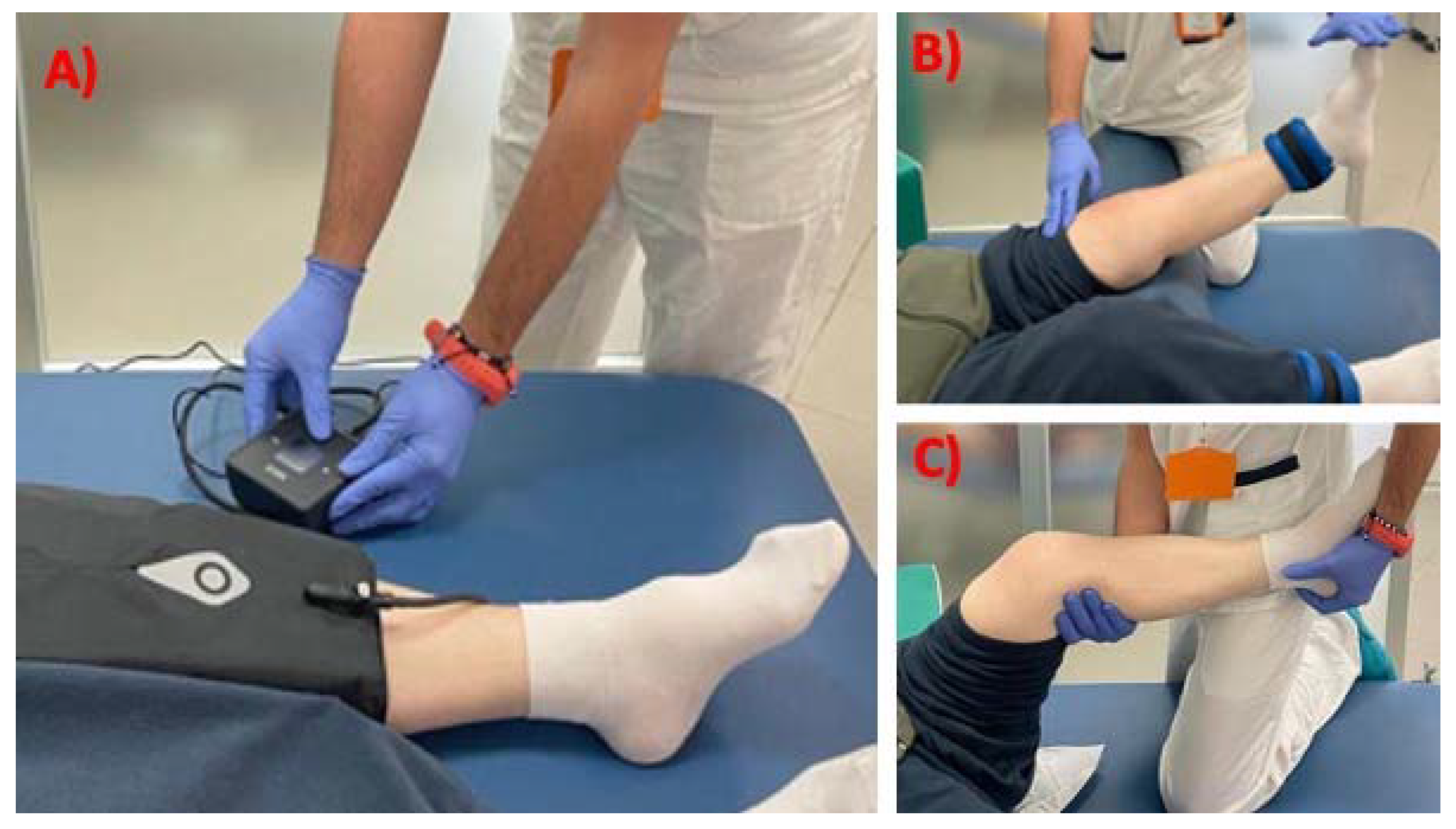
2.4. Intervention
2.5. Statistical Analysis
3. Results
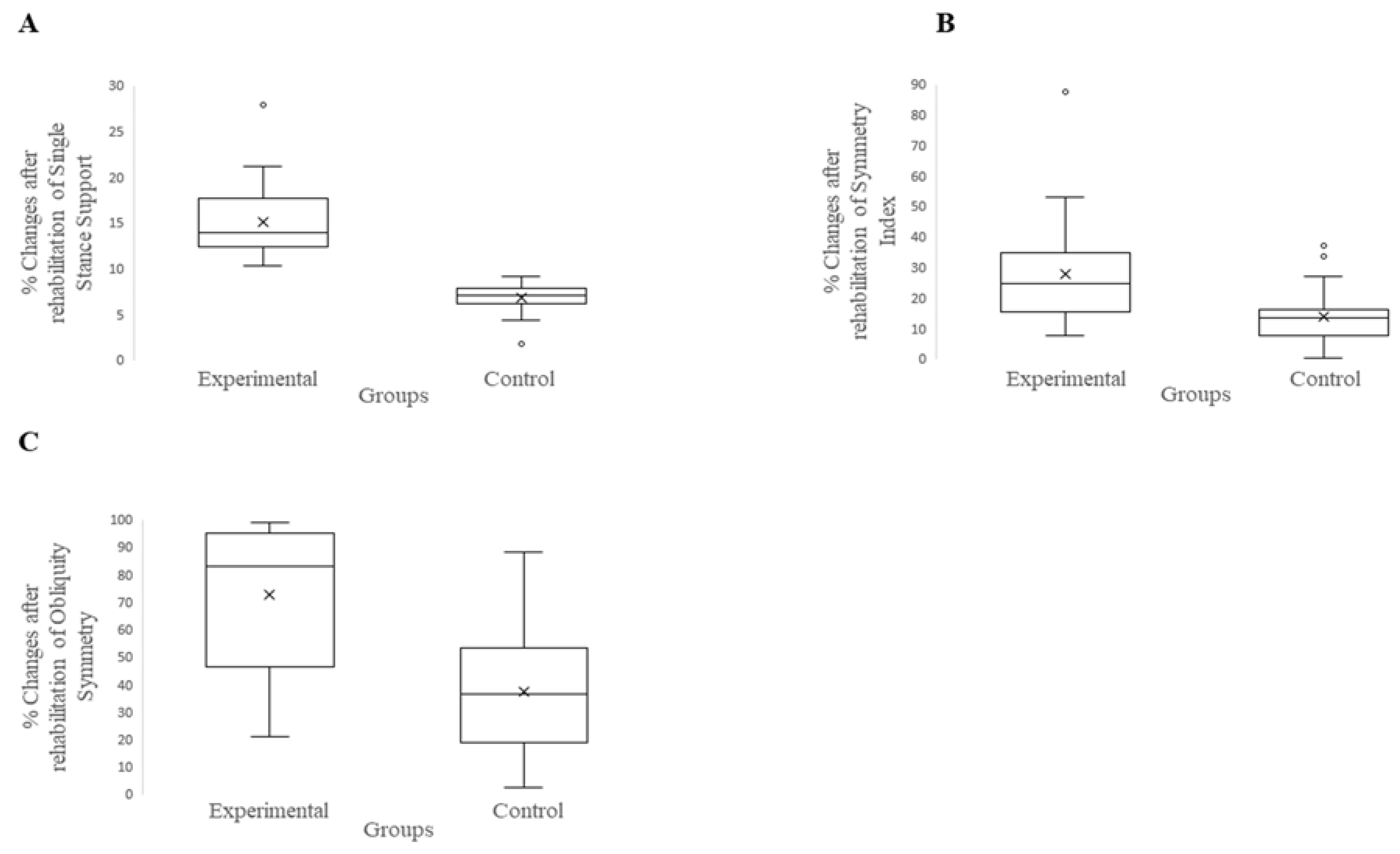
Visual Feedback versus Traditional Treatments
4. Discussion
Limitations
5. Conclusions
Author Contributions
Funding
Institutional Review Board Statement
Informed Consent Statement
Data Availability Statement
Acknowledgments
Conflicts of Interest
References
- Hussain, S.M.; Neilly, D.W.; Baliga, S.; Patil, S.; Meek, R. Knee Osteoarthritis: A Review of Management Options. Scott. Med. J. 2016, 61, 7–16. [Google Scholar] [CrossRef] [PubMed]
- Carr, A.J.; Robertsson, O.; Graves, S.; Price, A.J.; Arden, N.K.; Judge, A.; Beard, D.J. Knee Replacement. Lancet 2012, 379, 1331–1340. [Google Scholar] [CrossRef] [PubMed]
- Levinger, P.; Menz, H.B.; Morrow, A.D.; Feller, J.A.; Bartlett, J.R.; Bergman, N.R. Lower Limb Biomechanics in Individuals with Knee Osteoarthritis before and after Total Knee Arthroplasty Surgery. J. Arthroplasty 2013, 28, 994–999. [Google Scholar] [CrossRef] [PubMed]
- Bączkowicz, D.; Skiba, G.; Czerner, M.; Majorczyk, E. Gait and Functional Status Analysis before and after Total Knee Arthroplasty. Knee 2018, 25, 888–896. [Google Scholar] [CrossRef] [PubMed]
- Judge, A.; Cooper, C.; Williams, S.; Dreinhoefer, K.; Dieppe, P. Patient-Reported Outcomes One Year after Primary Hip Replacement in a European Collaborative Cohort. Arthritis Care Res. 2010, 62, 480–488. [Google Scholar] [CrossRef] [PubMed]
- Hawker, G.A.; Badley, E.M.; Borkhoff, C.M.; Croxford, R.; Davis, A.M.; Dunn, S.; Gignac, M.A.; Jaglal, S.B.; Kreder, H.J.; Sale, J.E.M. Which Patients Are Most Likely to Benefit from Total Joint Arthroplasty? Arthritis Rheum. 2013, 65, 1243–1252. [Google Scholar] [CrossRef]
- Beswick, A.D.; Wylde, V.; Gooberman-Hill, R.; Blom, A.; Dieppe, P. What Proportion of Patients Report Long-Term Pain after Total Hip or Knee Replacement for Osteoarthritis? A Systematic Review of Prospective Studies in Unselected Patients. BMJ Open 2012, 2, e000435. [Google Scholar] [CrossRef]
- Artz, N.; Elvers, K.T.; Lowe, C.M.; Sackley, C.; Jepson, P.; Beswick, A.D. Effectiveness of Physiotherapy Exercise Following Total Knee Replacement: Systematic Review and Meta-Analysis. BMC Musculoskelet. Disord. 2015, 16, 15. [Google Scholar] [CrossRef] [Green Version]
- Masaracchio, M.; Hanney, W.J.; Liu, X.; Kolber, M.; Kirker, K. Timing of Rehabilitation on Length of Stay and Cost in Patients with Hip or Knee Joint Arthroplasty: A Systematic Review with Meta-Analysis. PLoS ONE 2017, 12, e0178295. [Google Scholar] [CrossRef] [Green Version]
- Lai, Y.-F.; Lin, P.-C.; Chen, C.-H.; Chen, J.-L.; Hsu, H.-T. Current Status and Changes in Pain and Activities of Daily Living in Elderly Patients with Osteoarthritis Before and After Unilateral Total Knee Replacement Surgery. J. Clin. Med. 2019, 8, 221. [Google Scholar] [CrossRef]
- Zhu, Y.; Feng, Y.; Peng, L. Effect of Transcutaneous Electrical Nerve Stimulation for Pain Control after Total Knee Arthroplasty: A Systematic Review and Meta-Analysis. J. Rehabil. Med. 2017, 49, 700–704. [Google Scholar] [CrossRef] [PubMed] [Green Version]
- Bonnefoy-Mazure, A.; Favre, T.; Praplan, G.; Armand, S.; Sagawa Junior, Y.; Hannouche, D.; Turcot, K.; Lübbeke, A.; Miozzari, H.H. Associations between Gait Analysis Parameters and Patient Satisfaction One Year Following Primary Total Knee Arthroplasty. Gait Posture 2020, 80, 44–48. [Google Scholar] [CrossRef] [PubMed]
- Demeco, A.; Marotta, N.; Moggio, L.; Pino, I.; Marinaro, C.; Barletta, M.; Petraroli, A.; Palumbo, A.; Ammendolia, A. Quantitative Analysis of Movements in Facial Nerve Palsy with Surface Electromyography and Kinematic Analysis. J. Electromyogr. Kinesiol. 2021, 56, 102485. [Google Scholar] [CrossRef] [PubMed]
- Ammarullah, M.I.; Santoso, G.; Sugiharto, S.; Supriyono, T.; Kurdi, O.; Tauviqirrahman, M.; Winarni, T.I.; Jamari, J. Tresca Stress Study of CoCrMo-on-CoCrMo Bearings Based on Body Mass Index Using 2D Computational Model. J. Tribol. 2022, 33, 31–38. [Google Scholar]
- Zanasi, S. Innovations in Total Knee Replacement: New Trends in Operative Treatment and Changes in Peri-Operative Management. Eur. Orthop. Traumatol. 2011, 2, 21–31. [Google Scholar] [CrossRef] [PubMed] [Green Version]
- Mistry, J.B.; Elmallah, R.D.K.; Bhave, A.; Chughtai, M.; Cherian, J.J.; McGinn, T.; Harwin, S.F.; Mont, M.A. Rehabilitative Guidelines after Total Knee Arthroplasty: A Review. J. Knee Surg. 2016, 29, 201–217. [Google Scholar] [CrossRef]
- Levy, A.S.; Marmar, E. The Role of Cold Compression Dressings in the Postoperative Treatment of Total Knee Arthroplasty. Clin. Orthop. Relat. Res. 1993, 174–178. [Google Scholar] [CrossRef]
- Zhang, T.; Qui, B.; Liu, H.J.; Xu, J.; Xu, D.X.; Wang, Z.Y.; Niu, W. Effects of Visual Feedback During Balance Training on Knee Function and Balance Ability in Postoperative Patients After Knee Fracture: A Randomized Controlled Trial. J. Rehabil. Med. 2022, 54, jrm00281. [Google Scholar] [CrossRef]
- Rougier, P.; Boudrahem, S. Effects of Visual Feedback of Center-of-Pressure Displacements on Undisturbed Upright Postural Control of Hemiparetic Stroke Patients. Restor. Neurol. Neurosci. 2010, 28, 749–759. [Google Scholar] [CrossRef]
- Marotta, N.; Demeco, A.; Indino, A.; de Scorpio, G.; Moggio, L.; Ammendolia, A. Nintendo WiiTM versus Xbox KinectTM for Functional Locomotion in People with Parkinson’s Disease: A Systematic Review and Network Meta-Analysis. Disabil. Rehabil. 2022, 44, 331–336. [Google Scholar] [CrossRef]
- Schmid, A.; Duncan, P.W.; Studenski, S.; Lai, S.M.; Richards, L.; Perera, S.; Wu, S.S. Improvements in Speed-Based Gait Classifications Are Meaningful. Stroke 2007, 38, 2096–2100. [Google Scholar] [CrossRef] [PubMed]
- Pignolo, L.; Basta, G.; Carozzo, S.; Bilotta, M.; Todaro, M.R.; Serra, S.; Ciancarelli, I.; Tonin, P.; Cerasa, A. A Body-Weight-Supported Visual Feedback System for Gait Recovering in Stroke Patients: A Randomized Controlled Study. Gait Posture 2020, 82, 287–293. [Google Scholar] [CrossRef] [PubMed]
- Pfeufer, D.; Gililland, J.; Böcker, W.; Kammerlander, C.; Anderson, M.; Krähenbühl, N.; Pelt, C. Training with biofeedback devices improves clinical outcome compared to usual care in patients with unilateral TKA: A systematic review. Knee Surg. Sports Traumatol. Arthrosc. 2019, 27, 1611–1620. [Google Scholar] [CrossRef]
- Marshall, A.N.; Hertel, J.; Hart, J.M.; Russell, S.; Saliba, S.A. Visual Biofeedback and Changes in Lower Extremity Kinematics in Individuals With Medial Knee Displacement. J. Athl. Train. 2020, 55, 255–264. [Google Scholar] [CrossRef] [PubMed] [Green Version]
- Hawker, G.A.; Mian, S.; Kendzerska, T.; French, M. Measures of Adult Pain: Visual Analog Scale for Pain (VAS Pain), Numeric Rating Scale for Pain (NRS Pain), McGill Pain Questionnaire (MPQ), Short-Form McGill Pain Questionnaire (SF-MPQ), Chronic Pain Grade Scale (CPGS), Short Form-36 Bodily Pain Scale (SF-36 BPS), and Measure of Intermittent and Constant Osteoarthritis Pain (ICOAP). Arthritis Care Res. 2011, 63 (Suppl. S11), S240–S252. [Google Scholar] [CrossRef]
- Bouwstra, H.; Smit, E.B.; Wattel, E.M.; van der Wouden, J.C.; Hertogh, C.M.P.M.; Terluin, B.; Terwee, C.B. Measurement Properties of the Barthel Index in Geriatric Rehabilitation. J. Am. Med. Dir. Assoc. 2019, 20, 420–425.e1. [Google Scholar] [CrossRef]
- De Ridder, R.; Lebleu, J.; Willems, T.; De Blaiser, C.; Detrembleur, C.; Roosen, P. Concurrent Validity of a Commercial Wireless Trunk Triaxial Accelerometer System for Gait Analysis. J. Sport Rehabil. 2019, 28, jsr.2018-0295. [Google Scholar] [CrossRef] [Green Version]
- Yazıcı, M.V.; Çobanoğlu, G.; Yazıcı, G. Test-retest reliability and minimal detectable change for wearable gait analysis system (G-Walk) measurements in children with cerebral palsy. Turk. J. Med. Sci. 2022, 52, 658–666. [Google Scholar] [CrossRef]
- Vagnini, A.; Furone, R.; Zanotti, G.; Adamo, P.; Temporiti, F.; Gatti, R. Agreement between Inertial Measurement Unit and Optoelectronic System to Measure Postural Sway. Technol. Health Care 2022, 30, 757–762. [Google Scholar] [CrossRef]
- Arshad, M.Z.; Jamsrandorj, A.; Kim, J.; Mun, K.-R. Gait Events Prediction Using Hybrid CNN-RNN-Based Deep Learning Models through a Single Waist-Worn Wearable Sensor. Sensors 2022, 22, 8226. [Google Scholar] [CrossRef]
- Christensen, J.C.; LaStayo, P.C.; Marcus, R.L.; Stoddard, G.J.; Bo Foreman, K.; Mizner, R.L.; Peters, C.L.; Pelt, C.E. Visual knee-kinetic biofeedback technique normalizes gait abnormalities during high-demand mobility after total knee arthroplasty. Knee 2018, 25, 73–82. [Google Scholar] [CrossRef] [PubMed]
- Izzo, R.; Bertoni, M.; Cejudo, A.; Giovannelli, M.; Hosseini Varde’i, C. The global symmetry index, symmetry index, quality index and kinematics of the gait cycle with the synchronized contribution of the latest generation magnetic- inertial and electromyographic technology. Practical surveys and planning hypotheses for the revision of gesture. J. Phy. Edu. Sport 2022, 22, 1258–1270. [Google Scholar] [CrossRef]
- Demeco, A.; de Sire, A.; Marotta, N.; Palumbo, A.; Fragomeni, G.; Gramigna, V.; Pellegrino, R.; Moggio, L.; Petraroli, A.; Iona, T.; et al. Effectiveness of Rehabilitation through Kinematic Analysis of Upper Limb Functioning in Wheelchair Basketball Athletes: A Pilot Study. Appl. Sci. 2022, 12, 2929. [Google Scholar] [CrossRef]
- Croce, U.D.; Riley, P.O.; Lelas, J.L.; Kerrigan, D.C. A Refined View of the Determinants of Gait. Gait Posture 2001, 14, 79–84. [Google Scholar] [CrossRef] [PubMed]
- Neumann, D.A. Kinesiology of the Musculoskeletal System, 3rd ed.; Mosby: London, UK, 2010. [Google Scholar]
- Bragonzoni, L.; Rovini, E.; Barone, G.; Cavallo, F.; Zaffagnini, S.; Benedetti, M.G. How Proprioception Changes before and after Total Knee Arthroplasty: A Systematic Review. Gait Posture 2019, 72, 1–11. [Google Scholar] [CrossRef] [PubMed]
- Riva, D.; Bianchi, R.; Rocca, F.; Mamo, C. Proprioceptive Training and Injury Prevention in a Professional Men’s Basketball Team: A Six-Year Prospective Study. J. Strength Cond. Res. 2016, 30, 461–475. [Google Scholar] [CrossRef] [Green Version]
- Hatfield, G.L.; Hubley-Kozey, C.L.; Astephen Wilson, J.L.; Dunbar, M.J. The Effect of Total Knee Arthroplasty on Knee Joint Kinematics and Kinetics during Gait. J. Arthroplast. 2011, 26, 309–318. [Google Scholar] [CrossRef]


| Experimental Group (n = 20) | Control Group (n = 20) | Between Groups Comparison | ||||||
|---|---|---|---|---|---|---|---|---|
| Mdn | Range | Mdn | Range | U | W | Z | p-Value | |
| Age (yr) | 70.5 | 66.3–76.5 | 71 | 69–73 | 196 | 406 | −0.109 | 0.920 |
| Height (cm) | 161 | 160–164.5 | 160 | 159–161 | 153.5 | 363.5 | −1.271 | 0.804 |
| Body mass (kg) | 67 | 62.5–69 | 66 | 64–67.75 | 172.5 | 382.5 | −0.748 | 0.463 |
| BMI | 25.9 | 24.9–26.2 | 25.5 | 25–26.05 | 190.5 | 400.5 | −0.257 | 0.804 |
| Therapy duration (days) | 37.5 | 33.3–39.8 | 36.5 | 31–40 | 191.5 | 401.5 | −0.231 | 0.825 |
| Experimental Group | Control Group | Statistics between Groups at T1 | |||||||||
|---|---|---|---|---|---|---|---|---|---|---|---|
| T0 | T1 | T0 | T1 | ||||||||
| mdn | IQR | mdn | IQR | mdn Delta (%) | mdn | IQR | mdn | IQR | mdn Delta (%) | p-Value | |
| Clinical Assessment | |||||||||||
| NRS | 60 | 50–77.5 | 10 | 10–20 | −75% | 60 | 52.5–70 | 20 | 10–20 | −71.43% | 0.278 |
| Barthel index | 40 | 35–63.75 | 100 | 95–100 | 118.75% | 45 | 40–53.7 | 95 | 91–100 | 102.2% | 0.381 |
| Kinematic Gait Assessment | |||||||||||
| Cadence | 64.75 | 59.7–70 | 82.6 | 78.8–90.3 | 27.76% | 61.1 | 48–80.4 | 82.35 | 76.4–89 | 30.37% | 0.389 |
| Velocity | 0.56 | 0.5–0.7 | 0.8 | 0.7–0.8 | 39.59% | 0.57 | 0.35–0.65 | 0.74 | 0.66–0.8 | 39.69% | 0.465 |
| Cycle duration | 2.21 | 1.7–2.3 | 1.53 | 1.4–1.6 | −28.51% | 2.17 | 1.8–2.6 | 1.57 | 1.3–1.9 | −27.37% | 0.285 |
| Cycle length | 1.23 | 1.2–1.3 | 1.02 | 0.97–1.1 | −12.57% | 1.22 | 1.1–1.3 | 1.07 | 0.94–1.1 | −12.79% | 0.492 |
| Symmetry index | 72.7 | 63.5–77.9 | 87.15 | 84–92.8 | 24.64% | 74.4 | 71.5–77.5 | 84 | 80.8–85.4 | 13.25% | 0.001 * |
| Single stance support | 32.88 | 31.9–34.1 | 37.8 | 36.6–38.9 | 13.89% | 33.2 | 31.4–34.3 | 34.9 | 34.1–36.5 | 7.08% | <0.0001 * |
| Propulsion | 3.3 | 2.73–3.8 | 4.8 | 4.25–6.1 | 55.59% | 3.55 | 3.1–4.6 | 5.05 | 4.1–6.2 | 32.81% | 0.007 |
| Tilt symmetry | 74.5 | 68.2–77.4 | 90.9 | 86.2–95 | 21.87% | 77.3 | 64.3–83.2 | 93.25 | 77.4–95.6 | 18.33% | 0.009 |
| Obliquity Symmetry | 42.65 | 36.5–50.1 | 73 | 62.3–82.1 | 83.16% | 44.8 | 44.9–46.3 | 58.65 | 51.3–70.3 | 36.27% | <0.0001 * |
| Rotation symmetry | 55.60 | 44.6–85.2 | 91.1 | 70.6–95 | 33.38% | 63.5 | 61–66.5 | 76.55 | 73.9–78.7 | 20.01% | 0.087 |
Publisher’s Note: MDPI stays neutral with regard to jurisdictional claims in published maps and institutional affiliations. |
© 2022 by the authors. Licensee MDPI, Basel, Switzerland. This article is an open access article distributed under the terms and conditions of the Creative Commons Attribution (CC BY) license (https://creativecommons.org/licenses/by/4.0/).
Share and Cite
Carozzo, S.; Vatrano, M.; Coschignano, F.; Battaglia, R.; Calabrò, R.S.; Pignolo, L.; Contrada, M.; Tonin, P.; Cerasa, A.; Demeco, A. Efficacy of Visual Feedback Training for Motor Recovery in Post-Operative Subjects with Knee Replacement: A Randomized Controlled Trial. J. Clin. Med. 2022, 11, 7355. https://doi.org/10.3390/jcm11247355
Carozzo S, Vatrano M, Coschignano F, Battaglia R, Calabrò RS, Pignolo L, Contrada M, Tonin P, Cerasa A, Demeco A. Efficacy of Visual Feedback Training for Motor Recovery in Post-Operative Subjects with Knee Replacement: A Randomized Controlled Trial. Journal of Clinical Medicine. 2022; 11(24):7355. https://doi.org/10.3390/jcm11247355
Chicago/Turabian StyleCarozzo, Simone, Martina Vatrano, Francesco Coschignano, Riccardo Battaglia, Rocco Salvatore Calabrò, Loris Pignolo, Marianna Contrada, Paolo Tonin, Antonio Cerasa, and Andrea Demeco. 2022. "Efficacy of Visual Feedback Training for Motor Recovery in Post-Operative Subjects with Knee Replacement: A Randomized Controlled Trial" Journal of Clinical Medicine 11, no. 24: 7355. https://doi.org/10.3390/jcm11247355