Review

Spontaneous Breathing and Pendelluft in Patients with Acute Lung Injury: A Narrative Review

1
Department of Biomedical Engineering, National Cheng Kung University, Tainan 704, Taiwan
2
Department of Internal Medicine, National Cheng Kung University Hospital, College of Medicine, National Cheng Kung University, Tainan 704, Taiwan
3
Department of Biomedical Engineering, Fourth Military Medical University, Xi’an 710032, China
4
Institute of Technical Medicine, Furtwangen University, 78120 VS-Schwenningen, Germany
5
College of Biomedical Engineering, China Medical University, Taichung 404, Taiwan
*
Author to whom correspondence should be addressed.
J. Clin. Med. 2022, 11(24), 7449; https://doi.org/10.3390/jcm11247449
Submission received: 25 October 2022 / Revised: 10 December 2022 / Accepted: 12 December 2022 / Published: 15 December 2022
(This article belongs to the Section Pulmonology)

Abstract

:
Acute respiratory distress syndrome (ARDS) is characterized by acute-onset rapid-deteriorating inflammatory lung injury. Although the preservation of spontaneous breathing may have physiological benefits in oxygenation, increasing evidence shows that vigorous spontaneous breathing may aggravate lung injury (i.e., patient self-inflicted lung injury). Increased lung stress and pendelluft, which is defined as intrapulmonary gas redistribution without a significant change in tidal volume, are important mechanisms of patient self-inflicted lung injury. The presence of pendelluft may be considered a surrogate marker of vigorous inspiratory effort, which can cause the dependent lung to overstretch. In this review, we summarized three major methods for electrical impedance tomography–based pendelluft monitoring. Future studies are warranted to compare and validate the different methods of pendelluft estimation in patients with ARDS.

1. Introduction

Acute respiratory distress syndrome (ARDS), which is characterized by acute-onset rapid-deteriorating inflammatory lung injury, affects approximately 10% of patients in the intensive care unit (ICU) [1]. Pathologically, it is characterized by extensive alveolar inflammation, increased vascular permeability, increased lung weight, and decreased aeration area [2]. The preservation of gentle spontaneous breathing during ARDS may have physiological benefits in oxygenation owing to the improved aeration of the dependent lung [3] and the redistribution of pulmonary blood flow toward the nondependent lung [4]. In addition, the maintenance of optimal diaphragm muscle contraction reduces the risk of ventilator-induced diaphragm dysfunction (VIDD) [5,6]. However, increasing evidence shows that spontaneous breathing may aggravate lung injury. In the present review, we summarize the beneficial and harmful effects of spontaneous breathing effort and also introduce pendelluft as a surrogate marker of vigorous breathing effort.

2. The Beneficial Effects of Spontaneous Breathing

2.1. Prevention of Diaphragm Dysfunction

In 2004, Vassilakopoulos and Petrof [7] first introduced VIDD, which is defined as decreased diaphragm muscle contractility secondary to the use of mechanical ventilation. The incidence of VIDD in patients who received mechanical ventilation ranges from 23% to 84% [8]. Diaphragm contractile activity decreases with increasing ventilator driving pressure and controlled ventilator mode [6]. To avoid the disuse atrophy of the diaphragm during mechanical ventilation, the preservation of spontaneous breathing would provide longer ventilator-free days and a shorter ICU length of stay [9]. These data indicate that the maintenance of optimal spontaneous breathing effort could be considered a method for preventing diaphragm dysfunction [10].

2.2. Increased Ventilation in the Dependent Lung

During spontaneous breathing, inspiratory muscle effort, which is mainly caused by diaphragm muscle contraction, will cause pleural pressure to become more negative [11]. The lowering of the pleural pressure will expand the lung by increasing transpulmonary pressure. Among patients with ARDS receiving pressure support ventilation, lower support level with higher inspiratory effort was associated with increased ventilation toward the dependent lung [12]. The difference in ventilation distribution could be explained by the cephalad movement of diaphragm in controlled ventilation, thus leading the ventilation to the nondependent region. By contrast, the dependent part of the diaphragm moves the most during spontaneous breathing, thus increasing the amount of ventilation to the dependent region [13]. Therefore, the maintenance of optimal spontaneous breathing effort may improve dependent lung ventilation and increase antero-posterior ventilation homogeneity and even oxygenation [12].

3. The Harmful Effects of Spontaneous Breathing—Patient Self-Inflicted Lung Injury (P-SILI)

3.1. Patient-Ventilator Asynchronies

Spontaneous breathing may occur in a vigorous form in patients with ARDS [14]. Protective lung ventilation remains the standard in patients with ARDS because pulmonary overdistention will further worsen the injured lung [15]. Breath stacking (double triggering) was found to occur frequently in nonparalyzed, sedated patients with acute lung injury under low tidal volume ventilation [16]. The number and duration of the cluster of double triggering and/or ineffective efforts are related to the duration of mechanical ventilation and ICU stay, as well as with hospital mortality in mechanically ventilated ICU patients [17]. The ACURASYS trial reported that breath stacking may offer one explanation for the beneficial effect of neuromuscular agent administration because breath stacking will not occur in paralyzed patients [18]. However, the Reevaluation of Systemic Early Neuromuscular Blockade trial reevaluated the benefits of cisatracurium in patients with ARDS and found that there was no difference in 90-day mortality when compared with sedation only [19]. Light sedation and higher positive end-expiratory pressure may be associated with less reverse triggering and less breath stacking [20]. The objective assessment of patient-ventilator asynchrony may provide the final answer.

3.2. Excessive Lung Perfusion

Vigorous inspiratory effort causes strong negative intrathoracic pressure, which results in excessive fluid shift from intrapulmonary vessels to interstitial and alveolar spaces [21,22]. This phenomenon has been described in patients with vigorous inspiratory effort against airway obstruction and is also called post-obstructive pulmonary edema [23]. In animal studies, abrupt deflation after sustained inflation, which is associated with a dramatic decrease in intrapulmonary pressure, could cause acute lung injury mediated by the significant vascular leakage and acute decompensation of the left ventricle [24,25]. These data imply that lung injury may also be caused by the abrupt and large change in intrathoracic pressure.

3.3. Increased Lung Stress and Pendelluft

The presence of a strong spontaneous breathing effort leads to a dramatic fluctuation in tidal volume. High tidal volume with high transpulmonary pressure would lead to lung injury [26]. Unfortunately, limiting tidal volume may not be safe. In an observational study of a patient with postoperative hypoxemia, high inspiratory effort caused early inflation in the dependent lung area and simultaneous deflation in the nondependent lung area [27]. The consolidative or atelectatic lung would attenuate the transmission of transpulmonary pressure and cause more local overdistension during diaphragm contraction [28]. This phenomenon was termed pendelluft, which is characterized by intrapulmonary gas redistribution without a significant change in tidal volume [27]. Pendelluft is clinically significant because it represents regional lung hyperdistention despite low tidal volume ventilation. The degree of pendelluft is proportional to the breathing effort even in reverse triggering [29]. However, a higher inspiratory effort is not always associated with a higher pendelluft volume [30]. Patients with high magnitude pendelluft also had increased inflammatory mediators related to acute lung injury independently of transpulmonary pressure [31]. Therefore, it would be better to monitor pendelluft and esophageal pressure simultaneously.
All three phenomena pertain to “P-SILI,” which is a lung injury caused by vigorous spontaneous breathing effort [32]. A high respiratory drive may also activate the expiratory muscles, thus resulting in negative expiratory transpulmonary pressure [22,33] and possibly causing subsequent lung derecruitment [22]. Asynchronies such as breath stacking and increased perfusion secondary to extreme effort may be easily assessed clinically, but pendelluft monitoring is difficult. The presence of pendelluft could only be assessed using electrical impedance tomography (EIT). In the following section, we introduce the three different methods of EIT-based pendelluft estimation.

4. Monitoring Pendelluft in Patients with Lung Injury

To accurately monitor intrapulmonary gas distribution, EIT, which allows for the non-invasive dynamic measurement of intrapulmonary ventilation distribution, has been implemented in many clinical and animal studies [34,35].

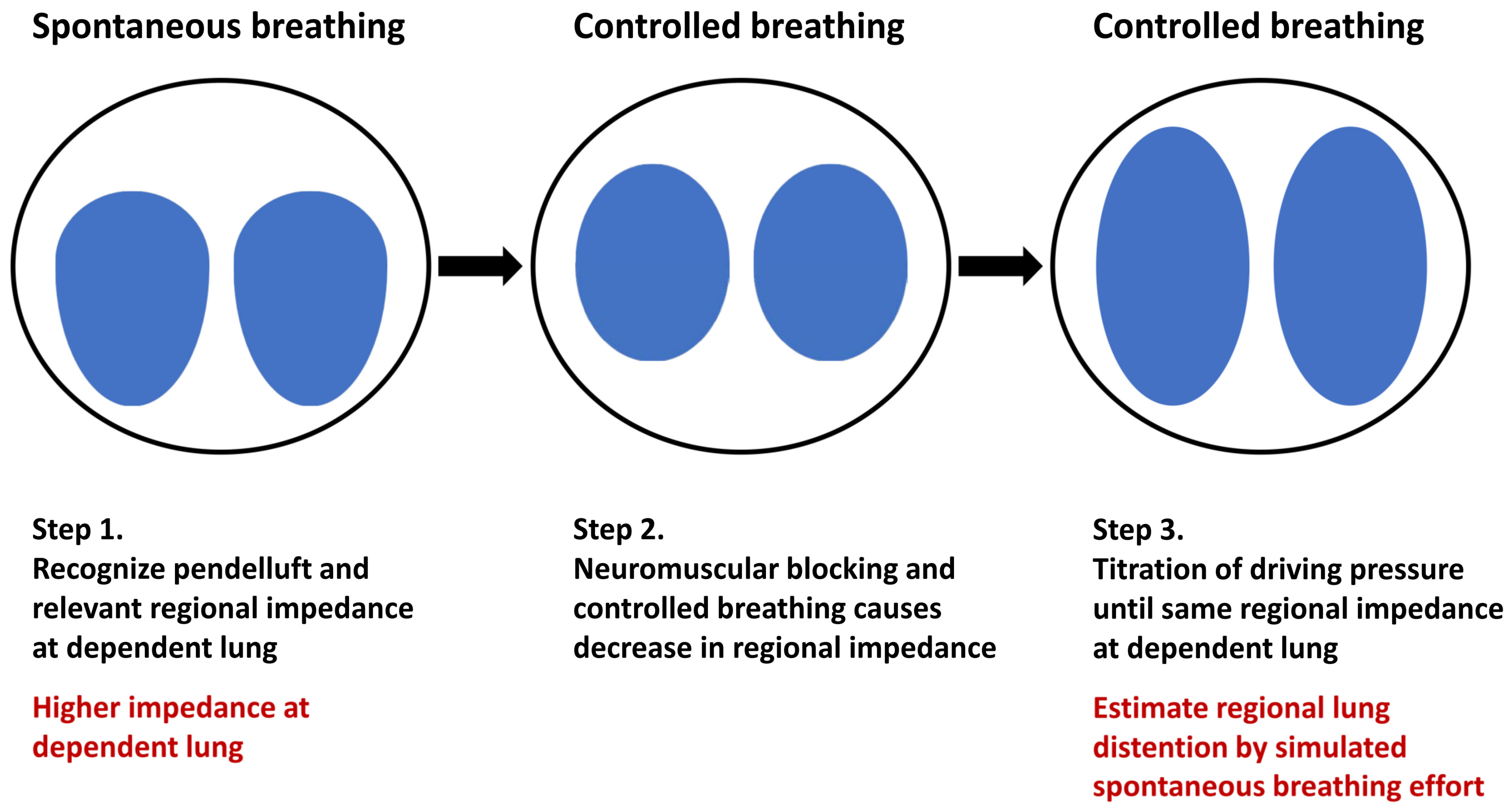
4.1. Estimation of Regional Lung Inflation during Spontaneous Breathing by Controlled Ventilation

Yoshida et al. [27] first described a method for estimating the regional lung inflation during spontaneous breathing. In an animal study, the maximal impedance change in the dependent lung region at the beginning of inspiration was recorded when pendelluft developed. After the administration of the muscle relaxant, the driving pressure was continuously titrated until the same impedance change developed in the dependent lung (Figure 1). In their study, a significantly larger tidal volume was observed after achieving comparable impedance changes in the dependent lung, thus indicating overdistention in the dependent lung. Although this method could only estimate regional lung inflation instead of the amount of pendelluft, it could still provide information on regional lung stress. However, this method may use an extremely high driving pressure, which might increase the risk of ventilator-induced lung injury and limit its clinical application.

4.2. Estimation of Intrapulmonary Gas Flow

On the basis of this real-time monitoring device, Coppadoro et al. [34] proposed a pendelluft estimation method. In brief, global and regional impedance–time curves were analyzed. To evaluate intrapulmonary gas flow across the dependent and nondependent lung regions, the regions of interest can be classified from ventral to dorsal into four layers of the same thickness. The time point at the nadir of the global impedance was considered the initiation of inspiration (T0). Similarly, the time point of the nadirs of each layer was identified (Ti). Subsequently, the volume change of each region between the time point of the regional nadir (Ti) and global nadir (T0) was calculated (Figure 2) [30]. In their study, the amount of pendelluft increased significantly with decremental titration of pressure support ventilation in some of their patients. This method is practical for clinical use but has a drawback, namely, the assumption of a homogeneous lung zones in each layer, which may result in an underestimation of the pendelluft volume. The threshold of a high pendelluft and its correlation with regional lung stress should be validated in a future study.

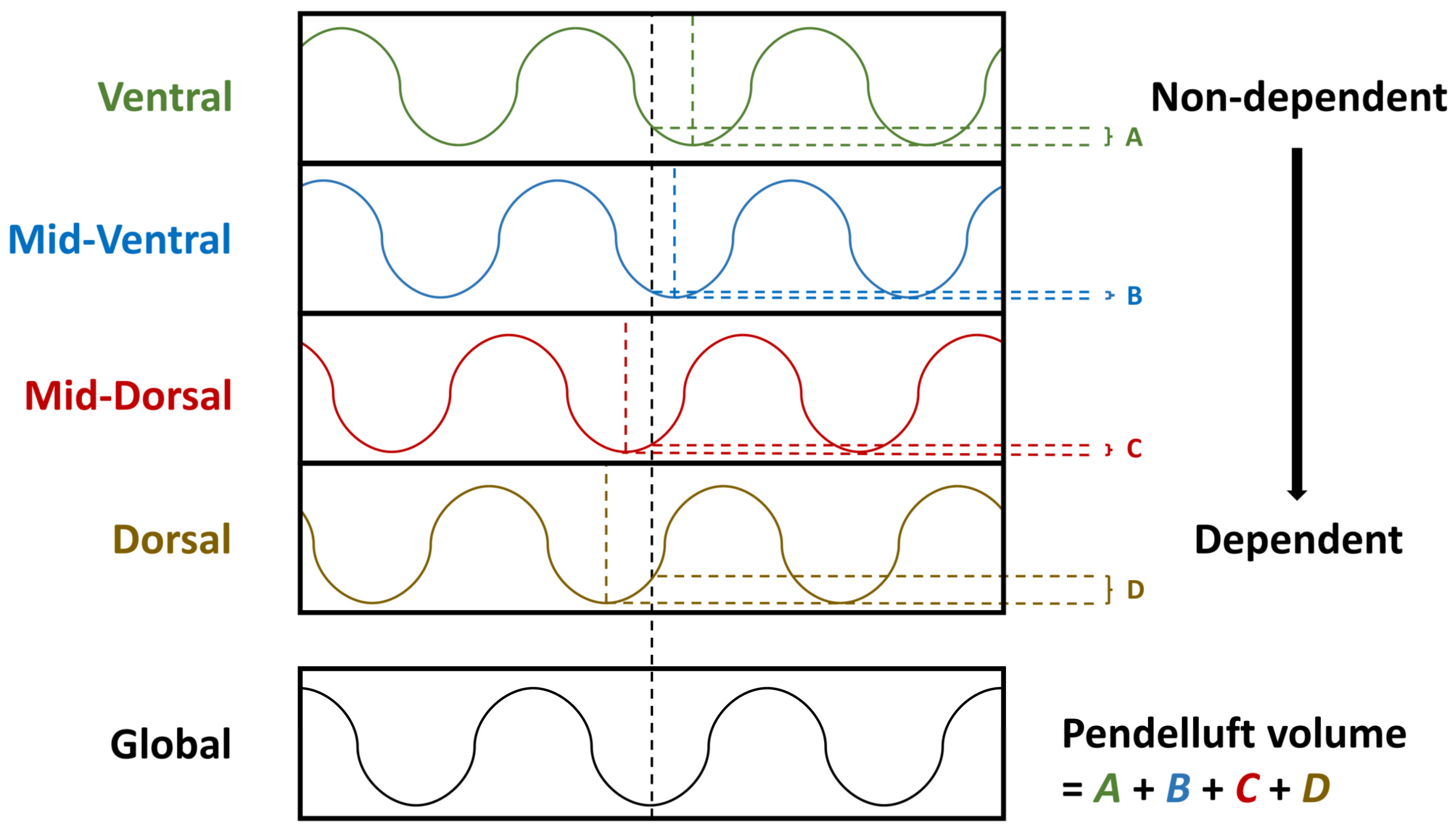
4.3. Measurement of Pendelluft Amplitude

Recently, Sang et al. [36] introduced another estimation method based on electrical impedance tomography, which can measure the pendelluft for each pixel. First, the timepoints of nadir (Tnadir) and peak (Tpeak) in each breathing cycle of the global impedance–time curve are identified as the starting time points of inspiration and expiration, respectively. The impedance differences regarding the global ventilation time point (Zglobal) were established by subtracting the impedance at Tnadir from that at Tpeak. Second, the regional impedance–time curve of each pixel was created. The impedance difference between the peak and nadir of each breathing cycle for each pixel is calculated (Zregional). If ventilation is homogenous during tidal breathing, the global impedance change (Zglobal) will be close to the regional impedance change (Zregional). The pendelluft amplitude is estimated using the following formula (Figure 3):
P e n d e l l u f t   a m p l i t u d e = 1 N i = 1 N Z r e g i o n a l , i Z g l o b a l , i t i d a l   v o l u m e × 100 %
Measurement errors and noise may also cause differences between Zregional and Zglobal values. A retrospective observational study that evaluated 30 healthy volunteers defined a pendelluft amplitude that is >2.5% of the tidal volume as a clinically significant pendelluft [37]. In a subsequent validation cohort of 200 patients with ARDS, a clinically significant pendelluft was associated with longer ventilator-dependent days in patients with severe hypoxemia (PaO2/FiO2 ratio < 200 mmHg) [35]. However, given that the threshold was defined on the basis of healthy volunteers, different cut-off points might have occurred in patients with ARDS.

5. Conclusions

Appropriate spontaneous breathing may prevent VIDD and improve ventilation homogeneity but may cause P-SILI when breathing effort is excessive. By using standard protective lung ventilation, patient-ventilator asynchronies and extreme effort can be easily detected. However, pendelluft could only be assessed by EIT monitoring. Future studies are warranted to compare and validate the different methods of pendelluft estimation in patients with ARDS.

Author Contributions

P.-L.S. and C.-W.C.: conceptualization, literature search, and writing the article; Z.Z., Y.-F.K. and K.-S.C.: writing and reviewing the article. All authors have read and agreed to the published version of the manuscript.

Funding

This research was funded by the National Science and Technology Council (grant number 111-2314-B-006-092-MY3).

Institutional Review Board Statement

Not applicable.

Informed Consent Statement

Not applicable.

Data Availability Statement

Not applicable.

Conflicts of Interest

The authors declare no conflict of interest.

References

  1. Bellani, G.; Laffey, J.G.; Pham, T.; Fan, E.; Brochard, L.; Esteban, A.; Gattinoni, L.; van Haren, F.; Larsson, A.; McAuley, D.F.; et al. Epidemiology, patterns of care, and mortality for patients with acute respiratory distress syndrome in intensive care units in 50 countries. JAMA 2016, 315, 788–800. [Google Scholar] [CrossRef] [PubMed]
  2. Fan, E.; Brodie, D.; Slutsky, A.S. Acute respiratory distress syndrome: Advances in diagnosis and treatment. JAMA 2018, 319, 698–710. [Google Scholar] [CrossRef] [PubMed]
  3. Fredericks, A.S.; Bunker, M.P.; Gliga, L.A.; Ebeling, C.G.; Ringqvist, J.R.; Heravi, H.; Manley, J.; Valladares, J.; Romito, B.T. Airway pressure release ventilation: A review of the evidence, theoretical benefits, and alternative titration strategies. Clin. Med. Insights Circ. Respir. Pulm. Med. 2020, 14, 1179548420903297. [Google Scholar] [CrossRef] [PubMed] [Green Version]
  4. Carvalho, A.R.; Spieth, P.M.; Pelosi, P.; Beda, A.; Lopes, A.J.; Neykova, B.; Heller, A.R.; Koch, T.; Gama de Abreu, M. Pressure support ventilation and biphasic positive airway pressure improve oxygenation by redistribution of pulmonary blood flow. Anesth. Analg. 2009, 109, 856–865. [Google Scholar] [CrossRef] [PubMed]
  5. Levine, S.; Nguyen, T.; Taylor, N.; Friscia, M.E.; Budak, M.T.; Rothenberg, P.; Zhu, J.; Sachdeva, R.; Sonnad, S.; Kaiser, L.R.; et al. Rapid disuse atrophy of diaphragm fibers in mechanically ventilated humans. N. Engl. J. Med. 2008, 358, 1327–1335. [Google Scholar] [CrossRef]
  6. Goligher, E.C.; Fan, E.; Herridge, M.S.; Murray, A.; Vorona, S.; Brace, D.; Rittayamai, N.; Lanys, A.; Tomlinson, G.; Singh, J.M.; et al. Evolution of diaphragm thickness during mechanical ventilation. Impact of inspiratory effort. Am. J. Respir. Crit. Care Med. 2015, 192, 1080–1088. [Google Scholar] [CrossRef]
  7. Vassilakopoulos, T.; Petrof, B.J. Ventilator-induced diaphragmatic dysfunction. Am. J. Respir. Crit. Care Med. 2004, 169, 336–341. [Google Scholar] [CrossRef] [Green Version]
  8. Dres, M.; Goligher, E.C.; Heunks, L.M.A.; Brochard, L.J. Critical illness-associated diaphragm weakness. Intensive Care Med. 2017, 43, 1441–1452. [Google Scholar] [CrossRef]
  9. Van Haren, F.; Pham, T.; Brochard, L.; Bellani, G.; Laffey, J.; Dres, M.; Fan, E.; Goligher, E.C.; Heunks, L.; Lynch, J.; et al. Spontaneous breathing in early acute respiratory distress syndrome: Insights from the large observational study to UNderstand the Global Impact of Severe Acute Respiratory FailurE Study. Crit. Care Med. 2019, 47, 229–238. [Google Scholar] [CrossRef]
  10. Yoshida, T.; Amato, M.B.P.; Kavanagh, B.P.; Yuji Fujino, Y. Impact of spontaneous breathing during mechanical ventilation in acute respiratory distress syndrome. Curr. Opin. Crit. Care 2019, 25, 192–198. [Google Scholar] [CrossRef]
  11. Güldner, A.; Pelosi, P.; Gama de Abreu, M. Spontaneous breathing in mild and moderate versus severe acute respiratory distress syndrome. Curr. Opin. Crit. Care 2014, 20, 69–76. [Google Scholar] [CrossRef]
  12. Mauri, T.; Bellani, G.; Confalonieri, A.; Tagliabue, P.; Turella, M.; Coppadoro, A.; Citerio, G.; Patroniti, N.; Pesenti, A. Topographic distribution of tidal ventilation in acute respiratory distress syndrome: Effects of positive end-expiratory pressure and pressure support. Crit. Care Med. 2013, 41, 1664–1673. [Google Scholar] [CrossRef]
  13. Radke, O.C.; Schneider, T.; Heller, A.R.; Koch, T. Spontaneous breathing during general anesthesia prevents the ventral redistribution of ventilation as detected by electrical impedance tomography: A randomized trial. Anesthesiology 2012, 116, 1227–1234. [Google Scholar] [CrossRef] [Green Version]
  14. Mauri, T.; Langer, T.; Zanella, A.; Grasselli, G.; Pesenti, A. Extremely high transpulmonary pressure in a spontaneously breathing patient with early severe ARDS on ECMO. Intensive Care Med. 2016, 42, 2101–2103. [Google Scholar] [CrossRef]
  15. Acute Respiratory Distress Syndrome Network; Brower, R.G.; Matthay, M.A.; Morris, A.; Schoenfeld, D.; Thompson, B.T.; Wheeler, A. Ventilation with lower tidal volumes as compared with traditional tidal volumes for acute lung injury and the acute respiratory distress syndrome. N. Engl. J. Med. 2000, 342, 1301–1308. [Google Scholar]
  16. Pohlman, M.C.; McCallister, K.E.; Schweickert, W.D.; Pohlman, A.S.; Nigos, C.P.; Krishnan, J.A.; Charbeneau, J.T. Excessive tidal volume from breath stacking during lung-protective ventilation for acute lung injury. Crit. Care Med. 2008, 36, 3019–3023. [Google Scholar] [CrossRef]
  17. Magrans, R.; Ferreira, F.; Sarlabous, L.; López-Aguilar, J.; Gomà, G.; Fernandez-Gonzalo, S.; Navarra-Ventura, G.; Fernández, R.; Montanyà, J.; Kacmarek, R.; et al. The effect of clusters of double triggering and ineffective efforts in critically ill patients. Crit. Care Med. 2022, 50, e619–e629. [Google Scholar] [CrossRef]
  18. Papazian, L.; Forel, J.-M.; Gacouin, A.; Penot-Ragon, C.; Perrin, G.; Loundou, A.; Jaber, S.; Arnal, J.-M.; Perez, D.; Seghboyen, J.M.; et al. Neuromuscular blockers in early acute respiratory distress syndrome. N. Engl. J. Med. 2010, 363, 1107–1116. [Google Scholar] [CrossRef] [Green Version]
  19. National Heart, Lung, and Blood Institute PETAL Clinical Trials Network; Moss, M.; Huang, D.T.; Brower, R.G.; Ferguson, N.D.; Ginde, A.A.; Gong, M.N.; Grissom, C.K.; Gundel, S.; Hayden, D.; et al. Early neuromuscular blockade in the acute respiratory distress syndrome. N. Engl. J. Med. 2019, 380, 1997–2008. [Google Scholar]
  20. Slutsky, A.S.; Villar, J. Early Paralytic Agents for ARDS? Yes, no, and sometimes. N. Engl. J. Med. 2019, 380, 2061–2063. [Google Scholar] [CrossRef]
  21. Bhattacharya, M.; Kallet, R.H.; Ware, L.B.; Matthay, M.A. Negative-pressure pulmonary edema. Chest 2016, 150, 927–933. [Google Scholar] [CrossRef] [PubMed] [Green Version]
  22. Yoshida, T.; Fujino, Y.; Amato, M.B.P.; Kavanah, B.P. Fifty Years of Research in ARDS. Spontaneous breathing during mechanical ventilation. Risks, mechanisms, and management. Am. J. Respir. Crit. Care Med. 2017, 195, 985–992. [Google Scholar] [CrossRef] [PubMed]
  23. Yoshida, T.; Roldan, R.; Beraldo, M.A.; Torsani, V.; Gomes, S.; De Santis, R.R.; Costa, E.L.V.; Tucci, M.R.; Lima, R.G.; Kavanagh, B.P.; et al. Spontaneous effort during mechanical ventilation: Maximal injury with less positive end-expiratory pressure. Crit. Care Med. 2016, 44, e678–e688. [Google Scholar] [CrossRef] [PubMed]
  24. Katira, B.H.; Engelberts, D.; Otulaowski, G.; Giesinger, R.E.; Yoshida, T.; Post, M.; Kuebler, W.M.; Connelly, K.A.; Kavanagh, B.P. Abrupt deflation after sustained inflation causes lung injury. Am. J. Respir. Crit. Care Med. 2018, 198, 1165–1176. [Google Scholar] [CrossRef] [PubMed]
  25. Katira, B.H.; Engelberts, D.; Bouch, S.; Fliss, J.; Bastia, L.; Osada, K.; Connelly, K.A.; Amato, M.B.P.; Ferguson, N.D.; Kuebler, W.M.; et al. Repeated endo-tracheal tube disconnection generates pulmonary edema in a model of volume overload: An experimental study. Crit. Care 2022, 26, 47. [Google Scholar] [CrossRef]
  26. Leray, V.; Bourdin, G.; Flandreau, G.; Bayle, F.; Wallet, F.; Richard, J.C.; Guérin, C. A Case of pneumomediastinum in a patient with acute respiratory distress syndrome on pressure support ventilation. Respir. Care 2010, 55, 770–773. [Google Scholar]
  27. Yoshida, T.; Torsani, V.; Gomes, S.; De Santis, R.R.; Beraldo, M.A.; Costa, E.L.V.; Tucci, M.R.; Zin, W.A.; Kavanagh, B.P.; Amato, M.B.P. Spontaneous effort causes occult pendelluft during mechanical ventilation. Am. J. Respir. Crit. Care Med. 2013, 188, 1420–1427. [Google Scholar] [CrossRef] [Green Version]
  28. Yoshida, T.; Grieco, D.L.; Brochard, L.; Fujino, Y. Patient self-inflicted lung injury and positive end-expiratory pressure for safe spontaneous breathing. Curr. Opin. Crit. Care 2020, 26, 59–65. [Google Scholar] [CrossRef]
  29. Yoshida, T.; Nakamura, M.A.M.; Morais, C.C.A.; Amato, M.B.P.; Kavanagh, B.P. Reverse triggering causes an injurious inflation pattern during mechanical ventilation. Am. J. Respir. Crit. Care Med. 2018, 198, 1096–1099. [Google Scholar] [CrossRef]
  30. Coppadoro, A.; Grassi, A.; Giovannoni, C.; Rabboni, F.; Eronia, N.; Bronco, A.; Foti, G.; Fumagalli, R.; Bellani, G. Occurrence of pendelluft under pressure support ventilation in patients who failed a spontaneous breathing trial: An observational study. Ann. Intensive Care 2020, 10, 39. [Google Scholar] [CrossRef] [Green Version]
  31. Cornejo, R.A.; Arellano, D.H.; Ruiz-Rudolph, P.; Guiñez, D.V.; Morais, C.C.A.; Gajardo, A.I.J.; Lazo, M.T.; Brito, R.E.; Cerda, M.A.; González, S.J.; et al. Inflammatory biomarkers and pendelluft magnitude in ards patients transitioning from controlled to partial support ventilation. Sci. Rep. 2022, 12, 20233. [Google Scholar] [CrossRef]
  32. Brochard, L.; Slutsky, A.; Pesenti, A. Mechanical ventilation to minimize progression of lung injury in acute respiratory failure. Am. J. Respir. Crit. Care Med. 2017, 195, 438–442. [Google Scholar] [CrossRef] [Green Version]
  33. Guervilly, C.; Bisbal, M.; Forel, J.M.; Mechati, M.; Lehingue, S.; Bourenne, J.; Perrin, G.; Rambaud, R.; Adda, M.; Hraiech, S.; et al. Effects of neuromuscular blockers on transpulmonary pressures in moderate to severe acute respiratory distress syndrome. Intensive Care Med. 2017, 43, 408–418. [Google Scholar] [CrossRef]
  34. Frerichs, I.; Amato, M.B.P.; van Kaam, A.H.; Tingay, D.G.; Zhao, Z.; Grychtol, B.; Bodenstein, M.; Gagnon, H.; Böhm, S.H.; Teschner, E.; et al. Electrical impedance tomography examination, data analysis, terminology, clinical use and recommendations: Consensus statement of the TRanslational EIT developmeNt stuDy group. Thorax 2017, 72, 83–93. [Google Scholar] [CrossRef] [Green Version]
  35. Bachmann, M.C.; Morais, C.; Bugedo, G.; Bruhn, A.; Morales, A.; Borges, J.B.; Costa, E.; Retamal, J. Electrical impedance tomography in acute respiratory distress syndrome. Crit. Care 2018, 22, 263. [Google Scholar] [CrossRef] [Green Version]
  36. Sang, L.; Zhao, Z.; Yun, P.J.; Frerichs, I.; Möller, K.; Fu, F.; Liu, X.; Zhong, N.; Li, Y. Qualitative and quantitative assessment of pendelluft: A simple method based on electrical impedance tomography. Ann. Transl. Med. 2020, 8, 1216. [Google Scholar] [CrossRef]
  37. Chi, Y.; Zhao, Z.; Frerichs, I.; Lon, Y.; He, H. Prevalence and prognosis of respiratory pendelluft phenomenon in in mechanically ventilated ICU patients with acute respiratory failure: A retrospective cohort study. Ann. Intensive Care 2022, 12, 22. [Google Scholar] [CrossRef]
Figure 1. The estimation of the regional lung inflation of spontaneous breathing by controlled ventilation. During the whole estimation process, the regional ventilation of the patient is monitored by the EIT system (Pulmovista 500®, Draeger medical GmbH, Luebeck, Germany). At the beginning of spontaneous breathing, the strong inspiratory effort causes significant inflation and impedance change at the dependent lung region. After controlled breathing with neuromuscular blocking agents, the regional impedance changes at the dependent lung decreases. To estimate the regional lung inflation of the spontaneous breathing effort, incremental titration of the driving pressure is applied until the same regional impedance change at the dependent lung is achieved.
Jcm 11 07449 g001
Figure 2. The quantitative analysis of pendelluft. The global impedance–time curve is presented in the bottom column and the timepoint of nadir impedance was recognized as the starting point of inspiration (black dash line). The impedance–time curve of each region of interest is presented in the upper column, and the timepoints of nadir impedance were also defined, including ventral (green dash line), mid-ventral (blue dash line), mid-dorsal (red dash line), and dorsal (brown dash line) parts. The impedance difference of each region between the global and regional nadir timepoints were calculated and summed as the pendelluft volume.
Jcm 11 07449 g002
Figure 3. The estimation of pendelluft amplitude. The global impedance–time curve is presented in the upper column, and the timepoint of nadir impedance and peak impedance are recognized as the starting and ending points of inspiration (black dash line). The bottom column presents the impedance–time curve from two consecutive pixels. The regional impedance change of the pixel was defined by the impedance change in a breath cycle. Meanwhile, the global impedance change of the pixel was defined by the impedance difference in each pixel between the starting and ending timepoints of inspiration. The difference between regional and global impedance changes is summed as the pendelluft amplitude.
Jcm 11 07449 g003
Publisher’s Note: MDPI stays neutral with regard to jurisdictional claims in published maps and institutional affiliations.

