Mean Velocity of the Pulmonary Artery as a Clinically Relevant Prognostic Indicator in Patients with Heart Failure with Preserved Ejection Fraction
Abstract
:1. Introduction
2. Materials and Methods
2.1. Study Population
2.2. Transthoracic Echocardiography
2.3. Cardiac Magnetic Resonance
2.4. Invasive Pressure Assessment
2.5. Clinical Follow-Up
2.6. Statistical Analysis
3. Results
3.1. Baseline Characteristics According to mvPA
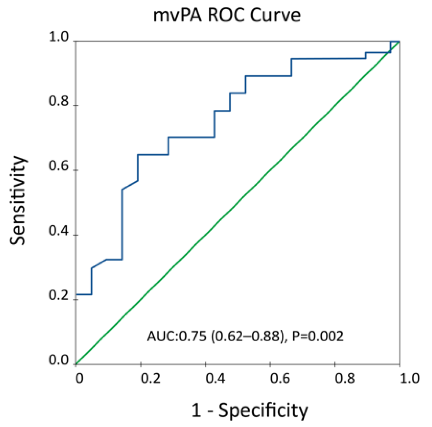
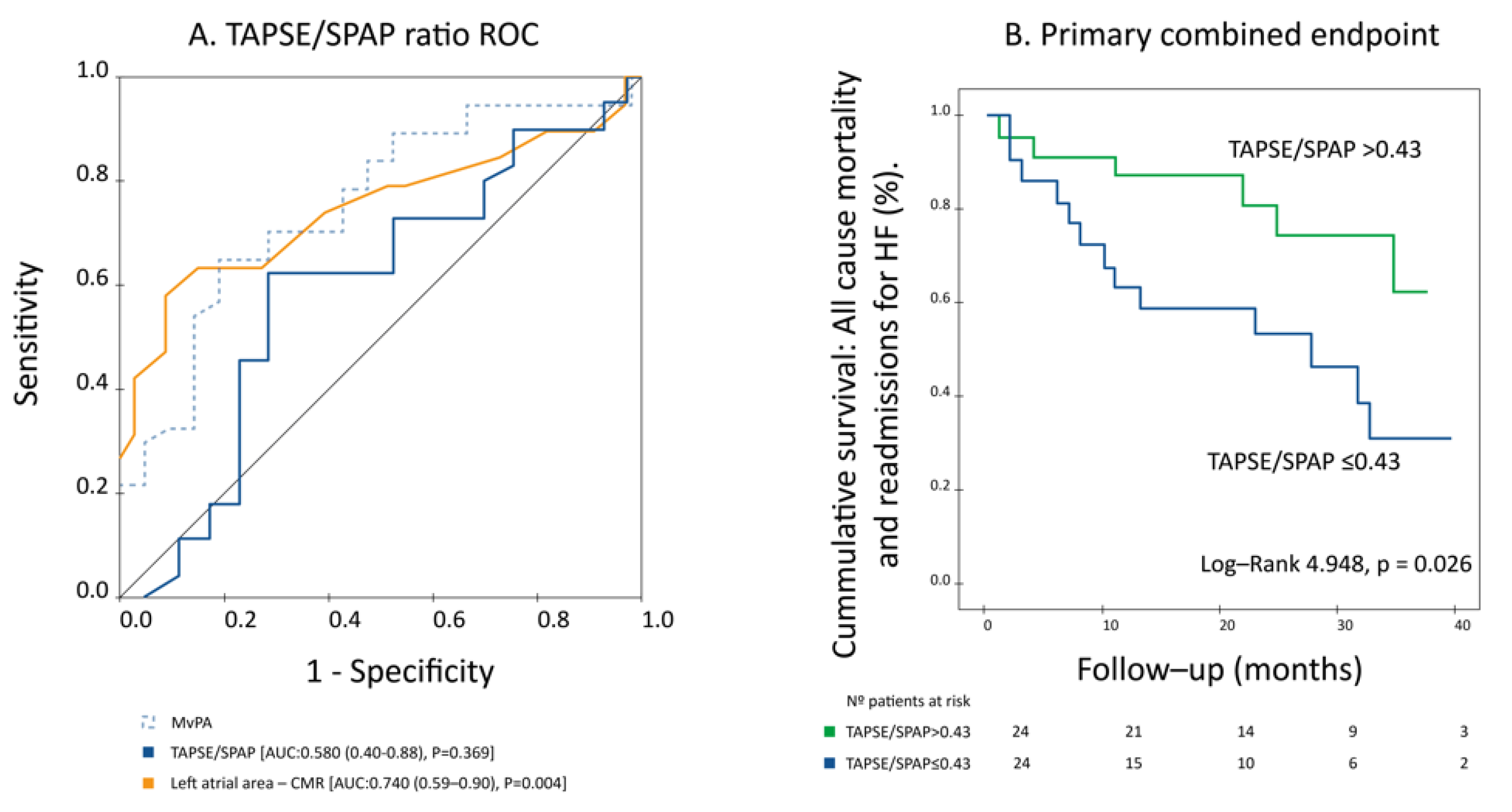
3.2. Prognostic Performance of mvPA and Non-Invasive RV–PC Coupling Parameters
4. Discussion
Limitations
5. Conclusions
Supplementary Materials
Author Contributions
Funding
Institutional Review Board Statement
Informed Consent Statement
Data Availability Statement
Acknowledgments
Conflicts of Interest
References
- McDonagh, T.A.; Metra, M.; Adamo, M.; Gardner, R.S.; Baumbach, A.; Böhm, M.; Burri, H.; Butler, J.; Čelutkienė, J.; Chioncel, O.; et al. 2021 ESC Guidelines for the diagnosis and treatment of acute and chronic heart failure. Eur. Heart J. 2021, 42, 3599–3726. [Google Scholar] [CrossRef]
- Shah, S.J.; Katz, D.H.; Selvaraj, S.; Burke, M.A.; Yancy, C.W.; Gheorghiade, M.; Bonow, R.O.; Huang, C.C.; Deo, R.C. Phenomapping for novel classification of heart failure with preserved ejection fraction. Circulation 2015, 131, 269–279. [Google Scholar] [CrossRef] [Green Version]
- Ghio, S.; Gavazzi, A.; Campana, C.; Inserra, C.; Klersy, C.; Sebastiani, R.; Arbustini, E.; Recusani, F.; Tavazzi, L. Independent and additive prognostic value of right ventricular systolic function and pulmonary artery pressure in patients with chronic heart failure. J. Am. Coll. Cardiol. 2001, 37, 183–188. [Google Scholar] [CrossRef] [Green Version]
- Gorter, T.M.; Hoendermis, E.S.; van Veldhuisen, D.J.; Voors, A.A.; Lam, C.S.; Geelhoed, B.; Willems, T.P.; van Melle, J.P. Right ventricular dysfunction in heart failure with preserved ejection fraction: A systematic review and meta-analysis. Eur. J. Heart Fail. 2016, 18, 1472–1487. [Google Scholar] [CrossRef]
- Guazzi, M.; Ghio, S.; Adir, Y. Pulmonary hypertension in HFpEF and HFrEF: JACC review topic of the week. J. Am. Coll. Cardiol. 2020, 76, 1102–1111. [Google Scholar] [CrossRef]
- Lam, C.S.; Roger, V.L.; Rodeheffer, R.J.; Borlaug, B.A.; Enders, F.T.; Redfield, M.M. Pulmonary hypertension in heart failure with preserved ejection fraction: A community-based study. J. Am. Coll. Cardiol. 2009, 53, 1119–1126. [Google Scholar] [CrossRef] [PubMed] [Green Version]
- Guazzi, M.; Bandera, F.; Pelissero, G.; Castelvecchio, S.; Menicanti, L.; Ghio, S.; Temporelli, P.L.; Arena, R. Tricuspid annular plane systolic excursion and pulmonary arterial systolic pressure relationship in heart failure: An index of right ventricular contractile function and prognosis. Am. J. Physiol. Circ. Physiol. 2013, 305, H1373–H1381. [Google Scholar] [CrossRef] [PubMed]
- Vonk Noordegraaf, A.; Westerhof, B.E.; Westerhof, N. The relationship between the right ventricle and its load in pulmonary hypertension. J. Am. Coll. Cardiol. 2017, 69, 236–243. [Google Scholar] [CrossRef] [PubMed]
- Nakagawa, A.; Yasumura, Y.; Yoshida, C.; Okumura, T.; Tateishi, J.; Yoshida, J.; Abe, H.; Tamaki, S.; Yano, M.; Hayashi, T.; et al. Prognostic importance of right ventricular-vascular uncoupling in acute decompensated heart failure with preserved ejection fraction. Circ. Cardiovasc. Imaging 2020, 13, e011430. [Google Scholar] [CrossRef] [PubMed]
- Santas, E.; Palau, P.; Guazzi, M.; de la Espriella, R.; Miñana, G.; Sanchis, J.; Bayes-Genís, A.; Lupón, J.; Chorro, F.J.; Núñez, J. Usefulness of right ventricular to pulmonary circulation coupling as an indicator of risk for recurrent admissions in heart failure with preserved ejection fraction. Am. J. Cardiol. 2019, 124, 567–572. [Google Scholar] [CrossRef] [PubMed]
- Guazzi, M.; Dixon, D.; Labate, V.; Beussink-Nelson, L.; Bandera, F.; Cuttica, M.J.; Shah, S.J. RV contractile function and its coupling to pulmonary circulation in heart failure with preserved ejection fraction: Stratification of clinical phenotypes and outcomes. JACC Cardiovasc. Imaging 2017, 10, 1211–1221. [Google Scholar] [CrossRef]
- Trejo-Velasco, B.; Fabregat-Andrés, Ó.; García-González, P.M.; Perdomo-Londoño, D.C.; Cubillos-Arango, A.M.; Ferrando-Beltrán, M.I.; Belchi-Navarro, J.; Pérez-Boscá, J.L.; Payá-Serrano, R.; Ridocci-Soriano, F. Prognostic value of mean velocity at the pulmonary artery estimated by cardiovascular magnetic resonance as a prognostic predictor in a cohort of patients with new-onset heart failure with reduced ejection fraction. J. Cardiovasc. Magn. Reson. 2020, 22, 28. [Google Scholar] [CrossRef] [PubMed]
- Trejo-Velasco, B.; Ridocci-Soriano, F.; García-González, M.P.; Cubillos-Arango, A.M.; Payá-Soriano, R.; Fabregat-Andrés, Ó. Mean velocity of the pulmonary artery estimated by cardiac magnetic resonance as an early prognostic predictor in heart failure. Med. Clin. 2019, 153, 232–238. [Google Scholar] [CrossRef] [PubMed]
- Nagueh, S.F.; Smiseth, O.A.; Appleton, C.P.; Byrd, B.F., 3rd; Dokainish, H.; Edvardsen, T.; Flachskampf, F.A.; Gillebert, T.C.; Klein, A.L.; Lancellotti, P.; et al. Recommendations for the evaluation of left ventricular diastolic function by echocardiography: An update from the american society of echocardiography and the European Association of Cardiovascular Imaging. J. Am. Soc. Echocardiogr. 2016, 29, 277–314. [Google Scholar] [CrossRef] [Green Version]
- Rudski, L.G.; Lai, W.W.; Afilalo, J.; Hua, L.; Handschumacher, M.D.; Chandrasekaran, K.; Solomon, S.D.; Louie, E.K.; Schiller, N.B. Guidelines for the echocardiographic assessment of the right heart in adults: A report from the American Society of Echocardiography endorsed by the European Association of Echocardiography, a registered branch of the European Society of Cardiology, and the Canadian Society of Echocardiography. J. Am. Soc. Echocardiogr. 2010, 23, 685–713. [Google Scholar] [CrossRef]
- Barreiro-Pérez, M.; Tundidor-Sanz, E.; Martín-García, A.; Díaz-Peláez, E.; Íscar-Galán, A.; Merchán-Gómez, S.; Gallego-Delgado, M.; Jiménez-Candil, J.; Cruz-González, I.; Sánchez, P.L. First magnetic resonance managed by a cardiology department in the spanish public healthcare system. experience and difficulties of an innovative model. Rev. Esp. Cardiol. 2018, 71, 365–372. [Google Scholar] [CrossRef]
- Petersen, S.E.; Aung, N.; Sanghvi, M.M.; Zemrak, F.; Fung, K.; Paiva, J.M.; Francis, J.M.; Khanji, M.Y.; Lukaschuk, E.; Lee, A.M.; et al. Reference ranges for cardiac structure and function using cardiovascular magnetic resonance (CMR) in Caucasians from the UK Biobank population cohort. J. Cardiovasc. Magn. Reson. 2017, 19, 18. [Google Scholar] [CrossRef] [Green Version]
- Aschauer, S.; Kammerlander, A.A.; Zotter-Tufaro, C.; Ristl, R.; Pfaffenberger, S.; Bachmann, A.; Duca, F.; Marzluf, B.A.; Bonderman, D.; Mascherbauer, J. The right heart in heart failure with preserved ejection fraction: Insights from cardiac magnetic resonance imaging and invasive haemodynamics. Eur. J. Heart Fail. 2016, 18, 71–80. [Google Scholar] [CrossRef]
- Sanz, J.; Kariisa, M.; Dellegrottaglie, S.; Prat-González, S.; Garcia, M.J.; Fuster, V.; Rajagopalan, S. Evaluation of pulmonary artery stiffness in pulmonary hypertension with cardiac magnetic resonance. JACC Cardiovasc. Imaging 2009, 2, 286–295. [Google Scholar] [CrossRef]
- García-Alvarez, A.; Fernández-Friera, L.; Mirelis, J.G.; Sawit, S.; Nair, A.; Kallman, J.; Fuster, V.; Sanz, J. Non-invasive estimation of pulmonary vascular resistance with cardiac magnetic resonance. Eur. Heart J. 2011, 32, 2438–2445. [Google Scholar] [CrossRef] [PubMed] [Green Version]
- Sanz, J.; García-Alvarez, A.; Fernández-Friera, L.; Nair, A.; Mirelis, J.G.; Sawit, S.T.; Pinney, S.; Fuster, V. Right ventriculo-arterial coupling in pulmonary hypertension: A magnetic resonance study. Heart 2012, 98, 238–243. [Google Scholar] [CrossRef]
- Guazzi, M.; Naeije, R. Pulmonary hypertension in heart failure: Pathophysiology, pathobiology, and emerging clinical perspectives. J. Am. Coll. Cardiol. 2017, 69, 1718–1734. [Google Scholar] [CrossRef] [PubMed]
- Tedford, R.J.; Hassoun, P.M.; Mathai, S.C.; Girgis, R.E.; Russell, S.D.; Thiemann, D.R.; Cingolani, O.H.; Mudd, J.O.; Borlaug, B.A.; Redfield, M.M.; et al. Pulmonary capillary wedge pressure augments right ventricular pulsatile loading. Circulation 2012, 125, 289–297. [Google Scholar] [CrossRef] [Green Version]
- Ghio, S.; Schirinzi, S.; Pica, S. Pulmonary arterial compliance: How and why should we measure it? Glob. Cardiol. Sci. Pract. 2015, 2015, 58. [Google Scholar] [CrossRef] [PubMed]
- Borlaug, B.A.; Kane, G.C.; Melenovsky, V.; Olson, T.P. Abnormal right ventricular-pulmonary artery coupling with exercise in heart failure with preserved ejection fraction. Eur. Heart J. 2016, 37, 3293–3302. [Google Scholar] [CrossRef] [PubMed] [Green Version]
- Tello, K.; Dalmer, A.; Axmann, J.; Vanderpool, R.; Ghofrani, H.A.; Naeije, R.; Roller, F.; Seeger, W.; Sommer, N.; Wilhelm, J.; et al. Reserve of right ventricular-arterial coupling in the setting of chronic overload. Circ. Heart Fail. 2019, 12, e005512. [Google Scholar] [CrossRef]
- Vanderpool, R.R.; Pinsky, M.R.; Naeije, R.; Deible, C.; Kosaraju, V.; Bunner, C.; Mathier, M.A.; Lacomis, J.; Champion, H.C.; Simon, M.A. RV-pulmonary arterial coupling predicts outcome in patients referred for pulmonary hypertension. Heart 2015, 101, 37–43. [Google Scholar] [CrossRef] [PubMed] [Green Version]
- Kazimierczyk, R.; Kazimierczyk, E.; Knapp, M.; Sobkowicz, B.; Malek, L.A.; Blaszczak, P.; Ptaszynska-Kopczynska, K.; Grzywna, R.; Kaminski, K.A. Echocardiographic assessment of right ventricular-arterial coupling in predicting prognosis of pulmonary arterial hypertension patients. J. Clin. Med. 2021, 10, 2995. [Google Scholar] [CrossRef]
- Esposito, A.; Palmisano, A.; Toselli, M.; Vignale, D.; Cereda, A.; Rancoita, P.M.V.; Leone, R.; Nicoletti, V.; Gnasso, C.; Monello, A.; et al. Chest CT-derived pulmonary artery enlargement at the admission predicts overall survival in COVID-19 patients: Insight from 1461 consecutive patients in Italy. Eur. Radiol. 2021, 31, 4031–4041. [Google Scholar] [CrossRef]


| No Event (n = 37) | Primary Combined Endpoint (n = 21) | Total Sample (n = 58) | p-Value | |
|---|---|---|---|---|
| Age (years) | 67.2 ± 12.7 | 68 ± 15.2 | 67.5 ± 13.5 | 0.391 |
| Sex, male (n, %) | 20 (54.1) | 14 (66.7) | 34 (58.6) | 0.349 |
| BSA (m2) | 1.8 ± 0.16 | 1.8 ± 0.18 | 1.8 ± 0.17 | 0.512 |
| Arterial hypertension (n, %) | 17 (45.9) | 13 (61.9) | 30 (51.7) | 0.242 |
| Diabetes mellitus (n, %) | 7 (18.9) | 3 (14.3) | 10 (17.2) | 0.653 |
| Dislipidemia (n, %) | 16 (43.2) | 8 (38.1) | 24 (41.4) | 0.702 |
| Atrial fibrillation (n, %) | 17 (45.9) | 10 (47.6) | 27 (46.6) | 0.902 |
| LBBB | 2 (5.4) | 3 (14.3) | 5 (8.6) | 0.247 |
| Previous coronary artery disease (n, %) | 5 (13.5) | 3 (14.3) | 8 (13.8) | 0.935 |
| Glomerular filtration rate (mL/min/1.73 m2) | 72.3 ± 17.6 | 73.9 ± 18.5 | 72.9 ± 17.8 | 0.691 |
| Stage 3–4 chronic kidney disease (n, %) | 5 (13.5) | 1 (4.8) | 6 (10.3) | 0.293 |
| NT-proBNP (pg/mL) | 841.3 ± 343.5 | 1341.1 ± 474.2 | 1281.8 ± 1164.5 | 0.310 |
| Prior HF hospitalization (n, %) | 7 (18.9) | 5 (23.8) | 12 (20.7) | 0.659 |
| NYHA functional class (n, %) | ||||
| I | 17 (45.9) | 2 (9.5) | 19 (32.8) | 0.002 |
| II | 14 (37.8) | 10 (47.6) | 24 (41.4) | |
| III | 6 (16.2) | 4 (19) | 10 (17.2) | |
| IV | 0 | 5 (23.8) | 5 (8.6) | |
| NYHA III–IV/IV (n, %) | 6 (16.2) | 9 (42.9) | 15 (25.9) | 0.026 |
| Cerebrovascular disease (n, %) | 5 (13.5) | 3 (14.3) | 8 (13.8) | 0.935 |
| No Event (n = 37) | Primary Combined Endpoint (n = 21) | Total Sample (n = 58) | p-Value | |
|---|---|---|---|---|
| Echocardiography parameters | ||||
| LVEF (%) | 58.9 ± 9.5 | 57.2 ± 8.4 | 58.3 ± 9.1 | 0.914 |
| LV septum width (mm) | 12.2 ± 1.7 | 12.8 ± 1.8 | 12.4 ± 1.7 | 0.267 |
| LV posterior wall width (mm) | 10.9 ± 2 | 11.5 ± 1.7 | 11.1 ± 1.9 | 0.244 |
| LVEDD (mm) | 47.5 ± 7.1 | 46.9 ± 5.9 | 47.2 ± 6.6 | 0.739 |
| LVESD (mm) | 33.7 ± 11.1 | 30.7 ± 9.7 | 32.5 ± 10.5 | 0.423 |
| Indexed left atrial volume (mL/m2) | 48.8 ± 18.7 | 43.9 ± 18.1 | 47.1 ± 18.5 | 0.405 |
| E/A ratio | 1.2 ± 0.7 | 1.0 ± 0.6 | 1.2 ± 0.7 | 0.440 |
| DT (ms) | 206.3 ± 55.1 | 212.2 ± 34.9 | 208.4 ± 48.1 | 0.773 |
| e’ (septal) | 6.6 ± 1.6 | 6.3 ± 2.5 | 6.5 ± 1.9 | 0.357 |
| e’ (lateral) | 10.4 ± 3.2 | 9.8 ± 4.2 | 10.2 ± 3.5 | 0.715 |
| E/e’ ratio (lateral) | 7.7 ± 3.5 | 7.1 ± 3.1 | 7.5 ± 3.3 | 0.631 |
| TAPSE (mm) | 21.5 ± 4.1 | 19.8 ± 5.3 | 20.9 ± 4.6 | 0.123 |
| S’ tricuspid (cm/s) | 11.3 ± 2.2 | 10.3 ± 4.1 | 10.9 ± 3 | 0.064 |
| Pulmonary acceleration time (ms) | 87.5 ± 26.3 | 83.6 ± 20.9 | 85.9 ± 24 | 0.646 |
| PAPs (mmHg) | 45.0 ± 16.6 | 46.6 ± 15.7 | 45.6 ± 16.1 | 0.532 |
| TAPSE/PAPs | 0.53 ± 0.2 | 0.46 ± 0.2 | 0.5 ± 0.2 | 0.369 |
| TR grade ≥ 3/4 | 7 (18.9) | 4 (19) | 11 (19) | 0.990 |
| CMR parameters | ||||
| LVEF (%) | 60.7 ± 8.1 | 57.4 ± 9.4 | 59.5 ± 8.7 | 0.165 |
| iLVEDV (mL/m2) | 82.2 ± 23.2 | 79.2 ± 34.2 | 81.1 ± 27.4 | 0.481 |
| iLVESV (mL/m2) | 34.5 ± 18.8 | 36.5 ± 22.8 | 35.2 ± 20.1 | 0.752 |
| Left ventricular mass (g) | 71.9 ± 21.1 | 72.1 ± 25.5 | 72 ± 22.5 | 0.984 |
| RVEF (%) | 55.5 ± 11.7 | 52.5 ± 9.1 | 54.4 ± 10.9 | 0.120 |
| iRVEDV (mL/m2) | 94.3 ± 24.7 | 106.3 ± 39.9 | 98.5 ± 31.1 | 0.233 |
| iRVESV (mL/m2) | 42.1 ± 15.9 | 52 ± 26.8 | 45.6 ± 20.7 | 0.160 |
| LGE (n, %) | 10 (27) | 13 (61.9) | 23 (39.7) | 0.009 |
| LGE ischemic pattern (n, %) | 3 (8.1) | 2 (9.5) | 5 (8.6) | 0.854 |
| LGE non-ischemic pattern (n, %) | 8 (21.6) | 12 (57.1) | 20 (34.5) | 0.006 |
| Left atrial area (mm2) | 15.4 ± 3.4 | 19.6 ± 5.5 | 16.9 ± 4.7 | 0.005 |
| Right atrial area (mm2) | 15.7 ± 4.9 | 18.7 ± 10.4 | 16.8 ± 7.5 | 0.252 |
| Maximal PA area (cm2) | 8.4 ± 2.5 | 11.4 ± 3.4 | 9.5 ± 3.2 | <0.001 |
| Minimal PA area (cm2) | 6.7 ± 2.0 | 9.2 ± 2.8 | 7.6 ± 2.6 | <0.001 |
| PA pulsatility (%) | 26.9 ± 14.2 | 25.8 ± 19.3 | 26.7 ± 16.1 | 0.382 |
| Right ventricular Ea/Emax | 0.91 ± 0.58 | 0.96 ± 0.38 | 0.93 ± 0.51 | 0.120 |
| mvPA (cm/s) | 10.9 ± 3.9 | 7.7 ± 2.7 | 9.8 ± 3.9 | 0.001 |
| PVR-CMR (Wood Units) | 4.2 ± 2.3 | 5.9 ± 1.8 | 4.8 ± 2.3 | 0.001 |
| RHC parameters * | ||||
| Mean PA pressure (mmHg) | 33.6 ± 15.6 | 35.7 ± 14.7 | 34.5 ± 14.9 | 0.728 |
| Pulmonary capillary pressure (mmHg) | 15.9 ± 4.7 | 14.8 ± 5.4 | 15.5 ± 4.5 | 0.347 |
| PA pulse pressure (mmHg) | 34.3 ± 16.2 | 31.2 ± 10.5 | 32.9 ± 13.9 | 0.873 |
| Cardiac index (mL/min/m2) | 2.5 ± 0.5 | 2.5 ± 1.1 | 2.5 ± 0.8 | 0.506 |
| PVR-CCD (UW) | 4.4 ± 3.1 | 5.2 ± 2.9 | 4.8 ± 3.0 | 0.494 |
| Transpulmonary gradient (mmHg) | 17.7 ± 13.7 | 20.8 ± 14.7 | 19.1 ± 13.9 | 0.478 |
| PA compliance (mL/mmHg) | 2.3 ± 1.7 | 1.8 ± 0.5 | 2.1 ± 1.9 | 0.882 |
| mvAP ≤ 9 cm/s (n = 30) | mvAP > 9 cm/s (n = 28) | Total Sample (n = 58) | p-Value | |
|---|---|---|---|---|
| Echocardiography parameters | ||||
| LVEF (%) | 59.1 ± 10.2 | 57.3 ± 7.7 | 58.3 ± 9.1 | 0.460 |
| LV septum width (mm) | 12.9 ± 1.8 | 11.7 ± 1.4 | 12.4 ± 1.7 | 0.010 |
| LV posterior wall width (mm) | 11.8 ± 1.8 | 10.3 ± 1.8 | 11.1 ± 1.9 | 0.011 |
| LVEDD (mm) | 47.5 ± 6.9 | 46.9 ± 6.3 | 47.2 ± 6.6 | 0.784 |
| LVESD (mm) | 31.8 ± 11.3 | 33.5 ± 9.4 | 32.5 ± 10.5 | 0.648 |
| Indexed left atrial volume (mL/m2) | 44.9 ± 18.6 | 49.9 ± 18.3 | 47.1 ± 18.5 | 0.362 |
| E/A ratio | 1.1 ± 0.5 | 1.4 ± 0.8 | 1.2 ± 0.7 | 0.154 |
| DT (ms) | 224 ± 38.7 | 185 ± 53.2 | 208.4 ± 48.1 | 0.044 |
| e’ (septal) | 5.9 ± 2.2 | 7.1 ± 1.5 | 6.5 ± 1.9 | 0.069 |
| e’ (lateral) | 9.1 ± 3.9 | 11.1 ± 2.9 | 10.2 ± 3.5 | 0.059 |
| E/e’ ratio (lateral) | 7.3 ± 3.4 | 7.7 ± 3.3 | 7.5 ± 3.3 | 0.732 |
| TAPSE (mm) | 20.8 ± 5 | 21.1 ± 4.1 | 20.9 ± 4.6 | 0.674 |
| S’ tricuspid (cm/s) | 11 ± 3.6 | 10.7 ± 1.8 | 10.9 ± 3 | 0.730 |
| Pulmonary acceleration time (ms) | 82.9 ± 28.1 | 90.8 ± 14.9 | 85.9 ± 24 | 0.358 |
| PAPs (mmHg) | 46.5 ± 17.2 | 44.6 ± 15.1 | 45.6 ± 16.1 | 0.673 |
| TAPSE/PAPs | 0.49 ± 0.3 | 0.50 ± 0.2 | 0.5 ± 0.2 | 0.974 |
| TR grade ≥ 3/4 | 7 (23.3) | 4 (14.3) | 11 (19) | 0.380 |
| CMR parameters | ||||
| LVEF (%) | 58.7 ± 9.5 | 60.4 ± 7.7 | 59.5 ± 8.7 | 0.732 |
| iLVEDV (mL/m2) | 82 ± 30.9 | 80.1 ± 23.6 | 81.1 ± 27.4 | 0.779 |
| iLVESV (mL/m2) | 36.9 ± 24.2 | 33.4 ± 14.8 | 35.2 ± 20.1 | 0.932 |
| Left ventricular mass (g) | 73.7 ± 21.9 | 70.2 ± 23.5 | 72 ± 22.5 | 0.486 |
| RVEF (%) | 51 ± 11.4 | 57.9 ± 9.3 | 54.4 ± 10.9 | 0.015 |
| iRVEDV (mL/m2) | 106 ± 30.1 | 90.8 ± 30.7 | 98.5 ± 31.1 | 0.053 |
| iRVESV (mL/m2) | 51.3 ± 16.6 | 39.7 ± 23 | 45.6 ± 20.7 | 0.002 |
| LGE (n, %) | 14 (46.7) | 9 (32.1) | 23 (39.7) | 0.215 |
| LGE ischemic pattern (n, %) | 2 (6.7) | 3 (10.7) | 5 (8.6) | 0.610 |
| LGE non-ischemic pattern (n, %) | 13 (43.3) | 7 (25) | 20 (34.5) | 0.117 |
| Left atrial area (mm2) | 18 ± 5.1 | 15.8 ± 4.1 | 16.9 ± 4.7 | 0.090 |
| Right atrial area (mm2) | 18.4 ± 9.4 | 15.2 ± 4.6 | 16.8 ± 7.5 | 0.128 |
| Maximal PA area (cm2) | 10.9 ± 3 | 7.8 ± 2.5 | 9.5 ± 3.2 | <0.001 |
| Minimal PA area (cm2) | 8.8 ± 2.4 | 6.2 ± 2.2 | 7.6 ± 2.6 | <0.001 |
| PA pulsatility (%) | 24.1 ± 14.1 | 29.3 ± 17.8 | 26.7 ± 16.1 | 0.194 |
| Right ventricular Ea/Emax | 1.1 ± 0.6 | 0.8 ± 0.4 | 0.93 ± 0.51 | 0.008 |
| mvPA (cm/s) | 6.8 ± 1.6 | 12.9 ± 3 | 9.8 ± 3.9 | <0.001 |
| PVR-CMR (Wood Units) | 6.5 ± 1.8 | 3.1 ± 1.1 | 4.8 ± 2.3 | <0.001 |
| RHC parameters * | ||||
| Mean PA pressure (mmHg) | 34.9 ± 14.8 | 33.6 ± 16.2 | 34.5 ± 14.9 | 0.823 |
| Pulmonary capillary pressure (mmHg) | 15.9 ± 5.5 | 14.6 ± 3.7 | 15.5 ± 4.5 | 0.513 |
| PA pulse pressure (mmHg) | 32.2 ± 13.6 | 34.4 ± 15.1 | 32.9 ± 13.9 | 0.698 |
| Cardiac index (mL/min/m2) | 2.5 ± 0.9 | 2.5 ± 0.4 | 2.5 ± 0.8 | 0.932 |
| PVR-CCD (UW) | 4.6 ± 2.8 | 5.2 ± 3.7 | 4.8 ± 3.0 | 0.804 |
| Transpulmonary gradient (mmHg) | 19.1 ± 13.5 | 19 ± 15.8 | 19.1 ± 13.9 | 0.993 |
| PA compliance (mL/mmHg) | 2.1 ± 1.3 | 2 ± 1.4 | 2.1 ± 1.9 | 0.904 |
| Cardiovascular events | ||||
| Readmission for decompensated heart failure (n, %) | 11 (36.7) | 4 (14.3) | 15 (25.9) | 0.049 |
| All-cause death (n, %) | 6 (20) | 2 (7.1) | 8 (13.8) | 0.156 |
| Primary combined endpoint (n, %) | 17 (56.7) | 4 (14.3) | 21 (36.2) | 0.001 |
| Hazard Ratio (95% CI) | p-Value | |
|---|---|---|
| NYHA functional class III–IV/IV | 0.67 (0.24–1.90) | 0.453 |
| Left atrial area—CMR (mL/m2) | 1.12 (1.01–1.24) | 0.034 |
| Late gadolinium enhancement—CMR | 0.72 (0.24–2.12) | 0.546 |
| mvPA < 9 cm/s | 4.11 (1.28–13.19) | 0.017 |
Publisher’s Note: MDPI stays neutral with regard to jurisdictional claims in published maps and institutional affiliations. |
© 2022 by the authors. Licensee MDPI, Basel, Switzerland. This article is an open access article distributed under the terms and conditions of the Creative Commons Attribution (CC BY) license (https://creativecommons.org/licenses/by/4.0/).
Share and Cite
Trejo-Velasco, B.; Cruz-González, I.; Barreiro-Pérez, M.; Díaz-Peláez, E.; García-González, P.; Martín-García, A.; Eiros, R.; Merchán-Gómez, S.; Pérez del Villar, C.; Fabregat-Andrés, O.; et al. Mean Velocity of the Pulmonary Artery as a Clinically Relevant Prognostic Indicator in Patients with Heart Failure with Preserved Ejection Fraction. J. Clin. Med. 2022, 11, 491. https://doi.org/10.3390/jcm11030491
Trejo-Velasco B, Cruz-González I, Barreiro-Pérez M, Díaz-Peláez E, García-González P, Martín-García A, Eiros R, Merchán-Gómez S, Pérez del Villar C, Fabregat-Andrés O, et al. Mean Velocity of the Pulmonary Artery as a Clinically Relevant Prognostic Indicator in Patients with Heart Failure with Preserved Ejection Fraction. Journal of Clinical Medicine. 2022; 11(3):491. https://doi.org/10.3390/jcm11030491
Chicago/Turabian StyleTrejo-Velasco, Blanca, Ignacio Cruz-González, Manuel Barreiro-Pérez, Elena Díaz-Peláez, Pilar García-González, Ana Martín-García, Rocío Eiros, Soraya Merchán-Gómez, Candelas Pérez del Villar, Oscar Fabregat-Andrés, and et al. 2022. "Mean Velocity of the Pulmonary Artery as a Clinically Relevant Prognostic Indicator in Patients with Heart Failure with Preserved Ejection Fraction" Journal of Clinical Medicine 11, no. 3: 491. https://doi.org/10.3390/jcm11030491
APA StyleTrejo-Velasco, B., Cruz-González, I., Barreiro-Pérez, M., Díaz-Peláez, E., García-González, P., Martín-García, A., Eiros, R., Merchán-Gómez, S., Pérez del Villar, C., Fabregat-Andrés, O., Ridocci-Soriano, F., & Sánchez, P. L. (2022). Mean Velocity of the Pulmonary Artery as a Clinically Relevant Prognostic Indicator in Patients with Heart Failure with Preserved Ejection Fraction. Journal of Clinical Medicine, 11(3), 491. https://doi.org/10.3390/jcm11030491


