Review

Pathophysiology of Heart Failure: A Role for Peripheral Blood Mononuclear Cells Mitochondrial Dysfunction?

1
University of Strasbourg, Translational Medicine Federation of Strasbourg (FMTS), Faculty of Medicine, Team 3072 “Mitochondria, Oxidative Stress and Muscle Protection”, 11 rue Humann, 67000 Strasbourg, France
2
University Hospital of Strasbourg, Physiology and Functional Exploration Service, 1 Place de l’Hôpital, 67091 Strasbourg, France
3
Internal Medicine, Diabete and Metabolic Diseases Service, University Hospital of Strasbourg, 1 Place de l’Hôpital, 67091 Strasbourg, France
*
Author to whom correspondence should be addressed.
J. Clin. Med. 2022, 11(3), 741; https://doi.org/10.3390/jcm11030741
Submission received: 20 December 2021 / Revised: 24 January 2022 / Accepted: 27 January 2022 / Published: 29 January 2022
(This article belongs to the Special Issue Current Challenges and Advances in Heart Failure and Heart Transplant)

Abstract

:
Heart failure (HF) is a leading cause of hospitalization in patients aged more than 65 years and is associated with high mortality rates. A better comprehension of its physiopathology is still needed, and, in addition to neurohormonal systems and sodium glucose co-transporter 2 modulations, recent studies focus on the mitochondrial respiration of peripheral blood circulating cells (PBMCs). Thus, cardiovascular metabolic risk factors and cellular switch with an increased neutrophil/lymphocytes ratio might favor the decreased PBMC mitochondrial respiration observed in relation with HF severity. PBMCs are implicated in the immune system function and mitochondrial dysfunction of PBMC, potentially induced by their passage through a damaged heart and by circulating mitoDAMPs, which can lead to a vicious circle, thus sustaining negative cardiac remodeling during HF. This new approach of HF complex pathophysiology appears to be a promising field of research, and further studies on acute and chronic HF with reduced or preserved LVEF are warranted to better understand whether circulating PBMC mitochondrial function and mitoDAMPs follow-ups in HF patients might show diagnosis, prognosis or therapeutic usefulness.

1. Introduction

Heart failure (HF) is a clinical syndrome defined by reduced cardiac output and/or elevated intracardiac pressures at rest or during exercise. It is characterized by typical symptoms, such as breathlessness, ankle swelling and fatigue, which may be accompanied by signs (elevated jugular venous pressure, pulmonary crackles and peripheral edema) caused by structural and/or functional cardiac abnormalities (ESC 2021). In Europe, there are 15 million HF patients and 120,000 new cases are reported each year in France. The mortality is about 10% and HF is the leading cause of hospitalizations in patients aged more than 65 years (177,000/year). Sympathetic nervous, renin–angiotensin–aldosterone and cardiac natriuretic system implications are well-known and their inhibition is the cornerstone of chronic HF management, which has been recently implemented with sodium glucose co-transporter 2 modulators, even in HF-preserved ejection fractions (HFpEFs) up to 60% [1,2,3,4,5]. Treatment of chronic HF is relatively consensual when the left ventricular ejection fraction (LVEF) is reduced (<40%), and management of acute HF is based on diuretics, triggering factor specific treatment and the early initiation of the recommended chronic therapy. Nevertheless, new targets are needed and might be identified through an improvement in HF pathophysiology knowledge. Interestingly, recent studies in HF have focused on inflammation and systemic oxidative stress potentially related to the mitochondrial dysfunction of peripheral circulating cells.
In this view, the mitochondrial respiratory function of peripheral blood mononuclear cells (PBMCs) (in extenso lymphocytes and monocytes) is easily available. These cells are involved in many inflammatory diseases, including those driven by ischemia-reperfusion episodes, which play a key role in cardiovascular alterations [6]. Whether their mitochondrial dysfunction could be used as a diagnostic, severity and/or prognosis biomarker in cardiovascular diseases is under evaluation, but the major role of mitochondria in cell energy and reactive oxygen species (ROSs) production support such a hypothesis. Thus, adequate ROS levels in terms of the amount and duration of secretion are considered as useful signaling factors. On the other hand, ROS secreted in excess and not buffered by antioxidants can lead to protein, lipid, and DNA damage and tissue dysfunctions. Accordingly, ROSs are increased in ischemic muscles and in pathological myocardium during HF [7,8], and circulating blood ROSs can also be interesting biomarkers of disease severity.
This brief review aims to report and discuss the data investigating the evolution and potential usefulness of PBMC mitochondrial respiration in the setting of human HF. This is a current challenge and a better comprehension of HF pathophysiology might lead to significant advances in HF management. Indeed, impaired PBMC mitochondrial respiration might be implicated in the development of inappropriate immune system responses and negative cardiovascular remodeling during HF.

2. Potential Relationships between PBMC, Mitochondrial Function and HF

T-cell recruitment and myocardial infiltration is well-described during ischemic and non-ischemic HF [9], with specific and enhanced abnormal interactions with cardiac antigens as compared to controls [10]. During infection, heart transplant or ischemic injury, these cells infiltrate the heart, in association with a disrupted self-tolerance of cardiac antigen. Altogether, these cellular crosstalks negatively affect cardiac remodeling and function [11]. Monocytes also play a central role, being recruited during a myocardial injury by a cell–cell interaction [12,13]. They favor fibroblasts proliferation, and in fine cardiac fibrosis. Moreover, neutrophils are described as a potential “weapon” for sterile inflammation and can lead to abnormal cardiac remodeling and HF development [14].
However, cellular mechanisms and bioenergetic dysfunctions leading to these pathological implications are poorly known, and PBMC mitochondrial function could play a significant role in this immune-related disorder during HF. Indeed, these easily available biomarkers are essential for adaptive immune system responses to organ injuries, and the recent data support an impaired mitochondrial respiration of PBMC in the setting of HF.
The first data were reported by Li et al. [15] who described a global decrease in mitochondrial respiratory function in 25 early stage, asymptomatic HFpEF patients, as compared to 24 controls (Table 1). The early stage asymptomatic patients were defined by cardiac remodeling (septal hypertrophy and/or a delayed relaxation) and the presence of a cardiometabolic risk factor (essential hypertension, dyslipidemia and type 2 diabetes). Moreover, the high sensitivity C-reactive protein, interleukin-6 (IL-6) and tumor necrosis factor-α (TNF-α), were significantly higher, and superoxide dismutase—a major antioxidant actor—transforming superoxide anion into hydrogen peroxide, was reduced in HF patients.
Then, Shirakawa et al. [16] observed in 31 compensated chronic HF patients with reduced EF (HFrEF) < 35% that the activities of several mitochondrial respiratory chain complexes were altered. Thus, complexes I + II and the maximal electron transfer system with complexes I + II and II alone were significantly reduced in functional class NYHA III (16 patients) versus the NYHA I or II groups (15 patients), (p < 0.05). This suggests that impaired PBMC mitochondrial respiration is related to the degree of severity of the disease. At the same time, mitochondrial ROSs were significantly higher in the NYHA III group than in the NYHA I or II groups.
Recently, data from 19 stage-D HFrEF patients also showed an impairment of maximal respiration as compared to the controls, with the basal oxygen consumption rate tending to be lower [17].
The causes potentially involved in PBMC-reduced mitochondrial respiration during HF are yet to be determined, but several hypotheses might be raised. Both cardiovascular metabolic risk factors and cellular switch might play a role.
Cardiovascular metabolic risk factors strongly associated with HF development [18] and chronic inflammation [19] might participate in such a depressed PBMC mitochondrial respiration during HF. Thus, a recent study showed that LDL-c is negatively associated with global mitochondrial respiration [20]. Altered mitochondrial respiration is also described in hypertension-related renal, cardiac and vascular disease, and age-related cardiac changes [21,22]. These abnormalities in PBMC could participate in systemic endothelial dysfunction [23].
Interestingly, there is a cellular switch (Figure 1) in the white cell count during HF development that could participate in the global decrease in mitochondrial respiration. Indeed, neutrophils are activated and increased and could facilitate cardiomyocytes’ apoptosis [24], whereas there is a progressive and relative lymphocytopenia due to the inflammation and down-regulation of the immune system [25]. This cellular switch in HF patients could lead to a decrease in global PBMC mitochondrial respiration, since neutrophils seem to have a minimal contribution to the oxygen consumption rate and cellular bioenergetics, as compared to lymphocytes and monocytes [26]. Indeed, studies show that the neutrophils-to-lymphocytes ratio (NLR) is increased in acute and chronic HF patients and is associated with frequent congestive HF decompensation, hospitalization readmission and one-year overall mortality [24,27,28,29].

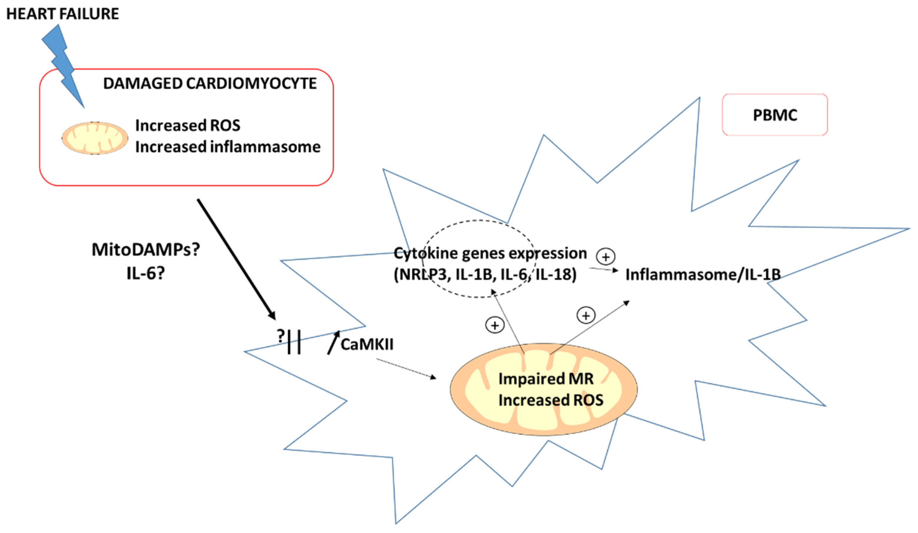
3. Potential Mechanisms Involved in Impaired Mitochondrial Respiration of PBMC in HF

Mitochondrial respiration is essential for PBMC homeostasis and its dysfunction could lead to numerous inflammatory responses in various diseases. On a mouse model with a defective mitochondrial function in CD4+ T lymphocytes, impaired mitochondrial respiration lead to lysosome dysfunction. Thus, this alteration exhibited in vitro and in vivo inflammatory responses with a higher level of cytokines (Interferon-γ) and T-cell proliferation [30]. During the first 7 days of a septic shock, a recent study found a global increase in the global mitochondrial respiration capacity and a paradoxical decrease in ATP-synthase activity [31]. These changes might be a mitochondrial adaptation to acute systemic stress. On the other hand, in a chronic state, such as chronic HF, the decreased non-adaptive mitochondrial respiration could have a negative impact. Indeed, Zhou et al. showed that the impaired mitochondrial respiration of the PBMC of patients with HF resulted in increased secretion of proinflammatory cytokines [17]. Furthermore, altered mitochondrial respiration is also associated with less effective T-cell migration [32].
Accordingly, a link between immune cell activation and immune cell metabolism has been demonstrated. In general, the activation of immune cells requires a rise in energy and most articles suggested that activated monocytes and T and B lymphocytes undergo metabolic reprogramming and an increased glucose uptake and glycolysis. On the contrary, long-living immune cells, such as macrophages or regulatory T cells, enhance oxidative phosphorylation and beta-oxidation to produce their energy [33]. The role of the different respiratory complexes is variable between the different types of immune cells, but there is a lot of evidence to suggest that modified mitochondrial respiration is involved in inflammatory responses.
Concerning HF, both the “proximity theory” and activation of the immune system cells by circulating factors arising from the heart might account for the relationships between PBMC and HF.
The “proximity theory” proposes that PBMCs are activated during their passage through the heart (Figure 2). Indeed, during non-ischemic stress (pressure overload, angiotensin II exposure), there is a communication between the immune system near the myocardium and damaged cardiomyocytes, especially through the calmoduline kinase II (CaMKII)/NRLP3 pathway (NRLP3 for nucleotide-binding, leucine-rich repeat and pyrin-domain-containing 3) [34]. The initial mitochondrial dysfunction in the cardiomyocytes leads to increased cellular stress, which further activates the inflammasome/interleukin-1β (IL-1β) pathway and immune cells’ recruitment [35]. Moreover, circulating IL-6 is increased and could participate in chronic HF development [36,37]. Interestingly, Zhou et al. [17] suggested that IL-6 acts as a signal, connecting the mitochondrial function and inflammation in PBMCs, leading to mitochondrial respiration impairment by inhibiting complex I activity.
The second hypothesis might be the circulation of factors arising from the heart and activating the immune system cells. Mitochondrial DNA (mtDNA) and oxidative stress are likely to be involved as circulating factors. Confirming such a hypothesis, Zhou et al. [17] demonstrated that mtDNA extracted from myocardial damaged mitochondria elicited an impairment of the maximal oxygen consumption rate in healthy PBMC. Moreover, in this study, mitochondrial ROS and inflammatory cytokine gene expression (NRLP3, IL-1B, IL-6 and IL-18) were increased.
Since ROS and mitochondrial dysfunction activate the immune system and inflammation, especially through NRLP3 activation [38], circulating mtDNA and extensively mitochondrial damage-associated molecular patterns (mitoDAMPs) could be systemic vectors of mitochondrial dysfunction in the heart, and can induce an impairment of PBMC mitochondrial respiration, which in turn might activate the immune system and inflammation (Figure 3). The impairment of mitochondrial respiration then leads to an ROS increase, which promotes the NLRP3 inflammasome activation [39]. One consequence is a higher production of IL-1β, a cytokine known for its negative effects on myocardium [40,41].
Interestingly in the CANTOS trial, the IL-1β blockade by canakinumab showed an HF-related decreases in hospitalization and mortality rates [42]. Similar to the IL-1β blockade by anakinra, Van Tassell et al. found an improvement of LV pressures as assessed by echocardiography after 12 weeks of treatment [43]. Corroborating a potential circulating factor and IL-1β implication, Bilchik et al. demonstrated that cardiac resynchronization therapy (CRT) in HF responder patients reduced the expression of inflammation-promoting genes related to IL-1 β in PBMC [44].
Recently, 2021 ESC guidelines on acute and chronic HF recommended the use of sodium glucose co-transporter 2 inhibitors (SGLT2i) as a safe and efficient first-choice treatment in HF with a reduced left ejection fraction (<40%) (1). One potential systemic effect is the reduction in inflammation through the inhibition of the NLRP3 inflammasome, independently of glucose lowering [45]. Accordingly, in the myocardium of diabetic rats, SGLT inhibition seems to increase mitochondrial respiration [46] and thus energy production. Moreover, these treatments improved the ketone level, a substrate of cardiac mitochondrial respiratory chains, in order to produce a high level of adenosine triphosphate (ATP) [47]. Other proposed mechanisms are the inhibition of HIF-1α negative effects, especially on the biogenesis of mitochondria [48,49] and on the activation of inflammatory macrophages [50]. Thus, these medications could have an interesting impact on PBMC mitochondrial respiration in HF, through the metabolic and non-metabolic pathways. Measurements of the mitochondrial respiration of PBMC from HF patients exposed to SGLTi will be useful to further investigate such issues.
Taken together, one can propose that PBMC mitochondrial impairment can lead to a vicious circle, finally altering the myocardium by activating fibrosis signaling and cardiac remodeling [51]. Accordingly, therapeutics studies targeting various cytokines (NLRP3, IL-1 β and IL-6) [52,53,54] showed promising results and support the need for a better comprehension of the activation of the immune system during HF.

4. Conclusions and Current Challenges

This new approach of HF complex pathophysiology appears to be a promising field of research. PBMCs are an easily available cell population implicated in the immune system function, and their mitochondrial dysfunction potentially induced by circulating mitoDAMPs may lead to a vicious circle sustaining cardiac dysfunction.
Nevertheless, most HF patients, whether with a reduced or preserved EF, have multiple comorbidities, and future studies trying to control comorbidities might be an important way to tease out the specific effects of these comorbidities, as they may produce differential molecular pathways to achieve the same vicious cycle of inflammation. Accordingly, individuals with a normal cardiac function but with type II diabetes, asthma exacerbation or pulmonary hypertension presented with an increased PBMC mitochondrial function [55,56,57].
Multisite clinical trials, in which large numbers of HF patients are studied with standardized PBMC mitochondrial function analysis measured alongside different blood biomarkers and clinical outcomes, and using linear regression modeling to control for individual comorbid conditions, are therefore warranted to better understand whether the circulating PBMC mitochondrial functions and mitoDAMPs follow-up in HF patients might show diagnosis, prognosis or therapeutic usefulness.

Author Contributions

Conceptualization, F.S., M.R., B.G. and S.T.; methodology, F.S., M.R., B.G. and S.T.; validation, F.S., M.R., A.-L.C., A.M., E.A., B.G. and S.T.; investigation, F.S., M.R., A.-L.C., A.M., E.A., B.G. and S.T.; writing—original draft preparation, F.S., M.R., A.M., B.G. and S.T. writing—review and editing, F.S., M.R., B.G. and S.T.; supervision, B.G. and S.T.; project administration, B.G. and S.T. All authors have read and agreed to the published version of the manuscript.

Funding

This research received no external funding.

Institutional Review Board Statement

Not applicable.

Informed Consent Statement

Not applicable.

Acknowledgments

We greatly thank Anne-Marie Kasprowicz for her expert secretarial assistance.

Conflicts of Interest

The authors declare no conflict of interest.

References

  1. McDonagh, T.A.; Metra, M.; Adamo, M.; Gardner, R.S.; Baumbach, A.; Böhm, M.; Burri, H.; Butler, J.; Čelutkienė, J.; Chioncel, O.; et al. 2021 ESC Guidelines for the diagnosis and treatment of acute and chronic heart failure. Eur. Heart J. 2021, 42, 3599–3726, Erratum in Eur. Heart J. 2021. [Google Scholar] [CrossRef]
  2. Maggioni, A.P.; Dahlström, U.; Filippatos, G.; Chioncel, O.; Crespo Leiro, M.; Drozdz, J.; Fruhwald, F.; Gullestad, L.; Logeart, D.; Fabbri, G.; et al. EURObservational Research Programme: Regional differences and 1-year follow-up results of the Heart Failure Pilot Survey (ESC-HF Pilot). Eur. J. Heart Fail. 2013, 15, 808–817. [Google Scholar] [CrossRef] [Green Version]
  3. Gabet, A.; Juillière, Y.; Lamarche-Vadel, A.; Vernay, M.; Olié, V. National trends in rate of patients hospitalized for heart failure and heart failure mortality in France, 2000–2012. Eur. J. Hear. Fail. 2015, 17, 583–590. [Google Scholar] [CrossRef]
  4. Lugnier, C.; Meyer, A.; Charloux, A.; Andrès, E.; Gény, B.; Talha, S. The Endocrine Function of the Heart: Physiology and Involvements of Natriuretic Peptides and Cyclic Nucleotide Phosphodiesterases in Heart Failure. J. Clin. Med. 2019, 8, 1746. [Google Scholar] [CrossRef] [Green Version]
  5. Anker, S.D.; Butler, J.; Filippatos, G.; Ferreira, J.P.; Bocchi, E.; Böhm, M.; Rocca, H.-P.B.; Choi, D.-J.; Chopra, V.; Chuquiure-Valenzuela, E.; et al. Empagliflozin in Heart Failure with a Preserved Ejection Fraction. N. Engl. J. Med. 2021, 385, 1451–1461. [Google Scholar] [CrossRef]
  6. Alfatni, A.; Riou, M.; Charles, A.-L.; Meyer, A.; Barnig, C.; Andres, E.; Lejay, A.; Talha, S.; Geny, B. Peripheral Blood Mononuclear Cells and Platelets Mitochondrial Dysfunction, Oxidative Stress, and Circulating mtDNA in Cardiovascular Diseases. J. Clin. Med. 2020, 9, 311. [Google Scholar] [CrossRef] [Green Version]
  7. Pottecher, J.; Guillot, M.; Belaidi, E.; Charles, A.-L.; Lejay, A.; Gharib, A.; Diemunsch, P.; Geny, B. Cyclosporine A normalizes mitochondrial coupling, reactive oxygen species production, and inflammation and partially restores skeletal muscle maximal oxidative capacity in experimental aortic cross-clamping. J. Vasc. Surg. 2013, 57, 1100–1108.e2. [Google Scholar] [CrossRef] [Green Version]
  8. Giordano, F.J. Oxygen, oxidative stress, hypoxia, and heart failure. J. Clin. Investig. 2005, 115, 500–508. [Google Scholar] [CrossRef]
  9. Nevers, T.; Salvador, A.M.; Grodecki-Pena, A.; Knapp, A.; Velázquez, F.; Aronovitz, M.; Kapur, N.K.; Karas, R.H.; Blanton, R.M.; Alcaide, P. Left Ventricular T-Cell Recruitment Contributes to the Pathogenesis of Heart Failure. Circ. Hear. Fail. 2015, 8, 776–787. [Google Scholar] [CrossRef] [Green Version]
  10. Blanton, R.M.; Carrillo-Salinas, F.J.; Alcaide, P. T-cell recruitment to the heart: Friendly guests or unwelcome visitors? Am. J. Physiol. Heart Circ. Physiol. 2019, 317, H124–H140. [Google Scholar] [CrossRef]
  11. Ismahil, M.A.; Hamid, T.; Bansal, S.S.; Patel, B.; Kingery, J.R.; Prabhu, S.D. Remodeling of the mononuclear phagocyte network underlies chronic inflammation and disease progression in heart failure: Critical importance of the cardiosplenic axis. Circ. Res. 2014, 114, 266–282. [Google Scholar] [CrossRef] [PubMed] [Green Version]
  12. Bajpai, G.; Bredemeyer, A.; Li, W.; Zaitsev, K.; Koenig, A.L.; Lokshina, I.; Mohan, J.; Ivey, B.; Hsiao, H.M.; Weinheimer, C.; et al. Tissue Resident CCR2- and CCR2+ Cardiac Macrophages Differentially Orchestrate Monocyte Recruitment and Fate Specifi-cation Following Myocardial Injury. Circ. Res. 2019, 124, 263–278. [Google Scholar] [CrossRef] [PubMed]
  13. Abplanalp, W.T.; John, D.; Cremer, S.; Assmus, B.; Dorsheimer, L.; Hoffmann, J.; Becker-Pergola, G.; Rieger, M.A.; Zeiher, A.M.; Vasa-Nicotera, M.; et al. Single-cell RNA-sequencing reveals profound changes in circulating immune cells in patients with heart failure. Cardiovasc. Res. 2020, 117, 484–494. [Google Scholar] [CrossRef] [Green Version]
  14. Bonaventura, A.; Montecucco, F.; Dallegri, F.; Carbone, F.; Lüscher, T.F.; Camici, G.; Liberale, L. Novel findings in neutrophil biology and their impact on cardiovascular disease. Cardiovasc. Res. 2019, 115, 1266–1285. [Google Scholar] [CrossRef]
  15. Li, P.; Wang, B.; Sun, F.; Li, Y.; Li, Q.; Lang, H.; Zhao, Z.; Gao, P.; Zhao, Y.; Shang, Q.; et al. Mitochondrial respiratory dysfunctions of blood mononuclear cells link with cardiac disturbance in patients with early-stage heart failure. Sci. Rep. 2015, 5, 10229. [Google Scholar] [CrossRef] [Green Version]
  16. Shirakawa, R.; Yokota, T.; Nakajima, T.; Takada, S.; Yamane, M.; Furihata, T.; Maekawa, S.; Nambu, H.; Katayama, T.; Fukushima, A.; et al. Mitochondrial reactive oxygen species generation in blood cells is associated with disease severity and exercise intolerance in heart failure patients. Sci. Rep. 2019, 9, 14709. [Google Scholar] [CrossRef]
  17. Zhou, B.; Wang, D.D.-H.; Qiu, Y.; Airhart, S.; Liu, Y.; Stempien-Otero, A.; O’Brien, K.D.; Tian, R. Boosting NAD level suppresses inflammatory activation of PBMCs in heart failure. J. Clin. Investig. 2020, 130, 6054–6063. [Google Scholar] [CrossRef]
  18. Visseren, F.L.J.; Mach, F.; Smulders, Y.M.; Carballo, D.; Koskinas, K.C.; Bäck, M.; Benetos, A.; Biffi, A.; Boavida, J.M.; Capodanno, D.; et al. ESC Scientific Document Group. 2021 ESC Guidelines on cardiovascular disease prevention in clinical practice. Eur. Heart J. 2021, 42, 3227–3337. [Google Scholar] [CrossRef]
  19. Tomiyama, H.; Shiina, K.; Matsumoto-Nakano, C.; Ninomiya, T.; Komatsu, S.; Kimura, K.; Chikamori, T.; Yamashina, A. The Contribution of Inflammation to the Development of Hypertension Mediated by Increased Arterial Stiffness. J. Am. Hear. Assoc. 2017, 6, e005729. [Google Scholar] [CrossRef]
  20. DeConne, T.M.; Muñoz, E.R.; Sanjana, F.; Hobson, J.C.; Martens, C.R. Cardiometabolic risk factors are associated with immune cell mitochondrial respiration in humans. Am. J. Physiol. Heart Circ. Physiol. 2020, 319, H481–H487. [Google Scholar] [CrossRef]
  21. Dai, D.-F.; Rabinovitch, P.S.; Ungvari, Z. Mitochondria and Cardiovascular Aging. Circ. Res. 2012, 110, 1109–1124. [Google Scholar] [CrossRef] [PubMed]
  22. Eirin, A.; Lerman, A.; Lerman, L.O. Enhancing Mitochondrial Health to Treat Hypertension. Curr. Hypertens. Rep. 2018, 20, 89. [Google Scholar] [CrossRef] [PubMed]
  23. Incalza, M.A.; D’Oria, R.; Natalicchio, A.; Perrini, S.; Laviola, L.; Giorgino, F. Oxidative stress and reactive oxygen species in endothelial dysfunction associated with cardiovascular and metabolic diseases. Vasc. Pharmacol. 2018, 100, 1–19. [Google Scholar] [CrossRef]
  24. Boralkar, K.A.; Kobayashi, Y.; Amsallem, M.; Ataam, J.A.; Moneghetti, K.J.; Cauwenberghs, N.; Horne, B.D.; Knowlton, K.U.; Maecker, H.; Kuznetsova, T.; et al. Value of Neutrophil to Lymphocyte Ratio and Its Trajectory in Patients Hospitalized With Acute Heart Failure and Preserved Ejection Fraction. Am. J. Cardiol. 2019, 125, 229–235. [Google Scholar] [CrossRef]
  25. Núñez, J.; Minana, G.; Bodi, V.; Nunez, E.; Sanchis, J.; Husser, O.; Llacer, A. Low Lymphocyte Count and Cardiovascular Diseases. Curr. Med. Chem. 2011, 18, 3226–3233. [Google Scholar] [CrossRef] [PubMed]
  26. Chacko, B.K.; Kramer, P.A.; Ravi, S.; Johnson, M.S.; Hardy, R.W.; Ballinger, S.W.; Darley-Usmar, V.M. Methods for defining distinct bioenergetic profiles in platelets, lymphocytes, monocytes, and neutrophils, and the oxidative burst from human blood. Lab. Investig. 2013, 93, 690–700. [Google Scholar] [CrossRef] [PubMed]
  27. Afari, M.E.; Bhat, T. Neutrophil to lymphocyte ratio (NLR) and cardiovascular diseases: An update. Expert Rev. Cardiovasc. Ther. 2016, 14, 573–577. [Google Scholar] [CrossRef]
  28. Curran, F.M.; Bhalraam, U.; Mohan, M.; Singh, J.S.; Anker, S.D.; Dickstein, K.; Doney, A.S.; Filippatos, G.; George, J.; Metra, M.; et al. Neutrophil-to-lymphocyte ratio and outcomes in patients with new-onset or worsening heart failure with reduced and preserved ejection fraction. ESC Heart Fail. 2021, 8, 3168–3179. [Google Scholar] [CrossRef]
  29. Durmus, E.; Kivrak, T.; Gerin, F.; Sunbul, M.; Sari, I.; Erdogan, O. Neutrophil-to-Lymphocyte Ratio and Platelet-to-Lymphocyte Ratio are Predictors of Heart Failure. Arq. Bras. Cardiol. 2015, 105, 606–613. [Google Scholar] [CrossRef]
  30. Baixauli, F.; Acin-Perez, R.; Villarroya-Beltrí, C.; Mazzeo, C.; Nuñez-Andrade, N.; Gabande-Rodriguez, E.; Ledesma, M.D.; Blázquez, A.; Martín, M.A.; Falcón-Pérez, J.M.; et al. Mitochondrial Respiration Controls Lysosomal Function during Inflammatory T Cell Responses. Cell Metab. 2015, 22, 485–498. [Google Scholar] [CrossRef] [Green Version]
  31. Clere-Jehl, R.; Helms, J.; Kassem, M.; Le Borgne, P.; Delabranche, X.; Charles, A.-L.; Geny, B.; Meziani, F.; Bilbault, P. Septic Shock Alters Mitochondrial Respiration of Lymphoid Cell-Lines and Human Peripheral Blood Mononuclear Cells: The Role of Plasma. Shock 2019, 51, 97–104. [Google Scholar] [CrossRef] [PubMed]
  32. Amitrano, A.M.; Berry, B.J.; Lim, K.; Kim, K.-D.; Waugh, R.E.; Wojtovich, A.P.; Kim, M. Optical Control of CD8+ T Cell Metabolism and Effector Functions. Front. Immunol. 2021, 12, 2309. [Google Scholar] [CrossRef] [PubMed]
  33. Faas, M.M.; De Vos, P. Mitochondrial function in immune cells in health and disease. Biochim. Biophys. Acta Mol. Basis Dis. 2020, 1866, 165845. [Google Scholar] [CrossRef] [PubMed]
  34. Suetomi, T.; Miyamoto, S.; Brown, J.H. Inflammation in nonischemic heart disease: Initiation by cardiomyocyte CaMKII and NLRP3 inflammasome signaling. Am. J. Physiol. Circ. Physiol. 2019, 317, H877–H890. [Google Scholar] [CrossRef] [PubMed]
  35. West, A.P. Mitochondrial dysfunction as a trigger of innate immune responses and inflammation. Toxicology 2017, 391, 54–63. [Google Scholar] [CrossRef] [PubMed]
  36. Chow, S.L.; Maisel, A.S.; Anand, I.; Bozkurt, B.; de Boer, R.A.; Felker, G.M.; Fonarow, G.C.; Greenberg, B.; Januzzi, J.L., Jr.; Kiernan, M.S.; et al. Role of biomarkers for the prevention, assessment, and management of heart failure: A scientific statement from the American heart association. Circulation 2017, 135, e1054–e1091. [Google Scholar] [CrossRef]
  37. Fontes, J.A.; Rose, N.R.; Cihakova, D. The varying faces of IL-6: From cardiac protection to cardiac failure. Cytokine 2015, 74, 62–68. [Google Scholar] [CrossRef] [Green Version]
  38. Kelley, N.; Jeltema, D.; Duan, Y.; He, Y. The NLRP3 Inflammasome: An Overview of Mechanisms of Activation and Regulation. Int. J. Mol. Sci. 2019, 20, 3328. [Google Scholar] [CrossRef] [Green Version]
  39. Zhou, R.; Yazdi, A.S.; Menu, P.; Tschopp, J. A role for mitochondria in NLRP3 inflammasome activation. Nature 2011, 469, 221–225, Erratum in Nature 2011, 475, 122. [Google Scholar] [CrossRef]
  40. Gulick, T.; Chung, M.K.; Pieper, S.J.; Lange, L.G.; Schreiner, G.F. Interleukin 1 and tumor necrosis factor inhibit car-diac myocyte beta-adrenergic responsiveness. Proc. Natl. Acad. Sci. USA 1989, 86, 6753–6757. [Google Scholar] [CrossRef] [Green Version]
  41. Tatsumi, T.; Matoba, S.; Kawahara, A.; Keira, N.; Shiraishi, J.; Akashi, K.; Kobara, M.; Tanaka, T.; Katamura, M.; Nakagawa, C.; et al. Cytokine-induced nitric oxide production inhibits mitochondrial energy production and impairs con-tractile function in rat cardiac myocytes. J. Am. Coll. Cardiol. 2000, 35, 1338–1346. [Google Scholar] [CrossRef] [Green Version]
  42. Everett, B.M.; Cornel, J.H.; Lainscak, M.; Anker, S.D.; Abbate, A.; Thuren, T.; Libby, P.; Glynn, R.J.; Ridker, P.M. Anti-Inflammatory Therapy With Canakinumab for the Prevention of Hospitalization for Heart Failure. Circulation 2019, 139, 1289–1299. [Google Scholar] [CrossRef] [PubMed]
  43. Van Tassell, B.W.; Canada, J.; Carbone, S.; Trankle, C.; Buckley, L.; Erdle, C.O.; Abouzaki, N.A.; Dixon, D.; Kadariya, D.; Christopher, S.; et al. Interleukin-1 Blockade in Recently Decompensated Systolic Heart Failure: Results From REDHART (Recently Decompensated Heart Failure Anakinra Response Trial). Circ. Hear. Fail. 2017, 10, e004373. [Google Scholar] [CrossRef] [PubMed]
  44. Bilchick, K.; Kothari, H.; Narayan, A.; Garmey, J.; Omar, A.; Capaldo, B.; McNamara, C. Cardiac resynchronization therapy reduces expression of inflammation-promoting genes related to interleukin-1β in heart failure. Cardiovasc. Res. 2019, 116, 1311–1322. [Google Scholar] [CrossRef] [Green Version]
  45. Lopaschuk, G.D.; Verma, S. Mechanisms of Cardiovascular Benefits of Sodium Glucose Co-Transporter 2 (SGLT2) Inhibitors. JACC Basic Transl. Sci. 2020, 5, 632–644. [Google Scholar] [CrossRef]
  46. Shao, Q.; Meng, L.; Lee, S.; Tse, G.; Gong, M.; Zhang, Z.; Zhao, J.; Zhao, Y.; Li, G.; Liu, T. Empagliflozin, a sodium glucose co-transporter-2 inhibitor, alleviates atrial remodeling and improves mitochondrial function in high-fat diet/streptozotocin-induced diabetic rats. Cardiovasc. Diabetol. 2019, 18, 165. [Google Scholar] [CrossRef] [Green Version]
  47. Ferrannini, E.; Muscelli, E.; Frascerra, S.; Baldi, S.; Mari, A.; Heise, T.; Broedl, U.C.; Woerle, H.-J. Metabolic response to sodium-glucose cotransporter 2 inhibition in type 2 diabetic patients. J. Clin. Investig. 2014, 124, 499–508, Erratum in J. Clin. Investig. 2014, 124, 1868. [Google Scholar] [CrossRef] [Green Version]
  48. Packer, M. Critical examination of mechanisms underlying the reduction in heart failure events with SGLT2 inhibitors: Identification of a molecular link between their actions to stimulate erythrocytosis and to alleviate cellular stress. Cardiovasc. Res. 2020, 117, 74–84. [Google Scholar] [CrossRef]
  49. Schönenberger, M.J.; Kovacs, W.J. Hypoxia signaling pathways: Modulators of oxygen-related organelles. Front. Cell. Dev. Biol. 2015, 3, 42. [Google Scholar] [CrossRef]
  50. Wang, T.; Liu, H.; Lian, G.; Zhang, S.Y.; Wang, X.; Jiang, C. HIF1α-induced glycolysis metabolism is essential to the acti-vation of inflammatory macrophages. Mediat. Inflamm. 2017, 2017, 9029327. [Google Scholar] [CrossRef] [Green Version]
  51. Murphy, S.P.; Kakkar, R.; McCarthy, C.P.; Januzzi, J.L., Jr. Inflammation in Heart Failure. J. Am. Coll. Cardiol. 2020, 75, 1324–1340. [Google Scholar] [CrossRef] [PubMed]
  52. Harouki, N.; Nicol, L.; Remy-Jouet, I.; Henry, J.-P.; Dumesnil, A.; Lejeune, A.; Renet, S.; Golding, F.; Djerada, Z.; Wecker, D.; et al. The IL-1β Antibody Gevokizumab Limits Cardiac Remodeling and Coronary Dysfunction in Rats With Heart Failure. JACC Basic Transl. Sci. 2017, 2, 418–430. [Google Scholar] [CrossRef] [PubMed]
  53. Kobara, M.; Noda, K.; Kitamura, M.; Okamoto, A.; Shiraishi, T.; Toba, H.; Matsubara, H.; Nakata, T. Antibody against interleukin-6 receptor attenuates left ventricular remodelling after myocardial infarction in mice. Cardiovasc. Res. 2010, 87, 424–430. [Google Scholar] [CrossRef] [PubMed] [Green Version]
  54. Maayah, Z.H.; Takahara, S.; Dyck, J.R.B. The beneficial effects of reducing NLRP3 inflammasome activation in the cardiotoxicity and the anti-cancer effects of doxorubicin. Arch. Toxicol. 2020, 95, 1–9. [Google Scholar] [CrossRef]
  55. Hartman, M.-L.; Shirihai, O.S.; Holbrook, M.; Xu, G.; Kocherla, M.; Shah, A.; Fetterman, J.L.; A Kluge, M.; A Frame, A.; Hamburg, N.M.; et al. Relation of mitochondrial oxygen consumption in peripheral blood mononuclear cells to vascular function in type 2 diabetes mellitus. Vasc. Med. 2014, 19, 67–74. [Google Scholar] [CrossRef] [Green Version]
  56. Ederlé, C.; Charles, A.L.; Khayath, N.; Poirot, A.; Meyer, A.; Clere-Jehl, R.; Andres, E.; De Blay, F.; Geny, B. Mitochondrial Function in Peripheral Blood Mononuclear Cells (PBMC) Is Enhanced, Together with Increased Reactive Oxygen Species, in Severe Asthmatic Patients in Exacerbation. J. Clin. Med. 2019, 8, 1613. [Google Scholar] [CrossRef] [Green Version]
  57. Riou, M.; Alfatni, A.; Charles, A.L.; Andrès, E.; Pistea, C.; Charloux, A.; Geny, B. New Insights into the Implication of Mitochondrial Dysfunction in Tissue, Peripheral Blood Mononuclear Cells, and Platelets during Lung Diseases. J. Clin. Med. 2020, 9, 1253. [Google Scholar] [CrossRef]
Figure 1. A cellular switch with increased neutrophils and reduced lymphocyte counts can lead to a global decrease in PBMC mitochondrial respiration during heart failure. PBMC: peripheral blood mononuclear cell. ADP: adenosine diphosphate. ATP: adenosine triphosphate.
Jcm 11 00741 g001
Figure 2. The “proximity theory”: communication between the damaged cardiomyocytes and PBMC in the heart. CaMKII: calmoduline kinase II; MitoDAMPs: mitochondrial damaged-associated molecular patterns; MR: mitochondrial respiration and ROS: reactive oxygen species.
Jcm 11 00741 g002
Figure 3. Potential vicious circle and therapeutic options: the alteration of PBMC mitochondria could exacerbate and/or sustain cardiac dysfunction and disease severity. NRLP3: nucleotide-binding, leucine-rich repeat and pyrin-domain-containing 3; PBMC: peripheral blood mononuclear cells and SGLT2i: sodium glucose co-transporter 2 (SGLT2) inhibitors.
Jcm 11 00741 g003
Table 1. Studies investigating PBMC mitochondrial respiration during heart failure.
Study 1 [15]. (Li et al., 2015)Study 2 [16]. (Shirakawa et al., 2019)Study 3 [17]. (Zhou B et al., 2020)
TypeComparative (vs. controls)Comparative according to NYHAComparative (vs. controls)
Population25 chronic HFpEF (>50%) asymptomatic patients31 chronic HFrEF (<35%) patients19 stage-D HFrEF (<35%) patients
Mitochondrial respirationImpairedImpaired according to NYHAImpaired
Mitochondrial ROSs in PBMCDecreased antioxidant capacityIncreasedIncreased
NYHA: New York Heart association. HF: heart failure. rEF: reduced ejection fraction. PBMC: peripheral blood mononuclear cell. ROS: reactive oxygen species. pEF: preserved ejection fraction.
Publisher’s Note: MDPI stays neutral with regard to jurisdictional claims in published maps and institutional affiliations.

