Review

Narrative Review of Classification Systems Describing Laryngeal Vascularity Using Advanced Endoscopic Imaging

1
Department of Otorhinolaryngology and Head and Neck Surgery, University Hospital Ostrava, 708 52 Ostrava, Czech Republic
2
Department of Craniofacial Surgery, Faculty of Medicine, University of Ostrava, 701 03 Ostrava, Czech Republic
3
Department of Otorhinolaryngology and Head and Neck Surgery, University Hospital Hradec Králove, Faculty of Medicine in Hradec Králove, Charles University, 500 03 Hradec Králové, Czech Republic
*
Author to whom correspondence should be addressed.
J. Clin. Med. 2023, 12(1), 10; https://doi.org/10.3390/jcm12010010
Submission received: 14 November 2022 / Revised: 12 December 2022 / Accepted: 13 December 2022 / Published: 20 December 2022
(This article belongs to the Special Issue Head and Neck Surgery: Clinical Updates and Perspectives)

Abstract

:
Endoscopic methods are critical in the early diagnosis of mucosal lesions of the head and neck. In recent years, new examination methods and classification systems have been developed and introduced into clinical practice. All of these new techniques target the notion of optical biopsy, which tries to assess the nature of the lesion before histology examination. Many methods suffer from interpretation issues due to subjective interpretation of the findings. Therefore, multiple classification systems have been developed to assist the proper interpretation of mucosal findings and reduce the error rate. They provide various perspectives on the assessment and interpretation of mucosa changes. This article provides a comprehensive and critical view of the available classification systems as well as their advantages and disadvantages.

1. Introduction

Endoscopy of the upper aerodigestive tract has become a common practice in otolaryngology and remains an inseparable part of in-office diagnostics of head and neck cancer. Nowadays, laryngeal squamous cell carcinoma is the most common form of head and neck cancer [1]. Unfortunately, the mucosal changes caused by a malignant tumor in the early stages are usually small and similar to non-neoplastic lesions. Therefore, differentiating between neoplastic and non-neoplastic tissue changes remains a diagnostic challenge even for experienced clinicians. Moreover, every surgical intervention in the larynx may lead to the deterioration of the voice after surgery due to the scarring of the vocal cords [2]. If a malignant tumor is present, then a resection margin of the healthy tissue is often required to successfully remove the lesion [3]. Therefore, advanced endoscopy methods are needed to identify patients who can be treated with less aggressive surgery or who can even be managed without surgical intervention. Attempting to differentiate between malignant and benign changes with naked eye or regular white light endoscopy is very difficult and histology examination remains the gold standard for the identification of cancerous changes [4]. Thus, many new endoscopy techniques have been developed. These techniques strive towards the concept of pre-histology diagnosis, which tries to determine the lesion histology before the biopsy.
Most methods try to utilize metabolic or morphological tissue changes induced by the lesion. The most popular methods are Narrow Band Imaging® (NBI, Olympus, Tokyo, Japan) or IMAGE 1S® (Karl Storz, Tuttingen, Germany). These methods utilize morphological changes of mucosa vascularization. Changes are caused by the capability of malignant tumors to induce neoangiogenesis. When the tumor is very small, nutrients are supplied to it by simple diffusion from the surrounding extracellular fluid [5]. If the tumor continues to grow, then diffusion becomes insufficient in providing enough nutrients for further cell growth; the tumor thus begins to experience ischemia [5]. Tissue ischemia triggers neoangiogenesis growth factors such as vascular endothelial growth factor (VEGF) [5]. When VEGF comes into contact with endothelial cells, it triggers a signaling cascade initiating the process of neoangiogenesis [5]. The result of this process is the formation of pathological vascularization [5].
Advanced endoscopy imaging methods enhance mucosa vascularization. According to these changes, we can determine with a certain probability if the observed lesion is benign or malignant. A meta-analysis performed by Zhui et al. pooled 25 studies and reported a sensitivity of 88.5% and a specificity 95.6% [6]. Unfortunately, interpretations of the results of the examinations are subjective and therefore may be prone to interpretation errors. One of the possibilities to achieve relative objectivity is to use a classification system. Multiple classification systems have been developed and can be used to determine the character of the laryngeal lesions. They provide interpretation guidelines, which are very useful for the proper assessment of the lesion character. Unfortunately, these classification systems are not uniform, and each has advantages and drawbacks. Thus, the aim of the paper is to provide a complex and critical overview of available classification systems for mucosal laryngeal lesions.

2. Materials and Methods

PubMed, the Cochrane Library, and Google Scholar databases were searched using the term “endoscopy”, “head and neck cancer”, “larynx”, and “classification” to identify articles published on the topic within the period 2000–2022. The search was conducted by two independent authors during November 2022. All articles were reviewed and only those written in the English language, dealing with adult patients, and describing a classification system of laryngeal lesions were retained for analysis. All duplicates were removed. Identification of the relevant studies was conducted according to the PRISMA guidelines. The selection process of relevant articles can be seen on the Preferred Reporting Items for Systematic Reviews and Meta-Analyses (PRISMA) flow diagram (Figure 1) [7].

3. Results

3.1. Current Classification Systems Used in the Description of Laryngeal Mucosal Vascularization

  • Classification according to Ni et al. (2011) [2]
The first available classification system was described by Ni et al. This classification is widely used by many ENT clinicians and was originally designed to be used with the NBI technology. This classification can be used with other technologies such as IMAGE 1S (Karl Storz) with similar results [3].
This system classifies endoscopy findings according to the changes of intrapapillary capillary loops (IPCLs) into five categories [2]. Lesions in category I–IV are considered to be benign (Figure 2) [2]. Category V lesions are considered malignant lesions and are divided into three subcategories: Va, Vb, and Vc (Figure 3) [2]. Ni et al. reported a cancer lesion detection sensitivity of 88.9% and a specificity of 93.2% [2]. Many subsequent studies and meta-analyses have confirmed the diagnostic value of this classification system [4,5,6,8]. The overview of this classification can be seen in Table 1.
  • Classification proposed by the European Laryngological Society (2016) [9]
This classification system was published by Arens et al. in 2016 [9]. It separates lesions according to their vascular architecture into two categories: longitudinal or perpendicular [9]. Longitudinal vascularization passes parallel to the mucosa and is associated with benign lesions (Figure 2) [9]. Perpendicular vascularization runs upright in the mucosa and is interpreted as suspicious (Figure 3) [9]. Perpendicular vascularization is specific for papilloma, high-grade dysplastic lesions, carcinoma in situ, and invasive carcinoma [9].
The high diagnostic yield of the classification has been confirmed by other authors [10,11]. Šifrer et al. studied 104 patients and described perpendicular vascularization in only 9.3% of benign lesions [10]. Histologically verified papillomatosis and malignant lesions showed perpendicular vascularization in 96.2% of subjects [10]. Table 2 overviews this classification.
  • Classification according to Puxxedu et al. (2016) [12]
This classification system was designed exclusively for enhanced contact endoscopy [12]. This technology combines enhanced endoscopy imaging (such as NBI or IMAGE 1S) and a special magnifying endoscope with a magnification up to 150x. Magnification of the observed tissue allows precise description of the changes in vascular microarchitecture. This technology is suitable only for use under general anesthesia due to the lack of flexible magnifying endoscopes.
The classification separates mucosal findings into types 0-IV, where 0 means normal mucosa, type I is interpreted as an inflammatory lesion, and type II is hyperplasia or papillomatosis if the capillary loop is encased by mucosal papilloma (Figure 4) [12]. Type III implies mild to moderate dysplasia [12]. Type IV should be interpreted as either high-grade dysplasia, carcinoma in situ, or invasive carcinoma (Figure 5) [12]. The results provided by Puxxedu et al. are promising and suggest that the sensitivity and specificity of the method in differentiating normal tissue vs. histological alterations is 100% [12]. The same sensitivity and specificity were achieved for differentiation of normal and inflammatory lesions vs. invasive carcinoma [12]. To differentiate between normal tissue and hyperplasia vs. dysplasia and invasive carcinoma, Puxxedu found a sensitivity and specificity of 97.6% [12]. We could not find other studies that confirm or contradict the results of this study. The overview of this classification can be seen in Table 3.

3.2. Classification Systems Used in Examination of Leukoplakia

Leukoplakia represents a specific diagnostic and therapeutic problem, and thus particular classification systems for describing this distinct pathology have been developed. Leukoplakia is a descriptive term used to name white patch-like lesions present on the mucosa [13]. Leukoplakia of the larynx can be mostly observed on the vocal cords. It is caused by extensive irritation of the laryngeal mucosa by alcohol, smoking, voice overuse, or laryngopharyngeal reflux [13]. The irritation causes formation of a keratin layer. Another cause of laryngeal leukoplakia is the use of inhalation corticosteroids [14]. Even though the term leukoplakia has been used for decades, it is descriptive but not clinically useful because it does not provide the risk stratification of the lesion. Histologically, the lesions can vary from hyperkeratosis to invasive cancer [15]. Therefore, early identification of the character of the lesion is crucial for a good prognosis and outcome of the treatment.
The pre-histological diagnosis of leukoplakia is difficult. Even though as much as 50% of the samples return as non-dysplastic lesions from the histopathology exam, a diagnosis of invasive cancer is made in 6–22% of the samples [16,17,18]. Therefore, lesion biopsy under general anesthesia remains common practice.
A few classification systems have been developed, and some of them can be used with white light endoscopy while others require enhanced imagining such as NBI. However, the proper NBI examination is difficult and sometimes impossible due to the “umbrella effect” [13]. This phenomenon causes the reflection of the light emitted from the light source. Therefore, the emitted light does not reach the IPCLs in the mucosa, which limits examination [13]. Nevertheless, vascularization around the leukoplakia can be observed and can yield important information about the observed lesion. It can be classified according to one of the available classifications. According to multiple authors, changes in the vascular architecture surrounding the primary lesion yield valuable information about the features of the lesion [13,19]. Stanikova et al. reported that perpendicular vascularization surrounding the leukoplakia was associated with malignant lesions (carcinoma in situ or invasive carcinoma). This was histologically confirmed in 84.6% of cases [19]. Leukoplakia surrounded by longitudinal type of vascularization was histologically benign (hyperkeratosis or low-grade dysplasia) in 83.8% of cases [19]. The authors also suggest that leukoplakia with favorable surrounding findings in NBI endoscopy can be followed conservatively without surgical intervention [19].
  • Clinical scoring of leukoplakia according to Young et al. (2014) [20]
Young et al. proposed a scoring system of vocal cord leukoplakia based on their macroscopical appearance during white light endoscopy [20]. His classification stratifies leukoplakia by seven macroscopical features: color, texture, size, hyperemia, thickness, symmetry, and oedema [20]. Color, texture, size, and hyperemia significantly correlated with final histopathology and therefore were proposed as one of the possible ways to select high-risk patients. Interrater reliability of the classification was found to be from 68 to 79% [20]. Lesions with lower scores had very high probability to be less aggressive and should be managed conservatively [20]. Unfortunately, the study did not provide an optimal cut-off point that could be used to differentiate between low-risk and high-risk lesions. The overview of this classification can be seen in Table 4.
  • Clinical scoring of leukoplakia by Fang et al. (2016) [21]
Fang et al. continued the previous research and removed one of the criteria (edema) from the Young et al. scoring system. Therefore, a six-tier system was established. Observed morphological features of the leukoplakia were useful in differentiation between malignant and benign lesions [21]. The morphological features were color, texture, size, hyperemia, thickness, and symmetry. The scoring system achieved good sensitivity (80.4%) and specificity (81.5%) with good interrater reliability [21]. Unfortunately, this study did not provide a specific cut-off that could be used to differentiate between benign and malignant lesion. Rather, the authors advised clinicians to set the cut-off point for each institution individually [21]. The overview of this classification can be seen in Table 5.
  • Laryngoscopic classification of vocal cord leukoplakia by Zhang et al. (2017) [17]
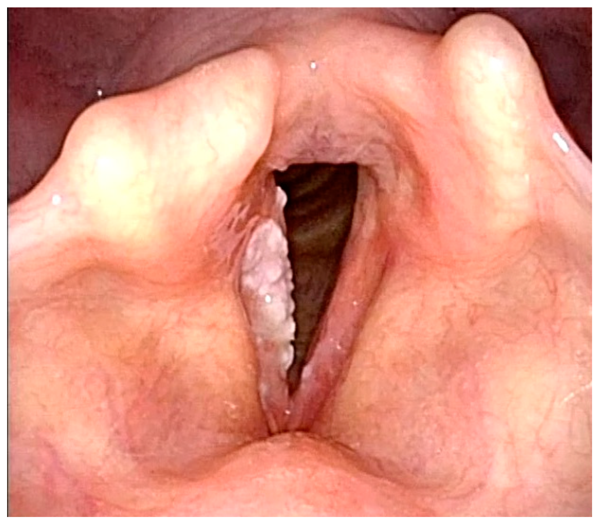
Zhang et al. tried to simplify classifications mentioned before by stratifying vocal cord leukoplakia into three subtypes: type I—flat and smooth; type II—bulged and smooth; and type III—bulged and rough [17]. According to the results, type I is mostly histologically interpreted as keratinization or hyperplasia without dysplastic changes (Figure 6) [17]. In type II, the dominant histology was mild to moderate dysplasia [17]. Type III presented the highest incidence of cancerous lesion (carcinoma in situ or invasive carcinoma), while incidence of non-cancerous lesions (keratosis or hyperplasia) was the lowest from all types (Figure 7) [17]. The authors further proposed conservative treatment in type I leukoplakia and surgical resection in type III leukoplakia [17]. Type II remains a grey zone, but the authors stated that leukoplakia in this stage is irreversible and may contain moderate or severe dysplasia [17]. The overview of this classification can be seen in Table 6.
A similar classification system was also proposed by Chen et al. [22]. This classification also used a three-tier classification system with similar categories: flat and smooth, elevated and smooth, and rough leukoplakia [22]. This study included 375 patients treated for vocal cord leukoplakia and confirmed that the morphology of the leukoplakia correlates significantly with the final histology examination [22].
  • Narrow-Band Imaging endoscopic classification of laryngeal leukoplakia according to Ni et al. (2019) [23]
Attempts to introduce advanced endoscopic methods used the modified Ni et al. classification. This classification stratifies leukoplakia into six types. Types 1–3 indicate benign leukoplakia (Figure 8) and types 4–6 suggest possibility of malignancy (Figure 9) [23]. The accuracy of the classification in judging the pathological nature of the leukoplakia was 90.8% [23]. The overview of this classification can be seen in Table 7.
An examination that can provide additional information about the lesion is laryngeal videostroboscopy. According to Rzepakowska et al., non-invasive leukoplakia (parakeratosis, low-grade dysplasia, etc.) tends to preserve the mucosal wave of the vocal cord [24]. On the other hand, the mucosal wave tends to diminish in the case of an invasive form of leukoplakia (high-grade dysplasia, invasive carcinoma, etc.) [24]. As stated by El-Demerdash, the overall accuracy of laryngeal videostroboscopy versus histology was 95% [25]. Those results were further confirmed by studies by other authors [25,26,27].

4. Discussion

Every classification system carries certain advantages and disadvantages. One of the major advantages of ELS classification is its simplicity. This two-stage system allows the examinator to classify mucosal vasculature findings as either perpendicular or longitudinal. Mehlum et al. found low interrater variability and suitability of the classification for inexperienced examinators [28]. A major disadvantage of this system is that it does not try to specify what the lesion is histologically according to endoscopy findings. The question is if specification of the histology of the lesion pre-operatively is required.
Ni classification provides ample information about the nature of the lesion. It also tries to state its histological character according to endoscopy findings. Unfortunately, the Ni classification has a few disadvantages. The major problem is the blurry cut-off line between malignant and benign lesions. According to the classification, Ni IV IPCLs have the appearance of small and dark brown spots, and should be interpreted as benign lesions [2]. Unfortunately, this appearance of IPCLs would be interpreted as perpendicular and therefore suspect according to ELS classification [9]. Therefore, a study determining whether Ni IV should be interpreted as a benign or suspect lesion is required. Another problem that affects multiple classification systems is the use of the old classification of laryngeal dysplasia. The terms “mild”, “moderate”, and “severe dysplasia” should no longer be used according to the new World Health Organization (WHO) revision of laryngeal dysplasia terms [29]. These terms should be replaced and reclassified to low-grade dysplasia and high-grade dysplasia according to the WHO [29].
The Puxxedu classification for ECE yields interesting data—it provides histological specifications of the examined lesion, and very high sensitivity and specificity are stated in the original work. Unfortunately, the sensitivity and specificity are calculated in a sub-optimal way in the original paper. Puxxedu stated his sensitivity and specificity rates according to healthy tissue vs. malignant tumors or inflammation changes vs malignant tumors [12]. These changes are usually very well pronounced and easy to differentiate even without ECE; therefore, the results may be biased. A study that compares sensitivity and specificity calculated according to the Puxxedu classification vs. final histology examination is therefore required. Also, the Puxxedu classification still uses the old classification of laryngeal dysplasia and should be modified to fit the new WHO recommendations.
Moreover, the situation is even more difficult if the patient has undergone radiotherapy. The vasculature is influenced by radiation and it is difficult to interpret vascular character properly. This makes differentiating between recurrence of the malignant tumors and post-radiation changes very difficult. The experience of the examiner comes into play more significantly. On the other hand, according to Zabrodsky et al., NBI is a good tool for follow-up of patients after radiotherapy for laryngeal and hypopharyngeal cancer with sensitivity of 92%, specificity of 76%, and overall accuracy of 88% [30].
Management of vocal cord leukoplakia remains a challenging topic in modern otolaryngology. Biopsy under general anesthesia and histological verification of the leukoplakia remains a common practice. Fortunately, clinicians have started to stratify the risk of malignancy of the leukoplakia using various classification systems as mentioned above to properly assess the risk of malignancy. A management algorithm combining the morphology of the leukoplakia, laryngeal videostroboscopy, and assessment of IPCLs around the lesion should be used to assess the risk of malignancy. If it remains low, then conservative treatment is suggested by some authors [19,20,22,31,32]. However, when managing the leukoplakia conservatively, clinicians should be very cautious and in case of any doubt examination under general anesthesia with histology examination of the leukoplakia should be performed.
Isenberg et al. provided a systematic review of 2188 biopsies of leukoplakia and showed that mild to moderate dysplasia was found in 33.5% of cases, and high-grade dysplasia or carcinoma in situ was found in 15.2% of cases [16]. According to Weller et al., laryngeal dysplasia carries a significant risk of malignant transformation [33]. The risk triples with increasing severity of dysplasia [33]. Therefore, clinicians should be aware of the possibility of malignant transformation of the vocal cord leukoplakia and patients should be tightly observed. Early discharge of patients with vocal cord leukoplakia should not be a common practice.
Unfortunately, we are still far from the concept of optical biopsy and pre-histology diagnosis. None of the advanced endoscopy methods can overcome histological verification of the lesion. The important point is that examinators should not assess laryngeal lesions solely according to their vasculature changes, appearance, or preservation of the mucosa wave. All available examinations should be performed to gather as much information as possible. Only complex and detailed examination allows the highest accuracy and diagnostic yield.
The future in differential diagnosis of laryngeal lesions is probably artificial intelligence (AI) and machine learning. These systems will probably be able to eliminate the problems with the subjective evaluation of the mentioned endoscopic classifications. Żurek et al. analyzed 11 studies that used AI in the early diagnosis of laryngeal lesions. Although various AI models were used, the overall accuracy was very high—from 80.6% to 99.7% [34]. The pooled sensitivity and specificity for differentiation between benign and malignant lesions were also very high: 91% and 94%, respectively [34].

5. Conclusions

ENT endoscopy remains a rapidly evolving and dynamic field of medicine, but the concept of optical biopsy and pre-histology diagnosis remains a challenging problem. The available classification systems provide very good sensitivity and specificity. However, the non-coherence of the classification systems remains an issue, and therefore a unified classification system is needed. Further research is needed to determine whether the Ni IV should be interpreted as a benign or a suspicious lesion. Also, research on the field of leukoplakia risk assessment is required. Artificial intelligence will probably be a valuable assistant in laryngeal examination in the future.

Author Contributions

Conceptualization, P.K. (Peter Kántor) and A.Š.; methodology, L.S.; validation, K.Z. and P.K. (Pavel Komínek); formal analysis, L.S.; investigation, P.K. (Peter Kántor); resources, L.S.; data curation, A.Š.; writing—original draft preparation, P.K. (Peter Kántor); writing—review and editing, L.S., K.Z. and P.K. (Pavel Komínek); supervision, P.K. (Pavel Komínek); project administration, K.Z.; funding acquisition, P.K. (Peter Kántor). All authors have read and agreed to the published version of the manuscript.

Funding

This research was funded by University of Ostrava, grant number SGS08/LF/2022.

Institutional Review Board Statement

The study was conducted in accordance with the Declaration of Helsinki, and approved by the Ethics Committee of University hospital Ostrava (protocol code 08/LF/2022, date of approval 31 March 2022) for studies involving humans.

Informed Consent Statement

Patient consent was waived due to the review nature of the study.

Data Availability Statement

Data sharing not applicable.

Conflicts of Interest

The authors declare no conflict of interest.

References

  1. Tsetsos, N.; Poutoglidis, A.; Vlachtsis, K.; Stavrakas, M.; Nikolaou, A.; Fyrmpas, G. Twenty-Year Experience with Salvage Total Laryngectomy: Lessons Learned. J. Laryngol. Otol. 2021, 135, 729–736. [Google Scholar] [CrossRef] [PubMed]
  2. Ni, X.-G.; He, S.; Xu, Z.-G.; Gao, L.; Lu, N.; Yuan, Z.; Lai, S.-Q.; Zhang, Y.-M.; Yi, J.-L.; Wang, X.-L.; et al. Endoscopic Diagnosis of Laryngeal Cancer and Precancerous Lesions by Narrow Band Imaging. J. Laryngol. Otol. 2011, 125, 288–296. [Google Scholar] [CrossRef] [PubMed]
  3. Abdullah, B.; Rasid, N.S.A.; Lazim, N.M.; Volgger, V.; Betz, C.S.; Mohammad, Z.W.; Hassan, N.F.H.N. Ni Endoscopic Classification for Storz Professional Image Enhancement System (SPIES) Endoscopy in the Detection of Upper Aerodigestive Tract (UADT) Tumours. Sci. Rep. 2020, 10, 6941. [Google Scholar] [CrossRef] [PubMed] [Green Version]
  4. Zhou, H.; Zhang, J.; Guo, L.; Nie, J.; Zhu, C.; Ma, X. The Value of Narrow Band Imaging in Diagnosis of Head and Neck Cancer: A Meta-Analysis. Sci. Rep. 2018, 8, 515. [Google Scholar] [CrossRef] [PubMed] [Green Version]
  5. Bertino, G.; Cacciola, S.; Fernandes, W.B.; Fernandes, C.M.; Occhini, A.; Tinelli, C.; Benazzo, M. Effectiveness of Narrow Band Imaging in the Detection of Premalignant and Malignant Lesions of the Larynx: Validation of a New Endoscopic Clinical Classification: Validation of a New Endoscopic Clinical Classification. Head Neck 2015, 37, 215–222. [Google Scholar] [CrossRef] [PubMed]
  6. Sun, C.; Han, X.; Li, X.; Zhang, Y.; Du, X. Diagnostic Performance of Narrow Band Imaging for Laryngeal Cancer: A Systematic Review and Meta-Analysis. Otolaryngol. Neck Surg. 2017, 156, 589–597. [Google Scholar] [CrossRef]
  7. Page, M.J.; McKenzie, J.E.; Bossuyt, P.M.; Boutron, I.; Hoffmann, T.C.; Mulrow, C.D.; Shamseer, L.; Tetzlaff, J.M.; Akl, E.A.; Brennan, S.E.; et al. The PRISMA 2020 Statement: An Updated Guideline for Reporting Systematic Reviews. BMJ 2021, 10, 89. [Google Scholar] [CrossRef]
  8. Ahmadzada, S.; Tseros, E.; Sritharan, N.; Singh, N.; Smith, M.; Palme, C.E.; Riffat, F. The Value of Narrowband Imaging Using the Ni Classification in the Diagnosis of Laryngeal Cancer. Laryngoscope Investig. Otolaryngol. 2020, 5, 665–671. [Google Scholar] [CrossRef]
  9. Arens, C.; Piazza, C.; Andrea, M.; Dikkers, F.G.; Tjon Pian Gi, R.E.A.; Voigt-Zimmermann, S.; Peretti, G. Proposal for a Descriptive Guideline of Vascular Changes in Lesions of the Vocal Folds by the Committee on Endoscopic Laryngeal Imaging of the European Laryngological Society. Eur. Arch. Otorhinolaryngol. 2016, 273, 1207–1214. [Google Scholar] [CrossRef]
  10. Šifrer, R.; Rijken, J.A.; Leemans, C.R.; Eerenstein, S.E.J.; van Weert, S.; Hendrickx, J.-J.; Bloemena, E.; Heuveling, D.A.; Rinkel, R.N.P.M. Evaluation of Vascular Features of Vocal Cords Proposed by the European Laryngological Society. Eur. Arch. Otorhinolaryngol. 2018, 275, 147–151. [Google Scholar] [CrossRef]
  11. Missale, F.; Taboni, S.; Carobbio, A.L.C.; Mazzola, F.; Berretti, G.; Iandelli, A.; Fragale, M.; Mora, F.; Paderno, A.; Del Bon, F.; et al. Validation of the European Laryngological Society Classification of Glottic Vascular Changes as Seen by Narrow Band Imaging in the Optical Biopsy Setting. Eur. Arch. Otorhinolaryngol. 2021, 278, 2397–2409. [Google Scholar] [CrossRef] [PubMed]
  12. Puxeddu, R.; Sionis, S.; Gerosa, C.; Carta, F. Enhanced Contact Endoscopy for the Detection of Neoangiogenesis in Tumors of the Larynx and Hypopharynx: ECE for Detection of Neoangiogenesis. Laryngoscope 2015, 125, 1600–1606. [Google Scholar] [CrossRef] [PubMed]
  13. Klimza, H.; Jackowska, J.; Tokarski, M.; Piersiala, K.; Wierzbicka, M. Narrow-Band Imaging (NBI) for Improving the Assessment of Vocal Fold Leukoplakia and Overcoming the Umbrella Effect. PLoS ONE 2017, 12, e0180590. [Google Scholar] [CrossRef] [Green Version]
  14. Mirza, N.; Kasper Schwartz, S.; Antin-Ozerkis, D. Laryngeal Findings in Users of Combination Corticosteroid and Bronchodilator Therapy. Laryngoscope 2004, 114, 1566–1569. [Google Scholar] [CrossRef]
  15. Kostev, K.; Jacob, L.; Kalder, M.; Sesterhenn, A.; Seidel, D. Association of Laryngeal Cancer with Vocal Cord Leukoplakia and Associated Risk Factors in 1,184 Patients Diagnosed in Otorhinolaryngology Practices in Germany. Mol. Clin. Oncol. 2018, 8, 689–693. [Google Scholar] [CrossRef] [PubMed] [Green Version]
  16. Isenberg, J.S.; Crozier, D.L.; Dailey, S.H. Institutional and Comprehensive Review of Laryngeal Leukoplakia. Ann. Otol. Rhinol. Laryngol. 2008, 117, 74–79. [Google Scholar] [CrossRef] [PubMed]
  17. Zhang, N.; Cheng, L.; Chen, M.; Chen, J.; Yang, Y.; Xie, M.; Li, C.; Chen, X.-L.; Zhou, L.; Wu, H.-T. Relationship between Laryngoscopic and Pathological Characteristics of Vocal Cords Leukoplakia. Acta Otolaryngol. 2017, 137, 1199–1203. [Google Scholar] [CrossRef] [PubMed]
  18. Ricci, G.; Molini, E.; Faralli, M.; Simoncelli, C. Retrospective Study on Precancerous Laryngeal Lesions: Long-Term Follow-Up. Acta Otorhinolaryngol. Ital. 2003, 23, 362–367. [Google Scholar]
  19. Staníková, L.; Šatanková, J.; Kučová, H.; Walderová, R.; Zeleník, K.; Komínek, P. The Role of Narrow-Band Imaging (NBI) Endoscopy in Optical Biopsy of Vocal Cord Leukoplakia. Eur. Arch. Oto-Rhino-Laryngol. 2017, 274, 355–359. [Google Scholar] [CrossRef]
  20. Young, C.-K.; Lin, W.-N.; Lee, L.-Y.; Lee, L.-A.; Hsin, L.-J.; Liao, C.-T.; Li, H.-Y.; Chen, I.-H.; Fang, T.-J. Laryngoscopic Characteristics in Vocal Leukoplakia: Inter-Rater Reliability and Correlation with Histology Grading: Characteristics in Vocal Leukoplakia. Laryngoscope 2015, 125, E62–E66. [Google Scholar] [CrossRef]
  21. Fang, T.-J.; Lin, W.-N.; Lee, L.-Y.; Young, C.-K.; Lee, L.-A.; Chang, K.-P.; Liao, C.-T.; Li, H.-Y.; Yen, T.-C. Classification of Vocal Fold Leukoplakia by Clinical Scoring: Clinical Predictors in Vocal Leukoplakia. Head Neck 2016, 38, E1998–E2003. [Google Scholar] [CrossRef] [PubMed]
  22. Chen, M.; Li, C.; Yang, Y.; Cheng, L.; Wu, H. A Morphological Classification for Vocal Fold Leukoplakia. Braz. J. Otorhinolaryngol. 2019, 85, 588–596. [Google Scholar] [CrossRef] [PubMed]
  23. Ni, X.-G.; Zhu, J.-Q.; Zhang, Q.-Q.; Zhang, B.-G.; Wang, G.-Q. Diagnosis of Vocal Cord Leukoplakia: The Role of a Novel Narrow Band Imaging Endoscopic Classification: NBI for Diagnosis of Vocal Cord Leukoplakia. Laryngoscope 2019, 129, 429–434. [Google Scholar] [CrossRef] [PubMed]
  24. Rzepakowska, A.; Sobol, M.; Sielska-Badurek, E.; Niemczyk, K.; Osuch-Wójcikiewicz, E. Morphology, Vibratory Function, and Vascular Pattern for Predicting Malignancy in Vocal Fold Leukoplakia. J. Voice 2020, 34, 812.e9–812.e15. [Google Scholar] [CrossRef] [PubMed]
  25. El-Demerdash, A.; Fawaz, S.A.; Sabri, S.M.; Sweed, A.; Rabie, H. Sensitivity and Specificity of Stroboscopy in Preoperative Differentiation of Dysplasia from Early Invasive Glottic Carcinoma. Eur. Arch. Otorhinolaryngol. 2015, 272, 1189–1193. [Google Scholar] [CrossRef]
  26. Cui, W.; Xu, W.; Yang, Q.; Hu, R. Clinicopathological Parameters Associated with Histological Background and Recurrence after Surgical Intervention of Vocal Cord Leukoplakia. Medicine 2017, 96, e7033. [Google Scholar] [CrossRef]
  27. Gugatschka, M.; Kiesler, K.; Beham, A.; Rechenmacher, J.; Friedrich, G. Hyperplastic Epithelial Lesions of the Vocal Folds: Combined Use of Exfoliative Cytology and Laryngostroboscopy in Differential Diagnosis. Eur. Arch. Otorhinolaryngol. 2008, 265, 797–801. [Google Scholar] [CrossRef]
  28. Mehlum, C.S.; Døssing, H.; Davaris, N.; Giers, A.; Grøntved, Å.M.; Kjaergaard, T.; Möller, S.; Godballe, C.; Arens, C. Interrater Variation of Vascular Classifications Used in Enhanced Laryngeal Contact Endoscopy. Eur. Arch. Otorhinolaryngol. 2020, 277, 2485–2492. [Google Scholar] [CrossRef]
  29. El-Naggar, A.K.; Chan, J.K.C.; Takata, T.; Grandis, J.R.; Slootweg, P.J. The Fourth Edition of the Head and Neck World Health Organization Blue Book: Editors’ Perspectives. Hum. Pathol. 2017, 66, 10–12. [Google Scholar] [CrossRef]
  30. Zabrodsky, M.; Lukes, P.; Lukesova, E.; Boucek, J.; Plzak, J. The Role of Narrow Band Imaging in the Detection of Recurrent Laryngeal and Hypopharyngeal Cancer after Curative Radiotherapy. BioMed Res. Int. 2014, 2014, 175398. [Google Scholar] [CrossRef] [Green Version]
  31. Chen, M.; Cheng, L.; Li, C.; Chen, J.; Shu, Y.; Wu, H. Nonsurgical Treatment for Vocal Fold Leukoplakia: An Analysis of 178 Cases. BioMed Res. Int. 2017, 2017, 6958250. [Google Scholar] [CrossRef] [PubMed]
  32. Staníková, L.; Kučová, H.; Walderová, R.; Zeleník, K.; Komínek, P. Úloha Narrow Band Imaging (NBI) v Hodnocení Leukoplakií Hrtanu. Otorinolaryngol. Foniatr. 2015, 64, 186–190. [Google Scholar]
  33. Weller, M.D.; Nankivell, P.C.; McConkey, C.; Paleri, V.; Mehanna, H.M. The Risk and Interval to Malignancy of Patients with Laryngeal Dysplasia; a Systematic Review of Case Series and Meta-Analysis: Risk and Interval to Malignancy of Patients with Laryngeal Dysplasia. Clin. Otolaryngol. 2010, 35, 364–372. [Google Scholar] [CrossRef] [PubMed]
  34. Żurek, M.; Jasak, K.; Niemczyk, K.; Rzepakowska, A. Artificial Intelligence in Laryngeal Endoscopy: Systematic Review and Meta-Analysis. J. Clin. Med. 2022, 11, 2752. [Google Scholar] [CrossRef] [PubMed]
Figure 1. PRISMA flow diagram of the selection process of the relevant articles.
Jcm 12 00010 g001
Figure 2. Histologically verified polyp of the right vocal cord, Ni type II of the mucosal vascularization, ELS classification—longitudinal type of vascularization.
Jcm 12 00010 g002
Figure 3. Histologically verified supraglottic squamous cell carcinoma. Ni type Vb of the mucosal vascularization, ELS classification—perpendicular type of vascularization.
Jcm 12 00010 g003
Figure 4. Histologically verified polyp of the left vocal cord. Puxxedu classification type I.
Jcm 12 00010 g004
Figure 5. Histologically verified supraglottic squamous cell carcinoma. Puxxedu classification type IV.
Jcm 12 00010 g005
Figure 6. Histologically-verified parakeratosis of the right vocal cord presenting as leukoplakia. Young scoring system—3 points, Fang scoring system—3 points, Zhang type I—flat and smooth.
Jcm 12 00010 g006
Figure 7. Histologically-verified squamous cell carcinoma of the right vocal cord presenting as leukoplakia. Young scoring system—5 points, Fang scoring system—5 points, Zhang type III—bulge and rough.
Jcm 12 00010 g007
Figure 8. Histologically-verified low-grade dysplasia presenting as leukoplakia of the left vocal cord. Ni classification of laryngeal leukoplakia—type I.
Jcm 12 00010 g008
Figure 9. Histologically-verified squamous cell carcinoma presenting as leukoplakia of both vocal cords. IPCLs can be seen around leukoplakia. Ni classification of laryngeal leukoplakia—type III–IV.
Jcm 12 00010 g009
Table 1. Narrow-band imaging endoscopic classification of the laryngeal lesions according to Ni et al. (2011) [2].
Endoscopic PatternMorphology of VesselsIPCLs
Type ISmall, oblique, and arborescentNot visible
Type IIEnlarged, oblique, and arborescentNot visible
Type IIIObscured or seen indistinctly by white mucosaNot visible
Type IVOblique and arborescent vessels not visibleSmall and dark brown spots
Type VaOblique and arborescent vessels not visibleDilated, solid, or hollow, with a brownish, speckled pattern, and various shapes
Type VbOblique and arborescent vessels not visibleTortuous, irregular, with a snake, earthworm, tadpole, or branch-like shapes
Type VcOblique and arborescent vessels not visibleTortuous or brownish speckles with irregular distribution
Abbreviation: IPCLs—intrapapillary capillary loops.
Table 2. Classification according to the European Laryngological Society by Arens et al. (2016) [9].
Endoscopic PatternMorphology of Vessels
Longitudinal vascular changesEctasiaDilated vessels
MeanderMeandering, tortuous vessels
VaricoseAdvanced meandering and dilated vessels
ConvoluteOrganized coil/tangle of vessels
Number of vesselsIncreased vessels number
Branches of vesselsIncreased branches of vessels
Change of directionAbrupt change of vessels direction
Perpendicular vascular changesEnlarged vessel loopsAbnormal IPCLs with wide-angled turning points
Dot-like vessel loopsAbnormal IPCLs with narrow-angled turning points
A Worm-like vesselsAbnormal vessels with spiral morphology and bizarre course
Abbreviation: IPCLs—intraepithelial capillary loops.
Table 3. Classification according to Puxxedu et al. (2016) [12].
Vascular PatternDiagnosisDescription
Type 0Normal mucosaThin-end regular subepithelial vessels connecting with a thicker and deeper arborescent vascular network running parallel to the epithelium.
Type IInflammationThe subepithelial vessels are increased in number and size with irregular and sometimes crossing directions.
Type IIHyperplasiaIntra-CLs are visible running toward the surface when the hyperplasia is at the initial stage. In this phase, CLs are generally still very thin and short, arising from the underlying inflammatory vasculature with a scattered distribution. In the case of mature hyperplasia, the deeper inflammatory vascular network is not visible, and only the elongated CLs can be easily seen. In the case of vegetating keratosis, the deeper inflammatory vascular network is often not visible, and the elongated CLs are difficult to see. A particular type of “bobby-pin” can be seen in laryngeal papillomatosis. The typical papilla encases the “bobby-pin” inside the papilloma.
Type IIIMild–moderate dysplasiaVascular changes become progressively more consistent with elongated small vessels in the typical “bobby-pin” shape, but some arborescence appears at the end of the CLs.
Type IVHigh-grade dysplasia/carcinoma in situ/invasive carcinomaThe vascularity of the chorion is more evident and CLs appear significantly dilated with various shapes and a wide range of vascular architectural changes such as corkscrews or tree-like patterns.
Abbreviation: CLs—capillary loops.
Table 4. Clinical scoring of leukoplakia according to Young et al. (2014) [20].
FactorsCategoriesScoreDefinitions of the Vocal Cord Leukoplakia
ColorHomogenous0The color is distributed evenly.
Non-homogeneous1The color is not distributed evenly.
TextureRegular0The surface is smooth and flat.
Irregular1The surface showed granular appearance.
SizeSmall0The sum of all vocal cord leukoplakia is less than half a length of one true vocal cord.
Large1The sum of all vocal cord leukoplakia exceeds half a length of one true vocal cord.
HyperemiaAbsence0The vocal cord leukoplakia is without peripheral erythema or increased vascularity.
Presence1The vocal cord leukoplakia is associated with peripheral erythema or increased vascularity.
ThicknessThin0The lesion is thin and blood vessels beneath the lesion are visible.
Thick1The lesion is thick and blood vessels beneath the lesion are invisible.
SymmetrySymmetric0Lesions are distributed at similar sites of bilateral vocal cords.
Asymmetric1Lesions are located at one or unopposed sites.
EdemaExist0Existence of vocal edema.
Absence1Absence of vocal edema.
Table 5. Clinical scoring of leukoplakia according to Fang et al. (2016) [21].
FactorsScoreDefinitions
ColorHomogenous0The color of vocal cord leukoplakia is distributed evenly.
Heterogeneous1The color of vocal cord leukoplakia is not distributed evenly.
TextureRegular0The surface of vocal cord leukoplakia is smooth and flat.
Irregular1The surface of vocal cord leukoplakia showed granular appearance.
SizeSmall0The sum of all vocal cord leukoplakia is less than half length of one true vocal cord.
Large1The sum of all vocal cord leukoplakia exceeds the half length of one true vocal cord.
HyperemiaAbsence0The vocal cord leukoplakia is without peripheral erythema or increased vascularity.
Presence1The vocal cord leukoplakia is associated with peripheral erythema or increased vascularity.
ThicknessThin0The lesion is thin and blood vessels beneath the lesion are visible.
Thick1The lesion is thick and blood vessels beneath the lesion are invisible.
SymmetrySymmetric0Lesions are distributed at similar sites of the bilateral cords.
Asymmetric1Lesions are located at one or unopposed sites.
Table 6. Laryngoscopic classification of vocal cord leukoplakia by Zhang et al. (2017) [17].
Type of LesionDescription
Type Iflat and smoothLocalized white plaque lesion having a uniform thin smooth homogeneous surface or white patch is raised slightly, but the edge of the white patch is continuous with the surrounding mucosa.
Type IIbulge and smoothWhite plaque lesion is homogeneous and significantly bulged with a constant texture throughout. It is higher than the mucosa around the plaque. The edge of the white patch is discontinuous with the surrounding mucosa.
Type IIIbulge and roughGrayish-white, nodular, verrucous, granular, non-homogeneous, and (or) exophytic lesions with irregular blunt or sharp projections. They have an irregular surface associated with erosion or ulceration that is higher than the mucosa around the plaque.
Table 7. Narrow-band imaging endoscopic classification of laryngeal leukoplakia according to Ni et al. (2019) [23].
TypeInterpretationDescription
Type IBenign leukoplakiaThere are no IPCLs but white plaque can be observed on the vocal cord with obliquely running vessels and branching vessels indistinctly present under the white plaque.
Type IIBenign leukoplakiaThere are white patches on the vocal cord but neither IPCLs nor obliquely running vessels or branching vessels can be found.
Type IIIBenign leukoplakiaIPCLs can be seen at the surface of the vocal cord mucosa where the epithelium is not covered by the leukoplakia, thus showing small brown spots with a relatively regular arrangement without clear boundaries. No obliquely running vessels or branching vessel were seen.
Type IVMalignant leukoplakiaIPCLs can be observed on the vocal cord, showing large brown spots embedded at the surface of white plaque.
Type VMalignant leukoplakiaIPCLs on the vocal cord can be seen with large brown spots that appear at the surface of the vocal cord mucosa outside the leukoplakia with obvious boundaries.
Type VIMalignant leukoplakiaIPCLs are visible at the surface of the vocal cord and are characterized by large brown spots or twisted earthworm-like vessels distributed at the surface of the leukoplakia as well as on the surface of the vocal cord epithelium outside the leukoplakia.
Abbreviation: IPCLs—intrapapillary capillary loops.
Disclaimer/Publisher’s Note: The statements, opinions and data contained in all publications are solely those of the individual author(s) and contributor(s) and not of MDPI and/or the editor(s). MDPI and/or the editor(s) disclaim responsibility for any injury to people or property resulting from any ideas, methods, instructions or products referred to in the content.