Article

Neutrophil-to-Lymphocyte Ratio Adds Valuable Information Regarding the Presence of DKA in Children with New-Onset T1DM

by
Alexandra-Cristina Scutca
1,2,
Delia-Maria Nicoară
1,*,
Monica Mărăzan
2,
Giorgiana-Flavia Brad
1,2 and
Otilia Mărginean
1,2,3
1
Department of Pediatrics, University of Medicine and Pharmacy “Victor Babes”, 300040 Timisoara, Romania
2
Department of Pediatrics I, Children’s Emergency Hospital “Louis Turcanu”, 300011 Timisoara, Romania
3
Department XI Pediatrics, Discipline I Pediatrics, Disturbances of Growth and Development in Children–BELIVE, 300011 Timisoara, Romania
*
Author to whom correspondence should be addressed.
J. Clin. Med. 2023, 12(1), 221; https://doi.org/10.3390/jcm12010221
Submission received: 26 November 2022 / Revised: 22 December 2022 / Accepted: 26 December 2022 / Published: 28 December 2022
(This article belongs to the Special Issue The Prevention, Treatment, and Complications of Diabetes Mellitus)

Abstract

:
Diabetic ketoacidosis (DKA) is an acute life-threatening complication occurring mainly at the onset of type 1 diabetes mellitus. The neutrophil-to-lymphocyte ratio (NLR), a marker for systemic inflammation, has recently generated increasing interest in many chronic diseases. The aim of this cross-sectional study was to determine the value of the neutrophil-to-lymphocyte ratio (NLR) in association with DKA severity across these cases. A total of 155 children with new-onset type 1 DM from one large center were included in the study. Total and differential leukocyte counts were measured upon admission and calculation of the NLR was performed. Patients were classified into four groups: without DKA, mild, moderate, and severe DKA at disease onset. Total WBCs, neutrophils, and monocytes increased with DKA severity (p-value < 0.005), while eosinophiles displayed an inverse relationship (p-value < 0.001). Median NLR scores increased from those without ketoacidosis (1.11) to mild (1.58), moderate (3.71), and severe (5.77) ketoacidosis groups. The statistical threshold value of the NLR in predicting DKA was 1.84, with a sensitivity of 80.2% and a specificity of 80%. Study findings indicate that a higher NLR score adds valuable information regarding the presence of DKA in children with new-onset T1DM.

Graphical Abstract

1. Introduction

Diabetic ketoacidosis (DKA) is an acute life-threatening complication occurring mainly at the onset of type 1 diabetes mellitus [1,2], with an incidence rate that spans from 13 to 80% [3,4,5]. Being a form of systemic inflammatory state [6,7], inflammatory markers such as blood leukocytes and C-reactive protein (CRP) play a key role in the pathogenesis [8,9]. Although complete blood counts (CBCs) are a part of the routine evaluation in diabetic patients, white blood cell (WBC) fractions did not receive significant attention from diabetes specialists in the past [10]. In recent years, however, there has been growing interest regarding the neutrophil-to-lymphocyte ratio (NLR) as a marker of systemic inflammation in cardiac diseases, neoplasms, and obesity, as well as in diabetes-related complications such as diabetic foot ulcers and retinopathy [11,12,13,14]. Against this background, our aim was to study the association between the NLR and DKA severity among children with new-onset T1DM.

2. Materials and Methods

2.1. Patient Recruitment

This cross-sectional study included data from one of the largest Romanian reference centers for pediatric T1DM. We reviewed 181 consecutive T1DM patients charts from the Pediatric Emergency Hospital “Louis Turcanu” in Timisoara, Romania, between 1 January 2015 to 30 June 2022, in accordance with the principles of the Declaration of Helsinki (1975, revised in 2013). Ethical approval was obtained from the ethics committee.
Inclusion criteria for cases were noted as new-onset T1DM in children aged 0 to 18 years, with or without diabetic ketoacidosis. Diagnosis of type 1 DM was established according to the American Diabetes Association (ADA) criteria of 2021. Exclusion criteria were as follows: infectious states, any other medical conditions that could alter hematological parameters, and patients with other types of diabetes.
Patients with DKA had a plasma glucose level > 11 mmol/L, a urine ketone level defined as moderate to high (+ to +++), and an arterial pH value < 7.30 at the time of admission. The ADA (American Diabetes Association) criteria for DKA severity were used: mild DKA, 7.20 ≤ pH < 7.30; moderate DKA, 7.10 ≤ pH< 7.20; and severe DKA, pH < 7.10 [15].

2.2. Biochemical Assays

Laboratory tests, including routine biochemistry tests and arterial gas analysis, were performed in the hospital laboratory. Blood samples were drawn at admission before the initial therapy, to avoid posttreatment changes in CBC parameters, and collected for differential WBC counts in tubes with EDTA and processed using a Sysmex XN-550 (Sysmex Corporation, Kobe, Japan) automatic blood counting system. Glycated Hb (HbA1c) was measured using a high-performance liquid chromatography kit supplied by Cobas E 411–Roche, Japan. Peptide C was evaluated using automated chemiluminescent assay (Cobas E 411–Roche, Tokyo, Japan). Neutrophil-to-lymphocyte ratios (NLR) were calculated.

2.3. Statistical Analysis

All data analysis was performed using the standard computer program Statistical Package for the Social Sciences (SPSS) for Windows, version 28 (SPSS Inc., Chicago, IL, USA) and GraphPad Prism9. The Shapiro–Wilk test was used to test the normality of the data distribution. Normally distributed variables were expressed as mean ± standard deviation (SD) and non-normally distributed variables were expressed as medians with interquartile ranges. Intergroup comparisons were performed by using an independent-sample t test and one-way ANOVA for normally distributed continuous data and Chi-Square tests for categorical variables. Non-normally distributed data were compared among multiple groups using the Kruskal–Wallis test. GraphPad Prism version 9 was used for univariate analysis with a post hoc procedure regarding NLR scores in DKA patients. Multiple regression analysis was performed to evaluate the association between the NLR or WBC parameters and the occurrence of DKA in T1DM patients. Receiver operating characteristic (ROC) curve analysis was plotted to compare the discrimination performance of HbA1c, C peptide, and CBC parameters in predicting DKA severity. The optimal threshold values were obtained using Youden’s index (sensitivity + specificity − 1, ranging from 0 to 1) and the maximized area under the curve (AUC). A p value (two-tailed) < 0.05 was considered statistically significant.

3. Results

3.1. Patient Characteristics Stratified by DKA Grade

Following the retrospective revision of T1DM electronic charts, there were 181 newly diagnosticated children. We excluded 26 patients due to concomitant acute infections. The study group included 155 children (76 males, 79 females), with a mean age of 9.00 ± 4.39 years (range 0–18 years).
According to the onset characteristics, fasting blood glucose, islet autoantibodies, serum C-peptide, ketone bodies, and blood gas analysis results [16], children with new-onset T1DM were divided into four groups: the non-DKA (n = 35), mild DKA (n = 25), moderate DKA (n = 33), and severe DKA (n = 62) group.
There were no significant differences in terms of age among the four groups. Regarding gender, there were more female patients in the severe DKA group. HbA1c levels were approximately equal in the four groups (mean = 11.40 ± 2.01).

3.2. Differential WBC Counts

As shown in Table 1, there was a significant difference in the total and differential WBC counts regarding the four groups, especially regarding total WBCs, neutrophils, and monocytes which increased with DKA severity (p < 0.0005). Eosinophiles displayed an inverse relationship to DKA severity (p-value < 0.001), decreasing with DKA severity. Lymphocytes were statistically lower in severe DKA patients compared to those with mild and moderate DKA.

3.3. NLR Score

A Kruskal–Wallis H test was performed to determine if there were significant differences in NLR scores between children without ketoacidosis and those with mild, moderate, or severe ketoacidosis. The distributions of NLR scores were not similar for all groups, as assessed by visual inspection of a boxplot. Median NLR scores increased from those without ketoacidosis (1.11) to mild (1.58), moderate (3.71), and severe (5.77) ketoacidosis groups (Figure 1). The distributions of NLR scores were significantly different between groups: X2(3) = 97.681, p = 0.000. Subsequently, multiple comparisons were performed through post hoc analysis using Dunn’s (1964) procedure with a Bonferroni correction. Adjusted p-values are presented. This post hoc analysis revealed statistically significant differences in median NLR scores between those with severe DKA and those with moderate (p = 0.002), mild (p = 0.000), or no DKA (p = 0.000); between mild and moderate DKA (p = 0.012), but not between those with mild DKA and no DKA (p = 1.000).

3.4. Correlation and Regression Analyses

A multiple regression analysis was performed to determine the correlation between blood pH and age, gender, HbA1c, C peptide, and NLR. The multiple regression model was statistically associated with blood pH: F(5, 130) = 41.485, p < 0.001, adj. R2 = 0.600. NLR score and age added significantly to the association, p < 0.001. Regression coefficients and standard errors are listed in Table 2.

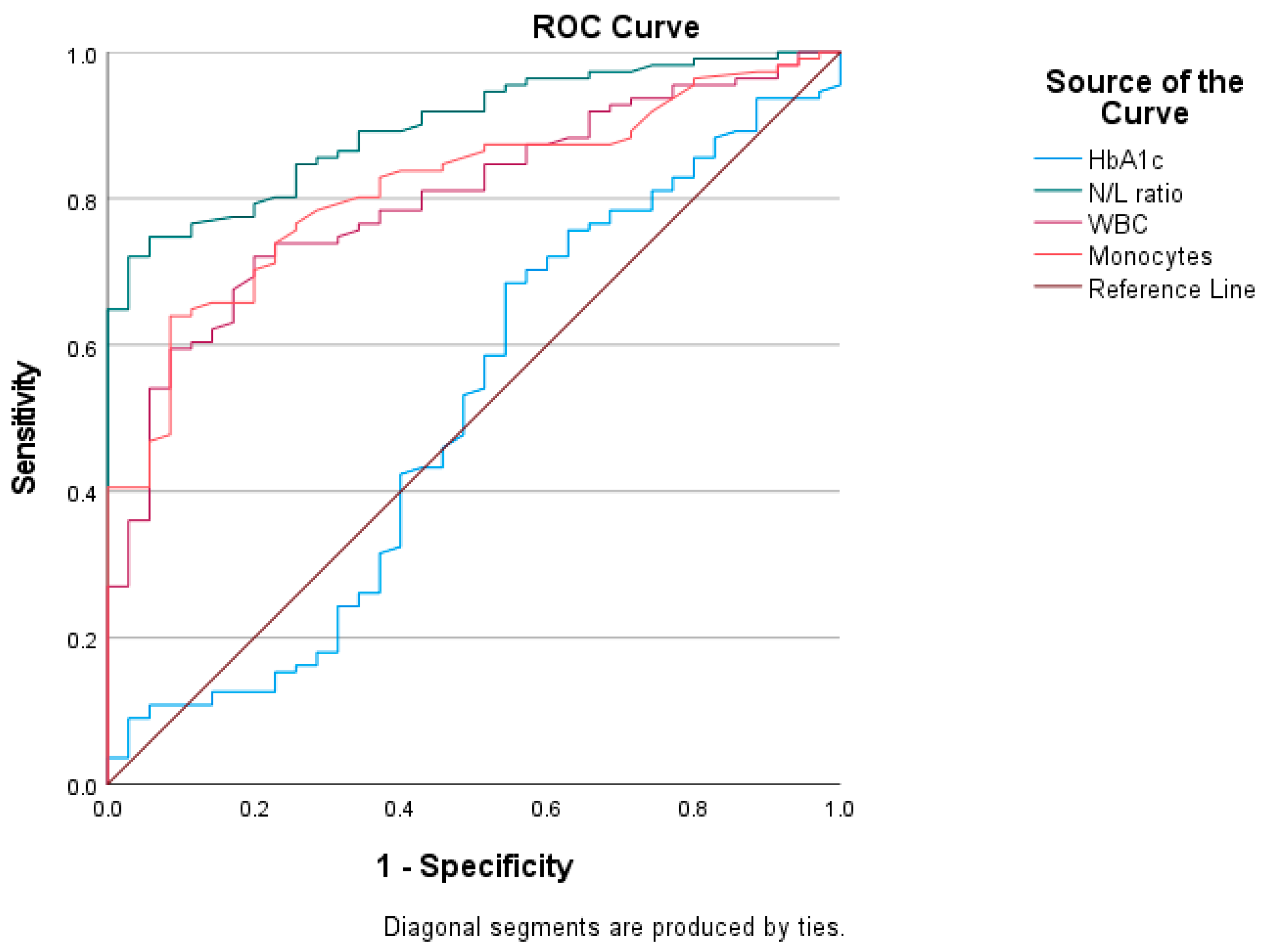
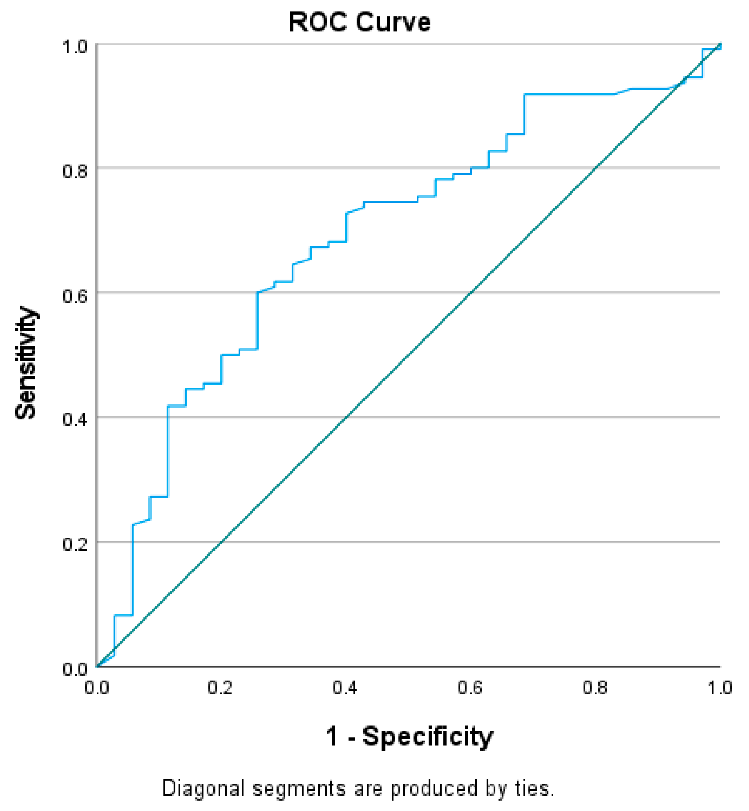
3.5. Receiver Operating Characteristics (ROC) Curve Analysis

The diagnostic ability of HbA1c, C peptide, WBCs, monocytes, and NLR in predicting DKA was analyzed by the ROC curve (Figure 2 and Figure 3). The AUCs and cut-off values were calculated according to their specificity and sensitivity as predictive factors. The most influential indicators for DKA patients were WBCs (AUC 0.800; 95% CI: 0.723–0.877, p < 0.000), monocytes (AUC 0.815; 95% CI: 0.742–0.887, p < 0.000), NLR (AUC = 0.903; 95% CI: 0.854–0.952, p < 0.000), and, to a lesser extent, C peptide (AUC = 0.690; 95% CI: 0.591–0.789, p = 0.001), as opposed to HbA1c (Table 2).
The statistical threshold value of the NLR in predicting DKA was 1.84, with a sensitivity of 80.2% and a specificity of 80% (Table 3).

4. Discussion

Type 1 diabetes mellitus (T1DM) represents one of the most frequent chronic illnesses affecting children [17]. Previous studies [16,17,18,19,20,21,22,23] have indicated an increase in both the frequency and severity of DKA cases in recent years. In our research, 81% of cases with T1DM presented with DKA, almost half of which were severe.
WBC counts, fractions, and indices, among which the NLR has received attention in recent years, were correlated with inflammation-associated diseases such as systemic hypertension [24], intracranial atherosclerosis [25], neoplasia [26], obesity [14], and type 2 diabetes [27,28,29].
The shifts in the percentage formula of white blood cells (increase in total WBCs, neutrophils, and monocytes; decrease in lymphocytes and eosinophiles) were similar to those cited in the literature [17,30].
Aside from systemic inflammation [31,32,33], the NLR, a well-characterized systemic inflammatory response marker [34], can also reflect both innate and adaptive immune (dys)function [9,35,36]. This simple ratio, which combines the predictive power of both increased neutrophil and decreased lymphocyte counts, has the advantage of being ubiquitous, cost effective, and also more stable compared with the absolute count [9,30,37]. Results from the present study are consistent with previous publications [10,30,38], in that WBC count and the NLR were found to be higher in patients with DKA.
Median NLR scores in our case were significantly different between groups, increasing from those without ketoacidosis (1.11; 0.80–1.80) to mild (1.58; 1.17–1.93), moderate (3.71; 1.98–4.85), and severe (5.77; 4.04–9.63) ketoacidosis groups. Our results regarding pediatric patients are consistent with a previous study addressing adults with DKA, which regards the NLR as a possible marker of the underlying severity of acute systemic inflammation in uninfected DKA patients [6]. Aside from the obvious effect of hemoconcentration on the NLR, the potential relationship between hyperglycemia and an increased NLR has been addressed in previous studies [39]. One possible explanation is that WBCs that are activated by advanced glycation end-products produce pro-inflammatory cytokines [29]. However, our study did not reveal statistical differences among the four groups in terms of mean HbA1c levels. This is consistent with some studies regarding children with DKA [40,41,42], and in opposition with other studies [17]. Another explanation is the fact that, in DKA, acute hyperglycemia promotes the accumulation of reactive oxygen species (ROS) which can damage peripheral blood lymphocytes’ DNA. This in turn may cause the apoptosis of lymphocytes and affect their proliferation [6,43,44].
In the present study, with new-onset T1DM children grouped according to blood pH, multivariate logistic regression analysis was performed in order to assess whether confounding exists between age, sex, HbA1c, C peptide, and NLR regarding blood pH. The NLR displayed a good discriminatory power regarding association with DKA, through correlation with blood pH.; age at onset, and, to a lesser extent, C peptide added statistically significantly to the prediction. This is consistent with a previous published study regarding adult T1DM patients [10] but, to our knowledge, was not yet reported in children. An upside to examining children is their lack of many confounding factors that can affect NLR levels, such as common medications and comorbidities present in adult patients with diabetes.
Assessing the ROC curve, the presence of DKA in our study lot was associated with an elevated NLR, monocytes, and WBCs. The area under the curve was largest for the NLR, with values above 1.84 being most frequently present in children with DKA (sensitivity of 80.2% and specificity of 80%). Regarding C peptide, plasma values were negatively correlated with the presence of DKA, mainly values below 0.690 ng/mL (sensitivity of 68.2% and specificity of 60%).
There were some limitations in the present study. Firstly, the sample size was relatively small, which could limit the power of the analyses. Secondly, our patients are only from one hospital, so that selection bias cannot be ruled out. Additionally, only one measurement of CBC and subsequent NLR calculation were used in the analysis: those upon admission. As such, there was no monitoring of the dynamic trend of the NLR. We look forward to additional multicenter studies with large samples.

5. Conclusions

This study adds complementary laboratory data regarding children with DKA at onset of T1DM [10,45]. It underlines the fact that higher NLR levels were associated with an increased prevalence of DKA in children with new-onset T1DM, and positively correlated with the DKA grade.
To the authors’ knowledge, it represents the first study to evaluate the NLR based on DKA severity in children with new-onset T1DM. This finding has clinical significance, especially in pre-hospital settings, where blood gas analysis is usually not part of routine investigations, because it may improve the early diagnosis of DKA in children with elevated glucose level and thereby facilitate proper care.

Author Contributions

Conceptualization, A.-C.S., D.-M.N. and O.M.; methodology, A.-C.S., D.-M.N. and G.-F.B.; software, D.-M.N.; validation, A.-C.S. and D.-M.N.; formal analysis, A.-C.S. and G.-F.B.; investigation, A.-C.S., D.-M.N. and M.M.; resources, A.-C.S. and M.M.; data curation A.-C.S. and D.-M.N.; writing—original draft preparation, D.-M.N. and G.-F.B.; writing—review and editing, A.-C.S. and O.M.; visualization, A.-C.S., D.-M.N., M.M., G.-F.B. and O.M; supervision, A.-C.S., D.-M.N. and O.M. All authors have read and agreed to the published version of the manuscript.

Funding

This research received no specific grant from any funding agency in the public, commercial, or not-for-profit sectors.

Institutional Review Board Statement

The study was conducted according to the guidelines of the Declaration of Helsinki and was approved by the Ethics Committee for Research of “Victor Babes” University of Medicine and Pharmacy, Timisoara (approval number 21264/17.11.2022).

Informed Consent Statement

Patient consent for further use of data was waived by the Ethics Committee for Research of “Victor Babes” University of Medicine and Pharmacy, Timisoara, due to the retrospective design of the study and the use of anonymized datasets.

Data Availability Statement

The data are not publicly available due to reasons of privacy.

Conflicts of Interest

The authors declare no conflict of interest.

References

  1. Alfayez, O.M.; Aldmasi, K.S.; Alruwais, N.H.; Bin Awad, N.M.; Al Yami, M.S.; Almohammed, O.A.; Almutairi, A.R. Incidence of Diabetic Ketoacidosis Among Pediatrics with Type 1 Diabetes Prior to and During COVID-19 Pandemic: A Meta-Analysis of Observational Studies. Front. Endocrinol. 2022, 13, 856958. [Google Scholar] [CrossRef] [PubMed]
  2. Duca, L.M.; Wang, B.; Rewers, M.; Rewers, A. Diabetic Ketoacidosis at Diagnosis of Type 1 Diabetes Predicts Poor Long-term Glycemic Control. Diabetes Care 2017, 40, 1249–1255. [Google Scholar] [CrossRef] [Green Version]
  3. Dhatariya, K.K.; Glaser, N.S.; Codner, E.; Umpierrez, G.E. Diabetic Ketoacidosis. Nat. Rev. Dis. Prim. 2020, 6, 1–20. [Google Scholar] [CrossRef] [PubMed]
  4. Umpierrez, G.E.; Kitabchi, A.E. Diabetic Ketoacidosis: Risk Factors and Management Strategies. Treat. Endocrinol. 2003, 2, 95–108. [Google Scholar] [CrossRef] [PubMed]
  5. Saydah, S.H.; Shrestha, S.S.; Zhang, P.; Zhou, X.; Imperatore, G. Medical Costs among Youth Younger than 20 Years of Age with and without Diabetic Ketoacidosis at the Time of Diabetes Diagnosis. Diabetes Care 2019, 42, 2256–2261. [Google Scholar] [CrossRef]
  6. Cheng, Y.; Yu, W.; Zhou, Y.; Zhang, T.; Chi, H.; Xu, C. Novel predictor of the occurrence of DKA in T1DM patients without infection: A combination of neutrophil/lymphocyte ratio and white blood cells. Open Life Sci. 2021, 16, 1365–1376. [Google Scholar] [CrossRef]
  7. Dalton, R.R.; Hoffman, W.H.; Passmore, G.G.; Martin, S.L. Plasma C-reactive protein levels in severe diabetic ketoacidosis. Ann. Clin. Lab. Sci. 2003, 33, 435–442. [Google Scholar]
  8. Gosmanov, A.R.; Gosmanova, E.O.; Kitabchi, A.E. Hyperglycemic Crises: Diabetic Ketoacidosis and Hyperglycemic Hyperosmolar State. [Updated 2021 May 9]. In Endotext; Feingold, K.R., Anawalt, B., Boyce, A., Eds.; MDText.com, Inc.: South Dartmouth, MA, USA, 2000; Available online: https://www.ncbi.nlm.nih.gov/books/NBK279052 (accessed on 15 October 2022).
  9. Russell, C.D.; Parajuli, A.; Gale, H.J.; Bulteel, N.S.; Schuetz, P.; de Jager, C.P.C.; Loonen, A.J.M.; Merekoulias, G.I.; Baillie, J.K. The utility of peripheral blood leucocyte ratios as biomarkers in infectious diseases: A systematic review and meta-analysis. J. Infect. 2019, 78, 339–348. [Google Scholar] [CrossRef] [Green Version]
  10. Xu, W.; Wu, H.F.; Ma, S.G.; Bai, F.; Hu, W.; Jin, Y.; Liu, H. Correlation between peripheral white blood cell counts and hyperglycemic emergencies. Int. J. Med. Sci. 2013, 10, 758–765. [Google Scholar] [CrossRef] [Green Version]
  11. Elsayed, A.M.; Araby, E. Neutrophil-Lymphocyte and Platelet-Lymphocyte ratios as a marker for diabetes control and complications. Benha Med. J. 2021, 38, 984–995. [Google Scholar] [CrossRef]
  12. Durmus, E.; Kivrak, T.; Gerin, F.; Sunbul, M.; Sari, I.; Erdogan, O. Neutrophil-to-Lymphocyte Ratio and Platelet-to-Lymphocyte Ratio are Predictors of Heart Failure. Arq. Bras. Cardiol. 2015, 105, 606–613. [Google Scholar] [CrossRef]
  13. Zhou, B.; Zhan, C.; Wu, J.; Liu, J.; Zhou, J.; Zheng, S. Prognostic Significance of Preoperative Neutrophil-to-Lymphocyte Ratio in Surgically Resectable Pancreatic Neuroendocrine Tumors. Med. Sci. Monit. 2017, 23, 5574–5588. [Google Scholar] [CrossRef] [Green Version]
  14. Rodríguez-Rodríguez, E.; López-Sobaler, A.M.; Ortega, R.M.; Delgado-Losada, M.L.; López-Parra, A.M.; Aparicio, A. Association between Neutrophil-to-Lymphocyte Ratio with Abdominal Obesity and Healthy Eating Index in a Representative Older Spanish Population. Nutrients 2020, 12, 855. [Google Scholar] [CrossRef] [Green Version]
  15. American Diabetes Association. Pharmacologic Approaches to Glycemic Treatment: Standards of Medical Care in Diabetes-2021. Diabetes Care 2021, 44 (Suppl. 1), S111–S124. [Google Scholar] [CrossRef]
  16. Ho, J.; Rosolowsky, E.; Pacaud, D.; Huang, C.; Lemay, J.A.; Brockman, N.; Rath, M.; Doulla, M. Diabetic Ketoacidosis at Type 1 Diabetes Diagnosis in Children During the COVID-19 Pandemic. Pediatr. Diabetes 2021, 22, 552–557. [Google Scholar] [CrossRef]
  17. Boboc, A.A.; Novac, C.N.; Ilie, M.T.; Ieșanu, M.I.; Galoș, F.; Bălgrădean, M.; Berghea, E.C.; Ionescu, M.D. The Impact of SARS-CoV-2 Pandemic on the New Cases of T1DM in Children. A Single-Centre Cohort Study. J. Pers. Med. 2021, 11, 551. [Google Scholar] [CrossRef]
  18. Kamrath, C.; Mönkemöller, K.; Biester, T.; Rohrer, T.R.; Warncke, K.; Hammersen, J.; Holl, R.W. Ketoacidosis in Children and Adolescents with Newly Diagnosed Type 1 Diabetes During the COVID-19 Pandemic in Germany. JAMA J. Am. Med. Assoc. 2020, 324, 801–804. [Google Scholar] [CrossRef]
  19. Dilek, S.Ö.; Gürbüz, F.; Turan, I.; Celiloǧlu, C.; Yüksel, B. Changes in the Presentation of Newly Diagnosed Type 1 Diabetes in Children During the COVID-19 Pandemic in a Tertiary Center in Southern Turkey. J. Pediatr. Endocrinol. Metab. 2021, 34, 1303–1309. [Google Scholar] [CrossRef]
  20. Dżygało, K.; Nowaczyk, J.; Szwilling, A.; Kowalska, A. Increased Frequency of Severe Diabetic Ketoacidosis at Type 1 Diabetes Onset Among Children During COVID-19 Pandemic Lockdown: An Observational Cohort Study. Pediatr. Endocrinol. Diabetes Metab. 2020, 26, 167–175. [Google Scholar] [CrossRef]
  21. Jacob, R.; Weiser, G.; Krupik, D.; Takagi, D.; Peled, S.; Pines, N.; Hashavya, S.; Gur-Soferman, H.; Gamsu, S.; Kaplan, O.; et al. Diabetic Ketoacidosis at Emergency Department Presentation During the First Months of the SARS-CoV-2 Pandemic in Israel: A Multicenter Cross-Sectional Study. Diabetes Ther. 2021, 12, 1569–1574. [Google Scholar] [CrossRef]
  22. Lawrence, C.; Seckold, R.; Smart, C.; King, B.R.; Howley, P.; Feltrin, R.; Smith, T.A.; Roy, R.; Lopez, P. Increased Paediatric Presentations of Severe Diabetic Ketoacidosis in an Australian Tertiary Centre During the COVID-19 Pandemic. Diabetes Med. 2021, 38, e14417. [Google Scholar] [CrossRef] [PubMed]
  23. McGlacken-Byrne, S.M.; Drew, S.E.V.; Turner, K.; Peters, C.; Amin, R. The SARS-CoV-2 Pandemic Is Associated with Increased Severity of Presentation of Childhood Onset Type 1 Diabetes Mellitus: A Multi-Centre Study of the First COVID-19 Wave. Diabetes Med. 2021, 38, e1464. [Google Scholar] [CrossRef] [PubMed]
  24. Jhuang, Y.H.; Kao, T.W.; Peng, T.C.; Chen, W.L.; Li, Y.W.; Chang, P.K.; Wu, L.W. Neutrophil to lymphocyte ratio as predictor for incident hypertension: A 9-year cohort study in Taiwan. Hypertens. Res. 2019, 42, 1209–1214. [Google Scholar] [CrossRef] [PubMed] [Green Version]
  25. Nam, K.W.; Kwon, H.M.; Jeong, H.Y.; Park, J.H.; Kim, S.H.; Jeong, S.M. High neutrophil to lymphocyte ratios predict intracranial atherosclerosis in a healthy population. Atherosclerosis 2018, 269, 117–121. [Google Scholar] [CrossRef] [PubMed]
  26. Proctor, M.J.; McMillan, D.C.; Morrison, D.S.; Fletcher, C.D.; Horgan, P.G.; Clarke, S.J. A derived neutrophil to lymphocyte ratio predicts survival in patients with cancer. Br. J. Cancer 2012, 107, 695–699. [Google Scholar] [CrossRef] [Green Version]
  27. Mertoglu, C.; Gunay, M. Neutrophil-Lymphocyte ratio and Platelet-Lymphocyte ratio as useful predictive markers of prediabetes and diabetes mellitus. Diabetes Metab. Syndr. 2017, 11 (Suppl. 1), S127–S131. [Google Scholar] [CrossRef]
  28. Imtiaz, F.; Shafique, K.; Mirza, S.S.; Ayoob, Z.; Vart, P.; Rao, S. Neutrophil lymphocyte ratio as a measure of systemic inflammation in prevalent chronic diseases in Asian population. Int. Arch. Med. 2012, 5, 2. [Google Scholar] [CrossRef]
  29. Wheelock, K.M.; Saulnier, P.J.; Tanamas, S.K.; Vijayakumar, P.; Weil, E.J.; Looker, H.C.; Hanson, R.L.; Lemley, K.V.; Yee, B.; Knowler, W.C.; et al. White blood cell fractions correlate with lesions of diabetic kidney disease and predict loss of kidney function in Type 2 diabetes. Nephrol. Dial. Transplant. 2018, 33, 1001–1009. [Google Scholar] [CrossRef] [Green Version]
  30. Ismail, N.A.; Abeer, M.; El Baky, N.E.D.A.; Kandil, M.E.; Rasshed, I.A.; Ahmed, A.N.; Ibrahim, M.H. Neutrophil-Lymphocyte Ratio, Platelet-Lymphocyte Ratio in Well Controlled and Uncontrolled Children and Adolescents with Type 1 Diabetes Mellitus. Res. J. Pharm. Biol. Chem. 2021, 12, 13. [Google Scholar] [CrossRef]
  31. Chen, M.; Zhu, Y.; Wang, J.; Wang, G.; Wu, Y. The Predictive Value of Neutrophil-to-Lymphocyte Ratio and Platelet-to-Lymphocyte Ratio Levels of Diabetic Peripheral Neuropathy. J. Pain Res. 2021, 14, 2049–2058. [Google Scholar] [CrossRef]
  32. Jambrik, Z.; Monti, S.; Coppola, V.; Agricola, E.; Mottola, G.; Miniati, M.; Picano, E. Usefulness of neutrophil/lymphocyte ratio as predictor of new-onset atrial fibrillation after coronary artery bypass grafting. Am. J. Cardiol. 2010, 105, 186–191. [Google Scholar] [CrossRef]
  33. Tamhane, U.U.; Aneja, S.; Montgomery, D.; Rogers, E.K.; Eagle, K.A.; Gurm, H.S. Association between admission neutrophil to lymphocyte ratio and outcomes in patients with acute coronary syndrome. Am. J. Cardiol. 2008, 102, 653–657. [Google Scholar] [CrossRef]
  34. Wang, J.R.; Chen, Z.; Yang, K.; Yang, H.J.; Tao, W.Y.; Li, Y.P.; Jiang, Z.J.; Bai, C.F.; Yin, Y.C.; Duan, J.M.; et al. Association between neutrophil-to-lymphocyte ratio, platelet-to-lymphocyte ratio, and diabetic retinopathy among diabetic patients without a related family history. Diabetol. Metab. Syndr. 2020, 12, 55. [Google Scholar] [CrossRef]
  35. Sawant, A.C.; Adhikari, P.; Narra, S.R.; Srivatsa, S.S.; Mills, P.K.; Srivatsa, S.S. Neutrophil to lymphocyte ratio predicts short- and long-term mortality following revascularization therapy for ST elevation myocardial infarction. Cardiol. J. 2014, 21, 500–508. [Google Scholar] [CrossRef] [Green Version]
  36. Rucker, A.J.; Rudemiller, N.P.; Crowley, S.D. Salt, Hypertension, and Immunity. Annu. Rev. Physiol. 2018, 80, 283–307. [Google Scholar] [CrossRef]
  37. Prats-Puig, A.; Gispert-Saüch, M.; Díaz-Roldán, F.; Carreras-Badosa, G.; Osiniri, I.; Planella-Colomer, M.; Mayol, L.; de Zegher, F.; Ibánez, L.; Bassols, J.; et al. Neutrophil-to-lymphocyte ratio: An inflammation marker related to cardiovascular risk in children. Thromb. Haemost. 2015, 114, 727–734. [Google Scholar] [CrossRef]
  38. Alamri, B.N.; Ferris, J.; Matheson, K.; De Tugwell, B. MON-638 The WBC Differential in Relation to DKA Severity. J. Endocr. Soc. 2020, 4 (Suppl. 1), 638. [Google Scholar] [CrossRef]
  39. Sefil, F.; Ulutas, K.T.; Dokuyucu, R.; Sumbul, A.T.; Yengil, E.; Yagiz, A.E.; Yula, E.; Ustun, I.; Gokce, C. Investigation of neutrophil lymphocyte ratio and blood glucose regulation in patients with type 2 diabetes mellitus. J. Int. Med. Res. 2014, 42, 581–588. [Google Scholar] [CrossRef]
  40. Lee, H.J.; Yu, H.W.; Jung, H.W.; Lee, Y.A.; Kim, J.H.; Chung, H.R.; Yoo, J.; Kim, E.; Yu, J.; Shin, C.H.; et al. Factors Associated with the Presence and Severity of Diabetic Ketoacidosis at Diagnosis of Type 1 Diabetes in Korean Children and Adolescents. J. Korean Med. Sci. 2017, 32, 303–309. [Google Scholar] [CrossRef]
  41. Khanolkar, A.R.; Amin, R.; Taylor-Robinson, D.; Viner, R.M.; Warner, J.; Gevers, E.F.; Stephenson, T. Diabetic Ketoacidosis Severity at Diagnosis and Glycaemic Control in the First Year of Childhood Onset Type 1 Diabetes-A Longitudinal Cohort Study. Int. J. Environ. Res. Public Health 2017, 15, 26. [Google Scholar] [CrossRef] [Green Version]
  42. Peng, W.; Yuan, J.; Chiavaroli, V.; Dong, G.; Huang, K.; Wu, W.; Ullah, R.; Jin, B.; Lin, H.; Derraik, J.G.B.; et al. 10-Year Incidence of Diabetic Ketoacidosis at Type 1 Diabetes Diagnosis in Children Aged Less Than 16 Years from a Large Regional Center (Hangzhou, China). Front. Endocrinol. 2021, 12, 653519. [Google Scholar] [CrossRef] [PubMed]
  43. Hu, H.; Xu, F.; Yang, W.; Ren, J.; Ge, W.; Yang, P. Apoptosis as an underlying mechanism in lymphocytes induced by riboflavin and ultraviolet light. Transfus. Apher. Sci. 2020, 59, 102899. [Google Scholar] [CrossRef] [PubMed]
  44. Mirzaei, S.; Hadadi, Z.; Attar, F.; Mousavi, S.E.; Zargar, S.S.; Tajik, A.; Saboury, A.A.; Rezayat, S.M.; Falahati, M. ROS-mediated heme degradation and cytotoxicity induced by iron nanoparticles: Hemoglobin and lymphocyte cells as targets. J. Biomol. Struct. Dyn. 2018, 36, 4235–4245. [Google Scholar] [CrossRef] [PubMed]
  45. Ma, S.G.; Jin, Y.; Xu, W.; Hu, W.; Bai, F.; Wu, X.J. Increased serum levels of ischemia-modified albumin and C-reactive protein in type 1 diabetes patients with ketoacidosis. Endocrine 2012, 42, 570–576. [Google Scholar] [CrossRef]
Figure 1. Univariate analysis with post hoc procedure regarding NLR scores in DKA patients; **** p = 0.000, *** p = 0.002.
Jcm 12 00221 g001
Figure 2. ROC curve analysis of HbA1c, WBCs, monocytes, and NLR; ROC, receiver operating characteristic. Significant differences were found (p < 0.000, respectively, for NLR, WBCs, and monocytes).
Jcm 12 00221 g002
Figure 3. ROC curve analysis of C peptide; ROC, receiver operating characteristic.
Jcm 12 00221 g003
Table 1. Demographic data and laboratory findings of all patients.
ParametersNon-DKA
(n = 35)
Mild DKA
(n = 25)
Moderate DKA
(n = 33)
Severe DKA
(n = 62)
p
Age (years)10 (5–13)9 (6.5–13)7.00 (3.50–11)9.00 (5–12)0.381
Males% (n)42 (15)76 (19)48 (16)41 (26) b0.028
HbA1c (%)11.37 ± 1.9511.68 ± 1.9411.37 ± 2.1611.52 ± 2.040.931
C-peptide (ng/mL)0.639 (0.41–0.94)0.481 (0.35–0.67)0.533 (0.29–0.77)0.330 (0.18–0.47) a, c<0.001
Blood pH7.36 (7.34–7.37)7.28 (7.23–7.29)7.17 (7.13–7.20) a6.97 (6.89–7.03) a, b, c0.000
WBCs (×103/mm3)8.53 (6.64–10.13)8.12 (6.68–8.90)12.27 (9.92–15.47) a, b18.78 (14.06–24.52) a, b, c<0.001
Neutrophils (×103/mm3)3.79 (2.99–5.24)4.58 (3.38–5.21)8.97 (6.24–12.6) a, b14.63 (11.06–18) a, b, c0.000
Lymphocytes (×103/mm3)2.92 (2.50–4.66)2.71 (1.96–3.49)2.86 (2.04–3.96)2.33 (1.59–3.2) a0.003
Thrombocytes (×103/mm3)299 (229–327)259 (223–344)347 (283–405) a342 (388–422) a, b<0.001
Monocytes (×103/mm3)0.60 (0.49–0.74)0.68 (0.51–0.77)0.87 (0.70–1.39) a1.71 (1.15–2.40) a, b, c<0.001
Eosinophiles (×103/mm3)0.12 (0.05–0.22)0.09 (0.03–0.13)0.06 (0.02–0.20)0.00 (0–0.02) a, b<0.001
NLR1.11 (0.80–1.80)1.58 (1.17–1.93)3.71 (1.98–4.85) a, b5.77 (4.04–9.63) a, b, c0.000
One-way ANOVA, Kruskal–Wallis H-test, and Chi-Square test. Data are expressed as mean ± standard deviation, median (interquartile range, IQR) or percentage (n, %). ICU, Intensive Care Unit; HbA1c, glycated hemoglobin; WBC, white blood cell count; NLR, neutrophil-to-lymphocyte ratio. Statistically significant differences, with a probability value of p < 0.05, are represented in bold. Compared with the non-DKA group, a p < 0.05. Compared with the mild DKA group, b p < 0.05. Compared with the moderate DKA group, c p < 0.05.
Table 2. Linear regression analysis of factors related to blood pH in new-onset T1DM patients.
pHB95% CI for BSE BßR2R2
LLUL
Model 0.6540.640
Age0.010 ***0.0040.0150.0030.239 ***
Gender0.003−0.0360.0420.0200.007
HbA1c0.001−0.0090.0110.0050.008
C peptide0.074 *−0.0140.1350.0300.145 *
NLR−0.038 ***−0.044−0.0320.003−0.770 ***
B = unstandardized regression coefficient; CI = confidence interval; LL = lower limit; UL = upper limit; SE B = standard error of the coefficient; ß = standardized coefficient; R2 = coefficient of determination; ∆R2 = adjusted R2. * p< 0.05, *** p < 0.001.
Table 3. ROC curve area and cut-off Values for predicting DKA. AUC = area under the curve; S.E. = standard error; CI = confidence interval.
VariableAUCS.E.95% CICut-OffSensitivity
%
Specificity
%
HbA1c0.5040.0600.386–0.62211.3849.551.4
C peptide0.6900.0500.591–0.7890.55468.260.0
WBC0.8000.0390.723–0.8778.86079.257.1
Monocytes0.8150.0370.742–0.8870.67580.262.9
NLR0.9030.0510.854–0.9521.8480.280.0
Disclaimer/Publisher’s Note: The statements, opinions and data contained in all publications are solely those of the individual author(s) and contributor(s) and not of MDPI and/or the editor(s). MDPI and/or the editor(s) disclaim responsibility for any injury to people or property resulting from any ideas, methods, instructions or products referred to in the content.

