1. Introduction
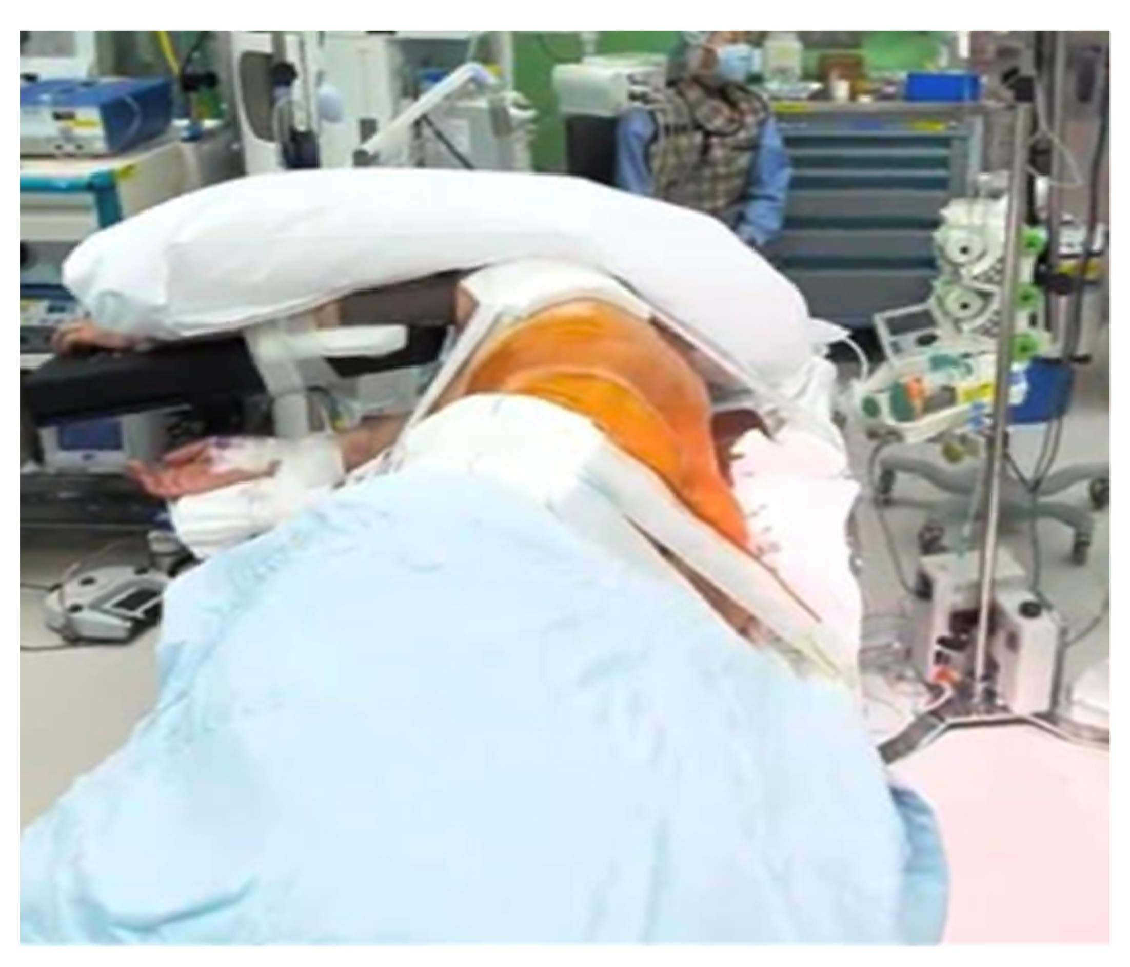
The oblique lumbar interbody fusion (OLIF) has gained popularity as a minimally invasive spinal fusion technique for the treatment of degenerative lumbar diseases including lumbar stenosis, spondylolisthesis, degenerative disc diseases, spinal instability, and spinal deformity. Compared to traditional posterior approaches, such as posterior lumbar interbody fusion (PLIF) and transforaminal lumbar interbody fusion (TLIF), OLIF provides favorable fusion by enabling a large fusion bed, and facilitates early recovery with less muscle damage, blood loss, and wound infection [
1,
2,
3,
4,
5]. Unlike PLIF and TLIF, OLIF traditionally requires repositioning the patient from supine to prone for supplemental pedicle screw fixation.
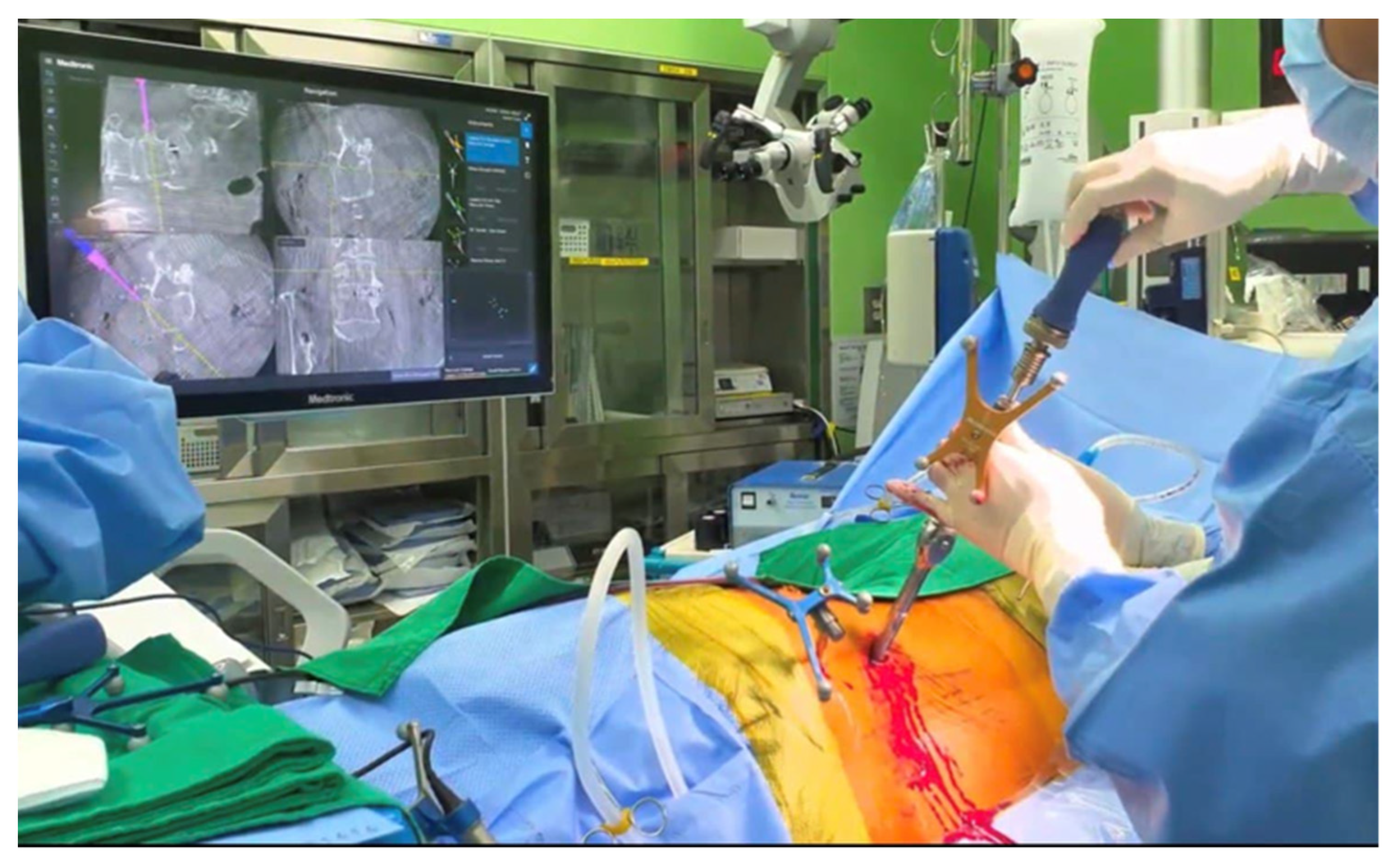
Recently, the O-arm and intraoperative navigation techniques have become increasingly important in spinal surgery [
6,
7]. These techniques increase the accuracy of pedicle screw placement and cage insertion, and reduce malposition compared with freehand and conventional fluoroscopy techniques [
8,
9,
10]. Fluoroscopy in the lateral position during pedicle screw insertion is awkward and inconvenient for surgeons. Because the O-arm system is removed from the operating field during virtual navigation, pedicle screws can be inserted from the lateral position without machine intervention. In addition, lower radiation exposure to surgeons and surgical teams is an advantage of the O-arm compared to the C-arm [
11,
12].
Previous studies on pedicle screw insertion in the lateral position without positional change have recently been reported [
13,
14,
15,
16,
17]. Therefore, we considered that performing OLIF in a single position by taking advantage of the O-arm would be very efficient in various clinical aspects and can be helpful for surgeons who are just starting out with single-segment OLIF. We aimed to evaluate the accuracy and efficiency of the single-position OLIF (SP-OLIF) using the O-arm procedure and to obtain clinical evidence supporting SP-OLIF by comparing two surgical methods: SP-OLIF using the O-arm and conventional OLIF (C-OLIF) using the C-arm.
4. Discussion
Since Amiot et al. first reported the use of a computer-assisted navigation system for pedicle screw fixation in 1995 [
20], navigation technology has been used in various spinal surgical procedures worldwide [
21,
22,
23]. O-arm navigation produces high-quality images comparable to those of conventional C-arm scans and provides the surgeon with clear intraoperative guidance. Three-dimensional (3D) real-time navigation, performed using an intraoperative O-arm system, can reveal 3D anatomic structures. The more intuitive 3D-position guidance provides a significant advantage in complex spine surgery [
24,
25].
Since Mayer first reported OLIF in 1997, it has become one of the most popular minimally invasive surgical procedures worldwide [
26]. OLIF spares the psoas and provides direct visualization of key structures, while minimizing the risk of injury to the lumbar plexus, ureter, and great vessels [
27]. However, conventional OLIF requires the repositioning of the patient from the lateral position to the prone position during surgery. Repositioning is time-consuming and has the potential to increase perioperative risk [
28]. To overcome this issue, several surgeons have attempted pedicle screw fixation in the lateral position, that is, SP-OLIF with PPSF [
29]. Favorable outcomes have been reported with clinical feasibility. Blizzard reported that the pedicle screw breach rate was 5.1% and the fusion rate at six months postoperatively was 87.5% in SP-OLIF with PPSF under C-arm fluoroscopy [
13]. Drazin et al. reported that the operation time was shorter in single-position lateral interbody fusion and PPSF than in repositioned patients (130 min vs. 190 min,
p = 0.009) [
30]. In our study, a shorter operation time and less EBL were observed in the SP-OLIF group than in the C-OLIF group, although the EBL was significantly different.
Pedicle screw placement accuracy was also high in this study, similar to the results of previous studies. Xi et al. reported that a total of 350 levels were operated upon using a navigation system, and 94.86% of cages were placed within the acceptable range [
12]. Tian and Xu reported that CT-based navigation systems had higher accuracy rates for pedicle screw placement than fluoroscopic guided screws (90.76% vs. 85.48%) [
31]. In a meta-analysis by Feng et al., O-arm navigation had significant advantages in terms of accuracy over conventional C-arm fluoroscopy [
32]. In addition, several studies have evaluated the accuracy of PPSF under navigation guidance in the lateral position [
12,
33,
34]. Ouchida et al. reported that the rate of screw misplacement in single-position OLIF using O-arm navigation is only 1.8% [
14,
33].
In contrast, Hiyama et al. raised the criticism that inserting screws while viewing fluoroscopy in a lateral position is unfamiliar to surgeons, and that a working space between the patient and fluoroscope cannot be secured [
29]. In addition, Mills et al. reported that pedicle screws placed in the lateral position had a higher rate of complications than those placed in the prone position [
16]. The oblique angle can also be disorienting for surgeons, and navigation may be a method to mitigate this disorientation and solve these problems [
35]. To offset this disorientation, we reduced the angle by 20° in the right lateral position and set it to 70°. Spatial perception can be secured using real-time position tracking, even in the awkward lateral position. Furthermore, the O-arm was retrieved after scanning; therefore, the working space was much wider. In our study, no screw placements were outside the clinically acceptable range. We believe that the use of the O-arm provides not only a precise view of the anatomy in an unfamiliar position but also high screw insertion accuracy.
In our study, which used SP-OLIF, there was one case (2.8%) of motor weakness, and three patients (8.3%) showed temporary radicular pain and numbness, but all recovered after three months. Lateral cage misplacement has been reported to range from 0.26 to 3.8% [
36]. However, our patient did not experience radicular pain due to cage misplacement. Radicular pain is thought to be caused by genitofemoral nerve irritation or nerve stretch caused by the elevation of the intervertebral space. Unlike lateral lumbar interbody fusion (LLIF), OLIF does not manipulate the psoas muscle, but orthogonal maneuvers may temporarily stretch the genitofemoral nerve and psoas muscle. However, these complications are very rare compared to LLIF [
37,
38]. In our study, there were no cases of wound revision or infection in the SP-OLIF group; however, there were also no cases of infection or revision in the C-OLIF group. The reasons for this may be as follows: less damage to the paraspinal muscles and facet joints [
39]; little stimulation of the nerve roots due to no laminectomy [
37]; and the increased accuracy of pedicle screw insertion using the navigation system.
The SP-OLIF group showed a higher fusion rate at one year after surgery than the C-OLIF group (94.4% vs. 90.0%,
p = 0.536), which was not significantly different but is consistent compared with previous studies. A meta-analysis by Tai-bang et al. revealed that postoperative fusion rates were similar between the OLIF and TLIF groups, with no statistical difference (mean difference = 1.55, 95% CI: -0.47 to 5.1,
p = 0.09) [
40]. Woods et al. reported that fusion rates of OLIF of L2–5 based on CT at six months was 95.3%, and there was successful fusion at 97.9% of surgical levels [
38]. Kotani reported a fusion rate of 96.8% (non-fusion was detected in three patients) in single-position OLIF with PPSF [
15]. Several studies report a slightly lower fusion rate of around 90%, and cage sinking and screw loosening often lead to pseudarthrosis [
41,
42,
43]. We believe that correct endplate preparation and proper cage placement can prevent these complications.
This study has some limitations, mainly due to its retrospective design, small sample size, and risk for confounding. A large multicenter randomized controlled trial is needed to obtain higher-level evidence. Our study was performed on L2–5 with single-level OLIF, excluding L5/S1; therefore, there may be differences from previous studies of multi-level OLIF, including L5/S1 or multi-level OLIF. We have considered including these factors in future studies.

 ,
, 
