Review

It Is Time for a Multidisciplinary Rehabilitation Approach: A Scoping Review on Stomatognathic Diseases in Neurological Disorders

by
Angela Militi
1,†,
Mirjam Bonanno
2,*,† and
Rocco Salvatore Calabrò
2
1
Department of Biomedical and Dental Sciences and Morphological and Functional Imaging, University of Messina, 98125 Messina, Italy
2
IRCCS Centro Neurolesi “Bonino-Pulejo”, Via Palermo, SS 113, C. Da Casazza, 98123 Messina, Italy
*
Author to whom correspondence should be addressed.
These authors contributed equally to this work.
J. Clin. Med. 2023, 12(10), 3528; https://doi.org/10.3390/jcm12103528
Submission received: 12 April 2023 / Revised: 15 May 2023 / Accepted: 16 May 2023 / Published: 17 May 2023
(This article belongs to the Special Issue Stomatognathic Diseases: State of the Art and Future Perspectives)

Abstract

:
Patients affected by neurological disorders can develop stomatognathic diseases (SD) related to decreased bite force and quality of mastication, bruxism, severe clicking and other temporomandibular disorders (TMD), which deeply affect patients’ swallowing, masticatory and phonation functions and, therefore, their quality of life. The diagnosis is commonly based on medical history and physical examination, paying attention to the temporomandibular joint (TMJ) range of movements, jaw sounds and mandibular lateral deviation. Diagnostic tools such as computed tomography and magnetic resonance imaging are used instead in case of equivocal findings in the anamnesis and physical evaluation. However, stomatognathic and temporomandibular functional training has not been commonly adopted in hospital settings as part of formal neurorehabilitation. This review is aimed at describing the most frequent pathophysiological patterns of SD and TMD in patients affected by neurological disorders and their rehabilitative approach, giving some clinical suggestions about their conservative treatment. We have searched and reviewed evidence published in PubMed, Google Scholar, Scopus and Cochrane Library between 2010 and 2023. After a thorough screening, we have selected ten studies referring to pathophysiological patterns of SD/TMD and the conservative rehabilitative approach in neurological disorders. Given this, the current literature is still poor and unclear about the administration of these kinds of complementary and rehabilitative approaches in neurological patients suffering from SD and/or TMD.

Graphical Abstract

1. Introduction

The stomatognathic system (SS) is defined as a functional complex including craniofacial structures with musculoskeletal and ligamentous components, the temporomandibular joint (TMJ), oral cavity, neck and masticatory muscles [1]. Patients affected by neurological disorders can develop stomatognathic diseases (SD) related to decreased bite force and quality of mastication, bruxism, severe clicking and other TMJ disorders (TMD), which deeply affect the patients’ quality of life [2]. In fact, the integrity of SS is fundamental in activating the neuromuscular chain that initiates the swallow reflex. On the other hand, SD can also cause myofascial pain that may irradiate in different regions, such as dental arches, ears, temples, forehead, occiput, cervical spine and shoulders [3] (Figure 1), resembling atypical headaches and facial pain.
Despite the presence of SD in neurological patients, functional training for SS and TMJ has not been commonly adopted in hospital settings as part of formal neurorehabilitation. Generally, the non-pharmacological treatment for SD and TMD aims to decrease pain, induce muscle release and stabilize muscle function and joint mobility through physical therapy (PT) and/or manual techniques (MT) [4]. Among the different PT modalities, electrophysical tools (ultrasound, LASER, TENS, interferential current) have analgesic and anti-inflammatory effects, as well as therapeutic exercise alone or in combination with MT. The latter consists of the administration of hands-on techniques to improve mobility and reduce pain in the cervical spine and its upper levels [5]. In this context, osteopathic manipulative treatment (OMT), which uses both direct and indirect MT, can be easily adapted to any type of patient [6]. The OMT can be useful in people complaining of orofacial pain as it induces muscle and fascial relaxation, promoting the release of endogenous opioids due to therapeutic touch [7,8]. Moreover, reticular formation and the brainstem are strictly involved in the coordination of the masticatory cycle, which is also controlled by a central pattern generator (CPG) which, in turn, influences gait rhythmicity too. In this way, the presence of SD and TMD is not only associated with pain or functional alterations, but it also involves the central nervous system, with a documented reduction of the grey matter volume in the somatosensory cortex and the premotor cortex [9,10]. Indeed, SS and TMJ dysfunction can be related to brain and muscle/joint alterations, so a multidisciplinary approach is required to deal with these complex problems. In current clinical practice, physiotherapists usually focus more on gait recovery and arm mobility, neglecting the role of TMJ in affecting posture and gait [11]. Patients affected by TMD could manifest a depressed head posture due to forces created by masticatory muscles. In this way, dysfunctional changes in the mandibular position, influenced by proprioceptive afferents, can have an impact on gait and balance stability through muscle connections provided by cervical muscles (i.e., sternocleidomastoid muscle, elevator scapulae), dorsal muscles (i.e., trapezius), lumbar spine (i.e., intrinsic spine muscles) and pelvic girdle [12,13,14]. Since this issue is often overlooked by professional figures of a neurorehabilitation team, our review is aimed to provide more awareness about this topic, describing the most frequent pathophysiological patterns of SD and TMD in patients affected by neurological disorders and their rehabilitative approach, and to give some clinical advice about their conservative interventions.

2. Methods

2.1. Search Strategy

The review was carried out by searching on PubMed, Google Scholar, Scopus and Cochrane Library using a combination of keywords that we have reported in Table 1.

2.2. PICO Evaluation

Search terms were defined according to the PICO model (population, intervention, comparison and outcome) [15]. The population includes patients affected by neurological disorders such as Parkinson’s disease, multiple sclerosis, spinocerebellar ataxia, oromandibular dystonia and stroke, and complaining about SD and/or TMD. Intervention included conservative and complementary therapies, such as physiotherapy, manual treatments, OMT and splint therapy. The comparison was referred to the absence of treatment and/or the administration of just one type of conservative treatment (as shown in Table 1). Lastly, the outcome included any improvements in pain perception and TMJ/SS function shown by the patients.

2.3. Inclusion and Exclusion Criteria

Then, the inclusion criteria were (i) neurological patients affected by SD/TMD; (ii) pain topic; (iii) English language; and (iv) publication in a peer-reviewed journal. We excluded articles that described theoretical models, methodological approaches, basic technical descriptions as well as animal studies and conference proceedings.

2.4. Literature Selection

We assessed the most relevant pilot studies, randomized controlled trials and case-control studies published between 2010 and 2023. Four hundred and twenty-nine articles were evaluated independently by two reviewers (MB and AM) according to title, abstract, text and scientific validity. The agreement assessment was performed using Cohen’s kappa coefficient [16]. In case of disagreement, an independent reviewer (RSC) mediated to achieve consensus. After removing duplicates (n = 264), 149 papers were initially screened, and only 32 were found eligible for a full assessment. Finally, only 10 articles fulfilled the inclusion criteria, as reported in the new PRISMA flowchart (Figure 2) [17].

2.5. Study Risk of Bias Assessment
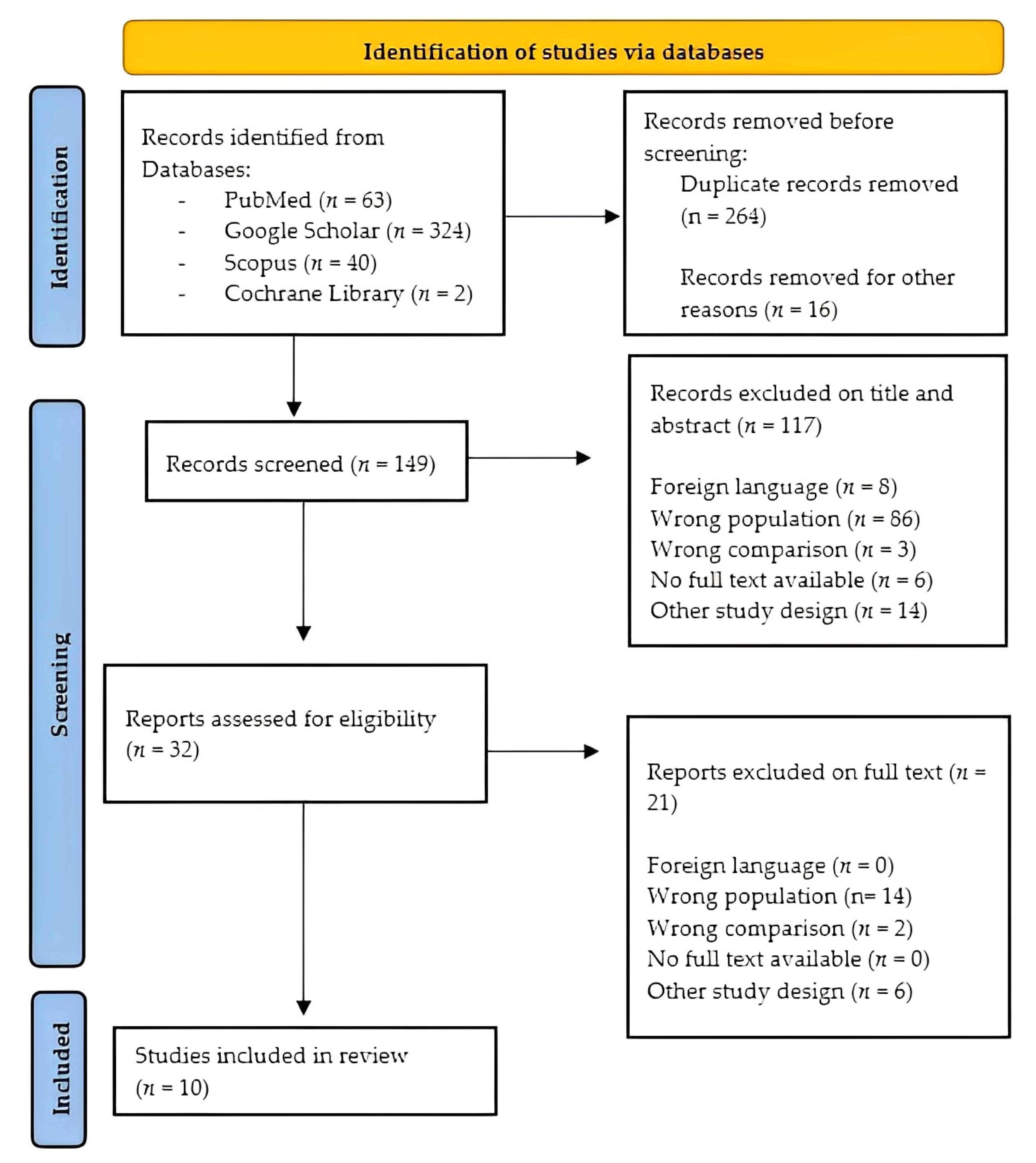
The risk of bias in controlled studies was assessed through a revised Cochrane risk of bias (RoB 2) [18], while cross-sectional and case-control studies were evaluated through Newcastle–Ottawa scale (NOS). Specifically, the risk of bias assessment was performed by two authors (A.M. and M.B.) without the use of automation tools.

3. Results

We analyzed the 10 selected pieces of evidence dealing with the presence and treatment of SD/TMD in patients affected by neurological disorders. The agreement of the judge’s decision for the inclusion of the studies was Cohen’s k = 0.82, which means a perfect agreement. In particular, we classified the studies according to the association between neurological disorders and TMD and its rehabilitation treatment, reporting a summary of results in a tabular form (see Table 2).
The risk of bias assessment of randomized controlled trials, cross-sectional studies and case-control studies was evaluated, respectively, with RoB 2 (Table 3) and NOS (Table 4).
The general quality of the included studies ranged from low [19,20,21,22,24,25,27,28] to moderate [27,29]. It should be considered that the low quality of the selected evidence is likely related to the great heterogeneity among studies for the methodologies, diagnostic tools and rehabilitation treatment administered. Additionally, we excluded one study from the risk of bias assessment [19], since it is a case report.

4. Pathophysiology of Stomatognathic and Temporomandibular Joint Disorders in Neurological Disorders

The etiology of SD and TMD is linked to a wide range of functional, psychological and environmental factors, especially in neurological disorders in which the underlying pathology is complex. People affected by multiple sclerosis (MS) are more susceptible to developing TMD disorders [30,31]. Indeed, the concomitant presence of psychological disturbances (i.e., anxiety, depression, behavioral alteration) can exacerbate TMD disorders, as confirmed by a systematic review [30]. In this vein, it has been recently reported that patients who suffer from psychological distress are less responsive to conventional treatments for TMD, requiring a longer duration of therapy [32]. MS patients can manifest three common orofacial alterations: facial palsy, trigeminal neuralgia and/or paresthesia. However, Costa, C. et al. [19] described an unusual pattern of SD in a MS patient, which included tooth hypersensitivity, hyposalivation associated with caries, halitosis and bruxism. The latter tends to increase when occlusion is impaired, also contributing to head and neck pain. In this context, some studies hypothesized that the augmented mobility of cranial bones due to reduced bone mass density, especially in the temporal ones, expands and contracts during bruxism, increasing intracranial pressure, which can favor brain damage [20]. Another mechanism that can be involved in the pathogenesis of TMD or SD in MS patients is cerebellar dysfunction. In fact, cerebellar plaques and proprioceptive changes may lead to an increased propensity to fatigue of TMJ structures in addition to a lack of coordination of mandibular movements [30,33]. In a similar way, SD is present in patients with spinocerebellar ataxia (SCA), who often present dysarthric speech and swallowing difficulties. According to Ferreira et al. [21], SCA subjects showed a decreased bite force and hypotrophy of the masseter and temporalis muscles, with an augmented electromyographic activity. The underlying hypothesis for these electromyographic changes can be related to an increase in the amplitude and duration of motor unit action potentials, in addition to reduced muscle recruitment [34]. Moreover, the lack of coordination, especially during lateral mandibular movements in SCA patients, can be explained by the pathological alteration in cerebellum pathways, affecting the synchrony and precision of movements [35]. Furthermore, patients affected by Parkinson’s disease (PD) can manifest SD and TMD due to the presence of rigidity. Body muscle rigidity could also affect masticatory muscles in association with augmented muscle tone during sleep [22]. This condition could favor the repetitive jaw muscle activity and grinding of the teeth, named bruxism, which is considered a factor for developing TMD. Bruxism can occur both during sleep (sleep bruxism) and wakefulness (awake bruxism) [36], and its pathogenesis seems to be related to central nervous structure alterations. In fact, some antidepressant drugs, such as Selective Serotonin Reuptake Inhibitors (SSRI), can cause bruxism as a side-effect of inhibiting dopaminergic neurons [23]. This could explain why bruxism is frequent in PD patients due to the reduction of dopamine presence in basal ganglia [29]. TMD and SD were also found in other movement disorders, including dystonia. The term “dystonia” refers to prolonged or intermittent muscle contractions, causing repetitive and abnormal movements and/or postures [37]. In this context, oromandibular dystonia (OMD) is often misdiagnosed due to shared clinical features with TMD [24]. In fact, OMD is associated with masticatory disturbances such as limited mouth opening, orofacial pain and TMJ dislocations that can simulate an isolated TMD, overlooking the real etiology of the disturbance or pain. Today, six subtypes of OMD are recognized: jaw closing (i), opening (ii), deviation (iii), protrusion (iv), lingual (v) and lip (vi) dystonia. In particular, the jaw-closing subtype is related to a loss of reciprocal muscle inhibition that greatly limits mouth opening, especially during speaking or eating, worsening the patient’s quality of life. In addition, dystonia tends to expand to other muscles, including orbicularis oculi, neck and shoulder muscles [38]. The pathophysiology of SD in OMD patients is still unclear, although some studies found that functional movement disorders, as well as dystonia, present a hypoactivation of the supplementary motor area and abnormal connectivity of those brain areas designed to select or inhibit movements [39]. The onset of SD and TMD in stroke patients depends on the extent and site of the vascular lesion, which can affect cortical areas, or motor-neuron pools of cranial nerves in the brain stem, causing sensorimotor deficits in SS. The presence of facial and masticatory muscle dysfunctions has been demonstrated in post-stroke patients, including weakness and hypotonus of the masseter, orbicularis oris, mylohyoid and digastric with an increase of thickness in these muscles [40]. In detail, the most common TMD in post-stroke patients seems to be related to disc displacement, which alters the structural relationship with condyle, thus producing a click sound when the mouth opens due to translation movements. This alteration could be chronic as it interferes with the simple opening of the mouth during speech or eating, and the disc becomes progressively more dislocated. Another hypothesis is that forward head posture, due to inefficacy to maintain postural alignment, causes an overload in posterior cervical muscles that can influence the TMJ by changing the position of the mandibular condyle and, consequentially, its functioning [25,41,42].

5. Diagnostic Methods and Tools for TMD

When orofacial pain occurs, patients commonly consult dentists or gnathologists, although osteopaths or physiotherapists can primarily identify a TMD during a physical examination and manual treatment [43]. In fact, the diagnosis of TMD is based on medical history and physical evaluation findings (Figure 3).
An in-depth anamnesis is the first fundamental step to guide the clinician to carry out the most relevant physical examination and diagnosis. A comprehensive history for a patient with orofacial pain includes the main complaint, medical history, dental history and psychosocial history. The patient should provide the dentist with a full description of the symptoms and the reason why the patient is seeking care. In this context, clinicians should investigate the quality of pain (i.e., burning, stabbing and blocking pain), which movements or activity exacerbates the pain and how long it has been present. In addition, clinicians should ask patients to indicate their pain point(s) with their fingers, figuring out the pain localization [44]. To more objectively rate the pain, different scales can be used in current clinical practice, such as the McGill Pain Questionnaire (MPQ), for the multidimensional detailed evaluation of orofacial pain. However, it takes a lot of time and requires good patient compliance. As an alternative, the short form of the MPQ (SF-MPQ) can be easily administered since it measures the pain intensity through the scores from the Present Pain Intensity (PPI) and the Visual Analogical Scale (VAS), also including sensory and affective scores that form the MPQ descriptors [44,45]. Otherwise, unidimensional pain scales are widely used in clinical evaluation routines and include VAS, the Verbal Rating Scale (VRS), which contains a list of adjectives marked up with a number describing five different levels of pain intensity; the Numerical Rating Scale (NRS), which ranges from 0 (no pain) to 10 (worst possible pain); and the Face Pain Scale (FPS), which uses seven emoticons to describe the pain sensation and is particularly useful in those patients with reduced compliance and/or aphasia [45]. As an adjunct, the Helkimo Clinical Dysfunction Index (HCDI) is a quick and simple test for the specific evaluation of TMD, assessing limitations in mandibular movements, joint function and also pain [46]. For research purposes, the neurophysiological methods, including laser-evoked potentials (LEPs), could be reliable assessment tools for painful syndromes, including TMD. According to de Tommaso [47], LEPs in trigeminal neuralgia and TMD present a smaller amplitude than healthy controls, suggesting trigeminal nociceptive system dysfunctions and neuropathic pain.
Furthermore, clinicians should observe the patient’s head and neck alignment with the whole body, hemifacial asymmetry, paying attention to abnormal mandibular movements, decreased joint range of motion and jaw sounds (i.e., clicking, popping, crepitus and grating), which can be related to anterior disc displacement (i.e., the click is produced during mouth opening) or to recapture the displaced disc (i.e., a second click is heard during mouth closing) [48]. In particular, the maximum mouth opening (MMO) is determined by measuring the interincisal distance, which is considered restricted when it is inferior to 35 mm. Restriction in MMO from 25 to 30 mm can be caused by intracapsular problems, such as disc displacement blocking the translation of the condyle. In this condition, the clinician describes the end-feel, which identifies the characteristics of TMJ restriction, as “hard”. Otherwise, a restricted mouth opening of 8 to 10 mm associated with a “soft” end-feel is most certainly of muscle origin. During MMO, two types of alteration can occur: deviations and deflections [47]. A deviation refers to any shift of the jaw midline that disappears during continued opening movement. Generally, it is caused by disc displacement with a reduction in one or both TMJs. A deflection consists of any shift of the midline to one side that becomes great during opening and does not return to the midline, and it reflects a restriction in one joint [49,50]. Indeed, lateral jaw movements should be about 12 mm. Moreover, dentists and gnathologists should consider both static and dynamic components of the patient’s occlusal scheme (see Table 5).
A static occlusal examination includes detecting teeth rotation, spacing, overjet and overbite (see Table 4) (including open-bites and cross-bites) that can reveal occlusal instability due to the presence of recurrently fracturing teeth and changes in tooth shape or position associated with indentations on lateral borders of the tongue and buccal mucosa related to bruxism tendency. In addition, dentists usually measure resting vertical dimension (RVD) and occlusal vertical dimension (OVD) through the Willis gauge, which is a tool that registers, in millimeters, the distance between the maxilla and mandibula. On the other hand, a dynamic occlusion examination refers to the study of teeth contact during mandibular movements, assessing the centric occlusion and intercuspal contacts marked up using articulating paper or a photographic record [51]. It should not be underestimated that physiological occlusion in adults can deviate in one or more occlusal parameters (Table 2) from the theoretically ideal one. Since this “well-adapted” occlusion is also aesthetically pleasing to the patient and has no pathological manifestations, it does not require any medical or orthodontic intervention. In fact, dentists should respect the biological variation in form and appearance of occlusion coherently with its function [52].
Actually, physical examination also includes palpation, following the direction of muscle fibers causing pain that can radiate into neighbor areas (periauricular, occiput, neck, shoulders) [53]. The palpation should address masticatory muscles (i.e., temporalis, superficial and deep masseter) and the surrounding neck and shoulder muscles that can highlight the location of pain and myogenous TMD. Notably, the temporalis muscle is divided into three portions (anterior, medial and posterior) that should be evaluated individually, as well as the digastric muscle during the opening movement, sub-occipital and sternocleidomastoid. Since lateral and medial pterygoids are not directly touchable, clinicians should therefore examine them using the resistance of hands during contractions. Interestingly, it has been hypothesized that medial pterygoid muscles could influence the opening pressure of the auditory tube, causing the “ear fullness” symptom in those patients with ear-related TMD. In detail, the dentist or therapist (both physiotherapist and osteopath) should bilaterally palpate masticatory muscles, placing one finger extra-orally and another one intra-orally, to detect hypertrophy, tenderness or pain, especially in muscle insertions. Moreover, postural evaluation should consider the upper cervical vertebral spine (C1, C2 and C3) and the cranial morphology of TMD patients since cervical dysfunctions could play a pivotal role in the development and maintenance of SD and TMD symptoms [54]. In fact, the TMJ degenerative process induces a backward-positioned jaw, which reduces pharyngeal airway capacity, altering the cervical posture in a compensatory forward head position for the decreased airway volume in the upright position [55]. Finally, clinicians should not overlook the psychological and stress status of the patients, since it can be involved in the etiology of some TMD, including bruxism [56]. When medical history and physical examination are equivocal, imaging instruments such as radiographic examinations (i.e., panoramic, planography and transcranial radiography), Computed Tomography (CT) and Magnetic Resonance Imaging (MRI) can be valid tools in the diagnostic path. TMJ radiographs are useful to collect information about morphological and anatomical characteristics between the condyle, articular tubercle and fossa. In detail, panoramic radiography is used to reveal osteophytes, fractures or other bone alterations, whereas planography is more accurate than panoramic radiography in spotting details in the styloid and mastoid process and the zygomatic arch. In the sagittal and coronal planes, the planography can also document the position of the condyle in relationship to the fossa during the MMO. Transcranial radiography provides a detailed evaluation imaging of the condyle, fossa and articular tubercle, with a large overlap in skull bones [57]. Notably, the most performed variation of CT in dentistry is the cone-beam (CBCT), which is useful to view skeletal and dental tissues involved in degenerative joint processes (osteoarthritis) using a low dose of radiation. MRI can instead confirm any disc-related TMD due to its ability to detect early abnormalities in the location and morphology of TMJ. Additionally, ultrasonography is less expensive than MRI and allows the diagnosis of an internal TMJ derangement [58,59]. However, these instruments are typically reserved for patients with persistent symptoms and for those in which conservative therapy has been ineffective.

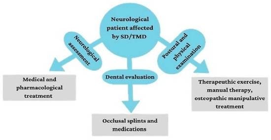
6. Cranial–Temporomandibular and Stomatognathic Rehabilitation Approach

Multidisciplinary management with a focus on conservative and complementary therapies is currently recommended for patients who present TMD or orofacial pain. However, evidence about cranial–temporomandibular and stomatognathic rehabilitation (CTS-R) in patients affected by neurological disorders is still lacking (see Table 2).
Conservative and rehabilitative management should aim to decrease orofacial pain and muscle contractures/spasms, improving TMJ function, despite the variety of TMD and SD types that clinicians could find in the population of neurological patients. Notably, Zapata-Soria et al. [60] identified some CTS-R interventions in post-stroke patients, including therapeutic jaw exercises. It seems that both jaw opening and head lift exercises can improve digastric and mylohyoid muscle thickness as well as hyoid bone movements. In fact, the digastric muscle assists the depression and retrusion of the mandible during the following breathing, swallowing and chewing activity [26]. According to Oh et al. [27], the administration of postural alignment exercises can be a promising approach to restore neck mobility and TMJ opening function, especially in post-stroke patients. It is not surprising that a postural re-education approach can be effective in TMJ pain relief because of the strictly biomechanical relationship among stomatognathic skeletal elements, the cervical spine and the shoulder girdle [26]. In this context, postural exercises, such as head posture adjustments and the correction of the mandibular position and tongue [61], can be easily adapted and administered to a variety of neurological diseases.
Indeed, specific coordination exercises, including open-close or lateral mandibular movements [62], can be more useful in SCA and MS patients with cerebellum impairments since these exercises are effective to promote balanced and synchronized muscle activity, reducing muscle pain. Moreover, muscle strengthening exercises contribute to increasing the range of motion of the mandibula by the administration of isotonic jaw opening exercises with resistance, which inhibits jaw-closing muscles (i.e., masseter and temporalis), improving TMJ opening range and pain relief [63]. In this vein, it has been [64] suggested that exercise therapy, in addition to manual treatments, postural exercises and jaw mobilization, can be the most effective conservative and complementary management for orofacial pain and TMJ mobility. In particular, TMJ can be manipulated through myofascial release (MFR), in which the operator palpates muscles and soft tissues, producing a compression in tenderness points. Balanced ligamentous tension (BLT) comprises a series of techniques that provides both compression and passive approaches to place a joint in “balance” when moved in different planes. Among the manual treatments, cranial–sacral therapy (CST) consists of hands-on gentle manipulation of the skull and sacrum, which are bidirectionally linked through dural attachments [65]. Using this light pressure, the osteopath should release myofascial restrictions, identified through palpation, and restore mobility and reduce pain for patients [66]. Generally, the five-finger bilateral grip or “Sutherland’s technique” is a common means for the evaluation and treatment of cranial dysfunctions. The patient is supine and relaxed, while the osteopath is sitting behind him/her, with his/her hands bilaterally placed on the head of the patient. In detail, the fingers are placed in the following manner: (i) the index finger on the pterion, (ii) the middle finger in front of the ear’s tragus, (iii) the ring finger on the mastoid of the temporal bone, (iv) the little finger in the inferior-posterior part of the occiput, (v) while the thumb is placed gently on the cranial vault [67] (as shown in Figure 4).
Actually, the use of the occlusal splint to treat TMD is common in dentistry. In fact, these devices promote the correction of vertical dimension, TMJ realignment and repositioning, also providing cognitive awareness [68]. The mechanism of action consists of reducing electromyographic jaw muscle activity in the short term; however, long-term outcomes are still unclear due to the adaptive mechanisms of muscles [69].
Interestingly, Umay et al. [28] performed a combined protocol in post-stroke patients using intra-oral cold stimulation, strengthening oral exercises and intermittent galvanic stimulation to the masseter muscles for thirty minutes a day. The administration of functional electric stimulation prevented muscle atrophy, promoting compensatory mechanisms and coordination.
Another conservative and complementary approach that needs to be mentioned is the psychological management of pain, which should be included in the multimodal rehabilitation approach of TMD patients [70]. In fact, neurological disorders can cause psychological sequelae (i.e., anxiety and depression symptoms), which can exacerbate the TMD’s or SD’s symptoms. Breathing and relaxing exercises in which therapists guide the diaphragmatic inspiration, through hands on the rectus abdominis, are useful to promote muscle release and psychological wellness [71,72]. This is why counseling and behavioral approaches and relaxation techniques to manage pain could be adjunctive promising treatments, personalizing rehabilitation in a more centered-care way.

7. Discussion

As far as we know, this is one of the few reviews [11,60] that deal with SD and TMD and their rehabilitative approach in patients affected by neurological disorders. In fact, this issue is often overlooked by clinicians and therapists when it would need more attention, especially for the possible improvements in the most important functions of life: speech, eating and breathing, besides the postural alignment. It is noteworthy that evidence highlighted the correlation and the relationship between the neurological disorder and the onset of SD or TMD [14,22,36,37,38], while the literature about CTS-R intervention remains poor or limited to physical exercises for the post-stroke population [25,26,27,60] (see Table 2). Indeed, we have focused not only on the pathophysiological mechanism but also on the diagnostic work-up and treatment, pointing out the importance of a multidisciplinary and personalized approach.
Understanding the loading of SS and the existence of myofascial tension, articular dysfunctions and parafunctions such as bruxism are fundamental in delivering the most tailored functional evaluation and training in these patients. In fact, in neurological patients, it is not easy to address the right diagnosis due to patient compliance and the common overlapping between neurological disorders and SD or TMD. Physical examination should be meticulously performed, investigating patterns of occlusal contacts, mandibular opening movement and muscle tenderness. In this way, clinicians can collect indispensable information for planning primary and tempestive treatment.
Additionally, Botox injection and dry needling have been suggested to manage orofacial pain in myogenous TMD, especially when other strategies have already failed [73]. In particular, dry needling is less invasive than Botox injection, and it seems to be superior in reducing pain and improving the jaw range of motion, as confirmed by Kütük et al. [74]. Other kinds of medications, including corticosteroids, benzodiazepines and antidepressants, can also be used in TMD patients [75]. However, neurological patients have already taken many of these drugs for their pathology, and when ineffective, non-pharmacological approaches such as therapeutic exercises, manual techniques and physical therapy, as well as occlusal splints, should be considered as the primary intervention in these patients. Given that most medications induce muscle relaxation to reduce spasms and contractures, some authors [76] proposed the administration of relaxation exercises of masticatory muscles to improve TMD range of motion and reduce pain. In this context, static relaxation exercises can be more effective than standard active exercises.
Today, clinicians and therapists should consider the whole person as a unique and global system in which muscular and fascial components can influence body posture, SS and its functions (chewing, speaking and swallowing), which are almost impaired in neurological patients. Some authors stated [77,78] that the SS should be considered a part of the proprioceptive system, among balance, sight and postural control of the whole body. This could explain why manual treatments (such as OMT), physical exercise and postural re-education can be more effective than other pharmacological or non-conservative treatments. In fact, these treatments have been shown to act on the proprioceptive system and then sensorimotor integration, including the brainstem, subcortical and cortical centers, cervical region, proprioception and body posture. Given that, posture and gait training should be used with CTS-R since cervical, TMJ structures and lower muscles could influence each other through the fascial system; thus, it forms a single body system [79,80].
Consequentially, an altered pattern of movements in TMJ may cause an overload in masticatory muscles (i.e., posterior temporalis, ipsilateral external pterygoid and contralateral temporal anterior pterygoid, contralateral internal pterygoid). These alterations have repercussions on the upper trapezius and contralateral sternocleidomastoid, which determine flexion and side deviation of the head, also involving the ipsilateral shoulder (levator scapulae, omohyoid) and spine muscles (such as gran dorsi and iliopsoas) that have insertions on the lumbar tract and ileum. In this way, the cranial–mandibular structures could influence lower body extremity, not only in static posture but also during gait [11,81]. However, the relationship between human posture and TMD remains one of the unsolved research questions. Future studies should consider the chance for conservative and complementary approaches to induce appropriate neuroplastic changes, integrating them with neurologic exams, monitoring of body balance and coordination control systems.
Furthermore, a multidisciplinary approach is strongly recommended, as patients can benefit from complementary therapies, including OMT, posture re-education, physical exercises and occlusal splint therapy, for SD and TMD. Finally, a co-work among gnathologists (or even dentists), physiotherapists, osteopaths and neurologists is extremely important in achieving better outcomes and avoiding unnecessary treatments.
Since a standardized protocol for the evaluation and treatment of SD/TMD in neurological disorders is still lacking, herein, we have reported some evidence-based clinical advice about the administration of multidisciplinary conservative approaches for neurological patients who have SD/TMD:
-
Parafunctional activities, such as bruxism and day clenching, have been found in PD and MS patients [11,19,22,23], causing muscle pain and harmful effects on tooth enamel. Currently, there are no treatment methods to make these alterations stop. Occlusal splint therapy can reduce bruxism and clenching symptoms, acting on a negative feedback mechanism that greatly decreases muscle activity, maintaining a normal activation threshold for the muscle protective reflex. In addition, MT and MFR could be useful to induce masseter, temporalis and neck muscle relaxation that are associated with these symptoms [65];
-
Reduction in TMJ movements due to painful muscle contractions is a common TMD feature in neurological conditions, especially in OMD, PD and SCA [21,24,29,35,37,38]. However, evidence-based treatments have been documented only for OMD subjects, suggesting the use of Botox injections in specific head and neck muscles, such as platysma, lateral pterygoid and temporalis, to reduce muscle spasms [38]. Coordination exercises of TMJ through opening and closing the mouth, using a mirror or fingers bilaterally, to promote symmetrical movements may also be of help [62,82,83]. Additionally, MT and/or OMT could induce muscle release and restore pain thanks to the discharge of endogenous opioids due to therapeutic touch [9,65];
-
Muscle weakness and reduced TMJ functions were found in post-stroke survivors as a result of the acute onset of the brain damage [25,26,27,28]. In this clinical condition, authors [26,27,60] suggested the administration of specific isometric and isotonic exercises to improve jaw muscle strength and postural programs [61,84], including exercises to increase TMJ and neck mobility. To restore oral muscle strength, some evidence supported the administration of specific exercises for labial, intrinsic tongue and masticatory muscles, which can be helpful in managing dysphagic symptoms [28].

8. Conclusions

To summarize, although SD and TMD in neurological patients do not seem to be uncommon, a standard diagnostic or rehabilitation approach is still lacking. Clinicians and therapists should consider the role of SS and TMJ structures during functional training, given their fundamental role in swallowing, chewing, breathing and speaking. To better manage SD/TMD, neurological patients should be seen as a single unit, in which the stomatognathic complex works together with the cervical spine and lower extremity in maintaining body posture and spinal alignment. Future studies are needed to fill this existing gap in the individuation and treatment of SD and/or TMD in the neurorehabilitation field.

Author Contributions

Conceptualization, A.M. and M.B.; methodology, A.M. and M.B.; software, M.B.; validation, all authors; formal analysis, M.B.; investigation, A.M. and M.B.; resources, R.S.C.; data curation, M.B.; writing—original draft preparation, A.M. and M.B.; writing—review and editing, R.S.C.; visualization, all authors; supervision, R.S.C.; project administration, A.M. and R.S.C.; funding acquisition, R.S.C. All authors have read and agreed to the published version of the manuscript.

Funding

This study was supported by Current Research Funds 2023, Ministry of Health, Italy.

Institutional Review Board Statement

Not applicable.

Informed Consent Statement

Not applicable.

Data Availability Statement

Not applicable.

Acknowledgments

The authors wish to thank Elena Barbera for her contribution to this work and Agata Grosso for English editing.

Conflicts of Interest

The authors declare no conflict of interest.

Abbreviations

SSStomatognathic system
TMJTemporomandibular joint
TMDTemporomandibular dysfunction
SDStomatognathic disease
PTPhysiotherapy
MTManual therapy
OMTOsteopathic manipulative treatment
CPGCentral pattern generator
MSMultiple sclerosis
SCASpinocerebellar ataxia
PDParkinson’s disease
OMDOromandibular dystonia
RoBRevised Cochrane risk of bias
NOSNewcastle–Ottawa Scale
MPQMcGill Pain Questionnaire
SF-MPQShort form McGill Pain Questionnaire
PPIPresent pain intensity
VASVisual analogical scale
VRSVerbal rating scale
NRSNumerical rating scale
FPSFaces pain scale
HCDIHelkimo Clinical Dysfunction Index
OVDOcclusal vertical dimension
RVDResting vertical dimension
MMOMaximum mouth opening
LEPLaser-evoked potential
CTComputed tomography
MRIMagnetic Resonance Imaging
CBCTCone-beam computed tomography
CTS-RCranial–temporomandibular and stomatognathic rehabilitation
MFRMyofascial release
CSTCranial–sacral therapy
BLTBalanced ligamentous tension

References

  1. Zieliński, G.; Filipiak, Z.; Ginszt, M.; Matysik-Woźniak, A.; Rejdak, R.; Gawda, P. The Organ of Vision and the Stomatognathic System—Review of Association Studies and Evidence-Based Discussion. Brain Sci. 2022, 12, 14. [Google Scholar] [CrossRef] [PubMed]
  2. Dai, R.; Lam, O.L.; Lo, E.C.; Li, L.S.; Wen, Y.; McGrath, C. Orofacial functional impairments among patients following stroke: A systematic review. Oral. Dis. 2015, 21, 836–849. [Google Scholar] [CrossRef] [PubMed]
  3. Wieckiewicz, M.; Boening, K.; Wiland, P.; Shiau, Y.-Y.; Paradowska-Stolarz, A. Reported concepts for the treatment modalities and pain management of temporomandibular disorders. J. Headache Pain 2015, 16, 106. [Google Scholar] [CrossRef] [PubMed]
  4. McNeely, M.; Armijo Olivo, S.; Magee, D. A systematic review of physical therapy intervention for temporomandibular dis-orders. Phys. Ther. 2006, 86, 710–720. [Google Scholar] [CrossRef] [PubMed]
  5. Bialosky, J.E.; Bishop, M.D.; Price, D.D.; Robinson, M.E.; George, S.Z. The mechanisms of manual therapy in the treatment of musculoskeletal pain: A comprehensive model. Man Ther. 2009, 14, 531–538. [Google Scholar] [CrossRef]
  6. Armijo-Olivo, S.; Pitance, L.; Singh, V.; Neto, F.; Thie, N.; Michelotti, A. Effectiveness of Manual Therapy and Therapeutic Exercise for Temporomandibular Disorders: Systematic Review and Meta-Analysis. Phys. Ther. 2016, 96, 9–25. [Google Scholar] [CrossRef]
  7. Easterbrook, S.; Keys, J.; Talsma, J.; Pierce-Talsma, S. Osteopathic Manipulative Treatment for Temporomandibular Disorders. J. Am. Osteopat. Assoc. 2019, 119, e29–e30. [Google Scholar] [CrossRef]
  8. Gesslbauer, C.; Vavti, N.; Keilani, M.; Mickel, M.; Crevenna, R. Effectiveness of osteopathic manipulative treatment versus osteopathy in the cranial field in temporomandibular disorders—A pilot study. Disabil. Rehabil. 2018, 40, 631–636. [Google Scholar] [CrossRef]
  9. McParlin, Z.; Cerritelli, F.; Friston, K.J.; Esteves, J.E. Therapeutic Alliance as Active Inference: The Role of Therapeutic Touch and Synchrony. Front. Psychol. 2022, 13, 783694. [Google Scholar] [CrossRef]
  10. Yin, Y.; He, S.; He, N.; Zhang, W.; Luo, L.; Chen, L.; Liu, T.; Tian, M.; Xu, J.; Chen, S.; et al. Brain alterations in sensorimotor and emotional regions associated with temporomandibular disorders. Oral. Dis. 2022. [Google Scholar] [CrossRef]
  11. Minervini, G.; Franco, R.; Marrapodi, M.M.; Crimi, S.; Badnjević, A.; Cervino, G.; Bianchi, A.; Cicciù, M. Correlation between Temporomandibular Disorders (TMD) and Posture Evaluated trough the Diagnostic Criteria for Temporomandibular Disorders (DC/TMD): A Systematic Review with Meta-Analysis. J. Clin. Med. 2023, 12, 2652. [Google Scholar] [CrossRef] [PubMed]
  12. An, J.-S.; Jeon, D.-M.; Jung, W.-S.; Yang, I.-H.; Lim, W.H.; Ahn, S.-J. Influence of temporomandibular joint disc displacement on craniocervical posture and hyoid bone position. Am. J. Orthod. Dentofac. Orthop. 2015, 147, 72–79. [Google Scholar] [CrossRef] [PubMed]
  13. Paço, M.; Duarte, J.; Pinho, T. Orthodontic Treatment and Craniocervical Posture in Patients with Temporomandibular Disor-ders: An Observational Study. Int. J. Environ. Res. Public. Health 2021, 18, 3295. [Google Scholar] [CrossRef] [PubMed]
  14. Garstka, A.A.; Brzózka, M.; Bitenc-Jasiejko, A.; Ardan, R.; Gronwald, H.; Skomro, P.; Lietz-Kijak, D. Cause-Effect Relationships between Painful TMD and Postural and Functional Changes in the Musculoskeletal System: A Preliminary Report. Pain Res. Manag. 2022, 2022, 1429932. [Google Scholar] [CrossRef] [PubMed]
  15. Brown, D. A Review of the PubMed PICO Tool: Using Evidence-Based Practice in Health Education. Health Promot. Pract. 2019, 21, 496–498. [Google Scholar] [CrossRef] [PubMed]
  16. Więckowska, B.; Kubiak, K.B.; Jóźwiak, P.; Moryson, W.; Stawińska-Witoszyńska, B. Cohen’s Kappa Coefficient as a Measure to Assess Classification Improvement following the Addition of a New Marker to a Regression Model. Int. J. Environ. Res. Public. Health 2022, 19, 10213. [Google Scholar] [CrossRef]
  17. Page, M.J.; McKenzie, J.E.; Bossuyt, P.M.; Boutron, I.; Hoffmann, T.C.; Mulrow, C.D.; Shamseer, L.; Tetzlaff, J.M.; Akl, E.A.; Brennan, S.E. The PRISMA 2020 statement: An updated guideline for reporting systematic reviews. BMJ 2021, 372, n71. [Google Scholar] [CrossRef] [PubMed]
  18. Sterne, J.A.C.; Savović, J.; Page, M.J.; Elbers, R.G.; Blencowe, N.S.; Boutron, I.; Cates, C.J.; Cheng, H.Y.; Corbett, M.S.; Eldridge, S.M.; et al. RoB 2: A revised tool for assessing risk of bias in randomised trials. BMJ 2019, 366, l4898. [Google Scholar] [CrossRef]
  19. Costa, C.; Santiago, H.; Pereira, S.; Castro, A.R.; Soares, S.C. Oral Health Status and Multiple Sclerosis: Classic and Non-Classic Manifestations—Case Report. Diseases 2022, 10, 62. [Google Scholar] [CrossRef]
  20. Williams, D.E.; Lynch, J.E.; Doshi, V.; Singh, G.D.; Hargens, A.R. Bruxism and Temporal Bone Hypermobility in Patients with Multiple Sclerosis. Cranio® 2011, 29, 178–186. [Google Scholar] [CrossRef]
  21. Ferreira, B.; Palinkas, M.; Gonçalves, L.; Da Silva, G.; Arnoni, V.; Regalo, I.H.; Vasconcelos, P.; Júnior, W.; Hallak, J.; Regalo, S.C.H.; et al. Spinocerebellar ataxia: Functional analysis of the stomatognathic system. Med. Oral Patol. Oral Cir. Bucal 2019, 24, e165–e171. [Google Scholar] [CrossRef]
  22. Choi, H.-G.; Yoon, J.-H.; Chung, T.-H.; Min, C.; Yoo, D.-M.; Wee, J.-H.; Kang, S.-Y.; Choi, Y.; Hong, S.-J.; Byun, S.-H. Association between Temporomandibular Joint Disorder and Parkinson’s Disease. Brain Sci. 2021, 11, 747. [Google Scholar] [CrossRef] [PubMed]
  23. Beers, E.; Van Grootheest, A.C. Bruxisme als bijwerking van serotonineheropnameremmers. Ned. Tijdschr. Tandheelkd. 2007, 114, 388–390. [Google Scholar] [PubMed]
  24. Handa, S.; Shaefer, J.R.; Keith, D.A. Oromandibular dystonia and temporomandibular disorders. J. Am. Dent. Assoc. 2022, 153, 899–906. [Google Scholar] [CrossRef] [PubMed]
  25. Ramos, M.A.; Moura, B.G.; Araujo, C.C.; Del Antonio, T.T.; Da Silva, J.K.M. Temporomandibular dysfunction in patients with a history of stroke. Man. Ther. Posturol. Rehabil. J. 2020, 17, 1–5. [Google Scholar] [CrossRef]
  26. Choi, J.B.; Jung, Y.J.; Park, J. Comparison of 2 types of therapeutic exercise: Jaw opening exercise and head lift exercise for dysphagic stroke: A pilot study. Medicine 2020, 99, e22136. [Google Scholar] [CrossRef]
  27. Oh, D.-W.; Kang, T.-W.; Kim, S.-J. Effect of Stomatognathic Alignment Exercise on Temporomandibular Joint Function and Swallowing Function of Stroke Patients with Limited Mouth Opening. J. Phys. Ther. Sci. 2013, 25, 1325–1329. [Google Scholar] [CrossRef]
  28. Umay, E.K.; Yilmaz, V.; Gundogdu, I.; Ozturk, E.; Gurcay, E.; Karaahmet, O.; Saylam, G.; Ceylan, T.; Cakci, A. What happens to swallowing muscles after stroke? A prospective randomized controlled electrophysiological study. Neurol. India 2019, 67, 1459–1466. [Google Scholar] [CrossRef]
  29. Verhoeff, M.C.; Lobbezoo, F.; Wetselaar, P.; Aarab, G.; Koutris, M. Parkinson’s disease, temporomandibular disorders and bruxism: A pilot study. J. Oral. Rehabil. 2018, 45, 854–863. [Google Scholar] [CrossRef]
  30. Minervini, G.; Mariani, P.; Fiorillo, L.; Cervino, G.; Cicciù, M.; Laino, L. Prevalence of temporomandibular disorders in people with multiple sclerosis: A systematic review and meta-analysis. Cranio® 2022, 1–9. [Google Scholar] [CrossRef]
  31. Manchery, N.; Henry, J.D.; Nangle, M.R. A systematic review of oral health in people with multiple sclerosis. Community Dent. Oral. Epidemiol. 2020, 48, 89–100. [Google Scholar] [CrossRef] [PubMed]
  32. Jung, W.; Lee, K.-E.; Suh, B.-J. Influence of psychological factors on the prognosis of temporomandibular disorders pain. J. Dent. Sci. 2021, 16, 349–355. [Google Scholar] [CrossRef] [PubMed]
  33. Carvalho, L.; Matta, A.; Nascimento, O. Temporomandibular disorders (TMD) and multiple sclerosis (MS)(P1. 120). Cranio 2015, 84, 120. [Google Scholar]
  34. Liang, L.; Chen, T.; Wu, Y. The electrophysiology of spinocerebellar ataxias. Neurophysiol. Clin. 2016, 46, 27–34. [Google Scholar] [CrossRef]
  35. Velázquez-Pérez, L.C.; Rodríguez-Labrada, R.; Fernandez-Ruiz, J. Spinocerebellar Ataxia Type 2: Clinicogenetic Aspects, Mechanistic Insights, and Management Approaches. Front. Neurol. 2017, 8, 472. [Google Scholar] [CrossRef] [PubMed]
  36. Verhoeff, M.C.; Koutris, M.; Berendse, H.W.; van Dijk, K.D.; Lobbezoo, F. Parkinson’s disease, temporomandibular disorder pain and bruxism and its clinical consequences: A protocol of a single-centre observational outpatient study. BMJ Open. 2022, 12, e052329. [Google Scholar] [CrossRef]
  37. Albanese, A.; Bhatia, K.; Bressman, S.B.; DeLong, M.R.; Fahn, S.; Fung, V.S.; Hallett, M.; Jankovic, J.; Jinnah, H.A.; Klein, C.; et al. Phenomenology and classification of dystonia: A consensus update. Mov. Disord. 2013, 28, 863–873. [Google Scholar] [CrossRef]
  38. Yoshida, K. Botulinum Toxin Therapy for Oromandibular Dystonia and Other Movement Disorders in the Stomatognathic System. Toxins 2022, 14, 282. [Google Scholar] [CrossRef]
  39. Yoshida, K. Clinical Characteristics of Functional Movement Disorders in the Stomatognathic System. Front. Neurol. 2020, 11, 123. [Google Scholar] [CrossRef]
  40. Schimmel, M.; Leemann, B.; Christou, P.; Kiliaridis, S.; Herrmann, F.R.; Müller, F. Quantitative assessment of facial muscle impairment in patients with hemispheric stroke. J. Oral. Rehabil. 2011, 38, 800–809. [Google Scholar] [CrossRef]
  41. Lau, K.T.; Cheung, K.Y.; Chan, K.B.; Chan, M.H.; Lo, K.Y.; Chiu, T.T.W. Relationships between sagittal postures of thoracic and cervical spine, presence of neck pain, neck pain severity and disability. Man. Ther. 2010, 15, 457–462. [Google Scholar] [CrossRef] [PubMed]
  42. Corrêa, E.C.; Bérzin, F. Temporomandibular disorder and dysfunctional breathing. Braz. J. Oral. Sci. 2004, 3, 498–502. [Google Scholar]
  43. Saran, S.; Saccomanno, S.; Petricca, M.T.; Carganico, A.; Bocchieri, S.; Mastrapasqua, R.F.; Caramaschi, E.; Levrini, L. Physiotherapists and Osteopaths’ Attitudes: Training in Management of Temporomandibular Disorders. Dent. J. 2022, 10, 210. [Google Scholar] [CrossRef] [PubMed]
  44. Bender, S.D. Assessment of the Orofacial Pain Patient. Dent. Clin. 2018, 62, 525–532. [Google Scholar] [CrossRef] [PubMed]
  45. Sirintawat, N.; Sawang, K.; Chaiyasamut, T.; Wongsirichat, N. Pain measurement in oral and maxillofacial surgery. J. Dent. Anesth. Pain Med. 2017, 17, 253–263. [Google Scholar] [CrossRef]
  46. Alonso-Royo, R.; Sánchez-Torrelo, C.M.; Ibáñez-Vera, A.J.; Zagalaz-Anula, N.; Castellote-Caballero, Y.; Obrero-Gaitán, E.; Rodríguez-Almagro, D.; Lomas-Vega, R. Validity and Reliability of the Helkimo Clinical Dysfunction Index for the Diagnosis of Temporomandibular Disorders. Diagnostics 2021, 11, 472. [Google Scholar] [CrossRef]
  47. De Tommaso, M. Laser-evoked potentials in primary headaches and cranial neuralgias. Expert. Rev. Neurother. 2008, 8, 1339–1345. [Google Scholar] [CrossRef]
  48. Małgorzata, P.; Małgorzata, K.-M.; Karolina, C.; Gala, A. Diagnostic of Temporomandibular Disorders and Other Facial Pain Conditions—Narrative Review and Personal Experience. Medicina 2020, 56, 472. [Google Scholar] [CrossRef]
  49. Krasińska-Mazur, M.; Homel, P.; Gala, A.; Stradomska, J.; Pihut, M. Differential diagnosis of temporomandibular disorders—A review of the literature. Folia Med. Cracov. 2022, 62, 121–137. [Google Scholar] [CrossRef]
  50. Okeson, J.P.; de Leeuw, R. Differential Diagnosis of Temporomandibular Disorders and Other Orofacial Pain Disorders. Dent. Clin. 2011, 55, 105–120. [Google Scholar] [CrossRef]
  51. Mehta, S.B.; Rizzo, D.; Paulose, B.; Botbol, A.; Vijay, S.; Arjuna, A.; Banerji, S. An evaluation of dental practitioner habits with occlusal assessment and the clinical application of practical techniques in occlusion, amongst a cohort of participants based in the UK, South Africa, Malta, and Malaysia. J. Oral. Rehabil. 2022, 49, 944–953. [Google Scholar] [CrossRef] [PubMed]
  52. de Kanter, R.J.A.M.; Battistuzzi, P.G.F.C.M.; Truin, G.-J. Temporomandibular Disorders: “Occlusion” Matters! Pain Res. Manag. 2018, 2018, 8746858. [Google Scholar] [CrossRef] [PubMed]
  53. De Rossi, S.S.; Stern, I.; Sollecito, T.P. Disorders of the Masticatory Muscles. Dent. Clin. 2013, 57, 449–464. [Google Scholar] [CrossRef] [PubMed]
  54. Kang, J.-H. Associations Among Temporomandibular Joint Osteoarthritis, Airway Dimensions, and Head and Neck Posture. J. Oral. Maxillofac. Surg. 2020, 78, 2183.e1–2183.e12. [Google Scholar] [CrossRef]
  55. Opris, H.; Baciut, M.; Bran, S.; Onisor, F.; Almasan, O.; Manea, A.; Tamas, T.; Stoia, S.; Gabriel, A.; Baciut, G.; et al. Lateral Cephalometric Analytical Uses for Temporomandibular Joint Disorders: The Importance of Cervical Posture and Hyoid Position. Int. J. Environ. Res. Public. Health 2022, 19, 11077. [Google Scholar] [CrossRef]
  56. Manfredini, D.; Lobbezoo, F. Role of psychosocial factors in the etiology of bruxism. J. Orofac. Pain 2009, 23, 153–166. [Google Scholar]
  57. Ferreira, L.A.; Grossmann, E.; Januzzi, E.; de Paula, M.V.Q.; Carvalho, A.C.P. Diagnosis of temporomandibular joint disorders: Indication of imaging exams. Braz. J. Otorhinolaryngol. 2016, 82, 341–352. [Google Scholar] [CrossRef]
  58. Gauer, R.L.; Semidey, M.J. Diagnosis and treatment of temporomandibular disorders. Am. Fam. Physician 2015, 91, 378–386. [Google Scholar]
  59. Chan, N.H.Y.; Ip, C.K.; Li, D.T.S.; Leung, Y.Y. Diagnosis and Treatment of Myogenous Temporomandibular Disorders: A Clinical Update. Diagnostics 2022, 12, 2914. [Google Scholar] [CrossRef]
  60. Zapata-Soria, M.; Cabrera-Martos, I.; López-López, L.; Ortiz-Rubio, A.; Granados-Santiago, M.; Ríos-Asín, I.; Valenza, M.C. Clinical Characteristics and Rehabilitation Strategies for the Stomatognathic System Disturbances in Patients with Stroke: A Systematic Review. Int. J. Environ. Res. Public. Health 2023, 20, 657. [Google Scholar] [CrossRef]
  61. Carini, F.; Mazzola, M.; Fici, C.; Palmeri, S.; Messina, M.; Damiani, P.; Tomasello, G. Posture and posturology, anatomical and physiological profiles: Overview and current state of art. Acta Bio Med. Atenei Parm. 2017, 88, 11–16. [Google Scholar] [CrossRef]
  62. Haketa, T.; Kino, K.; Sugisaki, M.; Takaoka, M.; Ohta, T. Randomized Clinical Trial of Treatment for TMJ Disc Displacement. J. Dent. Res. 2010, 89, 1259–1263. [Google Scholar] [CrossRef] [PubMed]
  63. Tuncer, A.B.; Ergun, N.; Tuncer, A.H.; Karahan, S. Effectiveness of manual therapy and home physical therapy in patients with temporomandibular disorders: A randomized controlled trial. J. Bodyw. Move Ther. 2013, 17, 302–308. [Google Scholar] [CrossRef] [PubMed]
  64. Shimada, A.; Ishigaki, S.; Matsuka, Y.; Komiyama, O.; Torisu, T.; Oono, Y.; Sato, H.; Naganawa, T.; Mine, A.; Yamazaki, Y.; et al. Effects of exercise therapy on painful temporomandibular disorders. J. Oral. Rehabil. 2019, 46, 475–481. [Google Scholar] [CrossRef]
  65. Roberts, A.; Harris, K.; Outen, B.; Bukvic, A.; Smith, B.; Schultz, A.; Bergman, S.; Mondal, D. Osteopathic Manipulative Medicine: A Brief Review of the Hands-On Treatment Approaches and Their Therapeutic Uses. Medicines 2022, 9, 33. [Google Scholar] [CrossRef]
  66. Brantingham, J.W.; Cassa, T.K.; Bonnefin, D.; Pribicevic, M.; Robb, A.; Pollard, H.; Tong, V.; Korporaal, C. Manipulative and Multimodal Therapy for Upper Extremity and Temporomandibular Disorders: A Systematic Review. J. Manip. Physiol. Ther. 2013, 36, 143–201. [Google Scholar] [CrossRef]
  67. Bordoni, B.; Zanier, E. Sutherland’s legacy in the new millennium: The osteopathic cranial model and modern osteopathy. Adv. Mind Body Med. 2015, 29, 15–21. [Google Scholar]
  68. Zhang, L.; Xu, L.; Wu, D.; Yu, C.; Fan, S.; Cai, B. Effectiveness of exercise therapy versus occlusal splint therapy for the treatment of painful temporomandibular disorders: A systematic review and meta-analysis. Ann. Palliat. Med. 2021, 10, 6122–6132. [Google Scholar] [CrossRef]
  69. de Paula Gomes CA, F.; El Hage, Y.; Amaral, A.P.; Politti, F.; Biasotto-Gonzalez, D.A. Effects of massage therapy and occlusal splint therapy on electromyographic activity and the intensity of signs and symptoms in individuals with temporomandibular disorder and sleep bruxism: A randomized clinical trial. Chiropr. Man. Ther. 2014, 22, 43. [Google Scholar] [CrossRef]
  70. Penlington, C.; Bowes, C.; Taylor, G.; Otemade, A.A.; Waterhouse, P.; Durham, J.; Ohrbach, R. Psychological therapies for temporomandibular disorders (TMDs). Cochrane Database Syst. Rev. 2022, 8, CD013515. [Google Scholar] [CrossRef]
  71. Urbański, P.; Trybulec, B.; Pihut, M. The Application of Manual Techniques in Masticatory Muscles Relaxation as Adjunctive Therapy in the Treatment of Temporomandibular Joint Disorders. Int. J. Environ. Res. Public. Health 2021, 18, 12970. [Google Scholar] [CrossRef] [PubMed]
  72. De Melo, L.A.; Medeiros, A.; Campos, M.D.F.T.P.; De Resende, C.M.B.M.; Barbosa, G.A.S.; De Almeida, E.O. Manual Therapy in the Treatment of Myofascial Pain Related to Temporomandibular Disorders: A Systematic Review. J. Oral. Facial Pain. Headache 2020, 34, 141–148. [Google Scholar] [CrossRef] [PubMed]
  73. Gil-Martinez, A.; Paris-Alemany, A.; López-De-Uralde-Villanueva, I.; La Touche, R. Management of pain in patients with temporomandibular disorder (TMD): Challenges and solutions. J. Pain Res. 2018, 11, 571–587. [Google Scholar] [CrossRef] [PubMed]
  74. Kütük, S.G.; Özkan, Y.; Kütük, M.; Özdaş, T. Comparison of the Efficacies of Dry Needling and Botox Methods in the Treatment of Myofascial Pain Syndrome Affecting the Temporomandibular Joint. J. Craniofacial Surg. 2019, 30, 1556–1559. [Google Scholar] [CrossRef]
  75. Ouanounou, A.; Goldberg, M.; Haas, D.A. Pharmacotherapy in Temporomandibular Disorders: A Review. J. Canadian Dent. Assoc. 2017, 83, h7. [Google Scholar]
  76. Bae, Y.; Park, Y. The Effect of Relaxation Exercises for the Masticator Muscles on Temporomandibular Joint Dysfunction (TMD). J. Phys. Ther. Sci. 2013, 25, 583–586. [Google Scholar] [CrossRef]
  77. Cuccia, A.M.; Caradonna, C.; Caradonna, D. Manual therapy of the mandibular accessory ligaments for the man-agement of temporomandibular joint disorders. J. Osteopath. Med. 2011, 111, 102–112. [Google Scholar]
  78. Yin, C.S.; Lee, Y.J. Neurological influences of the temporomandibular joint. J. Bodyw. Mov. Ther. 2007, 11, 285–294. [Google Scholar] [CrossRef]
  79. Schleip, R.; Klingler, W.; Lehmann-Horn, F. Active fascial contractility: Fascia may be able to contract in a smooth muscle-like manner and thereby influence musculoskeletal dynamics. Med. Hypotheses 2005, 65, 273–277. [Google Scholar] [CrossRef]
  80. Schleip, R.; Gabbiani, G.; Wilke, J.; Naylor, I.; Hinz, B.; Zorn, A.; Jäger, H.; Breul, R.; Schreiner, S.; Klingler, W. Fascia Is Able to Actively Contract and May Thereby Influence Musculoskeletal Dynamics: A Histochemical and Mechanographic Investigation. Front. Physiol. 2019, 10, 336. [Google Scholar] [CrossRef]
  81. Stecco, A.; Giordani, F.; Fede, C.; Pirri, C.; De Caro, R.; Stecco, C. From Muscle to the Myofascial Unit: Current Evidence and Future Perspectives. Int. J. Mol. Sci. 2023, 24, 4527. [Google Scholar] [CrossRef] [PubMed]
  82. Saito, E.T.; Akashi, P.M.H.; Sacco, I.D.C.N. Global Body Posture Evaluation in Patients with Temporomandibular Joint Disorder. Clinics 2009, 64, 35–39. [Google Scholar] [CrossRef] [PubMed]
  83. Moraes, A.D.R.; Sanches, M.L.; Ribeiro, E.C.; Guimarães, A.S. Therapeutic exercises for the control of temporomandibular disorders. Dent. Press J. Orthod. 2013, 18, 134–139. [Google Scholar] [CrossRef] [PubMed]
  84. Craane, B.; Dijkstra, P.U.; Stappaerts, K.; De Laat, A. Randomized Controlled Trial on Physical Therapy for TMJ Closed Lock. J. Dent. Res. 2012, 91, 364–369. [Google Scholar] [CrossRef] [PubMed]
Figure 1. Clinical presentation of SD and TMD in people affected by neurological disorders.
Jcm 12 03528 g001
Figure 2. PRISMA flowchart for study selection.
Jcm 12 03528 g002
Figure 3. Theoretical diagnostic and therapeutic paradigm for neurological patients affected by TMD.
Jcm 12 03528 g003
Figure 4. Illustration of how “Sutherland technique” is practically performed on a skeletal model.
Jcm 12 03528 g004
Table 1. Search strategy used for the attrition of studies.
Neurological Disorders Temporomandibular/Stomatognathic DiseasesConservative and Rehabilitative
Approaches
“Parkinson’s disease” OR “Multiple sclerosis” OR “Spino-cerebellar ataxia” OR “stroke” OR “oro-mandibular dystonia” OR “movement disorders”“Temporomandibular joint disorders” OR “temporomandibular joint dysfunctions” OR “stomatognathic disease” OR “bruxism” OR “disc displacement” OR “temporomandibular myofascial pain” OR “orofacial pain” “Physical exercise therapy” OR “manual therapy” OR “osteopathic manipulative treatment” OR “cranial-sacral therapy” OR “physical therapy” OR “occlusal splint therapy”
Table 2. Description of studies that dealt with SD/TMD in patients with neurological disorders.
Reference NumberAssociation between SD/TMD and Neurological Disorder
(Yes or No)
Stomatognathic DiseaseDiagnostic ToolsMusculoskeletal Structures InvolvedConservative and Complementary
Treatments
Major Findings
Multiple sclerosis (MS)
Costa et al. [19] Yes TMD, bruxism, tooth hypersensitivity and hyposalivation Clinical intra- and extra-oral examinationSuboccipital and cervical musclesEndodontic intervention, occlusal adjustment and behavioral education The endodontic treatment met the aesthetic pleasing of the patient
Williams et al. [20] Yes Jaw clenching/bruxism Ultrasonic pulsed phase-locked loop (PPLL) and change in acoustic pathlength (∆L) as the measure of intracranial distance Masticatory muscles, temporal bones and TMJ NAJaw clenching/bruxism was associated with the displacement of the temporal bones and expansion of the cranial cavity in MS patients compared to healthy control
Spinocerebellar ataxia (SCA)
Ferreira et al. [21]Yes Increased masticatory muscle activity and reduction of maximal molar bite force RDCTMD, electromyographic activity, muscle thickness and maximum bite forceTMJ structures,
masseter and temporalis
NA SCA is characterized by functional and electromyographic alterations in SS, especially in chewing and bite force
Parkinson’s disease (PD)
Choi et al. [22] YesJaw tremor, bruxism amd TMJ rigidity Diagnosis of TMD was considered using ICD-10 code K07.6Masticatory muscles, TMJ structures and
cervical spine
NAThe authors stated that PD patients have a high risk to develop TMD; conversely, individuals affected by TMD have more risk to develop PD in the future
Verhoeff et al. [23] Yes Bruxism (both sleep and awake bruxism), TMD and orofacial pain The authors created an 18-item questionnaire, reporting:
(i) chronic pain; (ii) the DC/TMD; (iii) oral behavior; (iv) DC/TMD symptom questionnaire; (v) TMD pain screener
Masticatory muscles, TMJ structures and cervical spine NA There are correlations between PD and bruxism, and PD with TMJ pain
Oromandibular dystonia (OMD)
Handa et al. [24] Yes Myofascial pain in masticatory muscles and dental problems Differential diagnosis between OMD and TMD by clinical examination and ICD-10Masticatory muscles, TMJ structures and dental arches NASince OMD shares clinical features with TMD, they are often misdiagnosed with the risk to receive unnecessary treatments
Stroke
Alvater Ramos et al. [25] Yes Disc displacement and myogenous TMD TMD diagnosis was performed using RDCTMD; physical mechanical pressure on trigger points was tested using the algometer Wagner PAIN TES and Pressure Pain Threshold Test, while cervical ROM was assessed using a Sanny FleximeterCervical lateral-flexors muscles, TMJ and masticatory muscles NAThe authors found that post-stroke patients manifested augmented muscle tone and reduced cervical ROM on the affected side, suggesting that the musculoskeletal alterations caused by a stroke can predispose to TMD
Choi et al. [26]Yes Dysphagia Dysphagia was confirmed by a video-fluoroscopic swallowing studySuprahyoid muscles (digastric and mylohyoid muscles) and hyoid bone movements Jaw opening exercise (isometric and isotonic) and head lift exercise JOE and HLE were useful to improve supra-hyoid muscle strength and thickness. However, JOE required less effort than HLF
Oh et al. [27] Yes Decreased TMJ function Clinical examination with craniomandibular index and limited range in opening mouth; swallowing function was assessed using MASATMJ structures, masticatory muscles, neck and shoulder muscles Stomatognathic alignment exercise program (exercises to increase the mobility of the neck and TMJ), head and neck posture exercises and anterior chest stretching exercise) Stomatognathic alignment exercises were useful to improve TMJ and swallowing functions
Umay et al. [28]YesSwallowing dysfunction,
masticatory and swallowing muscles weakness
Swallowing intervals and motor action potentials (MAPs) of trigeminal, facial and hypoglossal nerves were measuredSwallowing muscles, masticatory muscles, hyoid bone and neck structuresThermal stimulation
(to radix of tongue, palate, tonsillar plica, and oral mucosa); oral motor strength exercises for labial, intrinsic tongue and masticatory muscles; intermittent galvanic stimulation
After four weeks of treatment, significant recovery in swallowing, motor and general functional levels of the patients was provided
Legend: SD (stomatognathic disease), TMD (temporomandibular disorder), TMJ (temporomandibular joint), RDCTMD (Research Diagnostic Criteria for Temporomandibular Disorders), JOE (jaw opening exercise), HLF (head lift exercise), MASA (Mann assessment of swallowing ability).
Table 3. Risk of bias assessment of randomized trials with RoB 2.
ReferenceRandomization
Process
Effect of Assignment on Intervention Effect of Adhering to InterventionMissing Outcome DataMeasurement of the Outcome Selection of the Reported Results
Choi et al. [26]SCSCLSCSCSC
Oh et al. [27]LLLLSCSC
Umay et al. [28]LLLLLL
Legend: L (low); H (high); SC (some concerns).
Table 4. Risk of bias assessment of cross-section and case-control studies through Newcastle–Ottawa Scale (NOS).
ReferenceNOS
Williams et al. [20] 4
Ferreira et al. [21] 4
Choi et al. [22] 6
Verhoeff et al. [23] 7
Handa et al. [24] 5
Alvater Ramos [25] 3
Table 5. Description of the main occlusal physiological parameters that dentists can measure during physical examination.
Main Occlusal Physiological
Parameters
Description
Centric occlusion Consists of a full occlusal contact between upper and lower teeth in habitual occlusion.
Incisal guidance Consists of the influence of the contacting surfaces of the mandibular and maxillary anterior teeth on mandibular movements.
Canine guidance Vertical displacement of the mandible due to gliding contact of the canine teeth, preventing potential damages.
Overjet Defined as the horizontal overlap of the incisors, which can be augmented in the second occlusion class or reduced in the third class.
OverbiteDefined as the vertical distance between the incisal margins of the upper incisors and the incisal margins of the lower incisors. It can be increased in case of a deep bite or reduced in an open bite.
Occlusal vertical dimension (OVD)Also known as the vertical dimension of occlusion and indicates the occlusion position of teeth in maximum intercuspation. A common trick is to ask the patient to say the word “Emma”, and after completing the word, the clinician has an estimate of OVD.
Resting vertical dimension (RVD)Refers to a resting position of the mandibula. It happens when the maxillary and mandibular arches are not in contact with each other.
Freeway spaceDefined as the neutral position attained by the mandibula as it is involuntarily suspended by the reciprocal coordination of masticatory muscles, with the maxillary and mandibular teeth separated.
Disclaimer/Publisher’s Note: The statements, opinions and data contained in all publications are solely those of the individual author(s) and contributor(s) and not of MDPI and/or the editor(s). MDPI and/or the editor(s) disclaim responsibility for any injury to people or property resulting from any ideas, methods, instructions or products referred to in the content.

