
A Global Review of Publicly Available Datasets Containing Fundus Images: Characteristics, Barriers to Access, Usability, and Generalizability


Abstract
1. Introduction
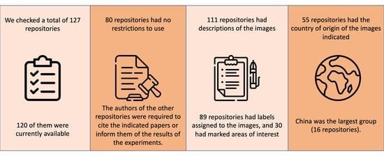
- The availability of the datasets;
- A breakdown of the legality of using the datasets;
- A classification of the image descriptions;
- Geographical distribution of the datasets that are available by continent and country.
2. Methods
2.1. Dataset Checking Strategy
- (1)
- Fully open;
- (2)
- Available after completing a form;
- (3)
- Available after account registration (and possible approval by the authors);
- (4)
- Available after sending an email to authors and approval from them;
- (5)
- Not available.
- After accessing, we manually checked each dataset described in 1 by downloading them to extract information about file status, sizes, and additional artifacts found in the images. Most available datasets were available as compressed files (ZIP/RAR/7Z), but some were available as separate files. We determined the sizes of the datasets in which the files were delivered separately by downloading all files and summing their sizes. We have prepared a tool to automatically generate the discussed information on repository contents and image samples—Ophthalmic Repository Sample Generator (Section 4.1). We also manually checked the content of each randomly selected image to see if there were any additional artifacts.
2.2. Image Descriptions and Legality of Use
- (1)
- Manually assigned labels corresponding to diagnosed ocular diseases, image quality, or described areas of interest;
- (2)
- Manual annotations on images indicating areas of interest;
- (3)
- No descriptions.
- (1)
- Notifying the authors of the datasets of the results and awaiting permission to publish the results;
- (2)
- References to the indicated articles or the dataset in the case of publication of the results;
- (3)
- No restrictions.
3. Data Availability
4. Code Availability
4.1. Ophthalmic Repository Sample Generator
- The sample size is an integer number representing the size of the random output sample of photos from the repository.
- A repository URL is a string representing a direct URL of the image file; e.g., for the Kaggle dataset, the schema is [username]/[dataset_id]. In the case of a Kaggle competition, it is ID.
- The data output file path is a string representing the output file with the downloaded repository content.
- The temporary full data path is a string representing the temporary path to which the repository will be extracted.
- The repository sample output path is a string representing the path where a randomly selected repository sample will be placed.
- The repository type is an integer representing the type of the repository source: 0 for classic URL, 1 for Kaggle competition, and 2 for Kaggle dataset.
- The output CSV file path is a string representing the path where the CSV file will be saved (separated by;) containing information about the repository.
5. Results
5.1. Data Access
5.2. Characteristics of Datasets
5.3. The Legality of Use and Image Descriptions
6. Discussion
7. Conclusions
Author Contributions
Funding
Institutional Review Board Statement
Informed Consent Statement
Data Availability Statement
Acknowledgments
Conflicts of Interest
References
- Ibrahim, H.; Liu, X.; Zariffa, N.; Morris, A.D.; Denniston, A.K. Health data poverty: An assailable barrier to equitable digital health care. Lancet Digit. Health 2021, 3, e260–e265. [Google Scholar] [CrossRef]
- Esteva, A.; Robicquet, A.; Ramsundar, B.; Kuleshov, V.; DePristo, M.; Chou, K.; Cui, C.; Corrado, G.; Thrun, S.; Dean, J. A guide to deep learning in healthcare. Nat. Med. 2019, 25, 24–29. [Google Scholar] [CrossRef] [PubMed]
- Norgeot, B.; Glicksberg, B.S.; Butte, A.J. A call for deep-learning healthcare. Nat. Med. 2019, 25, 14–15. [Google Scholar] [CrossRef]
- Grzybowski, A.; Brona, P.; Lim, G.; Ruamviboonsuk, P.; Tan, G.S.W.; Abramoff, M.; Ting, D.S.W. Artificial intelligence for diabetic retinopathy screening: A review. Eye 2019, 34, 451–460. [Google Scholar] [CrossRef]
- Grzybowski, A.; Brona, P. A pilot study of autonomous artificial intelligence-based diabetic retinopathy screening in Poland. Acta Ophthalmol. 2019, 97, e1149–e1150. [Google Scholar] [CrossRef] [PubMed]
- Miotto, R.; Wang, F.; Wang, S.; Jiang, X.; Dudley, J.T. Deep learning for healthcare: Review, opportunities and challenges. Briefings Bioinform. 2017, 19, 1236–1246. [Google Scholar] [CrossRef]
- Litjens, G.; Kooi, T.; Bejnordi, B.E.; Setio, A.A.A.; Ciompi, F.; Ghafoorian, M.; van der Laak, J.A.; van Ginneken, B.; Sánchez, C.I. A survey on deep learning in medical image analysis. Med. Image Anal. 2017, 42, 60–88. [Google Scholar] [CrossRef]
- Bluemke, D.A.; Moy, L.; Bredella, M.A.; Ertl-Wagner, B.B.; Fowler, K.J.; Goh, V.J.; Halpern, E.F.; Hess, C.P.; Schiebler, M.L.; Weiss, C.R. Assessing Radiology Research on Artificial Intelligence: A Brief Guide for Authors, Reviewers, and Readers from the Editorial Board. Radiology 2020, 294, 487–489. [Google Scholar] [CrossRef]
- Aggarwal, R.; Sounderajah, V.; Martin, G.; Ting, D.S.W.; Karthikesalingam, A.; King, D.; Ashrafian, H.; Darzi, A. Diagnostic accuracy of deep learning in medical imaging: A systematic review and meta-analysis. NPJ Digit. Med. 2021, 4, 65. [Google Scholar] [CrossRef] [PubMed]
- Ting, D.S.W.; Pasquale, L.R.; Peng, L.; Campbell, J.P.; Lee, A.Y.; Raman, R.; Tan, G.S.W.; Schmetterer, L.; Keane, P.A.; Wong, T.Y. Artificial intelligence and deep learning in ophthalmology. Br. J. Ophthalmol. 2018, 103, 167–175. [Google Scholar] [CrossRef]
- Khan, S.M.; Liu, X.; Nath, S.; Korot, E.; Faes, L.; Wagner, S.K.; Keane, P.A.; Sebire, N.J.; Burton, M.J.; Denniston, A.K. A global review of publicly available datasets for ophthalmological imaging: Barriers to access, usability, and generalisability. Lancet Digit. Health 2021, 3, e51–e66. [Google Scholar] [CrossRef]
- Available online: https://www.kaggle.com/datasets/linchundan/fundusimage1000 (accessed on 16 May 2023).
- Available online: https://data.mendeley.com/datasets/dh2x8v6nf8/1 (accessed on 16 May 2023).
- Available online: https://data.mendeley.com/datasets/trghs22fpg/1 (accessed on 16 May 2023).
- Available online: https://data.mendeley.com/datasets/trghs22fpg/2 (accessed on 16 May 2023).
- Available online: https://data.mendeley.com/datasets/trghs22fpg/3 (accessed on 16 May 2023).
- Available online: https://data.mendeley.com/datasets/trghs22fpg/4 (accessed on 16 May 2023).
- Available online: https://figshare.com/s/c2d31f850af14c5b5232 (accessed on 16 May 2023).
- Available online: https://www.kaggle.com/c/aptos2019-blindness-detection/data (accessed on 16 May 2023).
- Available online: https://people.eng.unimelb.edu.au/thivun/projects/AV_nicking_quantification/ (accessed on 16 May 2023).
- Available online: https://blogs.kingston.ac.uk/retinal/chasedb1/ (accessed on 16 May 2023).
- Available online: https://www.kaggle.com/datasets/gilescodes/cropped-train-diabetic-retinopathy-detection (accessed on 16 May 2023).
- Available online: https://www.kaggle.com/datasets/andrewmvd/retinal-disease-classification (accessed on 16 May 2023).
- Available online: https://www.kaggle.com/datasets/fareesamasroor/cardiacemboli (accessed on 16 May 2023).
- Available online: https://www.kaggle.com/datasets/mariaherrerot/ddrdataset (accessed on 16 May 2023).
- Available online: https://medicine.uiowa.edu/eye/abramoff/ (accessed on 16 May 2023).
- Available online: https://zenodo.org/record/4532361#.Yrr5sOzP1W4 (accessed on 16 May 2023).
- Available online: https://zenodo.org/record/4647952#.YtRttC-plQI (accessed on 16 May 2023).
- Available online: https://zenodo.org/record/4891308#.YtRwDS-plQI (accessed on 16 May 2023).
- Available online: https://www.kaggle.com/datasets/nikkich9/derbi-hackathon-retinal-fundus-image-dataset (accessed on 16 May 2023).
- Available online: https://data.mendeley.com/datasets/3csr652p9y/1 (accessed on 16 May 2023).
- Available online: https://data.mendeley.com/datasets/2rnnz5nz74/1 (accessed on 16 May 2023).
- Available online: https://data.mendeley.com/datasets/2rnnz5nz74/2 (accessed on 16 May 2023).
- Available online: https://www.kaggle.com/datasets/tanlikesmath/diabetic-retinopathy-resized (accessed on 16 May 2023).
- Available online: https://figshare.com/articles/dataset/Advancing_Bag_of_Visual_Words_Representations_for_Lesion_Classification_in_Retinal_Images/953671/2 (accessed on 16 May 2023).
- Available online: https://figshare.com/articles/dataset/Advancing_Bag_of_Visual_Words_Representations_for_Lesion_Classification_in_Retinal_Images/953671/3 (accessed on 16 May 2023).
- Available online: https://www.kaggle.com/datasets/mustaqimabrar/diabeticretinopathy (accessed on 16 May 2023).
- Available online: https://www.kaggle.com/datasets/rutujachaudhari/diabetic-retinopathy (accessed on 16 May 2023).
- Available online: https://www.kaggle.com/datasets/himanshuagarwal1998/diabetic-retinopathy (accessed on 16 May 2023).
- Available online: https://www.kaggle.com/datasets/analaura000/diabetic-retinopathy (accessed on 16 May 2023).
- Available online: https://www.kaggle.com/datasets/kameshwarandhayalan/diabetic-retinopathy (accessed on 16 May 2023).
- Available online: https://www.kaggle.com/datasets/sovitrath/diabetic-retinopathy-2015-data-colored-resized (accessed on 16 May 2023).
- Available online: https://www.kaggle.com/datasets/sovitrath/diabetic-retinopathy-224x224-2019-data (accessed on 16 May 2023).
- Available online: https://www.kaggle.com/datasets/sovitrath/diabetic-retinopathy-224x224-gaussian-filtered (accessed on 16 May 2023).
- Available online: https://www.kaggle.com/datasets/sovitrath/diabetic-retinopathy-224x224-grayscale-images (accessed on 16 May 2023).
- Available online: https://www.kaggle.com/datasets/amanneo/diabetic-retinopathy-resized-arranged (accessed on 16 May 2023).
- Available online: https://www.kaggle.com/datasets/kushagratandon12/diabetic-retinopathy-balanced (accessed on 16 May 2023).
- Available online: https://www.kaggle.com/datasets/makrovh/diabetic-retinopathy-blindness-detection-c-data (accessed on 16 May 2023).
- Available online: https://www.kaggle.com/datasets/dola1507108/diabetic-retinopathy-classified (accessed on 16 May 2023).
- Available online: https://www.kaggle.com/competitions/diabetic-retinopathy-classification-2/data (accessed on 16 May 2023).
- Available online: https://www.kaggle.com/competitions/retinopathy-classification-sai/data (accessed on 16 May 2023).
- Available online: https://www.kaggle.com/datasets/sachinkumar413/diabetic-retinopathy-dataset (accessed on 16 May 2023).
- Available online: https://www.kaggle.com/datasets/ahmedghazal54/diabetic-retinopathy-detection (accessed on 16 May 2023).
- Available online: https://www.kaggle.com/datasets/poojita2305/diabetic-retinopathy-detection (accessed on 16 May 2023).
- Available online: https://www.kaggle.com/datasets/mostafaeltalawy/diabetic-retinopathy-dataset (accessed on 16 May 2023).
- Available online: https://personalpages.manchester.ac.uk/staff/niall.p.mcloughlin/ (accessed on 16 May 2023).
- Available online: http://www.ia.uned.es/~ejcarmona/DRIONS-DB.html (accessed on 16 May 2023).
- Available online: https://drive.grand-challenge.org (accessed on 16 May 2023).
- Available online: https://www.kaggle.com/datasets/dola1507108/diabetic-retinopathy-organized (accessed on 16 May 2023).
- Available online: https://www.kaggle.com/datasets/sachinkumar413/diabetic-retinopathy-preprocessed-dataset (accessed on 16 May 2023).
- Available online: https://www.kaggle.com/datasets/shuvokkr/diabeticresized300 (accessed on 16 May 2023).
- Available online: https://www.kaggle.com/datasets/makjn10/diabetic-retinopathy-small (accessed on 16 May 2023).
- Available online: https://www.kaggle.com/datasets/dantealonso/diabeticretinopathytrainvalidation (accessed on 16 May 2023).
- Available online: https://www.kaggle.com/datasets/saipavansaketh/diabetic-retinopathy-unziped (accessed on 16 May 2023).
- Available online: https://www.kaggle.com/datasets/zhizhid/dr-2000 (accessed on 16 May 2023).
- Available online: https://www.kaggle.com/datasets/aviagarwal123/dr-201010 (accessed on 16 May 2023).
- Available online: https://github.com/deepdrdoc/DeepDRiD/blob/master/README.md (accessed on 16 May 2023).
- Available online: https://www.kaggle.com/datasets/nguyenhung1903/diaretdb1-v21 (accessed on 16 May 2023).
- Available online: https://www.kaggle.com/datasets/muhamedahmed/diabetic (accessed on 16 May 2023).
- Available online: https://www.kaggle.com/datasets/alisalen/diabetic-retinopathy-detection-processed (accessed on 16 May 2023).
- Available online: https://www.kaggle.com/datasets/lokeshsaipureddi/drishtigs-retina-dataset-for-onh-segmentation (accessed on 16 May 2023).
- Available online: http://cvit.iiit.ac.in/projects/mip/drishti-gs/mip-dataset2/Home.php (accessed on 16 May 2023).
- Available online: https://www.kaggle.com/datasets/diveshthakker/eoptha-diabetic-retinopathy (accessed on 16 May 2023).
- Available online: https://www.kaggle.com/c/diabetic-retinopathy-detection (accessed on 16 May 2023).
- Available online: https://www.kaggle.com/datasets/bishalbanerjee/eye-dataset (accessed on 16 May 2023).
- Available online: https://www.kaggle.com/datasets/iamachal/fundus-image-dataset (accessed on 16 May 2023).
- Available online: https://projects.ics.forth.gr/cvrl/fire/ (accessed on 16 May 2023).
- Available online: https://www.kaggle.com/datasets/izander/fundus (accessed on 16 May 2023).
- Available online: https://www.kaggle.com/datasets/klmsathishkumar/fundus-images (accessed on 16 May 2023).
- Available online: https://www.kaggle.com/datasets/spikeetech/fundus-dr (accessed on 16 May 2023).
- Available online: https://www.kaggle.com/datasets/balnyaupane/gaussian-filtered-diabetic-retinopathy (accessed on 16 May 2023).
- Available online: https://dataverse.harvard.edu/dataset.xhtml?persistentId=doi:10.7910/DVN/1YRRAC (accessed on 16 May 2023).
- Available online: https://github.com/lgiancaUTH/HEI-MED (accessed on 16 May 2023).
- Available online: https://www5.cs.fau.de/research/data/fundus-images/ (accessed on 16 May 2023).
- Available online: http://ai.baidu.com/broad/introduction (accessed on 16 May 2023).
- Available online: https://idrid.grand-challenge.org/Rules/ (accessed on 16 May 2023).
- Available online: https://medicine.uiowa.edu/eye/inspire-datasets (accessed on 16 May 2023).
- Available online: http://ai.baidu.com/broad/subordinate?dataset=pm (accessed on 16 May 2023).
- Available online: https://www.kaggle.com/datasets/dineswarreddy/indian-retina-classification (accessed on 16 May 2023).
- Available online: https://www.kaggle.com/datasets/bachaboos/isbi-2021-retina-23 (accessed on 16 May 2023).
- Available online: https://www.kaggle.com/datasets/bachaboos/isbi-retina-test (accessed on 16 May 2023).
- Available online: https://www.kaggle.com/linchundan/fundusimage1000 (accessed on 16 May 2023).
- Available online: https://journals.plos.org/plosone/article?id=10.1371/journal.pone.0179790#sec006 (accessed on 16 May 2023).
- Available online: https://github.com/smilell/AG-CNN (accessed on 16 May 2023).
- Available online: https://www.adcis.net/en/third-party/messidor2/ (accessed on 16 May 2023).
- Available online: https://odir2019.grand-challenge.org/Download/ (accessed on 16 May 2023).
- Available online: https://www.kaggle.com/datasets/andrewmvd/ocular-disease-recognition-odir5k (accessed on 16 May 2023).
- Available online: https://aistudio.baidu.com/aistudio/datasetdetail/122940 (accessed on 16 May 2023).
- Available online: https://figshare.com/articles/dataset/PAPILA/14798004/1 (accessed on 16 May 2023).
- Available online: https://figshare.com/articles/dataset/PAPILA/14798004 (accessed on 16 May 2023).
- Available online: https://www.kaggle.com/datasets/benjaminwarner/resized-2015-2019-blindness-detection-images (accessed on 16 May 2023).
- Available online: https://refuge.grand-challenge.org/ (accessed on 16 May 2023).
- Available online: https://ai.baidu.com/broad/download?dataset=gon (accessed on 16 May 2023).
- Available online: https://www.kaggle.com/datasets/kssanjaynithish03/retinal-fundus-images (accessed on 16 May 2023).
- Available online: https://deepblue.lib.umich.edu/data/concern/data_sets/3b591905z?locale=en (accessed on 16 May 2023).
- Available online: https://www.kaggle.com/datasets/andrewmvd/fundus-image-registration (accessed on 16 May 2023).
- Available online: http://medimrg.webs.ull.es/research/downloads/ (accessed on 16 May 2023).
- Available online: https://www.kaggle.com/datasets/priyanagda/ritedataset (accessed on 16 May 2023).
- Available online: http://www.rodrep.com/longitudinal-diabetic-retinopathy-screening—description.html (accessed on 16 May 2023).
- Available online: https://www.kaggle.com/datasets/aifahim/retinal-vassel-combine-same-format (accessed on 16 May 2023).
- Available online: https://www.kaggle.com/jr2ngb/cataractdataset (accessed on 16 May 2023).
- Available online: http://webeye.ophth.uiowa.edu/ROC/ (accessed on 16 May 2023).
- Available online: https://www.kaggle.com/datasets/beatrizsimoes/retina-quality (accessed on 16 May 2023).
- Available online: https://www.kaggle.com/datasets/hebamohamed/retinagen (accessed on 16 May 2023).
- Available online: http://bioimlab.dei.unipd.it/Retinal%20Vessel%20Tortuosity.htm (accessed on 16 May 2023).
- Available online: https://www.kaggle.com/datasets/ustinianw/retinal-tiny (accessed on 16 May 2023).
- Available online: http://cecas.clemson.edu/~ahoover/stare/ (accessed on 16 May 2023).
- Available online: https://www.kaggle.com/datasets/balnyaupane/small-diabetic-retinopathy-dataset (accessed on 16 May 2023).
- Available online: https://www.kaggle.com/datasets/mariaherrerot/the-sustechsysu-dataset (accessed on 16 May 2023).
- Available online: https://www.kaggle.com/competitions/innovation-challenge-2019/data (accessed on 16 May 2023).
- Available online: https://www.kaggle.com/competitions/vietai-advance-retinal-disease-detection-2020/data (accessed on 16 May 2023).
- Available online: http://www.varpa.es/research/ophtalmology.html#vicavr (accessed on 16 May 2023).
- Available online: http://people.duke.edu/~sf59/Estrada_TMI_2015_dataset.htm (accessed on 16 May 2023).
- Available online: https://novel.utah.edu/Hoyt/ (accessed on 16 May 2023).
- Available online: https://zenodo.org/record/3393265#.XazZaOgzbIV (accessed on 16 May 2023).
- Available online: https://www.kaggle.com/datasets/nawa393/dr15_test (accessed on 16 May 2023).
- Available online: https://www.kaggle.com/datasets/makorromanuel/merged-retina-datasets (accessed on 16 May 2023).
- Cen, L.P.; Ji, J.; Lin, J.W.; Ju, S.T.; Lin, H.J.; Li, T.P.; Wang, Y.; Yang, J.F.; Liu, Y.F.; Tan, S.; et al. Automatic detection of 39 fundus diseases and conditions in retinal photographs using deep neural networks. Nat. Commun. 2021, 12, 4828. [Google Scholar] [CrossRef]
- Yoo, T.K. A CycleGAN Deep Learning Technique for Artifact Reduction in Fundus Photography. Graefe’s Arch. Clin. Exp. Ophthalmol. 2020, 258, 1631–1637. [Google Scholar] [CrossRef]
- Hassan, T. A Composite Retinal Fundus and OCT Dataset along with Detailed Clinical Markings for Extracting Retinal Layers, Retinal Lesions and Screening Macular and Glaucomatous Disorders (dataset 1). 2021. [Google Scholar] [CrossRef]
- Hassan, T. A Composite Retinal Fundus and OCT Dataset along with Detailed Clinical Markings for Extracting Retinal Layers, Retinal Lesions and Screening Macular and Glaucomatous Disorders (dataset 2). 2021. [Google Scholar] [CrossRef]
- Hassan, T. A Composite Retinal Fundus and OCT Dataset along with Detailed Clinical Markings for Extracting Retinal Layers, Retinal Lesions and Screening Macular and Glaucomatous Disorders (dataset 3). 2021. [Google Scholar] [CrossRef]
- Hassan, T.; Akram, M.U.; Masood, M.F.; Yasin, U. Deep structure tensor graph search framework for automated extraction and characterization of retinal layers and fluid pathology in retinal SD-OCT scans. Comput. Biol. Med. 2019, 105, 112–124. [Google Scholar] [CrossRef] [PubMed]
- Hassan, T.; Akram, M.U.; Werghi, N.; Nazir, M.N. RAG-FW: A Hybrid Convolutional Framework for the Automated Extraction of Retinal Lesions and Lesion-Influenced Grading of Human Retinal Pathology. IEEE J. Biomed. Health Inform. 2021, 25, 108–120. [Google Scholar] [CrossRef] [PubMed]
- Diaz-Pinto, A.; Morales, S.; Naranjo, V.; Köhler, T.; Mossi, J.M.; Navea, A. CNNs for automatic glaucoma assessment using fundus images: An extensive validation. Biomed. Eng. Online 2019, 18, 29. [Google Scholar] [CrossRef] [PubMed]
- Pachade, S.; Porwal, P.; Thulkar, D.; Kokare, M.; Deshmukh, G.; Sahasrabuddhe, V.; Giancardo, L.; Quellec, G.; Mériaudeau, F. Retinal Fundus Multi-Disease Image Dataset (RFMiD): A Dataset for Multi-Disease Detection Research. Data 2021, 6, 14. [Google Scholar] [CrossRef]
- Li, T.; Gao, Y.; Wang, K.; Guo, S.; Liu, H.; Kang, H. Diagnostic assessment of deep learning algorithms for diabetic retinopathy screening. Inf. Sci. 2019, 501, 511–522. [Google Scholar] [CrossRef]
- Benítez, V.E.C.; Matto, I.C.; Román, J.C.M.; Noguera, J.L.V.; García-Torres, M.; Ayala, J.; Pinto-Roa, D.P.; Gardel-Sotomayor, P.E.; Facon, J.; Grillo, S.A. Dataset from fundus images for the study of diabetic retinopathy. Data Brief 2021, 36, 107068. [Google Scholar] [CrossRef]
- Akbar, S.; Sharif, M.; Akram, M.U.; Saba, T.; Mahmood, T.; Kolivand, M. Automated techniques for blood vessels segmentation through fundus retinal images: A review. Microsc. Res. Tech. 2019, 82, 153–170. [Google Scholar] [CrossRef]
- Raja, H. Data on OCT and Fundus Images. 2020. [Google Scholar] [CrossRef]
- Pires, R.; Jelinek, H.F.; Wainer, J.; Valle, E.; Rocha, A. Advancing Bag-of-Visual-Words Representations for Lesion Classification in Retinal Images. PLoS ONE 2014, 9, e96814. [Google Scholar] [CrossRef] [PubMed]
- Holm, S.; Russell, G.; Nourrit, V.; McLoughlin, N. DR HAGIS—a fundus image database for the automatic extraction of retinal surface vessels from diabetic patients. J. Med. Imaging 2017, 4, 014503. [Google Scholar] [CrossRef] [PubMed]
- Carmona, E.J.; Rincón, M.; García-Feijoó, J.; de-la Casa, J.M.M. Identification of the optic nerve head with genetic algorithms. Artif. Intell. Med. 2008, 43, 243–259. [Google Scholar] [CrossRef]
- Kauppi, T.; Kalesnykiene, V.; Kamarainen, J.K.; Lensu, L.; Sorri, I.; Raninen, A.; Voutilainen, R.; Uusitalo, H.; Kalviainen, H.; Pietila, J. The DIARETDB1 diabetic retinopathy database and evaluation protocol. In Proceedings of the British Machine Vision Conference 2007, Warwick, UK, 10–13 September 2007. [Google Scholar] [CrossRef]
- Sivaswamy, J.; Chakravarty, A.; Joshi, G.D.; Syed, T.A. A Comprehensive Retinal Image Dataset for the Assessment of Glaucoma from the Optic Nerve Head Analysis. JSM Biomed. Imaging Data Pap. 2015, 2, 1004. [Google Scholar]
- Sivaswamy, J.; Krishnadas, S.R.; Datt Joshi, G.; Jain, M.; Syed Tabish, A.U. Drishti-GS: Retinal image dataset for optic nerve head(ONH) segmentation. In Proceedings of the 2014 IEEE 11th International Symposium on Biomedical Imaging (ISBI), Beijing, China, 29 April–2 May 2014; pp. 53–56. [Google Scholar] [CrossRef]
- Hernandez-Matas, C.; Zabulis, X.; Triantafyllou, A.; Anyfanti, P.; Douma, S.; Argyros, A.A. FIRE: Fundus Image Registration dataset. Model. Artif. Intell. Ophthalmol. 2017, 1, 16–28. [Google Scholar] [CrossRef]
- Akbar, S.; Hassan, T.; Akram, M.U.; Yasin, U.; Basit, I. AVRDB: Annotated Dataset for Vessel Segmentation and Calculation of Arteriovenous Ratio. 2017. [Google Scholar]
- Giancardo, L.; Meriaudeau, F.; Karnowski, T.P.; Li, Y.; Garg, S.; Tobin, K.W.; Chaum, E. Exudate-based diabetic macular edema detection in fundus images using publicly available datasets. Med. Image Anal. 2012, 16, 216–226. [Google Scholar] [CrossRef] [PubMed]
- Kohler, T.; Budai, A.; Kraus, M.F.; Odstrcilik, J.; Michelson, G.; Hornegger, J. Automatic no-reference quality assessment for retinal fundus images using vessel segmentation. In Proceedings of the 26th IEEE International Symposium on Computer-Based Medical Systems, Porto, Portugal, 20–22 June 2013. [Google Scholar] [CrossRef]
- Budai, A.; Bock, R.; Maier, A.; Hornegger, J.; Michelson, G. Robust Vessel Segmentation in Fundus Images. Int. J. Biomed. Imaging 2013, 2013, 1–11. [Google Scholar] [CrossRef]
- Prasanna Porwal, S.P. Indian Diabetic Retinopathy Image Dataset (IDRiD). 2018. [Google Scholar] [CrossRef]
- Niemeijer, M.; Xu, X.; Dumitrescu, A.V.; Gupta, P.; van Ginneken, B.; Folk, J.C.; Abramoff, M.D. Automated Measurement of the Arteriolar-to-Venular Width Ratio in Digital Color Fundus Photographs. IEEE Trans. Med. Imaging 2011, 30, 1941–1950. [Google Scholar] [CrossRef]
- Tang, L.; Garvin, M.K.; Lee, K.; Alward, W.L.W.; Kwon, Y.H.; Abramoff, M.D. Robust Multiscale Stereo Matching from Fundus Images with Radiometric Differences. IEEE Trans. Pattern Anal. Mach. Intell. 2011, 33, 2245–2258. [Google Scholar] [CrossRef]
- Fu, H.; Li, F.; Orlando, J.I.; Bogunović, H.; Sun, X.; Liao, J.; Xu, Y.; Zhang, S.; Zhang, X. ADAM: Automatic Detection Challenge on Age-Related Macular Degeneration. 2020. [Google Scholar] [CrossRef]
- Li, L.; Xu, M.; Wang, X.; Jiang, L.; Liu, H. Attention Based Glaucoma Detection: A Large-scale Database and CNN Model. arXiv 2019, arXiv:1903.10831. [Google Scholar]
- Decenciere, E.; Zhang, X.; Cazuguel, G.; Lay, B.; Cochener, B.; Trone, C.; Gain, P.; Ordonez, R.; Massin, P.; Erginay, A.; et al. Feedback on a publicly distributed image database: The messidor database. Image Anal. Stereol. 2014, 33, 231. [Google Scholar] [CrossRef]
- Kovalyk, O.; Morales-Sánchez, J.; Verdú-Monedero, R.; Sellés-Navarro, I.; Palazón-Cabanes, A.; Sancho-Gómez, J.L. PAPILA. 2022. [Google Scholar] [CrossRef]
- Orlando, J.I.; Fu, H.; Breda, J.B.; van Keer, K.; Bathula, D.R.; Diaz-Pinto, A.; Fang, R.; Heng, P.A.; Kim, J.; Lee, J.; et al. REFUGE Challenge: A unified framework for evaluating automated methods for glaucoma assessment from fundus photographs. Med. Image Anal. 2020, 59, 101570. [Google Scholar] [CrossRef]
- Almazroa, A. Retinal Fundus Images for Glaucoma Analysis: The Riga Dataset. 2018. [Google Scholar] [CrossRef]
- Hu, Q.; Abràmoff, M.D.; Garvin, M.K. Automated Separation of Binary Overlapping Trees in Low-Contrast Color Retinal Images. In Advanced Information Systems Engineering; Springer: Berlin/Heidelberg, Germany, 2013; pp. 436–443. [Google Scholar] [CrossRef]
- Adal, K.M.; van Etten, P.G.; Martinez, J.P.; van Vliet, L.J.; Vermeer, K.A. Accuracy Assessment of Intra- and Intervisit Fundus Image Registration for Diabetic Retinopathy Screening. Investig. Ophthalmol. Vis. Sci. 2015, 56, 1805–1812. [Google Scholar] [CrossRef] [PubMed]
- Grisan, E.; Foracchia, M.; Ruggeri, A. A Novel Method for the Automatic Grading of Retinal Vessel Tortuosity. IEEE Trans. Med. Imaging 2008, 27, 310–319. [Google Scholar] [CrossRef] [PubMed]
- Lin, L.; Li, M.; Huang, Y.; Cheng, P.; Xia, H.; Wang, K.; Yuan, J.; Tang, X. The SUSTech-SYSU dataset for automated exudate detection and diabetic retinopathy grading. Sci. Data 2020, 7, 409. [Google Scholar] [CrossRef] [PubMed]
- Vázquez, S.G.; Cancela, B.; Barreira, N.; Penedo, M.G.; Rodríguez-Blanco, M.; Seijo, M.P.; de Tuero, G.C.; Barceló, M.A.; Saez, M. Improving retinal artery and vein classification by means of a minimal path approach. Mach. Vis. Appl. 2012, 24, 919–930. [Google Scholar] [CrossRef]
- Estrada, R.; Allingham, M.J.; Mettu, P.S.; Cousins, S.W.; Tomasi, C.; Farsiu, S. Retinal Artery-Vein Classification via Topology Estimation. IEEE Trans. Med. Imaging 2015, 34, 2518–2534. [Google Scholar] [CrossRef] [PubMed]
- Resnikoff, S.; Felch, W.; Gauthier, T.M.; Spivey, B. The number of ophthalmologists in practice and training worldwide: A growing gap despite more than 200,000 practitioners. Br. J. Ophthalmol. 2012, 96, 783–787. [Google Scholar] [CrossRef]
- Holst, C.; Sukums, F.; Radovanovic, D.; Ngowi, B.; Noll, J.; Winkler, A.S. Sub-Saharan Africa—the new breeding ground for global digital health. Lancet Digit. Health 2020, 2, e160–e162. [Google Scholar] [CrossRef]
- Müller, S.; Koch, L.M.; Lensch, H.; Berens, P. A Generative Model Reveals the Influence of Patient Attributes on Fundus Images. In Proceedings of the Medical Imaging with Deep Learning, Zurich, Switzerland, 6–8 July 2022. [Google Scholar]
- Ting, D.S.W.; Cheung, C.Y.L.; Lim, G.; Tan, G.S.W.; Quang, N.D.; Gan, A.; Hamzah, H.; Garcia-Franco, R.; Yeo, I.Y.S.; Lee, S.Y.; et al. Development and Validation of a Deep Learning System for Diabetic Retinopathy and Related Eye Diseases Using Retinal Images From Multiethnic Populations With Diabetes. JAMA 2017, 318, 2211. [Google Scholar] [CrossRef] [PubMed]
- Serener, A.; Serte, S. Geographic variation and ethnicity in diabetic retinopathy detection via deeplearning. Turk. J. Electr. Eng. Comput. Sci. 2020, 28, 664–678. [Google Scholar] [CrossRef]


| ID | Name | Access | Technical Details | Epidemiological Details | Legality | ||||||||
|---|---|---|---|---|---|---|---|---|---|---|---|---|---|
| Access Type | Ease of Access | Number of Photos | Photo Sizes | Photo Descriptions | Size of the Repository | Additional Artifacts in the Photos | Repository URL | Number of Patients | Eye Diseases | Country of Origin | |||
| 1 | 1000FIWC | OAAR | 2 | 1000 | Different: 3000 × 3152 px; 1728 × 2592 px; … | Labels | 777 MB | Absence | [12] | NR | DR, G | China | Citation: [128] |
| 2 | ACDLTFARIFP | OA | 1 | 2206 | 350 × 346 × 3 px | Absence | 294 MB | Absence | [13] | NR | NR | NR | Citation: [129] |
| 3 | ACRFAODAWDCMFERLRLASMAGD Version 1 | OA | 1 | 2560 | Different: 456 × 951 px; 576 × 760 px; … | Labels | 302 MB | Absence | [14] | NR | DME, CSR, AMD, G | UAE | Citation: [130] |
| 4 | ACRFAODAWDCMFERLRLASMAGD Version 2 | OA | 1 | 2988 | Different: 456 × 951 px; 576 × 760 px; … | Labels | 1597 MB | Absence | [15] | NR | G | UAE | Citation: [131] |
| 5 | ACRFAODAWDCMFERLRLASMAGD Version 3 | OA | 1 | 3100 | Different: 1934 × 2032 px; 576 × 760 px; … | Labels | 2969 MB | Absence | [16] | NR | DME, CSR, AMD, G | UAE | Citation: [132] |
| 6 | ACRFAODAWDCMFERLRLASMAGD Version 4 | OA | 1 | 2560 | Different: 1934 × 2032 px; 576 × 760 px; … | Labels | 1433.6 MB | Absence | [17] | NR | DME, CSR, AMD, G | UAE | Citation: [133,134] |
| 7 | ACRIMA | OA | 3 | 705 | Different: 349 × 349 px; 871 × 871 px; … | Labels | 23 MB | Absence | [18] | NR | G | Spain | Citation: [135] |
| 8 | APTOS | OAAL | 2 | 5590 | Different: 640 × 480 px; 2416 × 1736; … | Labels | 9748 MB | Absence | [19] | NR | DR | India | NR |
| 9 | Arteriovenous Nicking | OA | 3 | 90 | 401 × 401 px | Labels | 29 MB | Absence | [20] | NR | NR | NR | NR |
| 10 | CHHSIE | OA | 3 | 28 | 999 × 960 px | Annotations | 3 MB | Absence | [21] | 14 | HE | UK | NR |
| 11 | CTDRD | OAAR | 2 | 35,136 | Different: 4712 × 3163 px; 2213 × 2205 px; … | Absence | 40,755 MB | Absence | [22] | NR | DR | NR | NR |
| 12 | Calibration level 1 | OAAR | 2 | 3200 | Different: 1424 × 2144 px; 1536 × 2048 px; | Labels | 8181 MB | Absence | [23] | NR | DR | India | Citation: [136] |
| 13 | Cardiacemboli | OAAR | 2 | 943 | Different: 3264 × 4928 px; 2048 × 3072 px; … | Labels | 767 MB | Absence | [24] | NR | CE | NR | NR |
| 14 | DDR | OAAR | 2 | 12524 | Different: 512 × 512 px; 1536 × 2048 px; … | Labels | 3348 MB | Absence | [25] | NR | DR | China | Citation: [137] |
| 15 | DEFRIOVAA | OA | 3 | 50 | Different: 798 × 658 px; 886 × 719 px; … | Absence | 43.2 MB | Absence | [26] | 50 | DED | Netherlands | NR |
| 16 | DFFIFTSODR Version 0.1 | OA | 1 | 396 | 2124 × 2056 px | Labels | 238 MB | Absence | [27] | NR | DR | Paraguay | Citation: [138] |
| 17 | DFFIFTSODR Version 0.2 | OA | 1 | 757 | Different: 1444 × 1444 px; 2056 × 2124 px; … | Labels | 375 MB | Absence | [28] | NR | DR | Paraguay | Citation: [138] |
| 18 | DFFIFTSODR Version 0.3 | OA | 1 | 1437 | Different: 1028 × 1052 px; 1029 × 1062 px | Labels | 1536 MB | Absence | [29] | NR | DR | Paraguay | Citation: [138] |
| 19 | DHRFID | OAAR | 2 | 6542 | Different: 3456 × 5184 px; 1904 × 2460 px; … | Labels | 857 MB | Absence | [30] | NR | DR, G, O | NR | NR |
| 20 | DOFIFVSDOHRDRAP | OA | 1 | 100 | 1504 × 1000 px | Absence | 192 MB | Presence | [31] | NR | HR, DR, P | Pakistan | Citation: [139] |
| 21 | DOOAFI Version 1 | OA | 1 | 50 | 2032 × 1934 px | Absence | 33 MB | Absence | [32] | NR | G | NR | Citation: [140] |
| 22 | DOOAFI Version 2 | OA | 1 | 50 | 2032 × 1 934 px | Absence | 33 MB | Absence | [33] | NR | G | NR | Citation: [140] |
| 23 | DR (resized) | OAAR | 2 | 70,234 | Different: 278 × 278 px; 774 × 1024 px; … | Labels | 8038 MB | Absence | [34] | NR | DR | China | NR |
| 24 | DR 1 | OA | 3 | 1077 | 640 × 480 px | Labels | 200 MB | Absence | [35] | NR | DED | Brazil | Citation: [141] |
| 25 | DR 2 | OA | 3 | 520 | Different: 873 × 500 px; 877 × 582 px; 876 × 581 px; … | Labels | 200 MB | Absence | [36] | NR | DED | Brazil | Citation: [141] |
| 26 | DR 3 | OAAR | 2 | 13251 | Different: 1184 × 1792 px; 2048, 3072 px; … | Labels | 14,848 MB | Absence | [38] | NR | DR | NR | NR |
| 27 | DR 4 | OAAR | 2 | 105375 | 512 × 786 px | Labels | 12,697 MB | Absence | [37] | NR | DR | NR | NR |
| 28 | DR 5 | OAAR | 2 | 3662 | Different: 358 × 474 px; 1226 × 1844 px; … | Labels | 8908 MB | Absence | [39] | NR | DR | NR | NR |
| 29 | DR 6 | OAAR | 2 | 301 | 2056 × 2124 px | Labels | 128 MB | Absence | [40] | NR | DR | NR | NR |
| 30 | DR 7 | OAAR | 2 | 103 | Different: 3264 × 4928 px; 2000 × 3008 px; … | Absence | 101 MB | Absence | [41] | NR | NR | NR | NR |
| 31 | DR2015DCR | OAAR | 2 | 35,126 | 224 × 224 px | Labels | 1014 MB | Absence | [42] | NR | DR | NR | NR |
| 32 | DR224 × 2242019 | OAAR | 2 | 3662 | 224 × 224 px | Labels | 238 MB | Absence | [43] | NR | DR | India | NR |
| 33 | DR224 × 224GF | OAAR | 2 | 3662 | 224 × 224 px | Labels | 426 MB | Absence | [44] | NR | DR | India | NR |
| 34 | DR224 × 224GI | OAAR | 2 | 3662 | 224 × 224 px | Labels | 157 MB | Absence | [45] | NR | DR | India | NR |
| 35 | DRA | OAAR | 2 | 35,126 | Different: 1024 × 1024 px; 779 × 1024 px; … | Labels | 1024 MB | Absence | [46] | NR | DR | NR | NR |
| 36 | DRB | OAAR | 2 | 49,703 | 512 × 512 px | Labels | 2048 MB | Absence | [47] | NR | DR | NR | NR |
| 37 | DRBDC | OAAR | 2 | 88,700 | Different: 4752 × 3168 px; 2560 × 1920 px; … | Labels | 98,017 MB | Absence | [48] | NR | DR | NR | NR |
| 38 | DRC | OAAR | 2 | 70,234 | Different: 278 × 278 px; 727 × 1024 px; … | Labels | 8192 MB | Absence | [49] | NR | DR | NR | NR |
| 39 | DRC #2 | OAAR | 2 | 2608 | 224 × 224 px | Absence | 33 MB | Absence | [50] | NR | NR | NR | NR |
| 40 | DRC 3 | OAAR | 2 | 2608 | 224 × 224 px | Absence | 33 MB | Absence | [51] | NR | NR | NR | NR |
| 41 | DRD | OAAR | 2 | 2750 | 256 × 256 px | Labels | 350 MB | Absence | [52] | NR | DR | NR | NR |
| 42 | DRD 2 | OAAR | 2 | 2111 | Different: 554 × 512 px; 424 × 512 px; … | Labels | 269 MB | Absence | [53] | NR | DR | NR | NR |
| 43 | DRD 3 | OAAR | 2 | 3662 | 224 × 224 px | Labels | 349 MB | Absence | [54] | NR | DR | NR | NR |
| 44 | DRD 4 | OAAR | 2 | 12,844 | 224 × 224 px | Labels | 725 MB | Absence | [55] | NR | DR | NR | NR |
| 45 | DRHARMDAGI | OA | 3 | 39 | Different: 3216 × 2136 px; 2816 × 1880 px; … | Labels | 10 MB | Presence | [56] | 38 | DED, HR, G, AMD | UK | Citation: [142] |
| 46 | DRIFONS | OA | 3 | 110 | 600 × 400 px | Annotations | 2.5 MB | Presence | [57] | 55 | HR | Spain | Citation: [143] |
| 47 | DRIFVS | OAAR | 2 | 40 | 565 × 584 px | Annotations | 30 MB | Absence | [58] | 400 | DED | Netherlands | NR |
| 48 | DRO | OAAR | 2 | 35,128 | Different: 278 × 278 px; 738 × 1024 px; … | Labels | 6656 MB | Absence | [59] | NR | DR | NR | NR |
| 49 | DRPD | OAAR | 2 | 13,970 | 128 × 128 px | Labels | 46 MB | Absence | [60] | NR | DR | NR | NR |
| 50 | DRR300 × 300C | OAAR | 2 | 16,798 | Different: 300 × 300 px; 271 × 273 px; … | Labels | 123 MB | Absence | [61] | NR | DR | NR | NR |
| 51 | DRS | OAAR | 2 | 10024 | Different: 315 × 400 px; 1957 × 2196 px; … | Absence | 10,240 MB | Absence | [62] | NR | NR | NR | NR |
| 52 | DRTW | OAAR | 2 | 37254 | Different: 278 × 278 px; 899 × 1024 px; … | Labels | 4915 MB | Absence | [63] | NR | DR | NR | NR |
| 53 | DRU | OAAR | 2 | 34882 | Different: 3456 × 5184 px; 1957 × 2196 px; … | Labels | 32,768 MB | Absence | [64] | NR | DR | China | NR |
| 54 | DR_2000 | OAAR | 2 | 2000 | Different: 3456 × 5184; 2056 × 3088 px; … | Labels | 2048 MB | Absence | [65] | NR | DR | China | NR |
| 55 | DR_201010 | OAAR | 2 | 35,136 | Different: 2560 × 1920 px; 2592 × 1944 px; … | Labels | 38,809 MB | Absence | [66] | NR | DR | NR | NR |
| 56 | DeepDRiD | OA | 1 | 320 | Different: 1725 × 2230 px; 3072 × 3900; … | Absence | 1464 MB | Absence | [67] | NR | NR | NR | Citation: [138] |
| 57 | DiaRetDB1 V2.1 | OAAR | 2 | 89 | 1152 × 1500 px | Annotations | 137 MB | Absence | [68] | NR | DR | Finland | Citation: [144] |
| 58 | Diabetic | OAAR | 2 | 2769 | Different: 342 × 512 px; 434 × 512 px; … | Labels | 353 MB | Absence | [69] | NR | DR | NR | NR |
| 59 | Diabetic Retinopathy Detection Processed | OAAR | 2 | 51,500 | Different: 224 × 224 px; 400 × 400 px; … | Labels | 2764 MB | Absence | [70] | NR | DR | NR | NR |
| 60 | Drishti-GS | OAAR | 2 | 101 | Different: 1845 × 2050 px; 1763 × 2047 px; … | Annotations | 341 MB | Absence | [71] | NR | NR | NR | NR |
| 61 | Drishti-GS1 | OAAR | 2 | 101 | Different: 2048 × 1760 px; 2049 × 1749; … | Annotations, labels | 350 MB | Absence | [72] | NR | G | India | Citation: [145,146] |
| 62 | EOptha Diabetic Retinopathy | OAAR | 2 | 46 | Different: 1696 × 2544 px; 1000 × 1504 px; … | Annotations | 21 MB | Absence | [73] | NR | DR | NR | NR |
| 63 | EPACS | OA | 2 | 88702 | Different: 4752 × 3168 px; 4928 × 3264 px; … | Labels | 84,203 MB | Absence | [74] | NR | DED | USA | NR |
| 64 | Eye Dataset Workshop | OAAR | 2 | 12734 | Different: 3264 × 3264 px; 2592 × 2592 px; … | Labels | 672 MB | Absence | [75] | NR | DR | NR | NR |
| 65 | FID | OAAR | 2 | 32 | 605 × 700 px | Absence | 1 MB | Absence | [76] | NR | HE | NR | NR |
| 66 | FIRD | OA | 3 | 268 | 2912 × 2912 px | Annotations | 264 MB | Absence | [77] | 39 | NR | Greece | Citation: [147] |
| 67 | Fundus | OAAR | 2 | 1600 | Different: 1725 × 2230 px; 1727 × 2232 px; … | Labels | 890 MB | Absence | [78] | NR | DR | NR | NR |
| 68 | Fundus Images | OAAR | 2 | 650 | Different: 2048 × 3072 px; 2048 × 3085 | Labels | 206 MB | Absence | [79] | NR | G | NR | NR |
| 69 | Fundus_DR | OAAR | 2 | 61830 | 256 × 256 px | Labels | 1710 MB | Absence | [80] | NR | DR | NR | NR |
| 70 | Fundusvessels | OAAL | 1 | 3909 | 565 × 584 × 3 px | Absence | 100 MB | Absence | [98] | NR | NR | China | Citation: [148] |
| 71 | GFDR 2 | OAAR | 2 | 3662 | 224 × 224 px | Labels | 349 MB | Absence | [81] | NR | DR | NR | NR |
| 72 | Glaucoma Fundus | OA | 3 | 1542 | 240 × 240 px | Labels | 118.2 MB | Absence | [82] | 1542 | G | Republic of Korea | Citation |
| 73 | HEIME | OA | 3 | 169 | 2196 × 1958 px | Annotations | 300 MB | Absence | [83] | 910 | DED | USA | Citation: [149] |
| 74 | HRFQA | OA | 3 | 45 | 3888 × 2592 px | Labels | 68.1 MB | Absence | [84] | 45 | DED | Germany and Czech Republic | Citation: [150] |
| 75 | HRFQS | OA | 3 | 36 | 3504 × 2336 px | Annotations, labels | 73 MB | Absence | [84] | 18 | NR | Germany and Czech Republic | Citation: [151] |
| 76 | IARMD | OAAR | 2 | 1200 | 2124 × 2056 px | Labels | 606 MB | Absence | [85] | NR | AMD | China | NR |
| 77 | IDRID | OAAR | 2 | 516 | 4288 × 2848 px | Labels | 970 MB | Absence | [86] | NR | DED | India | Citation: [152] |
| 78 | INSFPIOTRAR | OAAF | 2 | 40 | 2392 × 2048 px | Annotations | 80 MB | Absence | [87] | NR | G | USA | Citation: [153] |
| 79 | INSFPIOTRS | OAAF | 2 | 30 | 768 × 1019 px | Annotations, labels | 80 MB | Presence | [87] | 15 | G | USA | Citation: [154] |
| 80 | IPM | OAAR | 2 | 1200 | 2124 × 2056 px | Labels | 608 MB | Absence | [88] | NR | M | China | Citation: [155] |
| 81 | IRC | OAAR | 2 | 1032 | Different: 2848 × 3408 px; 2848 × 3712 px; … | Labels | 486 MB | Absence | [89] | NR | DR, DME | India | NR |
| 82 | ISBI_2021_Retina_23 | OAAR | 2 | 2560 | Different: 1424 × 2144 px; 1536 × 2048 px; … | Labels | 6400 MB | Absence | [90] | NR | O | NR | NR |
| 83 | ISBI_RETINA_TEST | OAAR | 2 | 640 | Different: 1424 × 2144 px; 1536 × 2048 px; … | Labels | 1597 MB | Absence | [91] | NR | O | NR | NR |
| 84 | JSIEC | OA | 3 | 1000 | 3046 × 2572 px | Labels | 778 MB | Absence | [92] | NR | DED | China | Citation: [129] |
| 85 | Jichi DR | OA | 3 | 9939 | 1272 × 1272 px | Labels | 845 MB | Absence | [93] | 2740 | DED | Japan | NR |
| 86 | LSABG | OAAE | 1 | 4854 | 500 × 500 px | Labels | 300 MB | Absence | [94] | NR | G | China | Citation: [156] |
| 87 | Messidor-2 | OAAF | 2 | 1748 | 2240 × 1488 px | Absence | 2355 MB | Absence | [95] | 874 | DED | France | Citation: [157] |
| 88 | ODIR | OAAR | 1 | 8000 | 2048 × 1536 | Labels | 1228 MB | Absence | [96] | 5000 | DED, HR, G, AMD, C, M, O | China | NR |
| 89 | ODR | OAAR | 2 | 14,392 | Different: 188 × 250 px; 1607 × 2139 px; … | Labels | 2078 MB | Absence | [97] | 5000 | D, G | China | NR |
| 90 | OFOAOS | OAAR | 1 | 3909 | 740 × 740 × 3 px | Annotations | 100 MB | Absence | [98] | NR | NR | China | NR |
| 91 | Papila Version 1 | OA | 1 | 490 | 2576 × 1934 px | Annotations, labels | 563 MB | Absence | [99] | NR | M, H, A | Paraguay | Citation: [158] |
| 92 | Papila Version 2 | OA | 1 | 488 | 2576 × 1934 px | Annotations, labels | 563 MB | Absence | [100] | NR | M, H, A | Spain | Citation: [158] |
| 93 | R20152019BDI | OAAR | 2 | 94,292 | Different: 149 × 1024 px; 825 × 1024 px; … | Labels | 18,432 MB | Absence | [101] | NR | DR | Indie | NR |
| 94 | REFUGE Challenge 2020 | OAAR | 1 | 1200 | Different: 2124 × 2056 px; 1634 × 1634 px | Annotations, labels | NR | Absence | [102] | NR | G | China | Citation: [159] |
| 95 | RFGC | OAAR | 2 | 1200 | 1634 × 1634 px | Labels | 1433 MB | Absence | [103] | NR | G | China | NR |
| 96 | RFI | OAAR | 2 | 21,746 | Different: 314 × 336 px; 753 × 1024 px; … | Labels | 2048 MB | Absence | [104] | NR | DR, M, C | NR | NR |
| 97 | RFIFGA | OA | 3 | 750 | 2376 × 1584 px | Annotations | 13,209 MB | Absence | [105] | NR | G | Saudi Arabia, France | Citation: [160] |
| 98 | RFIR | OAAR | 2 | 270 | Different: 2912 × 2912 px; 2912 × 2912 px; … | Annotations | 456 MB | Presence | [106] | NR | NR | NR | Citation: [147] |
| 99 | RIM-ONE Version 2 | OA | 3 | 455 | Different: 398 × 401 px; 517 × 494 px; … | Annotations, labels | 12 MB | Absence | [107] | NR | G | Spain | NR |
| 100 | RIM-ONE Version 3 | OA | 3 | 159 | 2144 × 1424 px | Annotations, labels | 227 MB | Absence | [107] | NR | G | Spain | NR |
| 101 | RITE | OAAR | 2 | 100 | 512 × 512 px | Annotations | 33 MB | Absence | [108] | NR | NR | USA | Citation: [161] |
| 102 | RODRD | OAAF | 3 | 1120 | 2000 × 1312 px | Labels | 4710 MB | Absence | [109] | 70 | DED | Netherlands | Citation: [162] |
| 103 | RVCSF | OAAR | 2 | 226 | Different: 224 × 224 px; 605 × 700 px | Annotations | 6 MB | Absence | [110] | NR | NR | NR | NR |
| 104 | Retina | OAAL | 2 | 601 | Different: 1848 × 1224 px; 2592 × 1728; … | Labels | 3072 MB | Absence | [111] | NR | G, C, RD | NR | NR |
| 105 | Retina Online Challenge | OAAF | 2 | 100 | 768 × 576 px | Annotations | 26 MB | Absence | [112] | NR | DED | Netherlands | NR |
| 106 | Retina_Quality | OAAR | 2 | 26,052 | Different: 3246 × 3245 px; 2258 × 2257 px; … | Labels | 3788 MB | Absence | [113] | NR | DR | NR | NR |
| 107 | Retinagen | OAAR | 2 | 500 | Different: 605 × 700 px; 1106 × 1280 px; … | Absence | 23 MB | Absence | [114] | NR | NR | NR | NR |
| 108 | Retinal Vessel Tortuosity | OAAF | 2 | 60 | 1200 × 900 px | Annotations | 16 MB | Markers | [115] | 34 | HR | Italy | Citation, inform: [163] |
| 109 | Retinal_tiny | OAAR | 2 | 2062 | 512 × 512 | Labels | 104 MB | Absence | [116] | NR | DR | NR | NR |
| 110 | SAOTR | OA | 3 | 397 | 700 × 605 px | Annotations, labels | 361 MB | Absence | [117] | NR | DED | USA | NR |
| 111 | SDRD | OAAR | 2 | 1939 | 224 × 224 px | Labels | 188 MB | Absence | [118] | NR | DR | NR | NR |
| 112 | SUSTech-SYSU | OAAR | 2 | 1151 | 2136 × 2880 px | Labels | 400 MB | Absence | [119] | NR | DR | China | Citation: [164] |
| 113 | Sydney Innovation Challenge 2019 | OAAR | 2 | 14,145 | 2448 × 3264 px | Labels | 18,534 MB | Absence | [120] | NR | DR | Australia | NR |
| 114 | VACRDD | OAAR | 2 | 3785 | 512 × 512 px | Labels | 61 MB | Absence | [121] | NR | DR, G, O | NR | NR |
| 115 | VIFTCOTAVR | OAAE | 1 | 56 | 768 × 576 px | Annotations | 12 MB | Absence | [122] | NR | NR | Spain | Citation: [165] |
| 116 | WIDE | OA | 3 | 30 | Different: 731 × 1300 px; 977 × 1516 px; 854 × 1393 px… | Annotations | 63 MB | Absence | [123] | 30 | AMD | USA | Citation: [166] |
| 117 | William Hoyt | OA | 3 | 850 | Different: 601 × 600 px; 596 × 600 px; … | Labels | 170 MB | Presence | [124] | NR | P | NR | NR |
| 118 | Yangxi | OA | 3 | 18,394 | 297 × 297px | Labels | 5529 MB | Absence | [125] | 5825 | AMD | China | NR |
| 119 | dr15_test | OAAR | 2 | 34,043 | 512 × 512 px | Labels | 10,240 MB | Absence | [126] | NR | DR | NR | NR |
| 120 | merged_retina_datasets | OAAR | 2 | 2451 | Different: 2847 × 3925 px; 2847 × 3414 px; … | Labels | 5816 MB | Absence | [127] | NR | DR, O | NR | NR |
Disclaimer/Publisher’s Note: The statements, opinions and data contained in all publications are solely those of the individual author(s) and contributor(s) and not of MDPI and/or the editor(s). MDPI and/or the editor(s) disclaim responsibility for any injury to people or property resulting from any ideas, methods, instructions or products referred to in the content. |
© 2023 by the authors. Licensee MDPI, Basel, Switzerland. This article is an open access article distributed under the terms and conditions of the Creative Commons Attribution (CC BY) license (https://creativecommons.org/licenses/by/4.0/).
Share and Cite
Krzywicki, T.; Brona, P.; Zbrzezny, A.M.; Grzybowski, A.E. A Global Review of Publicly Available Datasets Containing Fundus Images: Characteristics, Barriers to Access, Usability, and Generalizability. J. Clin. Med. 2023, 12, 3587. https://doi.org/10.3390/jcm12103587
Krzywicki T, Brona P, Zbrzezny AM, Grzybowski AE. A Global Review of Publicly Available Datasets Containing Fundus Images: Characteristics, Barriers to Access, Usability, and Generalizability. Journal of Clinical Medicine. 2023; 12(10):3587. https://doi.org/10.3390/jcm12103587
Chicago/Turabian StyleKrzywicki, Tomasz, Piotr Brona, Agnieszka M. Zbrzezny, and Andrzej E. Grzybowski. 2023. "A Global Review of Publicly Available Datasets Containing Fundus Images: Characteristics, Barriers to Access, Usability, and Generalizability" Journal of Clinical Medicine 12, no. 10: 3587. https://doi.org/10.3390/jcm12103587
APA StyleKrzywicki, T., Brona, P., Zbrzezny, A. M., & Grzybowski, A. E. (2023). A Global Review of Publicly Available Datasets Containing Fundus Images: Characteristics, Barriers to Access, Usability, and Generalizability. Journal of Clinical Medicine, 12(10), 3587. https://doi.org/10.3390/jcm12103587