
Characteristics of Teenagers Presenting with Chest Pain after COVID-19 mRNA Vaccination

Abstract
:1. Introduction
2. Materials and Methods
2.1. Patients
2.2. Laboratory Tests
2.3. Transthoracic Echocardiography: Protocol
2.4. Echocardiography: Analysis
2.5. CMR: Protocol
2.6. CMR: Analysis
2.7. Statistical Analyses
3. Results
3.1. Basic Patient Characteristics
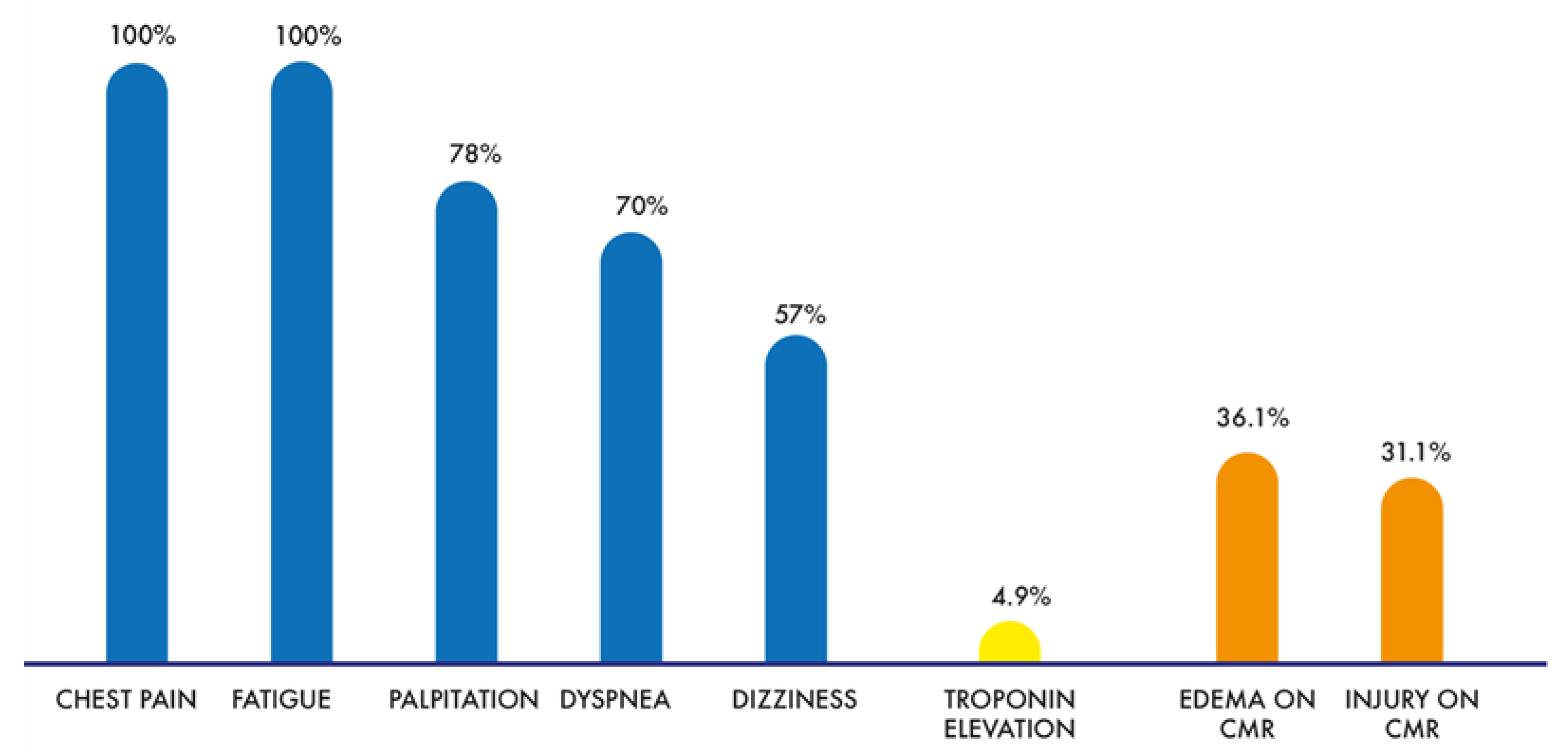
3.2. Clinical Presentation and Laboratory Results
3.3. Echocardiographic Findings
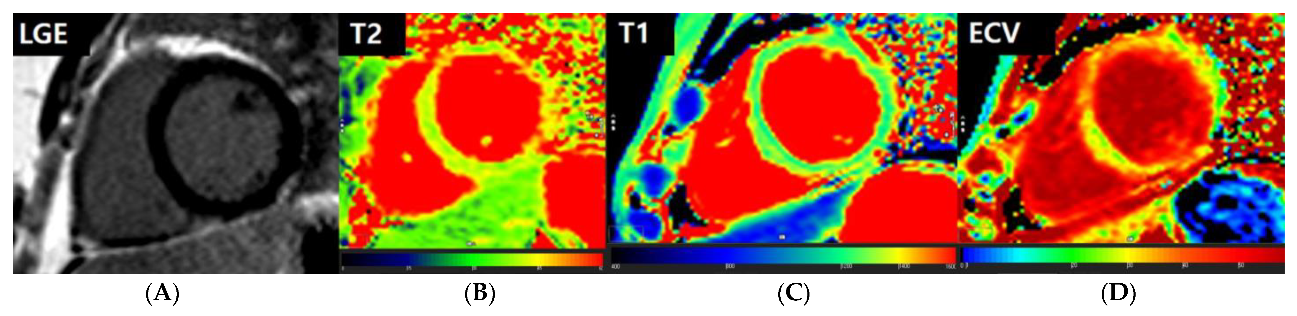
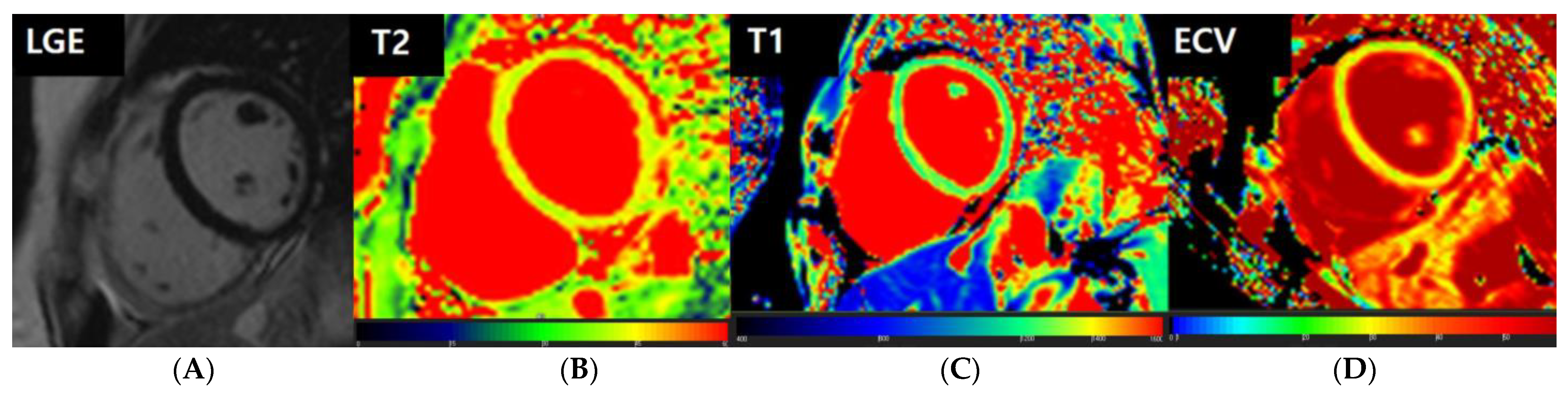
3.4. CMR Findings
3.5. Logistic Regression Analyses for Myocardial Abnormalities on CMR
3.6. Clinical Courses of Patients
4. Discussion
5. Conclusions
Author Contributions
Funding
Institutional Review Board Statement
Informed Consent Statement
Data Availability Statement
Conflicts of Interest
References
- Sharma, O.; Sultan, A.A.; Ding, H.; Triggle, C.R. A review of the progress and challenges of developing a vaccine for COVID-19. Front. Immunol. 2020, 11, 585354. [Google Scholar] [CrossRef]
- Das, B.B.; Kohli, U.; Ramachandran, P.; Nguyen, H.H.; Greil, G.; Hussain, T.; Tandon, A.; Kane, C.; Avula, S.; Duru, C.; et al. Myopericarditis after messenger RNA coronavirus disease 2019 vaccination in adolescents 12 to 18 years of age. J. Pediatr. 2021, 238, 26–32.e1. [Google Scholar] [CrossRef] [PubMed]
- Ambati, S.; Colon, M.; Mihic, M.; Sanchez, J.; Bakar, A. Acute myopericarditis after COVID-19 vaccine in teenagers. Case Rep. Cardiol. 2021, 2021, 8268755. [Google Scholar] [CrossRef] [PubMed]
- Patel, Y.R.; Louis, D.W.; Atalay, M.; Agarwal, S.; Shah, N.R. Cardiovascular magnetic resonance findings in young adult patients with acute myocarditis following mrna COVID-19 vaccination: A case series. J. Cardiovasc. Magn. Reason. 2021, 23, 101. [Google Scholar] [CrossRef] [PubMed]
- Dionne, A.; Sperotto, F.; Chamberlain, S.; Baker, A.L.; Powell, A.J.; Prakash, A.; Castellanos, D.A.; Saleeb, S.F.; de Ferranti, S.D.; Newburger, J.W.; et al. Association of myocarditis with bnt162b2 messenger rna COVID-19 vaccine in a case series of children. JAMA Cardiol. 2021, 6, 1446–1450. [Google Scholar] [CrossRef]
- Siripanthong, B.; Nazarian, S.; Muser, D.; Deo, R.; Santangeli, P.; Khanji, M.Y.; Cooper, L.T., Jr.; Chahal, C.A.A. Recognizing COVID-19-related myocarditis: The possible pathophysiology and proposed guideline for diagnosis and management. Heart Rhythm. 2020, 17, 1463–1471. [Google Scholar] [CrossRef] [PubMed]
- Qian, Z.; Travanty, E.A.; Oko, L.; Edeen, K.; Berglund, A.; Wang, J.; Ito, Y.; Holmes, K.V.; Mason, R.J. Innate immune response of human alveolar type ii cells infected with severe acute respiratory syndrome-coronavirus. Am. J. Respir. Cell Mol. Biol. 2013, 48, 742–748. [Google Scholar] [CrossRef] [Green Version]
- Goulter, A.B.; Goddard, M.J.; Allen, J.C.; Clark, K.L. Ace2 gene expression is up-regulated in the human failing heart. BMC Med. 2004, 2, 19. [Google Scholar] [CrossRef]
- Guo, J.; Wei, X.; Li, Q.; Li, L.; Yang, Z.; Shi, Y.; Qin, Y.; Zhang, X.; Wang, X.; Zhi, X.; et al. Single-cell rna analysis on ace2 expression provides insights into SARS-CoV-2 potential entry into the bloodstream and heart injury. J. Cell Physiol. 2020, 235, 9884–9894. [Google Scholar] [CrossRef]
- Weiss, S.R. Forty years with coronaviruses. J. Exp. Med. 2020, 217, e20200537. [Google Scholar] [CrossRef] [Green Version]
- Cooper, L.T., Jr. Myocarditis. N. Engl. J. Med. 2009, 360, 1526–1538. [Google Scholar] [CrossRef] [Green Version]
- Sagar, S.; Liu, P.P.; Cooper, L.T., Jr. Myocarditis. Lancet 2012, 379, 738–747. [Google Scholar] [CrossRef] [Green Version]
- Park, J.; Brekke, D.R.; Bratincsak, A. Self-limited myocarditis presenting with chest pain and st segment elevation in adolescents after vaccination with the bnt162b2 mrna vaccine. Cardiol. Young 2022, 32, 146–149. [Google Scholar] [CrossRef] [PubMed]
- Ferreira, V.M.; Schulz-Menger, J.; Holmvang, G.; Kramer, C.M.; Carbone, I.; Sechtem, U.; Kindermann, I.; Gutberlet, M.; Cooper, L.T.; Liu, P.; et al. Cardiovascular magnetic resonance in nonischemic myocardial inflammation: Expert recommendations. J. Am. Coll. Cardiol. 2018, 72, 3158–3176. [Google Scholar] [CrossRef]
- Kim, P.K.; Hong, Y.J.; Im, D.J.; Suh, Y.J.; Park, C.H.; Kim, J.Y.; Chang, S.; Lee, H.J.; Hur, J.; Kim, Y.J.; et al. Myocardial t1 and t2 mapping: Techniques and clinical applications. Korean J. Radiol. 2017, 18, 113–131. [Google Scholar] [CrossRef] [Green Version]
- Truong, D.T.; Dionne, A.; Muniz, J.C.; McHugh, K.E.; Portman, M.A.; Lambert, L.M.; Thacker, D.; Elias, M.D.; Li, J.S.; Toro-Salazar, O.H.; et al. Clinically suspected myocarditis temporally related to COVID-19 vaccination in adolescents and young adults: Suspected myocarditis after COVID-19 vaccination. Circulation 2022, 145, 345–356. [Google Scholar] [CrossRef]
- Caforio, A.L.; Pankuweit, S.; Arbustini, E.; Basso, C.; Gimeno-Blanes, J.; Felix, S.B.; Fu, M.; Helio, T.; Heymans, S.; Jahns, R.; et al. Current state of knowledge on aetiology, diagnosis, management, and therapy of myocarditis: A position statement of the european society of cardiology working group on myocardial and pericardial diseases. Eur. Heart J. 2013, 34, 2636–2648, 2648a–2648d. [Google Scholar] [CrossRef] [PubMed]
- Writing Committee, M.; Yancy, C.W.; Jessup, M.; Bozkurt, B.; Butler, J.; Casey, D.E., Jr.; Drazner, M.H.; Fonarow, G.C.; Geraci, S.A.; Horwich, T.; et al. 2013 accf/aha guideline for the management of heart failure: A report of the american college of cardiology foundation/american heart association task force on practice guidelines. Circulation 2013, 128, e240–e327. [Google Scholar]
- Law, Y.M.; Lal, A.K.; Chen, S.; Cihakova, D.; Cooper, L.T., Jr.; Deshpande, S.; Godown, J.; Grosse-Wortmann, L.; Robinson, J.D.; Towbin, J.A.; et al. Diagnosis and management of myocarditis in children: A scientific statement from the american heart association. Circulation 2021, 144, e123–e135. [Google Scholar] [CrossRef]
- Oster, M.E.; Shay, D.K.; Su, J.R.; Gee, J.; Creech, C.B.; Broder, K.R.; Edwards, K.; Soslow, J.H.; Dendy, J.M.; Schlaudecker, E.; et al. Myocarditis cases reported after mrna-based COVID-19 vaccination in the us from December 2020 to August 2021. JAMA 2022, 327, 331–340. [Google Scholar] [CrossRef]
- Sachdeva, S.; Song, X.; Dham, N.; Heath, D.M.; DeBiasi, R.L. Analysis of clinical parameters and cardiac magnetic resonance imaging as predictors of outcome in pediatric myocarditis. Am. J. Cardiol. 2015, 115, 499–504. [Google Scholar] [CrossRef] [PubMed]
- Luetkens, J.A.; Faron, A.; Isaak, A.; Dabir, D.; Kuetting, D.; Feisst, A.; Schmeel, F.C.; Sprinkart, A.M.; Thomas, D. Comparison of original and 2018 lake louise criteria for diagnosis of acute myocarditis: Results of a validation cohort. Radiol. Cardiothorac. Imaging 2019, 1, e190010. [Google Scholar] [CrossRef] [PubMed]
- Luetkens, J.A.; Doerner, J.; Thomas, D.K.; Dabir, D.; Gieseke, J.; Sprinkart, A.M.; Fimmers, R.; Stehning, C.; Homsi, R.; Schwab, J.O.; et al. Acute myocarditis: Multiparametric cardiac mr imaging. Radiology 2014, 273, 383–392. [Google Scholar] [CrossRef] [Green Version]
- Bohnen, S.; Radunski, U.K.; Lund, G.K.; Ojeda, F.; Looft, Y.; Senel, M.; Radziwolek, L.; Avanesov, M.; Tahir, E.; Stehning, C.; et al. Tissue characterization by t1 and t2 mapping cardiovascular magnetic resonance imaging to monitor myocardial inflammation in healing myocarditis. Eur. Heart J. Cardiovasc. Imaging 2017, 18, 744–751. [Google Scholar] [CrossRef] [PubMed]
- Bozkurt, B.; Kamat, I.; Hotez, P.J. Myocarditis with COVID-19 mrna vaccines. Circulation 2021, 144, 471–484. [Google Scholar] [CrossRef]
- Gargano, J.W.; Wallace, M.; Hadler, S.C.; Langley, G.; Su, J.R.; Oster, M.E.; Broder, K.R.; Gee, J.; Weintraub, E.; Shimabukuro, T.; et al. Use of mrna COVID-19 vaccine after reports of myocarditis among vaccine recipients: Update from the advisory committee on immunization practices—United States, June 2021. MMWR Morb. Mortal. Wkly Rep. 2021, 70, 977–982. [Google Scholar] [CrossRef]
- Kaul, R.; Sreenivasan, J.; Goel, A.; Malik, A.; Bandyopadhyay, D.; Jin, C.; Sharma, M.; Levine, A.; Pan, S.; Fuisz, A.; et al. Myocarditis following COVID-19 vaccination. Int. J. Cardiol. Heart Vasc. 2021, 36, 100872. [Google Scholar] [CrossRef]
- Jain, S.S.; Steele, J.M.; Fonseca, B.; Huang, S.; Shah, S.; Maskatia, S.A.; Buddhe, S.; Misra, N.; Ramachandran, P.; Gaur, L.; et al. COVID-19 vaccination-associated myocarditis in adolescents. Pediatrics 2021, 148, e2021053427. [Google Scholar] [CrossRef]
- Isaak, A.; Feisst, A.; Luetkens, J.A. Myocarditis following COVID-19 vaccination. Radiology 2021, 301, E378–E379. [Google Scholar] [CrossRef]
- Banka, P.; Robinson, J.D.; Uppu, S.C.; Harris, M.A.; Hasbani, K.; Lai, W.W.; Richmond, M.E.; Fratz, S.; Jain, S.; Johnson, T.R.; et al. Cardiovascular magnetic resonance techniques and findings in children with myocarditis: A multicenter retrospective study. J. Cardiovasc. Magn. Reason. 2015, 17, 96. [Google Scholar] [CrossRef] [Green Version]
- Shiyovich, A.; Witberg, G.; Aviv, Y.; Eisen, A.; Orvin, K.; Wiessman, M.; Grinberg, T.; Porter, A.; Kornowski, R.; Hamdan, A. Myocarditis following COVID-19 vaccination: Magnetic resonance imaging study. Eur. Heart J. Cardiovasc. Imaging 2021, 23, 1075–1082. [Google Scholar] [CrossRef] [PubMed]
- Bellos, I.; Karageorgiou, V.; Viskin, D. Myocarditis following mrna COVID-19 vaccination: A pooled analysis. Vaccine 2022, 40, 1768–1774. [Google Scholar] [CrossRef] [PubMed]


| All Patients (n = 61) | Myocardial Edema on CMR | Myocardial Injury on CMR | Myocardial Edema or Injury (Abnormality) on CMR | |||||||
|---|---|---|---|---|---|---|---|---|---|---|
| (+) (n = 22) | (−) (n = 39) | p-Value | (+) (n = 19) | (−) (n = 42) | p-Value | (+) (n = 24) | (−) (n = 37) | p-Value | ||
| Age (years) | 14.4 ± 1.9 | 15.0 ± 2.1 | 14.1 ± 1.8 | 0.0915 | 15.1 ± 2.2 | 14.2 ± 1.8 | 0.0983 | 15.0 ± 2.0 | 14.1 ± 1.8 | 0.0699 |
| Male:Female | 28:33 | 3:19 | 25:14 | 0.0001 | 3:16 | 25:17 | 0.0015 | 3:21 | 25:12 | <0.0001 |
| Height (cm) | 164.1 ± 9.4 | 161.3 ± 7.7 | 165.7 ± 9.9 | 0.0736 | 160.6 ± 8.5 | 165.7 ± 9.4 | 0.0494 | 161.1 ± 7.6 | 166.1 ± 10.0 | 0.0429 |
| Weight (kg) | 59.0 ± 14.7 | 52.9 ± 11.9 | 62.5 ± 15.1 | 0.0135 | 54.1 ± 12.0 | 61.3 ± 15.4 | 0.0749 | 53.0 ± 11.5 | 63.0 ± 15.3 | 0.0082 |
| BMI (kg/m2) | 21.7 ± 3.9 | 20.2 ± 3.7 | 22.5 ± 3.9 | 0.0291 | 20.8 ± 3.6 | 22.1 ± 4.0 | 0.2464 | 20.3 ± 3.5 | 22.6 ± 3.9 | 0.0239 |
| BSA (m2) | 1.64 ± 0.23 | 1.54 ± 0.19 | 1.69 ± 0.24 | 0.0144 | 1.56 ± 0.20 | 1.68 ± 0.24 | 0.0629 | 1.54 ± 0.19 | 1.70 ± 0.24 | 0.0083 |
| All Patients (n = 61) | Myocardial Edema on CMR | Myocardial Injury on CMR | Myocardial Edema or Injury (Abnormality) on CMR | |||||||
|---|---|---|---|---|---|---|---|---|---|---|
| (+) (n = 22) | (−) (n = 39) | p-Value | (+) (n = 19) | (−) (n = 42) | p-Value | (+) (n = 24) | (−) (n = 37) | p-Value | ||
| Vaccination (1st:2nd) | 26:35 | 8:14 | 18:21 | 0.4578 | 7:12 | 19:23 | 0.5392 | 9:15 | 17:20 | 0.5146 |
| Duration * (median, (1Q, 3Q)) | 7.0 (4.0, 16.0) | 7.0 (3.0, 14.0) | 7.0 (5.0, 22.0) | 0.4117 | 7.0 (4.0, 16.0) | 7.0 (4.0, 22.0) | 0.6619 | 7.0 (3.0, 14.0) | 7.0 (4.5, 19.5) | 0.5636 |
| CRP (mg/L) | 2.8 ± 4.9 | 2.9 ± 3.7 | 2.8 ± 5.5 | 0.896 | 3.0 ± 3.8 | 2.8 ± 5.3 | 0.839 | 2.7 ± 3.5 | 2.9 ± 5.6 | 0.880 |
| CK-MB (ng/mL) | 0.95 ± 1.53 | 1.20 ± 2.12 | 0.81 ± 1.10 | 0.356 | 1.31 ± 2.35 | 0.80 ± 1.06 | 0.244 | 1.12 ± 2.04 | 0.84 ± 1.13 | 0.499 |
| NTproBNP (pg/mL) | 30.5 ± 31.5 | 33.2 ± 24.9 | 29.0 ± 34.7 | 0.624 | 33.4 ± 26.1 | 29.3 ± 33.5 | 0.661 | 31.9 ± 24.1 | 29.6 ± 35.5 | 0.784 |
| Hemoglobin (g/dL) | 13.5 ± 1.1 | 12.8 ± 0.9 | 13.9 ± 1.0 | <0.0001 | 12.9 ± 0.9 | 13.8 ± 1.1 | 0.0017 | 12.8 ± 0.9 | 14.0 ± 1.0 | <0.0001 |
| Hematocrit (%) | 41.7 ± 3.4 | 39.6 ± 2.9 | 42.9 ± 3.1 | 0.0001 | 39.7 ± 2.8 | 42.6 ± 3.2 | 0.001 | 39.6 ± 2.8 | 43.1 ± 3.0 | <0.0001 |
| Serum iron (mcg/dL) | 80.1 ± 40.7 | 76.5 ± 43.2 | 82.1 ± 39.7 | 0.6052 | 84.8 ± 43.8 | 77.9 ± 39.6 | 0.5434 | 79.0 ± 43.0 | 80.8 ± 39.7 | 0.8688 |
| Iron saturation (%) | 22.7 ± 11.8 | 22.6 ± 12.9 | 22.7 ± 11.3 | 0.9946 | 25.4 ± 12.8 | 21.4 ± 11.2 | 0.2266 | 23.1 ± 12.6 | 22.4 ± 11.4 | 0.8183 |
| Ferritin (ng/mL) | 51.6 ± 37.8 | 46.2 ± 37.2 | 54.7 ± 38.3 | 0.4039 | 50.7 ± 39.0 | 52.1 ± 37.7 | 0.8999 | 45.2 ± 36.5 | 55.8 ± 38.6 | 0.2896 |
| All Patients (n = 61) | Myocardial Edema or Injury (Abnormality) on CMR | |||
|---|---|---|---|---|
| Abnormality (+) (n = 24) | Abnormality (−) (n = 37) | p-Value | ||
| LVEF (%) | 67.50 ± 5.48 | 68.99 ± 4.49 | 66.54 ± 5.90 | 0.0879 |
| LVEDD (mm) | 47.30 ± 4.13 | 46.83 ± 4.15 | 47.60 ± 4.14 | 0.4852 |
| LVESD (mm) | 28.98 ± 4.76 | 28.61 ± 2.89 | 29.21 ± 5.69 | 0.5906 |
| IVSd (mm) | 7.09 ± 1.40 | 7.01 ± 1.22 | 7.15 ± 1.51 | 0.7184 |
| IVSs (mm) | 9.96 ± 1.83 | 9.90 ± 1.83 | 10.00 ± 1.85 | 0.8433 |
| LVPWd (mm) | 6.63 ± 1.30 | 6.66 ± 1.28 | 6.62 ± 1.33 | 0.9027 |
| LVPWs (mm) | 11.39 ± 2.45 | 11.49 ± 3.06 | 11.32 ± 2.00 | 0.8053 |
| Mitral E (m/s) | 1.022 ± 0.16 | 1.012 ± 0.19 | 1.029 ± 0.134 | 0.6652 |
| Mitral A (m/s) | 0.489 ± 0.118 | 0.485 ± 0.096 | 0.491 ± 0.131 | 0.8631 |
| E/A | 2.20 ± 0.53 | 2.13 ± 0.46 | 2.24 ± 0.57 | 0.4130 |
| Lateral E′ (m/s) | 0.179 ± 0.036 | 0.180 ± 0.045 | 0.178 ± 0.029 | 0.8155 |
| Lateral A′ (m/s) | 0.079 ± 0.071 | 0.092 ± 0.111 | 0.071 ± 0.018 | 0.3650 |
| Lateral S′ (m/s) | 0.129 ± 0.097 | 0.117 ± 0.026 | 0.137 ± 0.122 | 0.3369 |
| Lateral E/E′ | 5.76 ± 1.23 | 5.50 ± 1.06 | 5.93 ± 1.31 | 0.1783 |
| Septal E′ (m/s) | 0.146 ± 0.024 | 0.146 ± 0.026 | 0.145 ± 0.023 | 0.9462 |
| Septal A′ (m/s) | 0.069 ± 0.083 | 0.057 ± 0.009 | 0.076 ± 0.106 | 0.2769 |
| Septal S′ (m/s) | 0.088 ± 0.016 | 0.089 ± 0.015 | 0.088 ± 0.017 | 0.8778 |
| Septal E/E′ | 7.25 ± 1.63 | 7.27 ± 2.06 | 7.24 ± 1.32 | 0.9615 |
| Tricuspid E′ (m/s) | 0.158 ± 0.029 | 0.163 ± 0.032 | 0.156 ± 0.027 | 0.3684 |
| Tricuspid A′ (m/s) | 0.093 ± 0.026 | 0.091 ± 0.019 | 0.095 ± 0.030 | 0.5780 |
| Tricuspid S′ (m/s) | 0.136 ± 0.018 | 0.137 ± 0.016 | 0.136 ± 0.019 | 0.8509 |
| TAPSE (mm) | 22.88 ± 3.31 | 22.81 ± 3.37 | 22.91 ± 3.33 | 0.9137 |
| PA velocity (m/s) | 0.99 ± 0.18 | 0.99 ± 0.21 | 0.99 ± 0.17 | 0.9996 |
| IVC diameter (mm) | 18.68 ± 3.30 | 18.66 ± 3.14 | 18.68 ± 3.45 | 0.9811 |
| All Patients (n = 61) | Myocardial Edema on CMR | Myocardial Injury on CMR | Myocardial Edema or Injury (Abnormality) on CMR | |||||||
|---|---|---|---|---|---|---|---|---|---|---|
| (+) (n = 22) | (−) (n = 39) | p-Value | (+) (n = 19) | (−) (n = 42) | p-Value | (+) (n = 24) | (−) (n = 37) | p-Value | ||
| LVEDV (mL) | 123.1 ± 31.1 | 112.5 ± 26.7 | 129.1 ± 32.1 | 0.0435 | 112.7 ± 27.9 | 127.8 ± 31.6 | 0.0777 | 112.3 ± 26.0 | 130.1 ± 32.4 | 0.0282 |
| LVESV (mL) | 44.1 ± 14.6 | 39.5 ± 11.0 | 46.7 ± 15.8 | 0.0656 | 39.5 ± 11.4 | 46.2 ± 15.5 | 0.0959 | 39.6 ± 10.8 | 47.0 ± 16.1 | 0.0369 |
| SV (mL) | 78.9 ± 19.8 | 73.0 ± 17.5 | 82.2 ± 20.4 | 0.0797 | 73.3 ± 18.5 | 81.4 ± 20.0 | 0.1369 | 72.7 ± 17.0 | 82.9 ± 20.6 | 0.05 |
| LVEF (%) | 64.6 ± 5.5 | 64.9 ± 3.8 | 64.5 ± 6.3 | 0.7584 | 65.0 ± 3.9 | 64.5 ± 6.1 | 0.718 | 64.8 ± 3.7 | 64.5 ± 6.5 | 0.8653 |
| LV mass (g) | 93.4 ± 24.0 | 81.2 ± 17.6 | 100.3 ± 24.4 | 0.0021 | 83.4 ± 18.6 | 98.0 ± 24.8 | 0.026 | 81.1 ± 17.6 | 101.4 ± 24.2 | 0.0008 |
| LVEDVi (mL/m2) | 74.6 ± 12.2 | 72.4 ± 12.4 | 75.8 ± 12.1 | 0.3085 | 71.8 ± 12.2 | 75.8 ± 12.2 | 0.2342 | 72.4 ± 12.0 | 76.0 ± 12.3 | 0.2606 |
| LVESVi (mL/m2) | 26.7 ± 6.9 | 25.5 ± 6.0 | 27.3 ± 7.3 | 0.3285 | 25.2 ± 5.8 | 27.3 ± 7.3 | 0.2599 | 25.6 ± 5.8 | 27.4 ± 7.5 | 0.3232 |
| SVi (mL/m2) | 47.9 ± 8.3 | 47.0 ± 7.8 | 48.4 ± 8.6 | 0.5261 | 46.7 ± 8.0 | 48.4 ± 8.4 | 0.446 | 46.8 ± 7.5 | 48.5 ± 8.8 | 0.4354 |
| LV mass-i (g/m2) | 56.4 ± 8.4 | 52.3 ± 7.2 | 58.6 ± 8.2 | 0.0036 | 53.2 ± 7.3 | 57.8 ± 8.5 | 0.0437 | 52.2 ± 7.3 | 59.0 ± 8.0 | 0.0014 |
| LGE | None | |||||||||
| T2 value (ms) | 39.5 ± 2.0 | 41.9 ± 0.8 | 38.2 ± 1.0 | <0.0001 | 41.3 ± 1.4 | 38.7 ± 1.7 | <0.0001 | 41.5 ± 1.4 | 38.2 ± 1.0 | <0.0001 |
| T1 value (ms) | 1225.0 ± 45.7 | 1268.7 ± 37.5 | 1200.3 ± 28.1 | <0.0001 | 1265.9 ± 37.6 | 1206.5 ± 36.2 | <0.0001 | 1265.6 ± 37.6 | 1198.6 ± 27.6 | <0.0001 |
| ECV fraction (%) | 27.1 ± 2.6 | 29.4 ± 2.5 | 25.8 ± 1.4 | <0.0001 | 30.2 ± 1.9 | 25.7 ± 1.3 | <0.0001 | 29.4 ± 2.4 | 25.6 ± 1.2 | <0.0001 |
| Myocardial Edema on CMR | Myocardial Injury on CMR | Myocardial Edema or Injury (Abnormality) on CMR | ||||
|---|---|---|---|---|---|---|
| OR (95% CI) | p-Value | OR (95% CI) | p-Value | OR (95% CI) | p-Value | |
| Demography | ||||||
| Age (years) | 1.314 (0.952–1.812) | 0.0964 | 1.326 (0.946–1.859) | 0.1013 | 1.334 (0.970–1.834) | 0.0762 |
| Sex (male:female) | 11.310 (2.839–45.057) | 0.0006 | 7.843 (1.976–31.128) | 0.0034 | 14.583 (3.626–58.657) | 0.0002 |
| Height (cm) | 0.947 (0.889–1.009) | 0.0899 | 0.940 (0.880–1.005) | 0.0680 | 0.939 (0.879–1.002) | 0.0586 |
| Weight (kg) | 0.947 (0.904–0.992) | 0.0209 | 0.962 (0.920–1.005) | 0.0825 | 0.944 (0.902–0.989) | 0.0145 |
| BMI (kg/m2) | 0.843 (0.718–0.989) | 0.0365 | 0.915 (0.788–1.063) | 0.2450 | 0.842 (0.719–0.984) | 0.0310 |
| BSA (m2) | 0.037 (0.002–0.618) | 0.0217 | 0.083 (0.006–1.234) | 0.0707 | 0.030 (0.002–0.496) | 0.0143 |
| Duration | 1.017 (0.985–1.051) | 0.3020 | 1.025 (0.990–1.061) | 0.1671 | 1.016 (0.984–1.049) | 0.3383 |
| Laboratory Test | ||||||
| Hemoglobin (g/dL) | 0.246 (0.108–0.560) | 0.0008 | 0.366 (0.182–0.738) | 0.0049 | 0.202 (0.083–0.492) | 0.0004 |
| Hematocrit (%) | 0.658 (0.510–0.851) | 0.0014 | 0.697 (0.545–0.890) | 0.0039 | 0.619 (0.470–0.814) | 0.0006 |
| Serum iron (mcg/dL) | 0.996 (0.983–1.010) | 0.5992 | 1.004 (0.991–1.018) | 0.5374 | 0.999 (0.986–1.012) | 0.8661 |
| Iron saturation (%) | 1.000 (0.956–1.046) | 0.9945 | 1.029 (0.982–1.078) | 0.2260 | 1.005 (0.962–1.051) | 0.8146 |
| Ferritin (ng/mL) | 0.994 (0.979–1.009) | 0.4 | 0.999 (0.985–1.014) | 0.8979 | 0.992 (0.977–1.007) | 0.2888 |
| Echocardiography | ||||||
| LVEF (%) | 1.067 (0.958–1.188) | 0.2370 | 1.130 (0.995–1.283) | 0.0593 | 1.101 (0.984–1.232) | 0.0942 |
| LVEDD (mm) | 0.982 (0.863–1.117) | 0.7835 | 0.904 (0.781–1.046) | 0.1738 | 0.955 (0.839–1.086) | 0.4789 |
| LVESD (mm) | 0.994 (0.891–1.110) | 0.9210 | 0.944 (0.839–1.062) | 0.3369 | 0.974 (0.873–1.086) | 0.6326 |
| IVSd (mm) | 0.994 (0.681–1.451) | 0.9761 | 1.050 (0.710–1.552) | 0.8067 | 0.932 (0.642–1.354) | 0.7131 |
| IVSs (mm) | 1.009 (0.757–1.347) | 0.9495 | 0.932 (0.687–1.264) | 0.6495 | 0.971 (0.730–1.291) | 0.8401 |
| LVPWd (mm) | 1.034 (0.689–1.550) | 0.8719 | 1.032 (0.678–1.570) | 0.8844 | 1.026 (0.689–1.528) | 0.9005 |
| LVPWs (mm) | 1.017 (0.819–1.263) | 0.8758 | 1.141 (0.893–1.457) | 0.2906 | 1.030 (0.832–1.276) | 0.7832 |
| Mitral E (m/s) | 1.117 (0.794–1.571) | 0.5270 | 0.757 (0.519–1.105) | 0.1493 | 0.927 (0.662–1.298) | 0.6593 |
| Mitral A (m/s) | 0.994 (0.636–1.555) | 0.9797 | 0.926 (0.577–1.485) | 0.7497 | 0.961 (0.618–1.495) | 0.8603 |
| E/A | 0.989 (0.895–1.093) | 0.8320 | 0.944 (0.849–1.051) | 0.2930 | 0.959 (0.868–1.059) | 0.4066 |
| Mitral E′ (m/s) | 1.422 (0.324–6.247) | 0.6407 | 0.507 (0.108–2.374) | 0.3881 | 1.212 (0.285–5.149) | 0.7945 |
| Mitral A′ (m/s) | 1.593 (0.587–4.323) | 0.3605 | 2.078 (0.484–8.932) | 0.3255 | 1.698 (0.534–5.404) | 0.37 |
| Mitral S′ (m/s) | 0.662 (0.176–2.490) | 0.5420 | 0.487 (0.057–4.182) | 0.5119 | 0.671 (0.198–2.277) | 0.5225 |
| Mitral E/E′ | 0.983 (0.940–1.029) | 0.4626 | 0.966 (0.918–1.017) | 0.1876 | 0.968 (0.924–1.015) | 0.1810 |
| Septal E′ (m/s) | 1.098 (0.121–9.927) | 0.9338 | 1.211 (0.124–11.844) | 0.8695 | 1.079 (0.124–9.405) | 0.9451 |
| Septal A′ (m/s) | 0.335 (0.006–19.32) | 0.5965 | 0.145 (0.002–12.897) | 0.3990 | 0.260 (0.004–16.351) | 0.5241 |
| Septal S′ (m/s) | 2.133 (0.076–59.978) | 0.6564 | 1.037 (0.032–33.406) | 0.9838 | 1.301 (0.049–34.856) | 0.8752 |
| Septal E/E′ | 1.013 (0.981–1.046) | 0.4480 | 0.981 (0.945–1.018) | 0.3038 | 1.001 (0.970–1.033) | 0.9567 |
| Tricuspid E′ (m/s) | 2.475 (0.347–17.658) | 0.3660 | 2.836 (0.367–21.892) | 0.3176 | 2.469 (0.352–17.325) | 0.3631 |
| Tricuspid A′ (m/s) | 0.514 (0.055–4.816) | 0.5603 | 0.481 (0.045–5.106) | 0.5439 | 0.568 (0.063–5.119) | 0.6143 |
| Tricuspid S′ (m/s) | 1.648 (0.062–43.754) | 0.7653 | 2.276 (0.071–72.749) | 0.6417 | 1.374 (0.054–34.790) | 0.8473 |
| TAPSE (mm) | 0.989 (0.828–1.181) | 0.9013 | 0.910 (0.749–1.106) | 0.3440 | 0.990 (0.831–1.179) | 0.9115 |
| PA velocity (m/s) | 1.210 (0.064–22.802) | 0.8989 | 0.253 (0.008–7.752) | 0.4311 | 1.000 (0.055–18.083) | >0.9999 |
| IVC diameter (mm) | 1.023 (0.871–1.201) | 0.7839 | 0.893 (0.751–1.060) | 0.1962 | 0.998 (0.852–1.169) | 0.9807 |
| Myocardial Edema on CMR | Myocardial Injury on CMR | Myocardial Edema or Injury (Abnormality) on CMR | ||||
|---|---|---|---|---|---|---|
| OR (95% CI) | p-Value | OR (95% CI) | p-Value | OR (95% CI) | p-Value | |
| Age (years) | 1.495 (1.031–2.168) | 0.0340 | 1.392 (0.976–1.985) | 0.0679 | 1.573 (1.066–2.321) | 0.0224 |
| Female | 10.190 (2.347–44.252) | 0.0019 | 7.741 (1.786–33.547) | 0.0062 | 14.323 (3.188–64.357) | 0.0005 |
| BMI | 0.855 (0.706–1.035) | 0.1083 | 0.950 (0.794–1.136) | 0.5750 | 0.853 (0.703–1.035) | 0.1068 |
Disclaimer/Publisher’s Note: The statements, opinions and data contained in all publications are solely those of the individual author(s) and contributor(s) and not of MDPI and/or the editor(s). MDPI and/or the editor(s) disclaim responsibility for any injury to people or property resulting from any ideas, methods, instructions or products referred to in the content. |
© 2023 by the authors. Licensee MDPI, Basel, Switzerland. This article is an open access article distributed under the terms and conditions of the Creative Commons Attribution (CC BY) license (https://creativecommons.org/licenses/by/4.0/).
Share and Cite
Park, C.H.; Yang, J.; Lee, H.S.; Kim, T.H.; Eun, L.Y. Characteristics of Teenagers Presenting with Chest Pain after COVID-19 mRNA Vaccination. J. Clin. Med. 2023, 12, 4421. https://doi.org/10.3390/jcm12134421
Park CH, Yang J, Lee HS, Kim TH, Eun LY. Characteristics of Teenagers Presenting with Chest Pain after COVID-19 mRNA Vaccination. Journal of Clinical Medicine. 2023; 12(13):4421. https://doi.org/10.3390/jcm12134421
Chicago/Turabian StylePark, Chul Hwan, Juyeon Yang, Hye Sun Lee, Tae Hoon Kim, and Lucy Youngmin Eun. 2023. "Characteristics of Teenagers Presenting with Chest Pain after COVID-19 mRNA Vaccination" Journal of Clinical Medicine 12, no. 13: 4421. https://doi.org/10.3390/jcm12134421
APA StylePark, C. H., Yang, J., Lee, H. S., Kim, T. H., & Eun, L. Y. (2023). Characteristics of Teenagers Presenting with Chest Pain after COVID-19 mRNA Vaccination. Journal of Clinical Medicine, 12(13), 4421. https://doi.org/10.3390/jcm12134421