1. Introduction
As the elderly population shows increased life expectancy without a longer frailty period, medical care aiming to maintain the quality of life for elderly patients is increasing [
1]. For similar reasons, implant treatment for elderly patients is increasing worldwide. A marked increase in the number of implant surgeries after the year 2000 for patients aged over 70 years has been reported by the University of Bern [
1]. The number of patients aged over 75 years with an implant-supported prosthesis has increased in Switzerland over the past 20 years [
2]. Recently, due to developments in implant surfaces, a procedure that allows application of a functional load faster than the traditional osseointegration period has been introduced in many studies. Since the differentiation rate of osteoblasts decreases with increasing age, older patients should be given a longer period for osseointegration [
3,
4,
5,
6].
In addition, clinical data on the implant osseointegration period according to the volume and density of the alveolar bone, and whether bone is transplanted, are also needed. It is of utmost importance to quantify implant stability at various time points and to determine and establish the timing of loading [
7,
8]. The changes in implant stability over time during the healing period could reveal more definite results upon comparing the physiological responses following implant placement in young and elderly patients. Implant stability occurs in two stages: primary and secondary [
7,
8]. Primary stability at implant installation is achieved by physical congruence between the surgically created bone bed and the implant, which is dependent on the macroscopic implant design, surgical technique, and bone density [
7]. During the osseointegration healing period, bone gradually forms inside the implant threads, and thus, secondary stability is attained by an incremental degree of bone-to-implant contact [
7]. The transition period in which the primary stability decreases while the secondary stability becomes established is a critical period with increased risk of osseointegration following micro-movement of the implant [
7]. Primary and secondary implant stability significantly relies on factors such as bone density, wound healing process, and bone metabolism, which could be affected by the age of the patient [
8].
Elderly patients undergoing implant surgery possess a higher risk compared to younger patients, owing to their medical condition and the adverse effects of medications being taken. Moreover, the differences in the bone quality and metabolism between elderly and young patients could result in differences in the osseointegration patterns following implant placement. The bone matrix is composed of bone mineral, collagen, bone water, and non-collagenous proteins [
3], and the components of the bone matrix change with aging, ultimately causing deterioration of the mechanical properties of bone, including increased brittleness and skeletal fragility [
4,
5]. In particular, the changes in non-collagenous proteins, which have molecular signaling functions regulating bone remodeling, alter the ability of bone to respond to external stimuli [
3,
6].
Despite age-related risk factors, previous studies did not demonstrate definite results that advanced age negatively affects osseointegration. Brocard et al. reported the lowest success rate in older patients [
9]. Bertl et al. reported that patients over 80 years of age showed a higher rate of early implant loss than younger patients [
10]. However, Schimmel et al. reported high implant survival rates in patients aged over 75 years (97.3% for 1 year and 96.1% for 3 years) based on a systematic review and meta-analysis [
11]. Jemt et al. demonstrated that younger patients, rather than older edentulous patients, had a higher risk of implant failure [
12]. As implant failure is a multifactorial complication, previous studies demonstrated that implant failure can be affected by the compliance method of the patient group and the selected statistical analysis methods based on age.
Therefore, the measurement of implant stability is even more necessary now, and two types of widely used non-invasive diagnostic methods have been developed and examined: resonance frequency analysis (RFA) and damping capacity analysis (DCA) [
13,
14]. In RFA, a method used by the Osstell ISQ Mentor (Osstell, Göteborg, Sweden), the stiffness of the implant–bone interface is outputted from the resonance frequency that is the response to oscillations applied to the implant–bone system [
14]. One DCA system device, Periotest M (Siemens AG, Bensheim, Germany), has been utilized to assess the mobility of natural teeth and is maintained to have the potential to reliably evaluate the stability of the implant–bone interface [
13,
14]. Recently, the new DCA system device Anycheck (Neobiotech, Seoul, Republic of Korea) has been released; it is a measuring device that uses percussion and has improved accuracy and reduced patient discomfort by reducing the intensity of the percussion [
15]. This system evaluates the duration of contact between an impacting rod and a healing abutment. It strikes the healing abutment six times over 2 s and converts the duration into implant stability test (IST) values [
15]. This system strikes a healing abutment with less force than Periotest M does and has a function that allows it to stop automatically if the stability is low to protect the implant [
15]. However, little is known about the factors affecting the IST values or the reliability of the device.
Multiple previous studies have reported on the correlation between RFA and DCA device results indicating the stability of the same implant. An in vitro study presented the strong correlation between the results from RFA and DCA devices [
16]. However, an in vivo study presented the relatively lower correlation between the results from RFA and DCA devices [
17]. The different results under experimental and clinical conditions suggest that there are clinical factors which affect the reliability of implant stability measuring devices. In the experimental condition, implant stability can be examined without any obstacles and the device can be positioned in relation to the implant in an ideal way. Clinically, examination of the stability of the implant in the oral cavity may have access difficulties due to the cheek, tongue, and contralateral teeth. These obstacles may unfavorably influence the factors needed for accurate measurement of implant stability, including exact contact between implant and device, the angle of the device to the implant, and the angle of the device against gravity. In addition, in the clinical condition, the accessibility and angle of the device are influenced by the location of the implant in the oral cavity (anterior/posterior, left/right, and mandible/maxilla).
Therefore, reliable implant stability measurement is required to evaluate the degree of osseointegration according to the implant placement site. However, there are few prospective clinical studies evaluating implant stability and measuring device reliability, according to the implant placement site and post-implantation duration in older patients. Therefore, the aim of this in vivo study was to evaluate implant stability and the reliability of different measuring devices, according to the implant placement site in the oral cavity and the duration of implantation in patients over 65 years. Additionally, one RFA device (Osstell) and two DCA devices (Periotest and Anycheck) were used to examine implants located in the maxillary right-posterior, maxillary anterior, maxillary left-posterior, mandibular right-posterior, mandibular anterior, and mandibular left-posterior positions.
The null hypotheses were that (1) the implant placement site and duration of implantation in the oral cavity do not affect implant stability and measuring device reliability in patients over 65 years, and (2) a correlation of 1 is shown by the three measuring devices.
3. Results
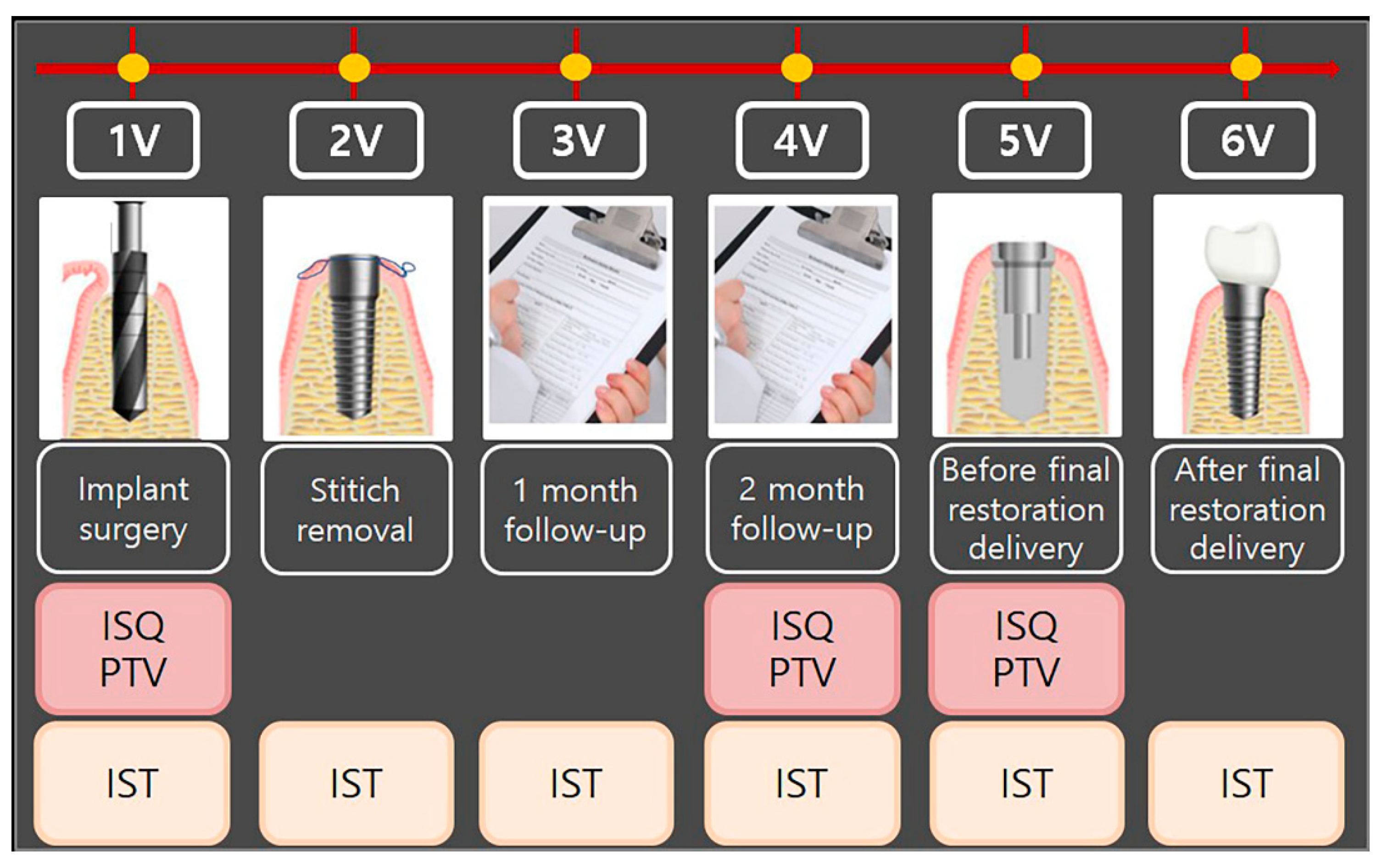
Implant stability measurement was performed using various devices. Mean values and standard deviations of ISQ, PTV, and IST among the groups, according to post-implantation duration, are shown in
Figure 3. For all the ISQ, PTV, and IST results, the implant stability results at the 2-month follow-up and before the final restoration delivery were significantly higher than those at the time of implant surgery. The significant differences in ISQ, PTV, and IST among the groups, according to post-implantation duration, are shown in
Table 5. For the ISQ and PTV, there were statistically significant differences between the first and fourth visits, first and fifth visits, and fourth and fifth visits (
p < 0.05). For the IST, there were significant differences at all points except those between the first and second visits, first and third visits, second and third visits, and third and fourth visits (
p < 0.05).
Mean values and standard deviations of implant stability measurements made with different devices according to dental implant placement site and duration are shown in
Figure 4. For the ISQ, there were statistically significant differences according to the post-implantation duration for each location of the inserted implants between the first and fourth visits, first and fifth visits, and fourth and fifth visits (
Table 6).
The results of Pearson’s correlation between the mean ISQ, mean PTV, and mean IST results are presented in
Table 7. At the first visit, the r between the ISQ and PTV results was −0.208, verifying the weak negative correlation (
p = 0.049). The r between the ISQ and IST results was 0.567, verifying the moderate positive correlation (
p < 0.001). Additionally, the r between the PTV and IST results was −0.490, verifying the moderate negative correlation (
p < 0.001). At the fourth visit, the r between the ISQ and PTV results was −0.298, verifying the weak negative correlation (
p = 0.001). The r between the ISQ and IST results was 0.367, verifying the weak positive correlation (
p = 0.003). Additionally, the r between the PTV and IST results was −0.701, verifying the strong negative correlation (
p < 0.001). At the fifth visit, the r between the ISQ and PTV results was −0.252, verifying the weak negative correlation (
p = 0.005). The r between the ISQ and IST results was 0.503, verifying the moderate positive correlation (
p < 0.001). Additionally, the r between the PTV and IST results was −0.479, demonstrating the moderate negative correlation (
p < 0.001).
The results of all groups for the implant stability values between the locations of implants and the positions of the arch in Osstell ISQ Mentor, Periotest M, and Anycheck are shown in
Table 8. The ISQ results showed statistically significant differences at the fourth (
p = 0.016) and fifth visits (
p = 0.042). In the PTV results, there were no significant differences in the correlations between the locations of the implants at all the visits (
p > 0.05). In the IST results, there was a statistically significant difference only at the fifth visit (
p = 0.044) in the correlations between the locations of implants.
4. Discussion
In the present study, the accuracy of implant stability measurement devices was evaluated under clinical conditions affecting the reliability of the devices. In addition to the results from the devices, the valid impacts and the angle formed by the handpiece with the horizontal plane were measured to analyze the reasons for inaccurate results. The results showed that the implant placement site and the post-implantation duration in the oral cavity affected implant stability and the reliability of the measuring devices in patients over 65 years of age.
Devices for evaluating implant stability are either RFA or DCA and each device has distinct characteristics depending on their operating principles. A DCA device is convenient for measurement without an additional process if a healing abutment is installed; more factors during measuring should be controlled to derive accurate results compared to RFA devices [
22]. According to previous studies, the PTV is influenced by the length of the fixture and the healing abutment, the position and direction of percussion, and the angle of the handpiece [
23,
24].
Between the DCA devices, Periotest M was significantly affected by the position of artificial bone model impact error, while Anycheck showed consistently low impact error in this study. The results showed that Anycheck was able to provide a relatively stable measurement under unfavorable access conditions. Anycheck measured while in contact with the implant, the device does not move minutely during measurement, and it is possible to measure stably at the desired position. However, Periotest M is unstable because it is measured at a certain distance from the implant. Faulkner et al. reported that Periotest M was very sensitive to the angulation of the handpiece and to the position at which the Periotest M impacted the abutment [
25]. A small change in the angle of the handpiece from 90 degrees to the abutment may cause a PTV difference between 2.5 and 4.0, as the rod hits an inconsistent point of abutment [
26,
27]. In addition, the variation in PTV was approximately 1.5 or 1 to 2 PTV depending on the height of the striking point per millimeter [
25,
26].
Some studies have investigated conflicting results for both RFA and DCA systems. Lee et al. investigated the strong correlation (0.981) between the ISQ and IST in an in vitro study [
13], while a systematic review showed a weak correlation (−0.294) between the ISQ and PTV [
15]. Additionally, the correlating ISQ and PTV readings of the buccal surface during implant installation were moderately negatively statistically significantly correlated (−0.466) between the two types of device for all 80 patients in the randomized clinical trial by Andreotti et al. [
14]. In this study, there were weak negative statistically significant correlations: −0.208 at 1V, −0.298 at 4V, and −0.252 at 5V, between ISQ and PTV. There were moderate positive statistically significant correlations: 0.567 at 1V, 0.367 at 4V, and 0.503 at 5V, between ISQ and IST. The results of this study are similar to the reported correlations in previous studies [
14,
15]. A factor that can affect the results is when using the DCA device clinically, the examiner is limited by patient cooperation, space, and access, unlike the laboratory study, where standardized models for measurement permit certain conditions. Thus, in vivo analyses have additional sources of error that could result in reduced accuracy of measurement. The results presented the weak or moderate statistically significant correlation between the three measuring devices.
Several studies have presented the strong correlation between ISQ and PTV, while others have presented no correlation [
28,
29,
30,
31]. Because of the discrepancies, standardized implant stability values have not yet been proved and analyses have been performed by other analytic methods, such as the measurement of ITV and radiographic and clinical examinations. There were moderate to high negative statistically significant correlations: −0.490 at 1V, −0.701 at 4V, and −0.479 at 5V, between PTV and IST. There was a moderate positive statistically significant correlation coefficient of 0.414 between the 5V and 6V, in IST (
p < 0.001).
Some studies have investigated whether both the Osstell ISQ and Periotest devices could reliably evaluate the stability of the implant [
13,
29,
32]. Lachmann et al. maintained that both the Osstell ISQ and Periotest presented acceptable reliability in expecting the implant stability in an in vitro study [
13]. Also, Pang et al. reported the strong correlation between the ISQ and PTV post-surgery and 2 months later [
29]. An animal study showed the strong correlation between ISQ and PTV [
31]. Additionally, some studies demonstrated that although both the Osstell ISQ and Periotest devices were useful for analyzing the stability of the implant, the Osstell ISQ was more accurate than the Periotest systems, presenting high reliability [
33,
34]. However, some studies have found conflicting results for both the Osstell ISQ and Periotest systems [
14,
31]. Considering the controversy, both the Osstell ISQ and Periotest were evaluated with the Anycheck in this study [
31].
There are well-known inconveniences and limitations of the Osstell ISQ and Periotest systems. The Osstell ISQ is a non-invasive system that can evaluate implant stability based on the structural analysis principle [
35]. This system could be fairly reliable when the bone–implant interface is rigid and the implants have achieved osseointegration. However, when the implant–bone interface is doubtful or is not rigid, the ISQ results tend to change [
36,
37]. Additionally, use of the Osstell ISQ requires removal of the upper fixture component and the smart-peg connection when evaluating implant stability and this could cause limitations and inconvenience. Long-term study on Periotest has shown that it could objectively measure implant stability [
38,
39]. However, some studies have reported that the devices lack sensitivity [
40,
41]. This is because the Periotest, designed for natural dentition, evaluates a wide dynamic range. However, the range used for evaluating implant stability is limited [
30]. Other studies have shown that an even narrower range of −4 to −2 or −4 to +2 is required for clinically osseointegrated implants [
42,
43]. Moreover, PTV was unable to identify implants with borderline stability or those in the process of osseointegration [
44]. PTVs have also been criticized for vulnerability to operator variables and lack of resolution [
45,
46]. The IST results were consistent with ISQ results. Additionally, the IST results range from 1 to 99. Usage of the Anycheck does not require unscrewing of the healing abutment and the procedure is therefore easier than that of the Osstell ISQ.
This clinical study investigated the reliability of each device by comparing RFA and DCA devices with different measurement principles. The results presented the effect of the implant placement site and the post-implantation duration in elderly patients on the reliability of each measuring device. In addition, an attempt was made to accurately obtain the angle the handpiece would make with the ground and the number of effective strokes when measuring stability with Anycheck.
Furthermore, Lombardi et al. demonstrated that early marginal bone remodeling was significantly influenced by implant insertion depth and factors related to biological width establishment, and reported that deep implant insertion, thin peri-implant mucosa, and short abutments were associated with greater marginal bone loss up to 6 months after prosthetic loading [
47]. Fu et al. reported that implant diameter had a more profound impact than length on ITV/primary ISQ, and bone density played a considerable role in ITV/primary ISQ determination [
48]. Bone density and ITV may have a greater effect than primary ISQ on marginal bone loss [
48]. All the sites of missing teeth were included for implant placement in this study. However, the bone density and volume of the alveolar bone, which may affect implant stability, differ based on the location of bone [
7]. Therefore, further investigation with implants placed in confined locations is necessary.
However, the limitation of this in vivo study was that the reliability of Anycheck was based on the correlations with the other systems and the agreement rate of each device was not evaluated in this study. Additionally, the design of the study could not compare the systems with the implant osseointegration and further large-scale in vivo studies are needed for clinical use. Additional studies are also needed to ascertain the reliability of the Anycheck system through analysis of the patient’s face shape, mouth size, and 3D structure of the oral cavity. In addition, the factors that may affect the measured values, such as implant length, implant diameters, bone quality, bone density, soft tissue, patient opening, and saliva, cannot be excluded during clinical use of this prospective multi-center study. Therefore, further in vivo studies are required to estimate the accuracy and accessibility of the devices in clinical use.


