Review

Genetics of Alzheimer’s Disease in the African American Population

by
Mark W. Logue
1,2,3,4,
Shoumita Dasgupta
3,5 and
Lindsay A. Farrer
3,4,6,7,8,9,*
1
National Center for PTSD, Behavioral Sciences Division, VA Boston Healthcare System, Boston, MA 02130, USA
2
Department of Psychiatry, Boston University Chobanian & Avedisian School of Medicine, Boston, MA 02118, USA
3
Department of Medicine (Biomedical Genetics), Boston University Chobanian & Avedisian School of Medicine, Boston, MA 02118, USA
4
Department of Biostatistics, Boston University School of Public Health, Boston, MA 02118, USA
5
Department of Medical Sciences and Education, Boston University Chobanian & Avedisian School of Medicine, Boston, MA 02118, USA
6
Department of Neurology, Boston University Chobanian & Avedisian School of Medicine, Boston, MA 02118, USA
7
Department of Ophthalmology, Boston University Chobanian & Avedisian School of Medicine, Boston, MA 02118, USA
8
Department of Epidemiology, Boston University School of Public Health, Boston, MA 02118, USA
9
Alzheimer’s Disease Research Center, Boston University Chobanian & Avedisian School of Medicine, Boston, MA 02118, USA
*
Author to whom correspondence should be addressed.
J. Clin. Med. 2023, 12(16), 5189; https://doi.org/10.3390/jcm12165189
Submission received: 26 June 2023 / Revised: 2 August 2023 / Accepted: 6 August 2023 / Published: 9 August 2023
(This article belongs to the Section Clinical Neurology)

Abstract

:
Black/African American (AA) individuals have a higher risk of Alzheimer’s disease (AD) than White non-Hispanic persons of European ancestry (EUR) for reasons that may include economic disparities, cardiovascular health, quality of education, and biases in the methods used to diagnose AD. AD is also heritable, and some of the differences in risk may be due to genetics. Many AD-associated variants have been identified by candidate gene studies, genome-wide association studies (GWAS), and genome-sequencing studies. However, most of these studies have been performed using EUR cohorts. In this paper, we review the genetics of AD and AD-related traits in AA individuals. Importantly, studies of genetic risk factors in AA cohorts can elucidate the molecular mechanisms underlying AD risk in AA and other populations. In fact, such studies are essential to enable reliable precision medicine approaches in persons with considerable African ancestry. Furthermore, genetic studies of AA cohorts allow exploration of the ways the impact of genes can vary by ancestry, culture, and economic and environmental disparities. They have yielded important gains in our knowledge of AD genetics, and increasing AA individual representation within genetic studies should remain a priority for inclusive genetic study design.

1. Introduction

Alzheimer’s disease (AD) is the most common form of dementia, comprising 60–80% of dementia cases [1]. There are environmental influences and health conditions that impact the rate of AD, including education [2], psychopathology, obesity and vascular-related health factors, trauma exposure, social engagement [3], and head injury. AD is approximately twice as prevalent in Black/African American (AA) individuals compared to White non-Hispanic/European ancestry (EUR) individuals [4,5,6]. The reasons for this disparity in risk are not fully understood [7]. For example, the prevalence of AD in AA individuals may be inflated due to biases in the methods for cognitive testing, which may systematically yield lower ratings in AA seniors [8,9]. The AA population also carries a higher burden of poor cardiovascular health [10] and diabetes [11] which are AD risk factors. Differences in education quality and environmental stressors related to economic disparities likely also contribute to the increased risk of AD in AA individuals [12,13,14]. In addition, some of the differences in AD risk may be due to genetic factors, which are the focus of this review.
In this review, we discuss findings from studies of individuals who self-reported as Black or AA, as well as those identified via cluster analysis of genetic profiles to be primarily of African ancestry. Most of these studies included persons living in the United States (US) and thus in similar Westernized social contexts. We also refer to genetic results obtained from African and other African ancestry populations where relevant. However, we acknowledge that both racial classifications and genetically clustered ancestry groups are poor proxies for biological variability [15,16,17]. Moreover, both self-reported race and genetic ancestry are correlated with many social determinants of health (SDOH), which also have a substantial influence on AD risk, presentation, and detection [18]. In the US, groups that differ by ancestry also will differ based on their life history, environment, opportunity, and exposure to systemic biases (e.g., [19]). Therefore, it is important to acknowledge that differences observed between studies in AA cohorts and EUR cohorts are not necessarily due to differences in the effects of specific variants but rather may represent a complex interaction between SDOH and genetics.
Currently, EUR cohorts are vastly overrepresented in genetic studies compared to other ancestry groups. For example, Sirugo et al. [20] found that over 50% of genome-wide association studies (GWAS) were focused on EUR cohorts, and 80% of participants in GWAS were primarily EUR. This difference limits our understanding of the genetic basis of AD in non-EUR populations because the impact of particular genetic loci on AD can vary by ancestry [21]. There are several reasons for this variability, including, but not limited to, ancestry-specific risk variants (alleles), different patterns of linkage disequilibrium (LD, i.e., correlations among proximal genetic variants), and modifying effects of AD risk variants by other genes and environmental factors.
Epidemiological studies comparing the risk of AD in EUR and AA families have found important differences and commonalities. Compared to individuals without AD, relatives of AD cases had an overall 1.5 times increased risk of AD in both AA and EUR families [22]. Green et al. compared the risk of AD in first-degree relatives of EUR and AA AD cases enrolled in the Multi-Institutional Research on Alzheimer Genetic Epidemiology (MIRAGE) Study to the risk for their spouses [23]. This comparison is informative because AD cases and spouses presumably also share many of the environmental risk factors for AD but none of the genetic risk factors. They found that the proportional increase of risk in first-degree relatives was approximately 2.5 times the risk in spouses of both EUR and AA cases. Therefore, although AA participants had a higher rate of AD overall, the relative increase due to genetics and possibly shared environment was consistent between AA and EUR subjects. Another study of MIRAGE participants showed a protective effect of education and a deleterious effect of head injury of similar magnitude in AA and EUR individuals [24]. A similar protective effect of alcohol use in both groups and no significant effect of smoking in either group were also observed, although caution should be taken when interpreting estimates of the effects of alcohol and smoking due to potential biases [25,26,27].

2. Identification of AD Risk Genes in EUR Cohorts

Most of the genes implicated in AD were identified in studies of EUR cohorts. Some early-onset (before age 60) cases of familial AD, specifically those that display an autosomal dominant inheritance pattern, are associated with rare, highly penetrant mutations primarily in the presenilin (PSEN1 and PSEN2) and amyloid precursor protein (APP) genes. In contrast, the genetic risk of the more common “late-onset” form of AD is determined by many genetic loci. The gene with the largest effect on AD risk, APOE, has three major isoform variants (alleles). Compared to the most common ε3 allele, the ε4 allele increases AD risk in a dose-dependent fashion [28] with odds ratios (ORs) of 3.2 and 14.9 for the ε3/ε4 and ε4/ε4 genotypes, respectively relative to the ε3/ε3 genotype [29]. In contrast, the ε2 allele is protective against AD with ORs of approximately 0.6 and 0.5 for the ε2/ε3 and ε2/ε2 genotypes, respectively [29,30]. However, the ε4 and ε2 effects appear much stronger among neuropathologically confirmed AD cases and controls with ORs of 31.2 for the ε4/ε4 genotype and 0.13 for the ε2/ε2 genotype [30]. Many other loci have been associated with AD in EUR GWAS using genome-wide genotyping arrays, which measure as many as several million single nucleotide polymorphisms (SNPs, i.e., single base-pair changes), and many million SNPs whose genotypes were imputed from the genotype array data using large population reference panels [31,32,33,34]. The most recent and largest of these studies included approximately 85,000 diagnosed AD cases and 296,000 controls and identified 75 independent AD risk loci [33]. These loci have a much lower impact than APOE on AD risk because the effect alleles are either rare or have smaller effect sizes. Most identified common (minor allele frequency [MAF] > 1%) variants have ORs in the range of 0.8 to 1.2. Higher ORs were observed with rare variants (MAF < 1%). For example, the TREM2 R47H missense mutation (rs75932628), which was initially identified as an AD risk variant in EUR cohorts by exome sequencing [35,36], has a MAF of 0.003 and an OR of 2.39 [33].
Improvements in the coverage of genome-wide genotyping arrays and multi-population whole genome sequence reference panels used for variant imputation have increased the accuracy and expanded the lower boundary of MAF of GWAS analyses, and indeed, several associations have been identified for variants with MAFs as small as 1%. However, analysis of genotypes derived from DNA sequence data remains the gold standard for assessing the impact of rare variants (MAF < 1%) on disease, and sequencing remains the only way to observe ultra-rare variants, which may be limited to a single family or a few individuals. Initial whole exome sequencing (WES) studies of relatively small samples containing several thousand individuals identified associations with missense variants in TREM2 and PLD3 [35,36,37]. Many additional rare variant associations were discovered by WES of much larger samples comprised of more than 10,000 individuals. Bis et al. conducted a genome-wide scan for AD using WES data obtained from a primarily EUR discovery cohort of 5740 cases and 5096 controls enrolled in the Alzheimer’s Disease Sequencing Project (ADSP) and several EUR replication cohorts that collectively included 12,121 cases and 18,789 controls who were assayed with a mix of WES and chip-based genotyping [38]. Genome-wide significant (GWS) associations were observed with variants in previously established AD loci, including TREM2, PILRA, MS46A, and multiple genes in the APOE region. GWS association was also identified with a rare variant in the non-coding RNA AC099552.4 and several variants in IGHG3. Gene-based tests, including only rare variants, implicated known AD risk genes, including ABCA7 and TREM2, as well as a novel gene ZNF655. Another ADSP study focusing on a subset of 507 genetically enriched EUR AD cases detected GWS association with a rare missense variant in CASP7 that encodes a caspase protein known to cleave a toxic fragment from the amyloid precursor protein [39]. Analyses of the ADSP WES dataset stratified by APOE ε4 carrier status yielded a novel GWS association of AD with a variant in GPAA1 among persons lacking ε4 and study-wide significant gene-based associations with OR8G5, IGHV3-7, and SLC24A3 among ε4 carriers [40]. Recently, Holstege et al. identified novel associations with rare predicted-damaging variants in ATP8B4 and ABCA1 in a WES study of 16,036 AD cases and 16,522 controls [41].
Collectively, the genes identified in these studies implicated the involvement of many biological pathways in AD pathogenesis, including misprocessing of amyloid-B and Tau, lipid processing and transport, neuroinflammation, immune function, neuronal development, and intracellular vesicular trafficking.

3. Association of AD with APOE Genotype in African American Cohorts

The APOE ε4 and ε2 alleles are more frequent in persons of African than European ancestry [42,43], but the estimated effect sizes for ε4 and ε2 differ between these groups. Early studies of the AD/APOE association conducted in relatively small AA cohorts yielded inconsistent findings with respect to the effect of ε4 heterozygosity on AD risk. Some studies reported a significantly increased risk of AD among ε3/ε4 compared to ε3/ε3 subjects with a smaller effect size than that observed in EUR cohorts (OR = 2.3–2.6 in AA cohorts versus > 3.0 in EUR cohorts) [29,30,44,45], but other studies did not observe a significantly elevated risk for AA ε4 heterozygotes [29,45,46]. Most studies found evidence of a deleterious effect of ε4 homozygosity, but the effect sizes varied substantially, with ORs ranging from 3.0 to 10.5 [29,45,46,47]. The protective effect of ε2 in AA cohorts has been confirmed in some studies (OR~0.42 for participants with ε2/ε2 and ε2/ε3 genotypes vs. those with ε3/ε3 [45,48] and OR = 0.41 for ε2/ε3 vs. ε3/ε3 [44]) but not in others [46]. Interestingly, ε2 OR estimates are consistently lower (0.41–0.43) in AA cohorts than those observed in EUR cohorts (OR~0.6; Table 1).
There is some evidence that the combination of APOE ε4 and exposure to environmental risk factors further increases AD risk. Recently, a large-scale examination of AD and related dementias (ADRD), APOE ε4, posttraumatic stress disorder (PTSD), and traumatic brain injury (TBI) in US veterans from the Million Veteran Program (MVP) observed that ε4, PTSD, and TBI were all major ADRD risk factors [49]. It was also found that the increased AD prevalence associated with TBI in an MVP AA cohort was further increased progressively among ε4 heterozygotes and homozygotes and that this increase was larger in the AA cohort than in the EUR cohort.
Several lines of evidence suggest that there might be multiple factors contributing to the lower impact of ε4 on AD risk in AA individuals compared to those in other populations. It has been shown that the AD/ε4 association is weaker in Yorubas from Ibadan, Nigeria, compared to AA individuals [45,50,51], suggesting a modification of the ε4 effect by genetic factors in European or other populations that are present in AA individuals due to admixture or variations in exposures and lifestyle [52].
Multiple studies have implicated other variants in the APOE region as an explanation for population differences in the magnitude of the effect of ε4 on AD risk. Rajabli et al. [53] examined the effect of genetic ancestry on the AD/APOE association in AA (1766 AD cases and 3730 controls) and Puerto Rican (220 AD cases and 169 controls) participants. Whereas the overall proportion of African ancestry had no effect on the pathogenicity of ε4 in either cohort, they found that the ε4 allele on an African-ancestry background haplotype (profile of variants in the immediate chromosomal region containing the two SNPs that determine the APOE isoform alleles) conferred less risk than ε4 present on a EUR haplotype background (ORAA = 2.34 vs. OREUR = 3.05, p = 0.005). This pattern was also observed in an analysis of African-ancestry and EUR ε4 haplotypes in a Puerto Rican sample (ORAA = 1.26 vs. OREUR = 4.49, p = 0.019). Recently, Le Guen et al. [54] identified an APOE coding variant, R145C (rs769455), which is very rare in non-African ancestry populations and occurs only on the haplotype background containing the ε3 allele. Stratified analyses in a discovery AA cohort of 2888 cases and 4957 controls indicated that R145C was associated with AD among ε3/ε4 heterozygotes (OR = 3.01, p = 6.0 × 10⁻⁶) but not among those with the ε2/ε3 or ε3/ε3 genotypes. Moreover, ε3/ε4 individuals with the R145C mutation had an earlier age of onset (p = 3.4 × 10⁻⁶).
Based on the observation that the ε4/AD association is progressively weaker in East Asian (EASN), EUR, and AA populations, respectively, Choi et al. [55] examined SNPs in the APOE region that showed a successive increase or decrease in allele frequency among 19,398 EASN individuals including Korean and Japanese participants, 15,836 EUR individuals, and 4985 AA individuals. Only one SNP, rs405509, located in the APOE promoter, showed a consistently large difference in genotype frequencies across populations that could account for the observed variability in the magnitude of the effect of ε4 on AD risk. Among those with the ε4/ε4 genotype, AD risk increased substantially in a dose-dependent manner with the number of rs405509 T alleles in the EASN (TT: OR = 27.02, p = 8.80 × 10−94; GT: OR = 15.87, p = 2.62 × 10⁻⁹) and EUR (TT: OR = 18.13, p = 2.69 × 10−108; GT: OR = 12.63, p = 3.44 × 10−64) groups and rs405509-T homozygotes had a younger onset and more severe cortical atrophy than those with G-allele. This association could not be evaluated in the AA cohort due to small samples for two rs405509 genotypes within APOE genotype subgroups. Subsequent functional experiments using APOE promoter fragments demonstrated that TT lowered APOE expression in human brain tissue and serum. These results suggest that the modifying effect of the rs405509 genotype explained much of the inter-population variability in the AD/ε4 association.
In summary, the APOE ε4 allele represents the largest single AD genetic risk factor in AA individuals, although the effect is attenuated relative to the risk observed in EUR cohorts. Multiple investigations have indicated that at least some of the differences across populations are due to local ancestry and other variants in the APOE region. Recent research in AA cohorts has also demonstrated that APOE ε4, in combination with environmental exposures, including TBI, worsens the impact of the exposures. A thorough understanding of the impact of the APOE locus is at the forefront of AD research because of its linkages to ancestry, environmental factors which may differ as a function of cultural and systematic biases, and the high impact of the APOE alleles on AD risk which makes it a compelling therapeutic target.

4. Candidate Gene Studies in African American Cohorts

Candidate gene studies examine the association with variants or genes based on a priori hypotheses about the role that they might have in disease pathogenesis. Several associations have been observed in candidate gene studies of AA cohorts (Table 2), which are briefly described here. Erlich et al. [56] investigated a cluster of PON genes, which have a role in lipid processing and are associated with cardiovascular disorders [57,58], in EUR and AA families from the MIRAGE Study. In the AA families, they found eight variants associated with AD risk, the strongest of which was rs987539 in PON2 (p~0.0001). Recognizing that vascular and inflammatory factors contribute to AD risk and the role of NOS genes in these pathways [59,60], Akomolafe et al. [61] evaluated the association of AD with the endothelial nitric oxide synthase (NOS3) Glu298Asp variant (rs1799983) and 10 other NOS variants in AA and EUR AD cases and controls from the MIRAGE Study and found a study-wide significant association with rs1799983 in the AA (p = 0.002) but not the EUR sample.
Studies of African ancestry cohorts, including AA and Caribbean-Hispanic individuals, contributed to the robust findings that dysregulation of cellular vesicular trafficking is an important pathway leading to AD. Motivated by the role of protein sorting genes in Aβ accumulation, Rogaeva et al. [62] examined the association of variants in members of the vacuolar protein sorting 10 (Vps10) domain-containing receptor protein family including the sortilin-related receptor (SORL1) gene in a multi-ancestry sample of EUR, AA, Caribbean Hispanic, and Israeli-Arab cohorts. They identified associations of AD with multiple SORL1 haplotypes, several of which were trans-ethnic and others specific to a single group. A haplotype comprised of three SNPs towards the 3′ end of SORL1 was nominally associated with AD (p = 0.0025 with ACT haplotype derived from rs3824968, rs2282649, and rs1010159) in the AA cohort, while in the Caribbean-Hispanic cohort, association was observed with a haplotype of three SNPs located near the 5′ end of the gene (p = 0.0053 with the CGC haplotype derived from rs668387, rs689021, and rs641120). These findings and other experiments from that study suggest that the associated variants impact AD risk through their effects on APP sorting and cleavage.
Table 2. Significant genetic associations for AD risk and AD-related traits in African American cohorts.
Study TypeSourceAfrican American Cohort CharacteristicsGeneVariant/
Trait
Ref/Alt AlleleRefAF AFR/
EUR *
Effect Size (OR or β)Uncorrected p-Value
AD Candidate Gene
(most significant locus reported)
Erlich et al. 2006 [56]Family cohort of 243 cases and 224 controlsPON2rs987539C/T29%/50%NA~0.0001
Akomolafe et al. 2006 [61]Family cohort of 241 cases and 226 controls.NOS3Glu298Asp (rs1799983)G/T93%/66%NA0.002
Reitz et al. 2011 [63]Family cohort of 310 cases and 327 controlsSORCS1rs1887635G/A90%/80%β = 0.300.053
Vardarajan et al. 2012 [64]Family cohort of 513 cases 504 controlsKIAA1033rs1196806A/G7%/15%OR = 1.50.03
Janicki et al. 2013. [65]Prospective Case/control cohort 185 cases and 389 controls.CYP19rs11070843G/A23%/15%OR = 0.60.027
McAninch et al. 2018 [66]Cohort of 3054 participants from multiple studiesDIO2Thr92AlaD2 (rs225014)C/T46%/34%OR = 1.3 #0.008 #
AD/Dementia GWAS
(excluding APOE region)
Reitz et al. 2013 [67]1968 AD cases and 3928 controlsABCA7 rs115550680 G/A7%/0% OR = 1.79 2.2 × 10⁻⁹
Kunkle et al. 2021 [68]2784 AD cases and 5222 controlsIGF1R/ARRDC4rs570487962C/A0.01%/0%OR= 0.101.6 × 10−9
Sherva et al. 2022 [69]4012 ADRD cases and 18,435 controls meta-analyzed with 6641 Proxy cases and 45,970 proxy controlsROBO1rs11919682C/G30%/26%Dir = −1.63 × 10⁻⁸
RP11-340A13.2rs148433063T/C97%/100%Dir = −8.65 × 10⁻⁹
Above meta-analyzed with Kunkle et al. 2021 [68] cases/controlsTREM2/TREML2rs73427293A/T86%/100%Dir = −2.95 × 10⁻⁹
RP11-157D6.1 /CD2APrs7738720T/C10%/0%Dir = −1.14 × 10⁻⁹
ABCA7rs73505251A/T15%/0%Dir = +3.26 × 10⁻¹⁰
Mez et al. 2017 [70]AD liability model for 1825 AD cases and 3784 controlsCOBLrs112404845T/A1.2%/0%β = 0.473.8 × 10−8
SLC10A2rs16961023G/C2.0%/0%β = 0.414.6 × 10−8
Candidate gene/Brain MRI TraitsCuenco et al. 2011 [71]158 Discordant sib pairsTTRrs3764476/
medial temporal atrophy
A/C35%/34%NA0.069
rs3794884/medial temporal atrophyA/C63%/66%NA0.075
Brain MRI Trait GWASMelville et al. 2012 [72]Family cohort including 188 cases and 231 controlsF5/SELPrs3917854/
hippocampal volume
C/T94%/69%Dir = +5.0 × 10⁻⁶
PICALMrs17148741/
hippocampal volume
C/T82%/97%Dir = −9.39 × 10⁻⁵
SYNPRrs935793/cerebral volumeA/G79%/80%Dir = +7.12 × 10⁻⁵
APOE-region effect modifying SNPsChoi et al. 2019 [55]Cohort of 1523 AD cases and 3462 controlsAPOErs405509T/G24%/48%Large effect in EUR but rare in AA; may explain population differences in ε4 effect
Le Guen et al. 2022 [54]Discovery cohort of 2888 cases and 4957 controlsAPOER145C (rs769455)T/C2.5%/0%OR = 3.01 among ε3/ε4 persons6.0 × 10−6
Targeted AD Gene SequencingN’Songo et al. 2017 [73]131 cases and 107 controls/Replication cohort of 67 cases and 233 controlsPSEN1Ser167Phe (rs777923890)T/CNAAlternate allele not present in controls
PSEN2Phe111LeuC/TNA
Cukier et al. 2016 [74]Variant discovery in 77 cases, Association in 531 cases and 527 controls/Replication cohort of 447 cases and 880 controlsABCA7rs14207605814 bp deletion6.7%/0%OR = 2.13
(Discovery)
OR = 1.65 (Replication)
0.0002 (Discovery) 0.012 (Replication)
Jin et al. 2015 [75]906 cases and 2487 controlsTREM2rs2234256G/A15%/0%OR = 1.270.01
rs2234258T/C5%/0%OR = 1.350.08
Logue et al. 2018 [76]Discovery cohort of 489 cases and 472 controls/Replication cohort of 484 cases and 484 controlsABCA7rs56722211111 bp LOF deletion0.99%/0%OR = 2.420.022
Whole exome sequencingN’Songo et al. 2017 [77]WES of 131 AD cases and 107 controls/
Combined discovery + Validation cohort of 198 cases and 538 controls
ABCA7rs3764647G/A26%/3.6%OR = 2.0/
OR = 1.41
0.0019/0.017
rs3752239C/A0.2%/4.1%OR = ^/
OR = 4.06
0.035/0.044
Logue et al. 2014 [78]Variant discovery WES of 7 cases and association/replication cohorts, including 1459 cases and 2263 controlsAKAP9rs144662445G/A0.5%/0%OR = 2.750.014/0.0022
rs149979685T/C0.5%/0%OR = 3.610.037/0.0022
* Allele frequencies for 1000 Genomes AFR (African ancestry) and EUR (European ancestry) reference populations, respectively. # Additive model results from meta-analysis. ^ Effect size not reported; Dir = direction of effect indicated as protective (−) or risk (+). NA = Not reported or not applicable.
A subsequent study showed that variants in the sortilin-related VPS10 domain-containing receptor 1 (SORCS1) were associated with AD in the same sample, although the evidence was more compelling in the EUR cohorts [63]. However, a nominally significant (p < 0.05) association between AD and a 3-SNP SORCS1 haplotype was observed in the AA cohort. One of the SNPs from that haplotype, rs1887635, was significantly associated with AD in the Caribbean-Hispanic cohort (p = 0.001). Vardarajan et al. [64] examined associations with SNPs in other genes involved in vesicular trafficking, particularly those that encode retromer and retromer-associated proteins, in EUR (8309 AD cases, 7366 controls) and AA (513 AD cases, 504 controls) cohorts assembled by the Alzheimer’s Disease Genetics Consortium (ADGC). They identified a nominally significant association of AD with SNPs in seven retromer genes in the EUR sample, of which three (KIAA1033, RAB7A, SNX1) were also significant in gene-based tests. Examination of these three genes in the AA dataset revealed no significant SNP associations in RAB7A or SNX1; however, nominally significant associations were observed with 10 SNPs in KIAA1033. Interestingly, these SNPs are distinct from other KIAA1033 SNPs showing association in the EUR dataset. Further studies are required to determine whether there are population-specific AD-related functional variants in KIAA1033.
Suggestive evidence for association with AD in the AA population has also been reported for two hormone-related genes. Reduced risk of AD (OR~0.6) was observed for a pair of variants in CYP19, a gene that codes for a protein involved in androgen processing, in the AA subset of a multi-ancestry cohort of females [65]. Another study found that the DIO2 Thr92AlaD2 (rs225014) missense variant, which is about 10% more frequent in African-ancestry populations compared to EUR populations, was associated with AD in a sample of 3054 AA individuals but not in a much larger group of 9304 EUR individuals [66]. DIO2 is involved in the activation of thyroid hormone, which has been linked to cognitive dysfunction [79].
Because many of these candidate gene associations have not been observed in AD GWAS focused on AA individuals with much larger sample sizes, it is unclear whether some results are false positives or reveal study design biases, including subject ascertainment. Alternatively, they may reflect small effect sizes or allelic heterogeneity that would require extremely large samples to detect in GWAS.

5. Genome-Wide Association Studies for AD Risk

GWAS has yielded relatively few GWS (p < 5.0 × 10⁻⁸) AD loci in AA cohorts, presumably because the sample sizes of these studies were much smaller than GWAS of EUR cohorts (Figure 1). The discovery samples in the first two published AD GWAS conducted in AA cohorts included fewer than 1300 AD cases and 1000 controls [47,80]. Neither study identified GWS associations with variants outside of the APOE region. However, a meta-analysis of the top-ranked results from the discovery sample and a much larger replication sample in the Kamboh et al. GWAS (4013 cases and 4274 controls) yielded suggestive associations (p < 1 × 10⁻⁴) with SNPs in two AD loci established in EUR cohorts (BIN1 and PICALM) and association with a novel locus (PPP1R3B) [80]. The first GWS association outside of the APOE region from an AA GWAS was identified with rs115550680 in ABCA7 in an ADGC dataset that contained 1968 AD cases and 3928 controls [69], including the subjects from prior GWAS [47,80]. This SNP is in weak LD with the peak ABCA7 SNP reported in the large GWAS conducted by Bellenguez et al. [33] in EUR cohorts (rs12151021, D′ = 1 and R2 = 0.053 in the 1KG AFR cohort). Notably, the effect size of the rs115550680 AD risk allele (OR = 1.79) approached the effect of the SNP defining the APOE ε4 isoform (rs429358, OR = 2.31). Gene-based aggregated variant tests implicated several other known AD risk genes, including BIN1, EPHA1, CR1, and CD33.
Many genes will escape detection in association studies because AD is a complex phenotype, and signals for genes that influence specific mechanisms are masked by influences of other genetic and non-genetic risk factors. To address this concern, Mez et al. developed a quantitative AD posterior-liability score of AD status controlling for APOE, ABCA7 SNP rs115550680, age, sex, and known other AD risk factors (including education, smoking, and diabetes) and the prevalence of AD given these risk factors [70]. A GWAS of this liability score in an AA cohort of 1825 AD cases and 3784 cognitively normal controls identified GWS evidence for association with two novel loci: rs112404845 (p = 3.8 × 10⁻⁸) upstream of COBL and rs16961023 (p = 4.6 × 10⁻⁸) downstream of SLC10A2.
More recently, additional GWS and suggestive associations have been identified in larger AA samples. An ADGC GWAS by Kunkle et al. [68] that included 2784 AA AD cases and 5222 controls did not observe GWS associations with any common variants outside of the APOE region (including the ABCA7 rs115550680 SNP), although suggestive evidence of association (p < 0.05 × 10⁻⁷) was obtained for common variants in ALCAM, EDEM1, GPC6, and VRK3, as well as for a rare variant in RBFOX1. GWS association was also found for a rare chromosome 15 intergenic variant (rs570487962) using a model which adjusted for APOE ε4 dosage. Suggestive gene-based associations (p < 10−4) were observed for STARD10 and ARAP11. Follow-up analyses in autopsied EUR AD cases and controls from the ROSMAP study [81] showed that ALCAM, ARAP1, GPC6, and RBFOX1 expression in the brain was associated with amyloid load, and STARD10 expression was associated with Tau pathology.
Sherva et al. [69] conducted a larger-scale AA dementia GWAS using a cohort from the Million Veteran Program (MVP) [82], a US Department of Veterans Affairs (VA) funded initiative that has generated genetic data linked to electronic medical record (EMR) data and survey responses [82] from more than 650,000 veterans to date [83]. Because of difficulties in discriminating AD from other forms of dementia (e.g., vascular, Lewy body, and frontotemporal dementias) based on EMR data, these investigators broadened the outcome to AD and related dementias (ADRD). An AA sample including 4012 ADRD cases and 18,435 controls was ascertained from the MVP cohort using International Classification of Diseases (ICD) codes. In addition, a GWAS for “proxy” dementia, defined as self-reported AD/dementia in the parents of the MVP participants, was performed in a manner analogous to proxy AD GWAS using EUR participants in the UK Biobank [31,32,33,34]. Analyses were performed separately for 4385 proxy maternal cases and 2256 proxy paternal cases, each compared with 45,970 controls. Results from the two AA proxy GWAS and the ADRD AA GWAS were combined by meta-analysis. GWS associations were found with many variants in the APOE region and with SNPs at two novel loci: rs11919682 near ROBO1 (p = 1.63 × 10⁻⁸) and rs148433063 near RP11-340A13.2 (p = 8.56 × 10⁻⁹). Sensitivity analysis with a more restrictive AD phenotype indicated that the associations are likely specific to AD rather than other forms of dementia. To obtain results from the largest possible sample, Sherva et al. combined results from the AD/proxy AD MVP GWAS with those from the Kunkle et al. AA AD GWAS [68]. GWS associations were obtained for several known AD risk loci, including ABCA7 (rs73505251, p = 3.26 × 10⁻¹⁰), TREM2 (rs73427293, p = 2.95 × 10⁻⁹), and CD2AP (rs7738720, p = 1.14 × 10⁻⁹). It is interesting that these peak SNPs differ from those observed in studies of EUR subjects and other AA cohorts. A suggestive association was also observed with several rare variants, including rs509334, which is located upstream of SORL1, a known AD risk gene [33,62].

6. AD Biomarker Studies

The development of AD biomarkers, especially ones that can identify prodromal indications of AD pathogenesis, is important and a major activity in academic and commercial laboratories. In particular, many efforts have focused on measures of amyloid-β (Aβ) and Tau in cerebrospinal (CSF) fluid [84] and peripheral blood [85], as well as the development of sensitive assays for neurofilament light chain (NFL) and glial fibrillary acidic protein (GFAP) in blood [85], and improvements in positron emission tomography (PET) scanning protocols for detection of Aβ and Tau accumulation in the brain [86]. However, recent work has indicated that baseline levels of some of these biomarkers and that the relationship between biomarker changes and symptomatology may differ between AA and EUR individuals [87,88]. Because some of the differences in the relationship between biomarkers and AD symptomatology may be due to a genetic effect, cross-ancestry biomarker studies are warranted. However, genetic studies of these biomarkers and other endophenotypes have so far primarily been performed using EUR cohorts (e.g., [89,90,91,92,93,94,95,96]), including studies that have relatively few AD cases (e.g., [97,98,99,100]). To date, there have been few published genetic association analyses of AD biomarkers in AA AD cohorts. A candidate gene study conducted in 158 AA and 469 EUR discordant sibships that focused on the transthyretin (TTR) gene identified a significant association of an MRI measure of medial temporal atrophy with several SNPs and a 3 SNP haplotype in the EUR families [71]. Although there were no significant associations in the AA sibships likely due to the three-fold smaller sample, two SNPs (rs3764476 and rs3794884) were nearly significant (0.05 < p < 0.08) with effect directions matching those in the EUR sibships. A subsequent GWAS of MRI traits conducted in an enlarged sample of MIRAGE AA and EUR families, as well as ADNI participants, identified GWS associations for hippocampal volume (HV) with SNPs in the APOE, F5/SELP, LHFP, and GCFC2 regions [72]. These associations were supported by evidence in each dataset, including the AA families. The most significant associations in the AA families were observed for HV with rs3917854 near F5 and SELP (p = 5.0 × 10⁻⁶) and rs17148741 in PICALM (p = 9.39 × 10⁻⁵) and for cerebral volume with rs935793 in SYNPR (p = 7.12 × 10⁻⁵). A recent multi-ancestry GWAS of PET-amyloid imaging included an African-ancestry cohort (n = 359) [101]. While most of the loci implicated in the joint analysis were likely accounted for the much larger EUR cohort (n > 11,000), the African-ancestry cohort analysis yielded one GWS association with rs2271774 near PTDS1 (p = 2.25 × 10⁻⁹).

7. Sequencing Studies in African American Cohorts

Many studies have used targeted sequencing of candidate genes, WES, or whole genome sequencing (WGS) to identify new AD risk loci or causal variants in established AD loci. Because of the larger expense for WGS compared to other sequencing approaches, coupled with the low power to detect rare variant associations in modest-size samples, many sequencing studies of AD in AA cohorts have focused on genes that were previously implicated in studies performed in larger EUR cohorts. However, greater use of WGS in studies of African ancestry populations is anticipated because of the recent rapid decline in sequencing costs and improvements in sequencing technology, as is already underway for the ADSP [102].
Sequencing studies have identified rare African ancestry-specific PSEN1 and APP mutations for early-onset AD [103,104,105,106]. Motivated by reports of association of variants in these genes with late-onset AD in EUR cohorts [107,108], N’Songo et al. sequenced the coding regions of PSEN1, PSEN2, and APP in an AA sample of 131 late-onset AD cases and 107 controls [73]. They identified 14 rare variants predicted to have a functional impact, including 6 that were observed only in AD cases. These 6 variants were examined in a replication AA sample of 67 AD cases and 233 controls, and two (PSEN2: NM_000447: Phe111Leu and PSEN1: rs777923890) were not observed in the controls, suggesting that they may contribute to late-onset AD risk.
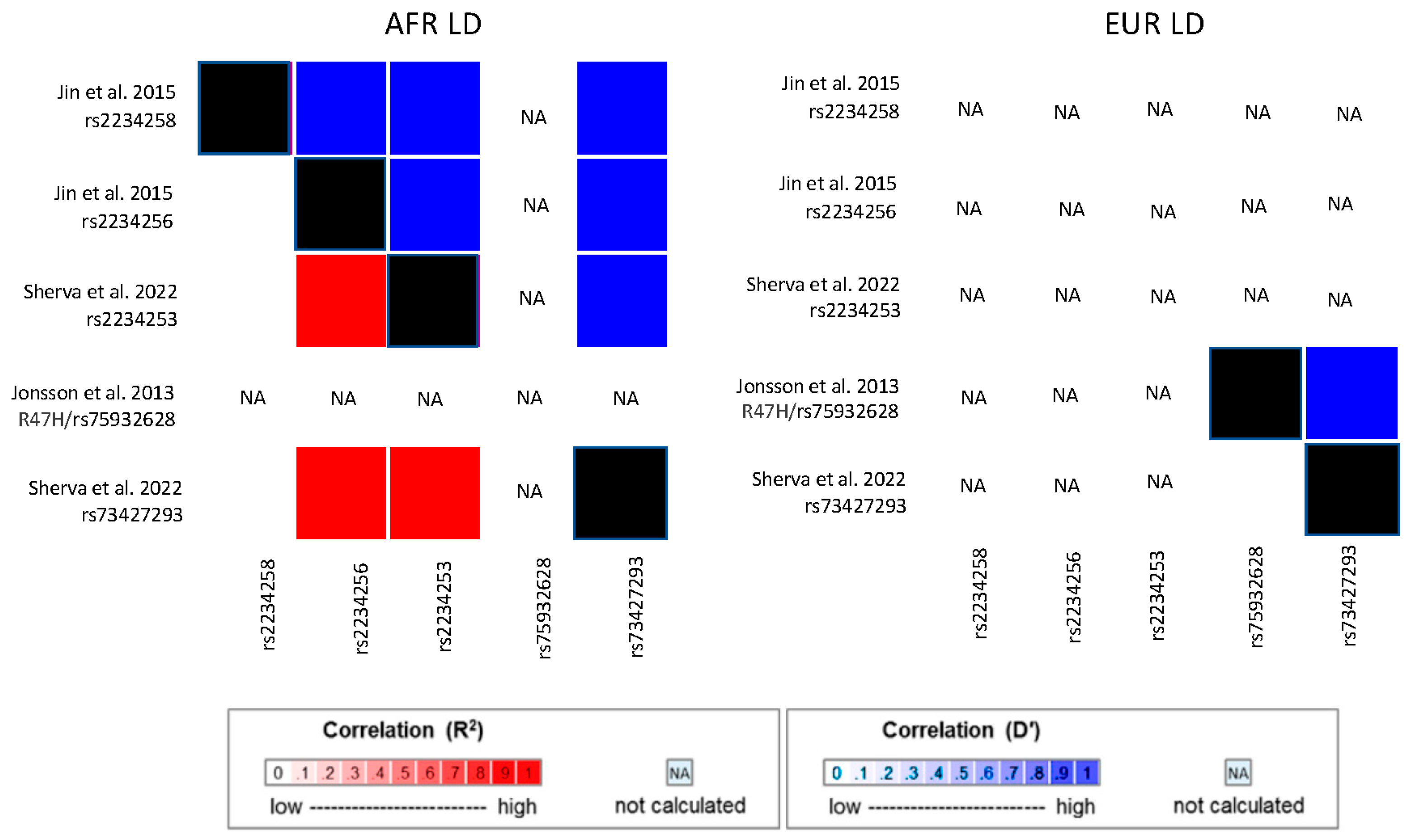
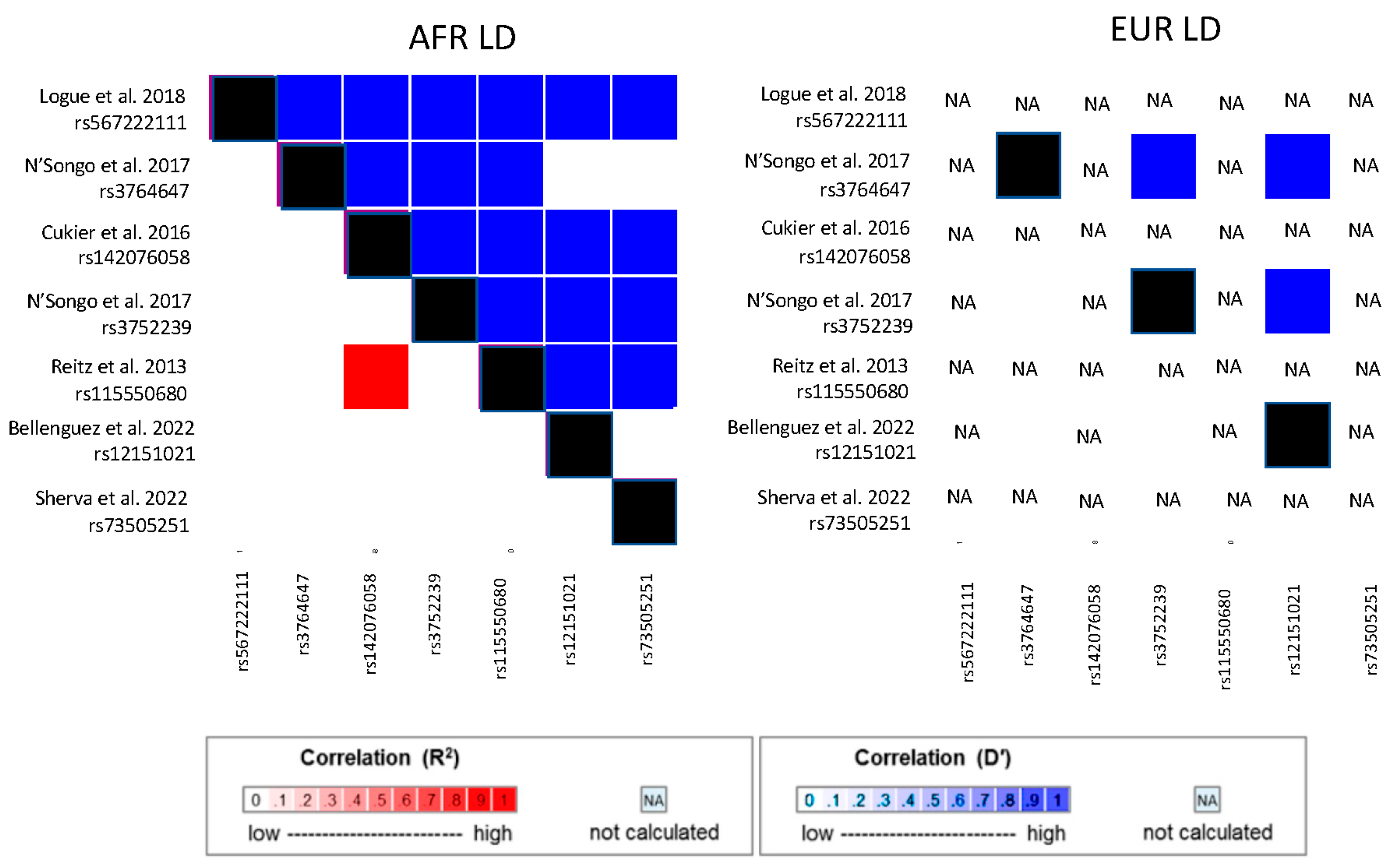
Potentially functional AD-related variants in TREM2 and ABCA7 were identified by targeted gene sequencing studies of AA cohorts. Cukier et al. [74] sequenced 77 subjects who have the intronic ABCA7 risk allele rs115550680, which was previously associated with AD risk in an ADGC AA cohort [67]. They identified a 44 bp deletion that is common in African ancestry populations (rs142076058), in strong LD with rs115550680, and associated with AD in two independent cohorts [69]. Sequencing of TREM2 in a pooled AA cohort of 202 AD cases and 136 controls and an independent AA cohort of 179 AD cases and 334 controls identified 6 coding variants that were subsequently genotyped and evaluated for association in an AA sample containing 906 cases and 2487 controls [75]. Although none of the findings were significant, the top-ranked variants, one likely to be benign (rs2234256, OR = 1.27, p = 0.01) and rs2234258, which was predicted to impact the short TREM2 isoform (OR = 1.35, p = 0.08), were significantly associated with AD/ADRD in the MVP GWAS (rs2234256, p = 4.64 × 10⁻⁹ and rs2234258, p = 5.48 × 10⁻⁷) [69] and are in high LD with each other and the peak TREM2 SNPs from the Kunkle et al. GWAS (rs73427293 and rs2234253; Figure 2). The TREM2 R47H missense variant (rs75932628) is rare in EUR populations (MAF = 0.005 in 1000 genomes EUR cohort) and is absent in African-ancestry populations (not observed in the 1000 genomes AFR cohort). Hence, rs75932628 is unlikely to underly the TREM2 associations observed in AA cohorts [75] or in a Caribbean Hispanic cohort that has substantial African ancestry [109]. Logue et al. sequenced coding and flanking proximate regions of approximately 100 genes associated with AD and AD-related traits in an AA discovery cohort of 489 AD cases and 472 controls [76]. They identified an association of AD with an 11 bp loss-of-function deletion in ABCA7 (OR = 3.57, p = 0.038), a finding which was bolstered by the inclusion of data from an AA replication cohort of 484 cases and 484 controls (p = 0.022). A low correlation between the deletion and other AD-associated ABCA7 variants indicates that there are likely multiple independent AA AD risk loci in ABCA7 (Figure 3).
A WES study conducted in a relatively small AA sample of 131 AD cases and 107 controls identified a nominally significant association of AD with ABCA7 missense variants rs3764647 and rs3752239 [77]. Aggregated rare variant tests also found nominally significant associations with several known AD genes, including ABCA7, MS4A6A, PTK2B, and ZCWPW1 [77]. In contrast, a very small WES study of seven AA AD cases (including two affected sib pairs) from the MIRAGE Study identified two rare missense mutations (rs144662445 and rs149979685) in AKAP9, a gene not previously associated with AD, that were present in more than one sequenced subject with AD [78]. Subsequent genotyping of these variants in the entire MIRAGE AA cohort of 422 cases and 394 controls and in an ADGC AA cohort of 1037 AD cases and 1869 controls revealed that they are in high LD and significantly associated with AD in each cohort: MIRAGE rs14466245, p = 0.037 and rs149979685, p = 0.014); ADGC: p = 0.0022 for both variants. Examination of African-ancestry haplotypes in 1000 Genomes Project [110] data indicated that these variants likely functionally impact the encoded Akap9 protein. A subsequent WGS study identified another rare AKAP9 coding mutation (p.R434W) that segregated in two large Caribbean Hispanic families and was nominally associated with a five-fold increased risk of AD [109]. No WGS studies of AD with a major focus on AAs have appeared in the literature at the time of this writing.

8. Linking AKAP9 to AD by Functional Studies

AKAP9 encodes a member of a family of proteins that bind signaling molecules to their targets [111]. The rs144662445 variant is located in the protein kinase alpha (PKA) binding subunit of AKAP9 [78,112]. This suggests an important potential risk mechanism, as activated PKA leads to phosphorylated Tau, which is a hallmark of AD neuropathology. To test this hypothesis, Ikezu et al. examined phosphorylated Tau and amyloid precursor protein (APP) levels in lymphoblastoid cell lines (LCLs) from 11 AA subjects with at least one of these variants (AKAP9+) and 17 AA subjects lacking these mutations (AKAP9-) [113]. LCLs were transduced by viral vectors expressing causative AD mutations in APP or human full-length wild-type Tau. Cell lysates were analyzed for total APP, Aβ40, and total and T181 phospho-Tau (pTau). AKAP9 mutations had no effect on Aβ40/APP but significantly increased pTau/Tau ratio in LCLs treated with phosphodiesterase-4 inhibitor rolipram, which activates protein kinase A. This study showed the impact of rare functional AKAP9 mutations on Tau, a central mechanism of AD pathogenesis, in LCLs derived from AD and control subjects.
To further the understanding of how the AKAP9 mutations influence Tau phosphorylation, You et al. performed a proteomic analysis of the Tau interactome using SH-SY5YP301L neuronal cells, which were CRISPR-Cas9-engineered to carry the AKAP9 rs144662445 mutation [114]. Their analyses revealed an enrichment of RNA binding proteins and a decrease of proteasomal molecules in rolipram-treated cells with the AKAP9 mutation. Those cells also displayed increased Tau phosphorylation at the S396 and S404 sites and increased oxidative stress. These changes in the Tau interactome mirrored disruptions noted in AD brain tissue vs. controls, and oxidative stress has been shown to play a role in neuronal dysfunction in AD brains [115,116].

9. Polygenic Risk Scores

One of the advances powered by large-scale GWAS is the development of methods for computing polygenic risk scores (PRSs) and using them to understand the shared genetic risk for multiple diseases and biological processes. A PRS summarizes an individual’s genetic risk for a particular disease or related trait based on GWAS summary results. They are computed as a weighted sum of the coefficient (effect size estimate) for each SNP generated from a GWAS, which is typically the log OR of the per-allele risk for binary disease outcomes. In EUR cohorts, these scores have been shown to predict AD risk better than APOE genotype and demographic risk factors alone [117]. AD PRSs have been associated with AD biomarkers [118] and neuroimaging traits [119,120,121]. The AD PRS has potential promise to distinguish early-stage AD among persons with mild cognitive impairment many years before AD symptoms typically appear [122]. However, the performance of a PRS is greatly influenced by the sample size of the GWAS used to determine the weights ascribed to the SNPs included in the PRS calculation [123]. Moreover, the strength of an association between a PRS and a trait depends on the correspondence between the effect sizes of the constituent SNPs in the GWAS and in the target sample, and the SNP effect sizes in AA cohorts will usually differ from those derived in EUR cohorts. Indeed, the accuracy of a PRS is reduced when there is a mismatch between the ancestry of the cohort used in the GWAS and the target cohort to which the PRS is applied [124,125,126]. However, there is some evidence that including multiple ancestry risk scores can improve PRS performance relative to using smaller-sample size individual ancestry GWAS results for PRS calculation [127]. Therefore, increasing sample sizes of AA cohorts and cohorts for other non-EUR populations in genetic studies has been identified as a priority for genetic research [124] as a means to more accurately determine genetic risk and to develop population-specific therapeutic strategies and subsequently reduce the growing health gap between populations.

10. Leveraging African American Cohorts to Increase Genetic Discovery

In addition to improving the accuracy of risk prediction for AA individuals, genetic studies conducted in AA cohorts have great potential to generate new and important insights into AD pathogenesis. Several studies have demonstrated that non-EUR cohorts can have a much greater impact relative to their sample size. For instance, a GWAS conducted by the PAIGE Study for multiple traits in 49,839 non-EUR individuals (a relatively small sample compared to GWAS for analogous traits in EUR cohorts) found 27 novel trait associations and secondary associations at 38 known trait loci and replicated over 1000 EUR GWAS associations [128]. African ancestry populations have more diverse genomes than EUR and EASN populations because African ancestry populations have evolved over a much longer period of time than other continental populations that were founded by small bands migrating from Africa. Consequently, the number of rare variants observed in any gene or other portion of the genome will be greater in African-ancestry cohorts than in EUR or EASN cohorts. There is a growing understanding that this greater genetic diversity can yield important insights into pathways to AD that would be missed by focusing on Europeans only. This is illustrated by a multi-population AD GWAS that included a large EUR sample and smaller groups of AA, EASN, and Israeli-Arabs that identified novel GWS associations with rs11168036 located between PFDN1 and HBEGF (p = 7 × 10⁻⁹), rs7920721 located between USP6NL and ECHDC3 (p = 3 × 10⁻⁸), and BZRAP1-AS1 SNP rs2632516 (p = 4 × 10⁻⁸) [21]. Analyses of models conditioned on the top SNP at the PFDN1/HBEGF, USP6NL/ECHDC3, and BZRAP1-AS1 loci confirmed a single association signal in each region. These GWS associations, except for rs7920721, were supported by evidence in multiple ethnic groups.

11. Importance of Racial/Ethnic Identity and Social Determinants of Health in Genetic Studies of AD

As discussed briefly in the Introduction, race is a social construct that is viewed as having little scientific value in representing distinct ancestral populations [15,16]. Therefore, many genetic studies focus on ancestry determined by clustering of genetic profiles relative to reference populations rather than on self-identified race or ethnicity. However, simply replacing ancestry, especially continental ancestry, for race does not accurately represent the diversity of human populations or the continuous nature of human genetic variation [17]. Moreover, both of these methods for grouping individuals are correlated with SDOH in similar but not identical ways [18]. Therefore, focusing on ancestral categorizations, scientists risk genetic essentialist framing of complex influences on the development of AD. Jointly considering race, concomitant social variables, and ancestry may be one way to incorporate a deeper understanding of the ways in which environmental pressures, such as poorer education and barriers to good nutrition, are genetically mediated [16,129]. However, care must be taken to ensure the use of race is appropriately contextualized as a proxy for SDOH until the more proximal factors themselves can be disentangled from race. The picture may be further muddled by the practice of race norming of assessments of patient cognition, which is incorporated by default into some tools that evaluate dementia severity, for instance, through the use of Heaton norms [130]. Race-norming attempts to compare patients within a particular group rather than across groups, but, in dementia assessment, can pathologize race, as AA individuals are compared against a lower baseline level of cognition than EUR individuals [131,132]. This can lead to differences in the type and severity of dementias between AA- and EUR-study cohorts and obscure systemic and environmental factors that lead to disparities across groups [133], thereby distorting study conclusions. Consideration of the interaction of SDOH and genetic factors is a complex but important endeavor in moving towards precision health.
Genetic research involving AA individuals should be undertaken with several interrelated factors in mind. As articulated by the National Academies committee “Use of Race, Ethnicity, and Ancestry as Population Descriptors in Genomics Research,” the foundational principles to the inclusive practice of genomics research are respect, equity and justice, beneficence, validity and reproducibility, transparency, and replicability [134]. Research must be performed and communicated in a way that promotes health without amplifying barriers to opportunities, exemplified by concerns surrounding genetic testing for sickle cell disease and carrier status [135]. To achieve this goal, scientists must not only simply include samples from diverse ancestral populations in large-scale genomic studies but truly partner with communities that have been historically excluded from research to ensure equitable benefit sharing [136,137] and assemble diverse teams to support these partnerships [138]. With this motivation, the ADSP prioritized the inclusion of diverse populations [139] and partnership with local investigators in many parts of the world, including the Caribbean, South America, Africa, Asia, and the Middle East, to enroll diverse cohorts that are sufficiently powered for genetic association studies [55,102,109,140,141,142,143]. In addition, while accurate risk prediction which takes into account SDOH and ancestry is important, and indeed this is one of the primary motivations of the priority to increase non-EUR representation in genetic studies, ancestry-specific protocols for treatment or identification of those at risk should be carefully evaluated and applied in a way that is respectful and does not limit access to care [16]. Identification of risk factors and risk markers in specific populations should not be the end goal but rather a way to refine hypotheses about the ways in which genetic, environmental, and cultural factors combine to influence health disparities. In summary, AD genetic research should be presented in a manner that avoids typological thinking promoting genetic determinism, stresses the impact of environmental and health factors, clearly indicates that the late-onset AD variants are neither necessary nor sufficient to cause AD [144,145], and promotes equitable benefits of research.

12. Conclusions

Increasing sample sizes of AA cohorts is and should remain a priority in genetic studies of AD and dementia. The studies highlighted in this review demonstrate the value of AA AD studies to increase our knowledge about the genetic underpinnings of AD risk. These studies will increase our understanding of the genetic architecture of AD in the AA population, help minimize health gaps that might develop because of the implementation of precision medicine approaches for AD, and lead to more accurate genetic testing and estimates of dementia risk. In elevating these goals, it is essential that we take care to be attentive to the distinctions between race and ancestry, avoid the use of harmful language and labels that reify notions of racial hierarchy, and form true partnerships between communities of participants and scientists to ensure identification of equitable, shared goals.

Author Contributions

All authors contributed to drafting and editing the manuscript. All authors have read and agreed to the published version of the manuscript.

Funding

This work was supported by VA BLR&D award BX004192 and National Institute on Aging awards R01-AG048927, RF1-AG057519, U54-AG052427, U01-AG058654, U01-AG062602, U19-AG068753, P30-AG072978 and U01-AG032984.

Institutional Review Board Statement

Not applicable.

Informed Consent Statement

Not applicable.

Data Availability Statement

Not applicable.

Conflicts of Interest

MWL, SD, and LAF have no conflict to disclose. This publication does not represent the views of the Department of Veteran Affairs or the United States Government.

References

  1. Alzheimer’s Association. 2023 Alzheimer’s disease facts and figures. Alzheimers Dement. 2023, 19, 1598–1695. [Google Scholar] [CrossRef] [PubMed]
  2. Sharp, E.S.; Gatz, M. Relationship between education and dementia: An updated systematic review. Alzheimer Dis. Assoc. Disord. 2011, 25, 289–304. [Google Scholar] [CrossRef] [PubMed] [Green Version]
  3. Evans, I.E.M.; Martyr, A.; Collins, R.; Brayne, C.; Clare, L. Social isolation and cognitive function in later life: A systematic review and meta-analysis. J. Alzheimers Dis. 2019, 70, S119–S144. [Google Scholar] [CrossRef] [Green Version]
  4. Tang, M.X.; Cross, P.; Andrews, H.; Jacobs, D.M.; Small, S.; Bell, K.; Merchant, C.; Lantigua, R.; Costa, R.; Stern, Y.; et al. Incidence of AD in African-Americans, Caribbean Hispanics, and Caucasians in northern Manhattan. Neurology 2001, 56, 49–56. [Google Scholar] [CrossRef] [PubMed] [Green Version]
  5. Demirovic, J.; Prineas, R.; Loewenstein, D.; Bean, J.; Duara, R.; Sevush, S.; Szapocznik, J. Prevalence of dementia in three ethnic groups: The South Florida program on aging and health. Ann. Epidemiol. 2003, 13, 472–478. [Google Scholar] [CrossRef]
  6. Mayeda, E.R.; Glymour, M.M.; Quesenberry, C.P.; Whitmer, R.A. Inequalities in dementia incidence between six racial and ethnic groups over 14 years. Alzheimers Dement 2016, 12, 216–224. [Google Scholar] [CrossRef] [Green Version]
  7. Barnes, L.L. Alzheimer disease in African American individuals: Increased incidence or not enough data? Nat. Rev. Neurol. 2022, 18, 56–62. [Google Scholar] [CrossRef]
  8. Gross, A.L.; Mungas, D.M.; Crane, P.K.; Gibbons, L.E.; MacKay-Brandt, A.; Manly, J.J.; Mukherjee, S.; Romero, H.; Sachs, B.; Thomas, M.; et al. Effects of education and race on cognitive decline: An integrative study of generalizability versus study-specific results. Psychol. Aging 2015, 30, 863–880. [Google Scholar] [CrossRef]
  9. Zahodne, L.B.; Manly, J.J.; Azar, M.; Brickman, A.M.; Glymour, M.M. Racial Disparities in Cognitive Performance in Mid- and Late Adulthood: Analyses of Two Cohort Studies. J. Am. Geriatr. Soc. 2016, 64, 959–964. [Google Scholar] [CrossRef] [Green Version]
  10. Clark, L.R.; Norton, D.; Berman, S.E.; Johnson, S.C.; Bendlin, B.B.; Wieben, O.; Turski, P.; Carlsson, C.; Asthana, S.; Gleason, C.E.; et al. Association of cardiovascular and Alzheimer’s disease risk factors with intracranial arterial blood flow in Whites and African Americans. J. Alzheimers Dis. 2019, 72, 919–929. [Google Scholar] [CrossRef] [PubMed]
  11. Harris, M.I.; Flegal, K.M.; Cowie, C.C.; Eberhardt, M.S.; Goldstein, D.E.; Little, R.R.; Wiedmeyer, H.M.; Byrd-Holt, D.D. Prevalence of diabetes, impaired fasting glucose, and impaired glucose tolerance in U.S. adults. The Third National Health and Nutrition Examination Survey, 1988–1994. Diabetes Care 1998, 21, 518–524. [Google Scholar] [CrossRef] [PubMed]
  12. Chin, A.L.; Negash, S.; Xie, S.; Arnold, S.E.; Hamilton, R. Quality, and not just quantity, of education accounts for differences in psychometric performance between african americans and white non-hispanics with Alzheimer’s disease. J. Int. Neuropsychol. Soc. 2012, 18, 277–285. [Google Scholar] [CrossRef] [PubMed] [Green Version]
  13. Sisco, S.; Gross, A.L.; Shih, R.A.; Sachs, B.C.; Glymour, M.M.; Bangen, K.J.; Benitez, A.; Skinner, J.; Schneider, B.C.; Manly, J.J. The role of early-life educational quality and literacy in explaining racial disparities in cognition in late life. J. Gerontol. B Psychol. Sci. Soc. Sci. 2015, 70, 557–567. [Google Scholar] [CrossRef] [PubMed] [Green Version]
  14. Yaffe, K.; Falvey, C.; Harris, T.B.; Newman, A.; Satterfield, S.; Koster, A.; Ayonayon, H.; Simonsick, E. Effect of socioeconomic disparities on incidence of dementia among biracial older adults: Prospective study. BMJ 2013, 347, 7051. [Google Scholar] [CrossRef] [PubMed] [Green Version]
  15. Williams, D.R. Race and health: Basic questions, emerging directions. Ann. Epidemiol. 1997, 7, 322–333. [Google Scholar]
  16. Borrell, L.N.; Elhawary, J.R.; Fuentes-Afflick, E.; Witonsky, J.; Bhakta, N.; Wu, A.H.B.; Bibbins-Domingo, K.; Rodriguez-Santana, J.R.; Lenoir, M.A.; Gavin, J.R., 3rd; et al. Race and genetic ancestry in medicine—A time for reckoning with racism. N. Engl. J. Med. 2021, 384, 474–480. [Google Scholar] [CrossRef]
  17. Lewis, A.C.F.; Molina, S.J.; Appelbaum, P.S.; Dauda, B.; Di Rienzo, A.; Fuentes, A.; Fullerton, S.M.; Garrison, N.A.; Ghosh, N.; Hammonds, E.M.; et al. Getting genetic ancestry right for science and society. Science 2022, 376, 250–252. [Google Scholar] [CrossRef]
  18. Babulal, G.M.; Quiroz, Y.T.; Albensi, B.C.; Arenaza-Urquijo, E.; Astell, A.J.; Babiloni, C.; Bahar-Fuchs, A.; Bell, J.; Bowman, G.L.; Brickman, A.M.; et al. Perspectives on ethnic and racial disparities in Alzheimer’s disease and related dementias: Update and areas of immediate need. Alzheimers Dement 2019, 15, 292–312. [Google Scholar] [CrossRef]
  19. Nguyen, A.B.; Moser, R.; Chou, W.Y. Race and health profiles in the United States: An examination of the social gradient through the 2009 CHIS adult survey. Public Health 2014, 128, 1076–1086. [Google Scholar]
  20. Sirugo, G.; Williams, S.M.; Tishkoff, S.A. The missing diversity in human genetic studies. Cell 2019, 177, 1080. [Google Scholar] [CrossRef] [Green Version]
  21. Jun, G.R.; Chung, J.; Mez, J.; Barber, R.; Beecham, G.W.; Bennett, D.A.; Buxbaum, J.D.; Byrd, G.S.; Carrasquillo, M.M.; Crane, P.K.; et al. Transethnic genome-wide scan identifies novel Alzheimer’s disease loci. Alzheimers Dement. 2017, 13, 727–738. [Google Scholar] [CrossRef] [PubMed]
  22. Devi, G.; Ottman, R.; Tang, M.X.; Marder, K.; Stern, Y.; Mayeux, R. Familial aggregation of Alzheimer disease among whites, African Americans, and Caribbean Hispanics in northern Manhattan. Arch. Neurol. 2000, 57, 72–77. [Google Scholar] [CrossRef]
  23. Green, R.C.; Cupples, L.A.; Go, R.; Benke, K.S.; Edeki, T.; Griffith, P.A.; Williams, M.; Hipps, Y.; Graff-Radford, N.; Bachman, D.; et al. Risk of dementia among white and African American relatives of patients with Alzheimer disease. JAMA 2002, 287, 329–336. [Google Scholar] [CrossRef] [PubMed] [Green Version]
  24. Bachman, D.L.; Green, R.C.; Benke, K.S.; Cupples, L.A.; Farrer, L.A.; MIRAGE Study Group. Comparison of Alzheimer’s disease risk factors in white and African American families. Neurology 2003, 60, 1372–1374. [Google Scholar] [CrossRef] [PubMed]
  25. Kukull, W.A. The association between smoking and Alzheimer’s disease: Effects of study design and bias. Biol. Psychiatry 2001, 49, 194–199. [Google Scholar] [CrossRef]
  26. Mukamal, K.J.; Kuller, L.H.; Fitzpatrick, A.L.; Longstreth, W.T., Jr.; Mittleman, M.A.; Siscovick, D.S. Prospective study of alcohol consumption and risk of dementia in older adults. JAMA 2003, 289, 1405–1413. [Google Scholar] [CrossRef]
  27. Hartz, S.M.; Oehlert, M.; Horton, A.C.; Grucza, R.A.; Fisher, S.L.; Culverhouse, R.C.; Nelson, K.G.; Sumerall, S.W.; Neal, P.C.; Regnier, P.; et al. Daily drinking is associated with increased mortality. Alcohol. Clin. Exp. Res. 2018, 42, 2246–2255. [Google Scholar] [CrossRef] [PubMed]
  28. Corder, E.H.; Saunders, A.M.; Strittmatter, W.J.; Schmechel, D.E.; Gaskell, P.C.; Small, G.W.; Roses, A.D.; Haines, J.L.; Pericak-Vance, M.A. Gene dose of apolipoprotein E type 4 allele and the risk of Alzheimer’s disease in late onset families. Science 1993, 261, 921–923. [Google Scholar] [CrossRef]
  29. Farrer, L.A.; Cupples, L.A.; Haines, J.L.; Hyman, B.; Kukull, W.A.; Mayeux, R.; Myers, R.H.; Pericak-Vance, M.A.; Risch, N.; van Duijn, C.M. Effects of age, sex, and ethnicity on the association between apolipoprotein E genotype and Alzheimer disease. A meta-analysis. APOE and Alzheimer Disease Meta Analysis Consortium. JAMA 1997, 278, 1349–1356. [Google Scholar] [CrossRef]
  30. Reiman, E.M.; Arboleda-Velasquez, J.F.; Quiroz, Y.T.; Huentelman, M.J.; Beach, T.G.; Caselli, R.J.; Chen, Y.; Su, Y.; Myers, A.J.; Hardy, J.; et al. Exceptionally low likelihood of Alzheimer’s dementia in APOE2 homozygotes from a 5000-person neuropathological study. Nat. Commun. 2020, 11, 667. [Google Scholar] [CrossRef] [Green Version]
  31. Jansen, I.E.; Savage, J.E.; Watanabe, K.; Bryois, J.; Williams, D.M.; Steinberg, S.; Sealock, J.; Karlsson, I.K.; Hagg, S.; Athanasiu, L.; et al. Genome-wide meta-analysis identifies new loci and functional pathways influencing Alzheimer’s disease risk. Nat. Genet. 2019, 51, 404–413. [Google Scholar] [CrossRef] [PubMed] [Green Version]
  32. Marioni, R.E.; Harris, S.E.; Zhang, Q.; McRae, A.F.; Hagenaars, S.P.; Hill, W.D.; Davies, G.; Ritchie, C.W.; Gale, C.R.; Starr, J.M.; et al. GWAS on family history of Alzheimer’s disease. Transl. Psychiatry 2018, 8, 99. [Google Scholar] [CrossRef] [PubMed] [Green Version]
  33. Bellenguez, C.; Küçükali, F.; Jansen, I.; Andrade, V.; Moreno-Grau, S.; Amin, N.; Naj, A.C.; Grenier-Boley, B.; Campos-Martin, R.; Holmans, P.A.; et al. New insights on the genetic etiology of Alzheimer’s and related dementia. Nat. Genet. 2022, 54, 412–436. [Google Scholar] [CrossRef] [PubMed]
  34. Wightman, D.P.; Jansen, I.E.; Savage, J.E.; Shadrin, A.A.; Bahrami, S.; Rongve, A.; Børte, S.; Winsvold, B.S.; Drange, O.K.; Martinsen, A.E.; et al. A genome-wide association study with 1,126,563 individuals identifies new risk loci for Alzheimer’s disease. Nat. Genet. 2021, 53, 1276–1282. [Google Scholar] [CrossRef] [PubMed]
  35. Jonsson, T.; Stefansson, H.; Steinberg, S.; Jonsdottir, I.; Jonsson, P.V.; Snaedal, J.; Bjornsson, S.; Huttenlocher, J.; Levey, A.I.; Lah, J.J.; et al. Variant of TREM2 associated with the risk of Alzheimer’s disease. N. Engl. J. Med. 2013, 368, 107–116. [Google Scholar] [CrossRef] [PubMed]
  36. Guerreiro, R.; Wojtas, A.; Bras, J.; Carrasquillo, M.; Rogaeva, E.; Majounie, E.; Cruchaga, C.; Sassi, C.; Kauwe, J.S.; Younkin, S.; et al. TREM2 variants in Alzheimer’s disease. N. Engl. J. Med. 2013, 368, 117–127. [Google Scholar]
  37. Cruchaga, C.; Karch, C.M.; Jin, S.C.; Benitez, B.A.; Cai, Y.; Guerreiro, R.; Harari, O.; Norton, J.; Budde, J.; Bertelsen, S.; et al. Rare coding variants in the phospholipase D3 gene confer risk for Alzheimer’s disease. Nature 2014, 505, 550–554. [Google Scholar] [CrossRef] [Green Version]
  38. Bis, J.C.; Jian, X.; Kunkle, B.W.; Chen, Y.; Hamilton-Nelson, K.L.; Bush, W.S.; Salerno, W.J.; Lancour, D.; Ma, Y.; Renton, A.E.; et al. Whole exome sequencing study identifies novel rare and common Alzheimer’s-Associated variants involved in immune response and transcriptional regulation. Mol. Psychiatry 2020, 25, 1859–1875. [Google Scholar] [CrossRef] [Green Version]
  39. Zhang, X.; Zhu, C.; Beecham, G.; Vardarajan, B.N.; Ma, Y.; Lancour, D.; Farrell, J.J.; Chung, J.; Alzheimer’s Disease Sequencing Project; Mayeux, R.; et al. A rare missense variant of CASP7 is associated with familial late-onset Alzheimer’s disease. Alzheimers Dement. 2019, 15, 441–452. [Google Scholar] [CrossRef]
  40. Ma, Y.; Jun, G.R.; Zhang, X.; Chung, J.; Naj, A.C.; Chen, Y.; Bellenguez, C.; Hamilton-Nelson, K.; Martin, E.R.; Kunkle, B.W.; et al. Analysis of whole-exome sequencing data for Alzheimer disease stratified by APOE genotype. JAMA Neurol. 2019, 76, 1099–1108. [Google Scholar] [CrossRef] [PubMed]
  41. Holstege, H.; Hulsman, M.; Charbonnier, C.; Grenier-Boley, B.; Quenez, O.; Grozeva, D.; van Rooij, J.G.J.; Sims, R.; Ahmad, S.; Amin, N.; et al. Exome sequencing identifies rare damaging variants in ATP8B4 and ABCA1 as risk factors for Alzheimer’s disease. Nat. Genet. 2022, 54, 1786–1794. [Google Scholar] [CrossRef] [PubMed]
  42. Howard, B.V.; Gidding, S.S.; Liu, K. Association of apolipoprotein E phenotype with plasma lipoproteins in African-American and white young adults. The CARDIA Study. Coronary Artery Risk Development in Young Adults. Am. J. Epidemiol. 1998, 148, 859–868. [Google Scholar] [CrossRef] [PubMed]
  43. Tang, M.X.; Stern, Y.; Marder, K.; Bell, K.; Gurland, B.; Lantigua, R.; Andrews, H.; Feng, L.; Tycko, B.; Mayeux, R. The APOE-epsilon4 allele and the risk of Alzheimer disease among African Americans, whites, and Hispanics. JAMA 1998, 279, 751–755. [Google Scholar] [CrossRef] [PubMed] [Green Version]
  44. Graff-Radford, N.R.; Green, R.C.; Go, R.C.; Hutton, M.L.; Edeki, T.; Bachman, D.; Adamson, J.L.; Griffith, P.; Willis, F.B.; Williams, M.; et al. Association between apolipoprotein E genotype and Alzheimer disease in African American subjects. Arch. Neurol. 2002, 59, 594–600. [Google Scholar] [CrossRef]
  45. Tang, M.X.; Maestre, G.; Tsai, W.Y.; Liu, X.H.; Feng, L.; Chung, W.Y.; Chun, M.; Schofield, P.; Stern, Y.; Tycko, B.; et al. Relative risk of Alzheimer disease and age-at-onset distributions, based on APOE genotypes among elderly African Americans, Caucasians, and Hispanics in New York City. Am. J. Hum. Genet. 1996, 58, 574–584. [Google Scholar] [PubMed]
  46. Sahota, A.; Yang, M.; Gao, S.; Hui, S.L.; Baiyewu, O.; Gureje, O.; Oluwole, S.; Ogunniyi, A.; Hall, K.S.; Hendrie, H.C. Apolipoprotein E-associated risk for Alzheimer’s disease in the African-American population is genotype dependent. Ann. Neurol. 1997, 42, 659–661. [Google Scholar] [CrossRef]
  47. Logue, M.W.; Schu, M.; Vardarajan, B.N.; Buros, J.; Green, R.C.; Go, R.C.; Griffith, P.; Obisesan, T.O.; Shatz, R.; Borenstein, A.; et al. A comprehensive genetic association study of Alzheimer disease in African Americans. Arch. Neurol. 2011, 68, 1569–1579. [Google Scholar] [CrossRef] [Green Version]
  48. Murrell, J.R.; Price, B.; Lane, K.A.; Baiyewu, O.; Gureje, O.; Ogunniyi, A.; Unverzagt, F.W.; Smith-Gamble, V.; Gao, S.; Hendrie, H.C.; et al. Association of apolipoprotein E genotype and Alzheimer disease in African Americans. Arch. Neurol. 2006, 63, 431–434. [Google Scholar] [CrossRef] [Green Version]
  49. Logue, M.W.; Miller, M.W.; Sherva, R.; ZHang, R.; Harrington, K.M.; Fonda, J.R.; Merritt, V.C.; Panizzon, M.S.; Hauger, R.L.; Wolf, E.J.; et al. Alzheimer’s disease and related dementias among aging veterans: Examining gene-by-environment interactions with posttraumatic stress disorder and traumatic brain injury. Alzheimers Dement. 2023, 19, 2549–2559. [Google Scholar] [CrossRef]
  50. Hendrie, H.C.; Murrell, J.; Baiyewu, O.; Lane, K.A.; Purnell, C.; Ogunniyi, A.; Unverzagt, F.W.; Hall, K.; Callahan, C.M.; Saykin, A.J.; et al. APOE epsilon4 and the risk for Alzheimer disease and cognitive decline in African Americans and Yoruba. Int. Psychogeriatr. 2014, 26, 977–985. [Google Scholar] [CrossRef] [Green Version]
  51. Gureje, O.; Ogunniyi, A.; Baiyewu, O.; Price, B.; Unverzagt, F.W.; Evans, R.M.; Smith-Gamble, V.; Lane, K.A.; Gao, S.; Hall, K.S.; et al. APOE epsilon4 is not associated with Alzheimer’s disease in elderly Nigerians. Ann. Neurol. 2006, 59, 182–185. [Google Scholar] [CrossRef]
  52. Farrer, L.A. Intercontinental epidemiology of Alzheimer disease: A global approach to bad gene hunting. JAMA 2001, 285, 796–798. [Google Scholar] [CrossRef]
  53. Rajabli, F.; Feliciano, B.E.; Celis, K.; Hamilton-Nelson, K.L.; Whitehead, P.L.; Adams, L.D.; Bussies, P.L.; Manrique, C.P.; Rodriguez, A.; Rodriguez, V.; et al. Ancestral origin of ApoE epsilon4 Alzheimer disease risk in Puerto Rican and African American populations. PLoS Genet. 2018, 14, e1007791. [Google Scholar] [CrossRef] [PubMed] [Green Version]
  54. Le Guen, Y.; Belloy, M.E.; Eger, S.J.; Chen, A.; Kennedy, G.; Thornton, T.A.; Farrer, L.A.; Napolioni, V.; He, Z.; Grecius, M.D. APOE missense variant R145C is associated with increased Alzheimer’s disease risk in African ancestry individuals with the APOE ε3/ε4 genotype. JAMA 2023, 329, 551–560. [Google Scholar] [CrossRef] [PubMed]
  55. Choi, K.Y.; Lee, J.J.; Gunasekaran, T.I.; Kang, S.; Lee, W.; Jeong, J.; Lim, H.J.; Zhang, X.; Zhu, C.; Won, S.Y.; et al. APOE promoter polymorphism-219T/G is an effect modifier of the influence of APOE ε4 on Alzheimer’s disease risk in a multiracial sample. J. Clin. Med. 2019, 8., 1236. [Google Scholar] [CrossRef] [Green Version]
  56. Erlich, P.M.; Lunetta, K.L.; Cupples, L.A.; Huyck, M.; Green, R.C.; Baldwin, C.T.; Farrer, L.A. MIRAGE Study Group. Polymorphisms in the PON gene cluster are associated with Alzheimer disease. Hum. Mol. Genet. 2006, 15, 77–85. [Google Scholar] [CrossRef] [PubMed]
  57. Shunmoogam, N.; Naidoo, P.; Chilton, R. Paraoxonase (PON)-1: A brief overview on genetics, structure, polymorphisms and clinical relevance. Vasc. Health Risk Manag. 2018, 14, 137–143. [Google Scholar] [CrossRef] [PubMed] [Green Version]
  58. Precourt, L.P.; Amre, D.; Denis, M.C.; Lavoie, J.C.; Delvin, E.; Seidman, E.; Levy, E. The three-gene paraoxonase family: Physiologic roles, actions and regulation. Atherosclerosis 2011, 214, 20–36. [Google Scholar] [CrossRef]
  59. Zamora, R.; Vodovotz, Y.; Billiar, T.R. Inducible nitric oxide synthase and inflammatory diseases. Mol. Med. 2000, 6, 347–373. [Google Scholar] [CrossRef] [Green Version]
  60. Krol, M.; Kepinska, M. Human nitric oxide synthase—Its functions, polymorphisms, and inhibitors in the context of inflammation, diabetes and cardiovascular diseases. Int. J. Mol. Sci. 2020, 22, 56. [Google Scholar] [CrossRef]
  61. Akomolafe, A.; Lunetta, K.L.; Erlich, P.M.; Cupples, L.A.; Baldwin, C.T.; Huyck, M.; Green, R.C.; Farrer, L.A. MIRAGE Study Group. Genetic association between endothelial nitric oxide synthase and Alzheimer disease. Clin. Genet. 2006, 70, 49–56. [Google Scholar] [CrossRef] [PubMed]
  62. Rogaeva, E.; Meng, Y.; Lee, J.H.; Gu, Y.; Kawarai, T.; Zou, F.; Katayama, T.; Baldwin, C.T.; Cheng, R.; Hasegawa, H.; et al. The neuronal sortilin-related receptor SORL1 is genetically associated with Alzheimer disease. Nat. Genet. 2007, 39, 168–177. [Google Scholar] [CrossRef] [PubMed]
  63. Reitz, C.; Tokuhiro, S.; Clark, L.N.; Conrad, C.; Vonsattel, J.P.; Hazrati, L.N.; Palotas, A.; Lantigua, R.; Medrano, M.; Jiménez-Velázquez, I.; et al. SORCS1 alters amyloid precursor protein processing and variants may increase Alzheimer’s disease risk. Ann. Neurol. 2011, 69, 47–64. [Google Scholar] [CrossRef] [PubMed] [Green Version]
  64. Vardarajan, B.N.; Bruesegem, S.Y.; Harbour, M.E.; Inzelberg, R.; Friedland, R.; St George-Hyslop, P.; Seaman, M.N.; Farrer, L.A. Identification of Alzheimer disease-associated variants in genes that regulate retromer function. Neurobiol. Aging 2012, 33, 2231.e2215–2231.e2230. [Google Scholar] [CrossRef] [PubMed] [Green Version]
  65. Janicki, S.C.; Park, N.; Cheng, R.; Schupf, N.; Clark, L.N.; Lee, J.H. Aromatase variants modify risk for Alzheimer’s disease in a multiethnic female cohort. Dement Geriatr. Cogn. Disord. 2013, 35, 340–346. [Google Scholar] [CrossRef] [Green Version]
  66. McAninch, E.A.; Rajan, K.B.; Evans, D.A.; Jo, S.; Chaker, L.; Peeters, R.P.; Bennett, D.A.; Mash, D.C.; Bianco, A.C. A common DIO2 polymorphism and Alzheimer disease dementia in African and European Americans. J. Clin. Endocrinol. Metab. 2018, 103, 1818–1826. [Google Scholar] [CrossRef]
  67. Reitz, C.; Jun, G.; Naj, A.; Rajbhandary, R.; Vardarajan, B.N.; Wang, L.S.; Valladares, O.; Lin, C.F.; Larson, E.B.; Graff-Radford, N.R.; et al. Variants in the ATP-binding cassette transporter (ABCA7), apolipoprotein E 4, and the risk of late-onset Alzheimer disease in African Americans. JAMA 2013, 309, 1483–1492. [Google Scholar] [CrossRef]
  68. Kunkle, B.W.; Schmidt, M.; Klein, H.U.; Naj, A.C.; Hamilton-Nelson, K.L.; Larson, E.B.; Evans, D.A.; De Jager, P.L.; Crane, P.K.; Buxbaum, J.D.; et al. Novel Alzheimer disease risk loci and pathways in African American individuals using the African Genome Resources Panel: A meta-analysis. JAMA Neurol. 2021, 78, 102–113. [Google Scholar] [CrossRef]
  69. Sherva, R.; Zhang, R.; Sahelijo, N.; Jun, G.; Anglin, T.; Chanfreu, C.; Cho, K.; Fonda, J.R.; Gaziano, J.M.; Harrington, K.M.; et al. African ancestry GWAS of dementia in a large military cohort identifies significant risk loci. Mol. Psychiatry 2023, 28, 1293–1302. [Google Scholar] [CrossRef]
  70. Mez, J.; Chung, J.; Jun, G.; Kriegel, J.; Bourlas, A.P.; Sherva, R.; Logue, M.W.; Barnes, L.L.; Bennett, D.A.; Buxbaum, J.D.; et al. Two novel loci, COBL and SLC10A2, for Alzheimer’s disease in African Americans. Alzheimers Dement. 2017, 13, 119–129. [Google Scholar] [CrossRef] [Green Version]
  71. Cuenco, K.T.; Friedland, R.; Baldwin, C.T.; Guo, J.; Vardarajan, B.; Lunetta, K.L.; Cupples, L.A.; Green, R.C.; DeCarli, C.; Farrer, L.A.; et al. Association of TTR polymorphisms with hippocampal atrophy in Alzheimer disease families. Neurobiol. Aging 2011, 32, 249–256. [Google Scholar] [CrossRef] [Green Version]
  72. Melville, S.A.; Buros, J.; Parrado, A.R.; Vardarajan, B.; Logue, M.W.; Shen, L.; Risacher, S.L.; Kim, S.; Jun, G.; DeCarli, C.; et al. Multiple loci influencing hippocampal degeneration identified by genome scan. Ann. Neurol. 2012, 72, 65–75. [Google Scholar] [CrossRef] [Green Version]
  73. N’Songo, A.; Carrasquillo, M.M.; Wang, X.; Nguyen, T.; Asmann, Y.; Younkin, S.G.; Allen, M.; Duara, R.; Custo, M.T.; Graff-Radford, N.; et al. Comprehensive screening for disease risk variants in early-onset Alzheimer’s disease genes in African Americans identifies novel PSEN variants. J. Alzheimers Dis. 2017, 56, 1215–1222. [Google Scholar] [CrossRef] [PubMed]
  74. Cukier, H.N.; Kunkle, B.W.; Vardarajan, B.N.; Rolati, S.; Hamilton-Nelson, K.L.; Kohli, M.A.; Whitehead, P.L.; Dombroski, B.A.; Van Booven, D.; Lang, R.; et al. ABCA7 frameshift deletion associated with Alzheimer disease in African Americans. Neurol. Genet. 2016, 2, e79. [Google Scholar] [CrossRef] [Green Version]
  75. Jin, S.C.; Carrasquillo, M.M.; Benitez, B.A.; Skorupa, T.; Carrell, D.; Patel, D.; Lincoln, S.; Krishnan, S.; Kachadoorian, M.; Reitz, C.; et al. TREM2 is associated with increased risk for Alzheimer’s disease in African Americans. Mol. Neurodegener. 2015, 10, 19. [Google Scholar] [CrossRef] [Green Version]
  76. Logue, M.W.; Lancour, D.; Farrell, J.; Simkina, I.; Fallin, M.D.; Lunetta, K.L.; Farrer, L.A. Targeted Sequencing of Alzheimer Disease Genes in African Americans Implicates Novel Risk Variants. Front. Neurosci. 2018, 12, 592. [Google Scholar] [CrossRef] [PubMed]
  77. N’Songo, A.; Carrasquillo, M.M.; Wang, X.; Burgess, J.D.; Nguyen, T.; Asmann, Y.W.; Serie, D.J.; Younkin, S.G.; Allen, M.; Pedraza, O.; et al. African American exome sequencing identifies potential risk variants at Alzheimer disease loci. Neurol. Genet. 2017, 3, e141. [Google Scholar] [CrossRef] [PubMed] [Green Version]
  78. Logue, M.W.; Schu, M.; Vardarajan, B.N.; Farrell, J.; Bennett, D.A.; Buxbaum, J.D.; Byrd, G.S.; Ertekin-Taner, N.; Evans, D.; Foroud, T.; et al. Two rare AKAP9 variants are associated with Alzheimer’s disease in African Americans. Alzheimers Dement. 2014, 10, 609–618.e611. [Google Scholar] [CrossRef] [Green Version]
  79. Dugbartey, A.T. Neurocognitive aspects of hypothyroidism. Arch. Intern. Med. 1998, 158, 1413–1418. [Google Scholar] [CrossRef] [Green Version]
  80. Kamboh, M.I.; Demirci, F.Y.; Wang, X.; Minster, R.L.; Carrasquillo, M.M.; Pankratz, V.S.; Younkin, S.G.; Saykin, A.J.; Jun, G.; Baldwin, C.; et al. Genome-wide association study of Alzheimer’s disease. Transl. Psychiatry 2012, 2, e117. [Google Scholar] [CrossRef] [Green Version]
  81. Yu, L.; Chibnik, L.B.; Srivastava, G.P.; Pochet, N.; Yang, J.; Xu, J.; Kozubek, J.; Obholzer, N.; Leurgans, S.E.; Schneider, J.A.; et al. Association of Brain DNA methylation in SORL1, ABCA7, HLA-DRB5, SLC24A4, and BIN1 with pathological diagnosis of Alzheimer disease. JAMA Neurol. 2015, 72, 15–24. [Google Scholar] [CrossRef] [Green Version]
  82. Gaziano, J.M.; Concato, J.; Brophy, M.; Fiore, L.; Pyarajan, S.; Breeling, J.; Whitbourne, S.; Deen, J.; Shannon, C.; Humphries, D.; et al. Million Veteran Program: A mega-biobank to study genetic influences on health and disease. J. Clin. Epidemiol. 2016, 70, 214–223. [Google Scholar] [CrossRef] [PubMed] [Green Version]
  83. Hunter-Zinck, H.; Shi, Y.; Li, M.; Gorman, B.R.; Ji, S.G.; Sun, N.; Webster, T.; Liem, A.; Hsieh, P.; Devineni, P.; et al. Genotyping Array Design and Data Quality Control in the Million Veteran Program. Am. J. Hum. Genet. 2020, 106, 535–548. [Google Scholar] [CrossRef] [PubMed]
  84. Bjerke, M.; Engelborghs, S. Cerebrospinal fluid biomarkers for early and differential Alzheimer’s disease diagnosis. J. Alzheimers Dis. 2018, 62, 1199–1209. [Google Scholar] [CrossRef] [PubMed] [Green Version]
  85. Teunissen, C.E.; Verberk, I.M.W.; Thijssen, E.H.; Vermunt, L.; Hansson, O.; Zetterberg, H.; van der Flier, W.M.; Mielke, M.M.; Del Campo, M. Blood-based biomarkers for Alzheimer’s disease: Towards clinical implementation. Lancet Neurol. 2022, 21, 66–77. [Google Scholar] [CrossRef]
  86. Bao, W.; Xie, F.; Zuo, C.; Guan, Y.; Huang, Y.H. PET Neuroimaging of Alzheimer’s disease: Radiotracers and their utility in clinical research. Front. Aging Neurosci. 2021, 13, 624330. [Google Scholar] [CrossRef]
  87. Howell, J.C.; Watts, K.D.; Parker, M.W.; Wu, J.; Kollhoff, A.; Wingo, T.S.; Dorbin, C.D.; Qiu, D.; Hu, W.T. Race modifies the relationship between cognition and Alzheimer’s disease cerebrospinal fluid biomarkers. Alzheimers Res. Ther. 2017, 9, 88. [Google Scholar] [CrossRef] [Green Version]
  88. Garrett, S.L.; McDaniel, D.; Obideen, M.; Trammell, A.R.; Shaw, L.M.; Goldstein, F.C.; Hajjar, I. Racial disparity in cerebrospinal fluid amyloid and tau biomarkers and associated cutoffs for mild cognitive impairment. JAMA Netw. Open 2019, 2, e1917363. [Google Scholar] [CrossRef]
  89. Hong, S.; Prokopenko, D.; Dobricic, V.; Kilpert, F.; Bos, I.; Vos, S.J.B.; Tijms, B.M.; Andreasson, U.; Blennow, K.; Vandenberghe, R.; et al. Genome-wide association study of Alzheimer’s disease CSF biomarkers in the EMIF-AD Multimodal Biomarker Discovery dataset. Transl. Psychiatry 2020, 10, 403. [Google Scholar] [CrossRef]
  90. Jansen, I.E.; van der Lee, S.J.; Gomez-Fonseca, D.; de Rojas, I.; Dalmasso, M.C.; Grenier-Boley, B.; Zettergren, A.; Mishra, A.; Ali, M.; Andrade, V.; et al. Genome-wide meta-analysis for Alzheimer’s disease cerebrospinal fluid biomarkers. Acta Neuropathol. 2022, 144, 821–842. [Google Scholar] [CrossRef]
  91. Chung, J.; Zhang, X.; Allen, M.; Wang, X.; Ma, Y.; Beecham, G.; Montine, T.J.; Younkin, S.G.; Dickson, D.W.; Golde, T.E.; et al. Genome-wide pleiotropy analysis of neuropathological traits related to Alzheimer’s disease. Alzheimers Res. Ther. 2018, 10, 22. [Google Scholar] [CrossRef] [PubMed]
  92. Kang, M.; Ang, T.F.A.; Devine, S.A.; Sherva, R.M.; Mukherjee, S.; Trittschuh, E.H.; Gibbons, L.E.; Scollard, P.; Lee, M.; Choi, S.E.; et al. A genome-wide search for pleiotropy in more than 100,000 harmonized longitudinal cognitive domain scores. Mol. Neurodegener. 2023, 18, 40. [Google Scholar] [CrossRef] [PubMed]
  93. Ramanan, V.K.; Risacher, S.L.; Nho, K.; Kim, S.; Swaminathan, S.; Shen, L.; Foroud, T.M.; Hakonarson, H.; Huentelman, M.J.; Aisen, P.S.; et al. APOE and BCHE as modulators of cerebral amyloid deposition: A florbetapir PET genome-wide association study. Mol. Psychiatry 2014, 19, 351–357. [Google Scholar] [CrossRef] [Green Version]
  94. Hohman, T.J.; Koran, M.E.; Thornton-Wells, T.A.; Alzheimer’s Disease Neuroimaging Initiative. Genetic modification of the relationship between phosphorylated tau and neurodegeneration. Alzheimers Dement 2014, 10, 637–645.e631. [Google Scholar] [CrossRef] [Green Version]
  95. Ramanan, V.K.; Nho, K.; Shen, L.; Risacher, S.L.; Kim, S.; McDonald, B.C.; Farlow, M.R.; Foroud, T.M.; Gao, S.; Soininen, H.; et al. FASTKD2 is associated with memory and hippocampal structure in older adults. Mol. Psychiatry 2015, 20, 1197–1204. [Google Scholar] [CrossRef] [Green Version]
  96. Mormino, E.C.; Betensky, R.A.; Hedden, T.; Schultz, A.P.; Ward, A.; Huijbers, W.; Rentz, D.M.; Johnson, K.A.; Sperling, R.A.; Alzheimer’s Disease Neuroimaging Initiative; et al. Amyloid and APOE epsilon4 interact to influence short-term decline in preclinical Alzheimer disease. Neurology 2014, 82, 1760–1767. [Google Scholar] [CrossRef] [PubMed] [Green Version]
  97. Hofer, E.; Roshchupkin, G.V.; Adams, H.H.H.; Knol, M.J.; Lin, H.; Li, S.; Zare, H.; Ahmad, S.; Armstrong, N.J.; Satizabal, C.L.; et al. Genetic correlations and genome-wide associations of cortical structure in general population samples of 22,824 adults. Nat. Commun. 2020, 11, 4796. [Google Scholar] [CrossRef]
  98. Grasby, K.L.; Jahanshad, N.; Painter, J.N.; Colodro-Conde, L.; Bralten, J.; Hibar, D.P.; Lind, P.A.; Pizzagalli, F.; Ching, C.R.K.; McMahon, M.A.B.; et al. The genetic architecture of the human cerebral cortex. Science 2020, 367, eaay6690. [Google Scholar] [CrossRef]
  99. Hibar, D.P.; Adams, H.H.H.; Jahanshad, N.; Chauhan, G.; Stein, J.L.; Hofer, E.; Renteria, M.E.; Bis, J.C.; Arias-Vasquez, A.; Ikram, M.K.; et al. Novel genetic loci associated with hippocampal volume. Nat. Commun. 2017, 8, 13624. [Google Scholar] [CrossRef] [Green Version]
  100. Elliott, L.T.; Sharp, K.; Alfaro-Almagro, F.; Shi, S.; Miller, K.L.; Douaud, G.; Marchini, J.; Smith, S.M. Genome-wide association studies of brain imaging phenotypes in UK Biobank. Nature 2018, 562, 210–216. [Google Scholar] [CrossRef] [Green Version]
  101. Ali, M.; Archer, D.B.; Gorijala, P.; Western, D.; Timsina, J.; Fernandez, M.V.; Wang, T.C.; Satizabal, C.L.; Yang, Q.; Beiser, A.S.; et al. Large multi-ethnic genetic analyses of amyloid imaging identify new genes for Alzheimer disease. Acta Neuropathol. Commun. 2023, 11, 68. [Google Scholar] [CrossRef]
  102. Rajabli, F.; Tosto, G.; Hamilton-Nelson, K.L.; Kunkle, B.W.; Vardarajan, B.N.; Naj, A.; Whitehead, P.G.; Gardner, O.K.; Bush, W.S.; Sariya, S.; et al. Admixture mapping identifies novel Alzheimer’s disease risk regions in African Americans. Alzheimers Dement. 2023, 19, 2538–2548. [Google Scholar] [CrossRef]
  103. Ting, S.K.; Benzinger, T.; Kepe, V.; Fagan, A.; Coppola, G.; Porter, V.; Hecimovic, S.; Chakraverty, S.; Alvarez-Retuerto, A.I.; Goate, A.; et al. A novel PSEN1 mutation (I238M) associated with early-onset Alzheimer’s disease in an African-American woman. J. Alzheimers Dis. 2014, 40, 271–275. [Google Scholar] [CrossRef]
  104. Edwards-Lee, T.; Ringman, J.M.; Chung, J.; Werner, J.; Morgan, A.; St George Hyslop, P.; Thompson, P.; Dutton, R.; Mlikotic, A.; Rogaeva, E.; et al. An African American family with early-onset Alzheimer disease and an APP (T714I) mutation. Neurology 2005, 64, 377–379. [Google Scholar] [CrossRef]
  105. Rippon, G.A.; Crook, R.; Baker, M.; Halvorsen, E.; Chin, S.; Hutton, M.; Houlden, H.; Hardy, J.; Lynch, T. Presenilin 1 mutation in an African American family presenting with atypical Alzheimer dementia. Arch. Neurol. 2003, 60, 884–888. [Google Scholar] [CrossRef] [Green Version]
  106. Ramirez Aguilar, L.; Acosta-Uribe, J.; Giraldo, M.M.; Moreno, S.; Baena, A.; Alzate, D.; Cuastumal, R.; Aguillon, D.; Madrigal, L.; Saldarriaga, A.; et al. Genetic origin of a large family with a novel PSEN1 mutation (Ile416Thr). Alzheimers Dement. 2019, 15, 709–719. [Google Scholar] [CrossRef]
  107. Cruchaga, C.; Haller, G.; Chakraverty, S.; Mayo, K.; Vallania, F.L.; Mitra, R.D.; Faber, K.; Williamson, J.; Bird, T.; Diaz-Arrastia, R.; et al. Rare variants in APP, PSEN1 and PSEN2 increase risk for AD in late-onset Alzheimer’s disease families. PLoS ONE 2012, 7, e31039. [Google Scholar] [CrossRef]
  108. Sassi, C.; Guerreiro, R.; Gibbs, R.; Ding, J.; Lupton, M.K.; Troakes, C.; Al-Sarraj, S.; Niblock, M.; Gallo, J.M.; Adnan, J.; et al. Investigating the role of rare coding variability in Mendelian dementia genes (APP, PSEN1, PSEN2, GRN, MAPT, and PRNP) in late-onset Alzheimer’s disease. Neurobiol. Aging 2014, 35, 2881.e2881–2881.e2886. [Google Scholar] [CrossRef] [PubMed] [Green Version]
  109. Vardarajan, B.N.; Barral, S.; Jaworski, J.; Beecham, G.W.; Blue, E.; Tosto, G.; Reyes-Dumeyer, D.; Medrano, M.; Lantigua, R.; Naj, A.; et al. Whole genome sequencing of Caribbean Hispanic families with late-onset Alzheimer’s disease. Ann. Clin. Transl. Neurol. 2018, 5, 406–417. [Google Scholar] [CrossRef] [PubMed] [Green Version]
  110. The 1000 Genomes Project Consortium. A map of human genome variation from population-scale sequencing. Nature 2010, 467, 1061–1073. [Google Scholar] [CrossRef] [Green Version]
  111. Lin, J.W.; Wyszynski, M.; Madhavan, R.; Sealock, R.; Kim, J.U.; Sheng, M. Yotiao, a novel protein of neuromuscular junction and brain that interacts with specific splice variants of NMDA receptor subunit NR1. J. Neurosci. 1998, 18, 2017–2027. [Google Scholar] [CrossRef] [PubMed]
  112. Terrin, A.; Monterisi, S.; Stangherlin, A.; Zoccarato, A.; Koschinski, A.; Surdo, N.C.; Mongillo, M.; Sawa, A.; Jordanides, N.E.; Mountford, J.C.; et al. PKA and PDE4D3 anchoring to AKAP9 provides distinct regulation of cAMP signals at the centrosome. J. Cell Biol. 2012, 198, 607–621. [Google Scholar] [CrossRef] [PubMed]
  113. Ikezu, T.; Chen, C.; DeLeo, A.M.; Zeldich, E.; Fallin, M.D.; Kanaan, N.M.; Lunetta, K.L.; Abraham, C.R.; Logue, M.W.; Farrer, L.A. Tau phosphorylation is impacted by rare AKAP9 mutations associated with Alzheimer disease in African Americans. J. Neuroimmune Pharmacol. 2018, 13, 254–264. [Google Scholar] [CrossRef] [PubMed] [Green Version]
  114. You, Y.; Hersh, S.W.; Aslebagh, R.; Shaffer, S.A.; Ikezu, S.; Mez, J.; Lunetta, K.L.; Logue, M.W.; Farrer, L.A.; Ikezu, T. Alzheimer’s disease associated AKAP9 I2558M mutation alters posttranslational modification and interactome of tau and cellular functions in CRISPR-edited human neuronal cells. Aging Cell 2022, 21, e13617. [Google Scholar] [CrossRef]
  115. Drummond, E.; Pires, G.; MacMurray, C.; Askenazi, M.; Nayak, S.; Bourdon, M.; Safar, J.; Ueberheide, B.; Wisniewski, T. Phosphorylated tau interactome in the human Alzheimer’s disease brain. Brain 2020, 143, 2803–2817. [Google Scholar] [CrossRef]
  116. Tonnies, E.; Trushina, E. Oxidative Stress, Synaptic Dysfunction, and Alzheimer’s Disease. J. Alzheimers Dis. 2017, 57, 1105–1121. [Google Scholar] [CrossRef] [Green Version]
  117. Escott-Price, V.; Sims, R.; Bannister, C.; Harold, D.; Vronskaya, M.; Majounie, E.; Badarinarayan, N.; Gerad/Perades; IGAP consortia; Morgan, K.; et al. Common polygenic variation enhances risk prediction for Alzheimer’s disease. Brain 2015, 138, 3673–3684. [Google Scholar] [CrossRef] [Green Version]
  118. Zettergren, A.; Lord, J.; Ashton, N.J.; Benedet, A.L.; Karikari, T.K.; Lantero Rodriguez, J.; Alzheimer’s Disease Neuroimaging Initiative; Snellman, A.; Suarez-Calvet, M.; Proitsi, P.; et al. Association between polygenic risk score of Alzheimer’s disease and plasma phosphorylated tau in individuals from the Alzheimer’s Disease Neuroimaging Initiative. Alzheimers Res. Ther. 2021, 13, 17. [Google Scholar] [CrossRef]
  119. Lupton, M.K.; Strike, L.; Hansell, N.K.; Wen, W.; Mather, K.A.; Armstrong, N.J.; Thalamuthu, A.; McMahon, K.L.; de Zubicaray, G.I.; Assareh, A.A.; et al. The effect of increased genetic risk for Alzheimer’s disease on hippocampal and amygdala volume. Neurobiol. Aging 2016, 40, 68–77. [Google Scholar] [CrossRef] [Green Version]
  120. Sabuncu, M.R.; Buckner, R.L.; Smoller, J.W.; Lee, P.H.; Fischl, B.; Sperling, R.A. Alzheimer’s Disease Neuroimaging Initiative The association between a polygenic Alzheimer score and cortical thickness in clinically normal subjects. Cereb. Cortex 2012, 22, 2653–2661. [Google Scholar] [CrossRef] [Green Version]
  121. Hayes, J.P.; Logue, M.W.; Sadeh, N.; Spielberg, J.M.; Verfaellie, M.; Hayes, S.M.; Reagan, A.; Salat, D.H.; Wolf, E.J.; McGlinchey, R.E.; et al. Mild traumatic brain injury is associated with reduced cortical thickness in those at risk for Alzheimer’s disease. Brain 2017, 140, 813–825. [Google Scholar] [CrossRef] [Green Version]
  122. Logue, M.W.; Panizzon, M.S.; Elman, J.A.; Gillespie, N.A.; Hatton, S.N.; Gustavson, D.E.; Andreassen, O.A.; Dale, A.M.; Franz, C.E.; Lyons, M.J.; et al. Use of an Alzheimer’s disease polygenic risk score to identify mild cognitive impairment in adults in their 50s. Mol. Psychiatry 2019, 24, 421–430. [Google Scholar] [CrossRef] [Green Version]
  123. Dudbridge, F. Power and predictive accuracy of polygenic risk scores. PLoS Genet. 2013, 9, e1003348. [Google Scholar] [CrossRef]
  124. Martin, A.R.; Kanai, M.; Kamatani, Y.; Okada, Y.; Neale, B.M.; Daly, M.J. Clinical use of current polygenic risk scores may exacerbate health disparities. Nat. Genet. 2019, 51, 584–591. [Google Scholar] [CrossRef]
  125. Duncan, L.; Shen, H.; Gelaye, B.; Meijsen, J.; Ressler, K.; Feldman, M.; Peterson, R.; Domingue, B. Analysis of polygenic risk score usage and performance in diverse human populations. Nat. Commun. 2019, 10, 3328. [Google Scholar] [CrossRef] [Green Version]
  126. Martin, A.R.; Gignoux, C.R.; Walters, R.K.; Wojcik, G.L.; Neale, B.M.; Gravel, S.; Daly, M.J.; Bustamante, C.D.; Kenny, E.E. Human demographic history impacts genetic risk prediction across diverse populations. Am. J. Hum. Genet. 2017, 100, 635–649. [Google Scholar] [CrossRef] [Green Version]
  127. Lake, J.; Warly Solsberg, C.; Kim, J.J.; Acosta-Uribe, J.; Makarious, M.B.; Li, Z.; Levine, K.; Heutink, P.; Alvarado, C.X.; Vitale, D.; et al. Multi-ancestry meta-analysis and fine-mapping in Alzheimer’s disease. Mol. Psychiatry 2023. [CrossRef] [PubMed]
  128. Wojcik, G.L.; Graff, M.; Nishimura, K.K.; Tao, R.; Haessler, J.; Gignoux, C.R.; Highland, H.M.; Patel, Y.M.; Sorokin, E.P.; Avery, C.L.; et al. Genetic analyses of diverse populations improves discovery for complex traits. Nature 2019, 570, 514–518. [Google Scholar] [CrossRef] [PubMed]
  129. Bonham, V.L.; Green, E.D.; Perez-Stable, E.J. Examining how race, ethnicity, and ancestry data are used in biomedical research. JAMA 2018, 320, 1533–1534. [Google Scholar] [CrossRef] [PubMed]
  130. Norman, M.A.; Moore, D.J.; Taylor, M.; Franklin, D., Jr.; Cysique, L.; Ake, C.; Lazarretto, D.; Vaida, F.; Heaton, R.K.; Hnrc Group. Demographically corrected norms for African Americans and Caucasians on the Hopkins Verbal Learning Test-Revised, Brief Visuospatial Memory Test-Revised, Stroop Color and Word Test, and Wisconsin Card Sorting Test 64-Card Version. J. Clin. Exp. Neuropsychol. 2011, 33, 793–804. [Google Scholar] [CrossRef] [PubMed]
  131. Carter, C.R. The NFL’s Racist ‘Race Norming’ Is an Afterlife of Slavery. Scientific American, 8 July; 2021.
  132. Belson, K. Black Former N.F.L. Players Say Racial Bias Skews Concussion Payouts. The New York Times, 26 August 2020; p. 7. [Google Scholar]
  133. Vyas, D.A.; Eisenstein, L.G.; Jones, D.S. Hidden in plain sight—Reconsidering the use of race correction in clinical algorithms. N. Engl. J. Med. 2020, 383, 874–882. [Google Scholar] [CrossRef]
  134. National Academies of Sciences, Engineering, and Medicine. Using Population Descriptors in Genetics and Genomics Research: A New Framework for an Evolving Field; National Academies of Sciences, Engineering, and Medicine: Washington, DC, USA, 2023. [Google Scholar]
  135. Naik, R.P.; Haywood, C., Jr. Sickle cell trait diagnosis: Clinical and social implications. Hematol. Am. Soc. Hematol. Educ. Program 2015, 2015, 160–167. [Google Scholar] [CrossRef] [PubMed] [Green Version]
  136. Devaney, S.A.; Malerba, L.; Manson, S.M. The “All of Us” program and indigenous peoples. N. Engl. J. Med. 2020, 383, 1892. [Google Scholar] [PubMed]
  137. Fox, K. The “All of Us” Program and Indigenous Peoples. Reply. N. Engl. J. Med. 2020, 383, 1982–1983. [Google Scholar] [CrossRef]
  138. Cho, M.K.; Fullerton, S.M.; Hammonds, E.M.; Lee, S.S.; Panofsky, A.; Reardon, J. Pangenomics: Prioritize diversity in collaborations. Nature 2023, 619, 698. [Google Scholar] [CrossRef] [PubMed]
  139. Mena, P.R.; Kunkle, B.W.; Faber, K.; Celis, K.; Adams, L.D.; Foroud, T.M.; Reyes-Dumeyer, D.; Kuzma, A.B.; Naj, A.C.; Martin, E.R.; et al. The Alzheimer’s disease sequencing project–follow up study (ADSP-FUS): Increasing ethnic diversity in Alzheimer’s genetics research with addition of potential new cohorts. Alzheimers Dement. 2020, 16, e046400. [Google Scholar] [CrossRef]
  140. Feliciano-Astacio, B.E.; Celis, K.; Ramos, J.; Rajabli, F.; Adams, L.D.; Rodriguez, A.; Rodriguez, V.; Bussies, P.L.; Sierra, C.; Manrique, P.; et al. The Puerto Rico Alzheimer Disease Initiative (PRADI): A multisource ascertainment approach. Front. Genet. 2019, 10, 538. [Google Scholar] [CrossRef]
  141. Nafikov, R.A.; Nato, A.Q., Jr.; Sohi, H.; Wang, B.; Brown, L.; Horimoto, A.R.; Vardarajan, B.N.; Barral, S.M.; Tosto, G.; Mayeux, R.P.; et al. Analysis of pedigree data in populations with multiple ancestries: Strategies for dealing with admixture in Caribbean Hispanic families from the ADSP. Genet. Epidemiol. 2018, 42, 500–515. [Google Scholar] [CrossRef]
  142. Li, D.; Farrell, J.J.; Mez, J.; Martin, E.R.; Bush, W.S.; Ruiz, A.; Boada, M.; de Rojas, I.; Mayeux, R.; Haines, J.L.; et al. Novel loci for Alzheimer’s disease identified by a genome-wide association study in Ashkenazi Jews. Alzheimers Dement. 2023. Online ahead of print. [Google Scholar] [CrossRef]
  143. Lee, J.; Petrosyan, S.; Khobragade, P.; Banerjee, J.; Chien, S.; Weerman, B.; Gross, A.; Hu, P.; Smith, J.A.; Zhao, W.; et al. Deep phenotyping and genomic data from a nationally representative study on dementia in India. Sci. Data 2023, 10, 45. [Google Scholar] [CrossRef] [PubMed]
  144. Lautenbach, D.M.; Christensen, K.D.; Sparks, J.A.; Green, R.C. Communicating genetic risk information for common disorders in the era of genomic medicine. Annu. Rev. Genom. Hum. Genet. 2013, 14, 491–513. [Google Scholar] [CrossRef] [PubMed] [Green Version]
  145. Visser, L.N.C.; Minguillon, C.; Sanchez-Benavides, G.; Abramowicz, M.; Altomare, D.; Fauria, K.; Frisoni, G.B.; Georges, J.; Ribaldi, F.; Scheltens, P.; et al. Dementia risk communication. A user manual for Brain Health Services-part 3 of 6. Alzheimers Res. Ther. 2021, 13, 170. [Google Scholar] [CrossRef] [PubMed]
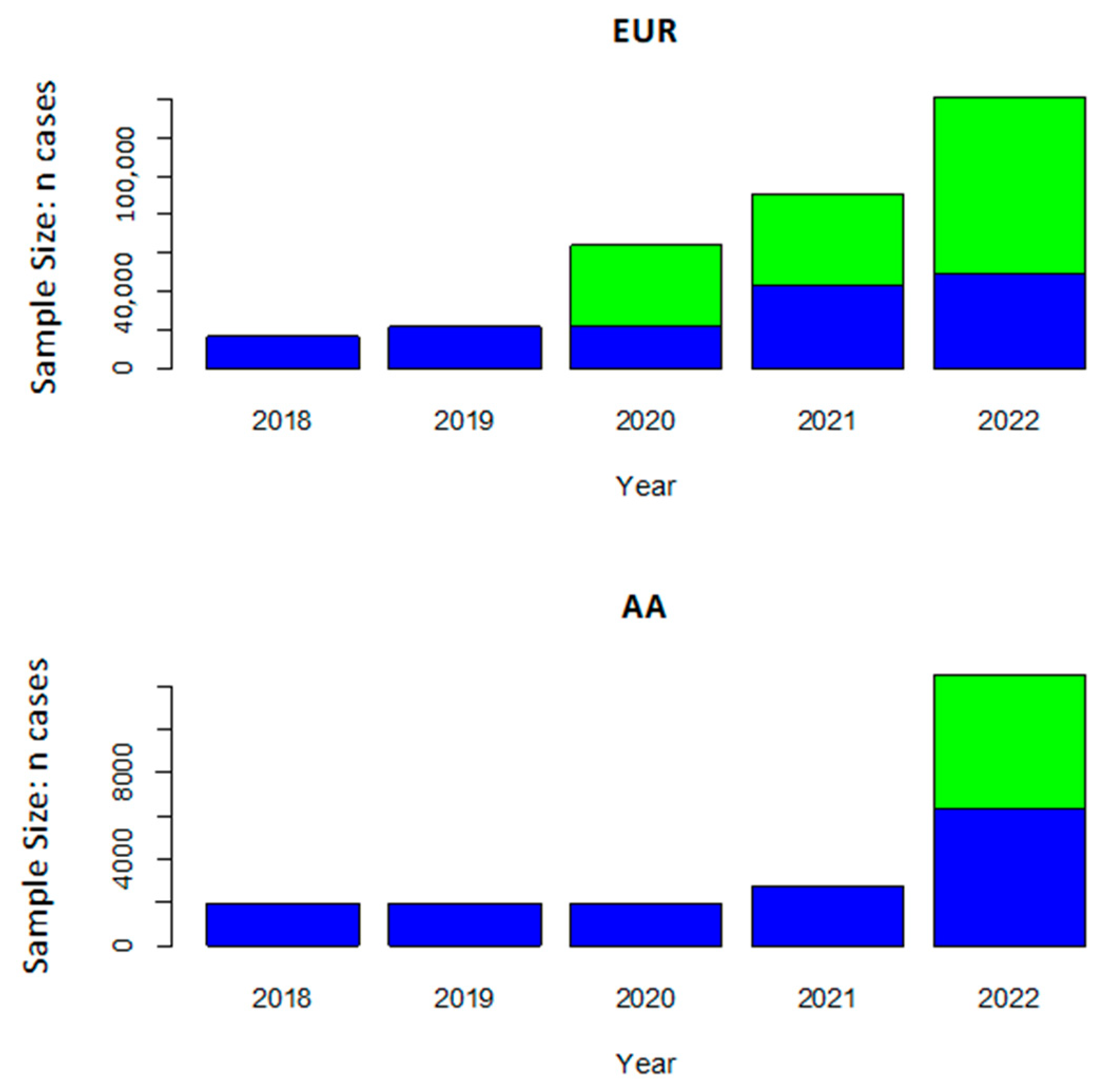
Figure 1. Discovery cohort sample sizes for AD cases in the largest European ancestry and African American AD GWAS by year. The number of cases in the AD case-control cohort GWAS is indicated in blue, and the number of proxy cases (i.e., AD status in the parents) is indicated in green. Although the sample size is much smaller for African American compared to European ancestry GWAS, the size of African American GWAS surged after the publication of a recent study [69].
Jcm 12 05189 g001
Figure 2. Linkage disequilibrium (LD) among TREM2 variants associated with AD risk in African American cohorts and the R47H variant associated with AD in European ancestry cohorts [35,69,75]. LD presented for both African-ancestry (AFR) and European ancestry (EUR) populations from the 1000 Genomes reference panel. NA indicates that LD was not estimated because one or both variants were too rare or absent in that population. Values on the diagonals which are necessarily 1.0 are shaded in black.
Jcm 12 05189 g002
Figure 3. Linkage disequilibrium (LD) of ABCA7 variants associated with AD risk in African American cohorts and the peak AD SNP (rs12151021) associated with AD in European ancestry cohorts [33,67,69,74,76,77]. LD presented for both African-ancestry (AFR) and European ancestry (EUR) populations from the 1000 Genomes reference panel. NA indicates that LD was not estimated because one or both of the variants were too rare or absent in that population. Values on the diagonals which are necessarily 1.0 are shaded in black.
Jcm 12 05189 g003
Table 1. Odds ratios (OR) or relative risk (RR) for AD for individual or aggregated APOE genotypes compared to the ε3/ε3 genotype among African American cohorts.
StudyCohort CharacteristicsAPOE Genotype
ε2/ε2 ε2/ε3 ε2/ε4ε2/ε2 ε2/ε3ε2/ε2ε2/ε3ε2/ε4ε3/ε4ε2/ε4 ε3/ε4ε2/ε4 ε3/ε4 ε4ε/4ε4/ε4
Tang et al. 1996 [45]New York community-based cohort of 106 AD cases and 154 controlsRR = 1.3 (p > 0.05).....RR = 0.6 (<0.05).RR = 3.0 (<0.05)
Farrer et al. 1997 [29]Multi-center Cohort of 235 cases and 240 controls..OR = 2.4 (>0.05)OR = 0.6 (>0.05)OR = 1.2 (>0.05)OR = 2.7 (<0.05)..OR = 12.5 (<0.05)
Sahota et al. 1997 [46]Indianapolis community-based cohort of 60 AD cases and 228 controls ...OR = 0.43
(>0.05)
OR = 0.34
(>0.05)
OR = 1.20
(>0.05)
..OR = 4.83
(<0.05)
Tang et al. 1998 [43]New York community-based cohort of 48 incident cases and 128 controls .......RR = 1.0
(p > 0.05)
.
Graff-Radford et al. 2002 [44]338 cases, 301 unrelated controls, and 108 siblings from the MIRAGE Study...OR = 0.41 (p < 0.05).OR = 2.6 (p < 0.05)..OR = 10.5 (p < 0.05)
Murell et al. 2006 [48]Indianapolis community-based cohort of 162 AD cases and 318 controls.OR = 0.42
(0.02)
.OR = 0.46
(0.047)
OR = 1.34
(0.61)
OR = 2.32
(<0.001)
..OR = 7.19
(<0.001)
Logue et al. 2011 [47]513 Cases and 496 controls from the MIRAGE and GenerAAtions Studies.OR = 0.43
(0.0094)
....OR = 2.08 (2 × 10⁻⁷).OR = 2.62
(3 × 10⁻¹⁴)
p-values indicated in parentheses; . = not calculated or not reported.
Disclaimer/Publisher’s Note: The statements, opinions and data contained in all publications are solely those of the individual author(s) and contributor(s) and not of MDPI and/or the editor(s). MDPI and/or the editor(s) disclaim responsibility for any injury to people or property resulting from any ideas, methods, instructions or products referred to in the content.