Review

Store-Operated Ca2+ Entry as a Putative Target of Flecainide for the Treatment of Arrhythmogenic Cardiomyopathy

1
Department of Biology and Biotechnology “Lazzaro Spallanzani”, University of Pavia, 27100 Pavia, Italy
2
Department of Health Sciences, University of Magna Graecia, 88100 Catanzaro, Italy
3
Department of Brain and Behavioral Sciences, University of Pavia, 27100 Pavia, Italy
4
Department of Biomedicine, School of Medicine, Benemérita Universidad Autónoma de Puebla, Puebla 72410, Mexico
*
Author to whom correspondence should be addressed.
J. Clin. Med. 2023, 12(16), 5295; https://doi.org/10.3390/jcm12165295
Submission received: 23 June 2023 / Revised: 4 August 2023 / Accepted: 12 August 2023 / Published: 14 August 2023
(This article belongs to the Section Cardiology)

Abstract

:
Arrhythmogenic cardiomyopathy (ACM) is a genetic disorder that may lead patients to sudden cell death through the occurrence of ventricular arrhythmias. ACM is characterised by the progressive substitution of cardiomyocytes with fibrofatty scar tissue that predisposes the heart to life-threatening arrhythmic events. Cardiac mesenchymal stromal cells (C-MSCs) contribute to the ACM by differentiating into fibroblasts and adipocytes, thereby supporting aberrant remodelling of the cardiac structure. Flecainide is an Ic antiarrhythmic drug that can be administered in combination with β-adrenergic blockers to treat ACM due to its ability to target both Nav1.5 and type 2 ryanodine receptors (RyR2). However, a recent study showed that flecainide may also prevent fibro-adipogenic differentiation by inhibiting store-operated Ca2+ entry (SOCE) and thereby suppressing spontaneous Ca2+ oscillations in C-MSCs isolated from human ACM patients (ACM C-hMSCs). Herein, we briefly survey ACM pathogenesis and therapies and then recapitulate the main molecular mechanisms targeted by flecainide to mitigate arrhythmic events, including Nav1.5 and RyR2. Subsequently, we describe the role of spontaneous Ca2+ oscillations in determining MSC fate. Next, we discuss recent work showing that spontaneous Ca2+ oscillations in ACM C-hMSCs are accelerated to stimulate their fibro-adipogenic differentiation. Finally, we describe the evidence that flecainide suppresses spontaneous Ca2+ oscillations and fibro-adipogenic differentiation in ACM C-hMSCs by inhibiting constitutive SOCE.

1. Introduction

Arrhythmogenic cardiomyopathy (ACM) is a genetic disease that leads to the progressive replacement of ventricular myocardium with fibrofatty scar tissue, thereby predisposing the patient to ventricular arrhythmias and sudden cardiac death (SCD) [1,2]. ACM may affect the right ventricle (arrhythmogenic right ventricular cardiomyopathy, ARVC), the left ventricle (arrhythmogenic left ventricular cardiomyopathy, ALVC), or both cardiac ventricles and is the main cause of SCD in young people and athletes [1,2]. A crucial role in the pathogenic mechanism of ACM can be played by the fibro-adipogenic differentiation of cardiac mesenchymal stromal cells (C-MSCs), which renders the cardiac tissue more likely to develop the arrhythmic events leading to the patient’s death [2,3]. Intriguingly, a recent investigation showed that the Ic antiarrhythmic drug (AAD) flecainide inhibited fibro-adipose differentiation in human C-MSCs (C-hMSCs) expanded from ACM patients (ACM C-hMSCs) by blocking store-operated Ca2+ entry (SOCE) [4], which represents the most important Ca2+ entry pathway in non-excitable cells [5,6,7], including MSCs [8,9,10]. Herein, we first describe the genetic background and therapeutic options of ACM. Then, we briefly recapitulate the main molecular mechanisms targeted by flecainide to mitigate arrhythmic events. Subsequently, we describe the role of intracellular Ca2+ oscillations in determining MSC fate. Next, we discuss our recent work showing that spontaneous Ca2+ oscillations in ACM C-hMSCs are accelerated to stimulate their fibro-adipogenic differentiation. Finally, we describe the evidence that flecainide suppresses spontaneous Ca2+ oscillations and fibro-adipogenic differentiation in ACM C-hMSCs by inhibiting constitutive SOCE and interfering with endoplasmic reticulum (ER) Ca2+ refilling.

2. Pathological Background and Treatment of ACM

Recently, an evidence-based reevaluation of published ARVC, the most common form of ACM, genes was carried out by experts in the field and showed that only a small number of genes encoding for desmosomal proteins, such as plakophilin-2 (PKP2), plakoglobin (JUP), and desmoglein 2 (DSG2), were definitively or moderately associated with ARVC (Table 1) [11]. Other genes that encode adherens junction proteins, such as cadherin 2 (CDH2) or catenin α3 (CTNNA3), that are also associated with ACM (Table 1) [1,2], only showed a moderate association with ACM [11]. Mutations in genes encoding proteins that are unrelated to intercellular junctions, such as cytoskeletal proteins, ion channels, and transporters, were also reported (Table 1) [1,2]; however, they were also shown to be weakly associated with ARVC [11]. This Gene Curation Expert Panel refuted the gene encoding for type 2 ryanodine receptor (RyR2), which has long been associated with ACM [1,2], as an ARVC gene [11].
Besides maintaining the mechanical stability of the heart and enabling the electrical coupling between ventricular cardiomyocytes, desmosomes may also play a crucial role in intracellular signal transduction and gene expression [2]. For instance, when the desmosomal assembly is disrupted by genetically defective proteins, plakoglobin 2 translocates from the intercalated discs to the nucleus, where it may compete with β-catenin to suppress the Wnt/β-catenin signalling pathway and elicit a gene transcriptional switch from myogenesis to fibrogenesis and adipogenesis [1,2,12]. A series of recent studies demonstrated that desmosomal mutations may also affect c-MSCs [13,14], which support the structural and functional integrity of the myocardium [15,16]. C-MSCs represent the primary source of myofibroblasts and adipocytes in the heart of ACM patients [13,14]. The fibrofatty replacement of ventricular myocardium may favour life-threatening arrhythmias, such as ventricular fibrillation and sustained ventricular tachycardia (VT), that ultimately cause SCD [2,3]. Patients who develop symptoms or have been diagnosed with ACM after a genetic screening or an electrocardiogram can be treated with implantable cardioverter-defibrillators (ICDs), radiofrequency catheter ablation, heart transplantation, β-blockers, or AADs [3,17]. Currently, ICDs represent the only therapeutic approach that can effectively reduce patient mortality, although they can be recommended only in the presence of clear arrhythmic risks, such as ventricular fibrillation and sustained VT [17]. AADs can be administered to reduce the burden of ACM-associated arrhythmias in symptomatic patients with non-sustained VT; moreover, AADs can be recommended as adjunctive therapy to mitigate the morbidities associated with ICDs (e.g., frequent device discharges and VT recurrences) and to facilitate catheter ablation [3,17]. Sotalol and amiodarone represent the first-line AADs in clinical practice, alone or in conjunction with β-blockers; however, there is no evidence that AAD therapy is effective at protecting patients from SCD. Furthermore, long-term treatment with amiodarone could induce extracardiac toxicity, so its use should be balanced in terms of effective arrhythmia suppression or reduction [3,17]. New hope for the pharmacological treatment of ACM has been sparked by the class Ic AAD flecainide ((RS)-N-(piperidin-2-ylmethyl)-2,5-bis(2,2,2-trifluoroethoxy) benzamide); C17H20F6N2O3) [18,19,20,21], which was approved by the Food and Drug Administration (FDA) in 1984 for the treatment of symptomatic sustained VT [18,20,22]. Flecainide targets a panel of ion channels and transporters that shape the cardiac action potential (AP; e.g., Nav1.5, Kv1.5, and Kv11.1) or the depolarization-induced Ca2+ transient responsible for cardiomyocyte contraction (e.g., type 2 ryanodine receptor, or RyR2) [22,23]. In addition, a recent investigation showed that flecainide may also inhibit fibro-adipose differentiation in ACM C-hMSCs by targeting SOCE [4].
Table 1. Genes whose mutations are associated with ACM.
GeneEncoded ProteinEstimated Frequency (%)Chromosomal Location
Desmosomes
PKP2Plakophilin-219–4612p11.21
DSPDesmoplakin1–166p24.3
DSG2Desmoglein-2 2.5–1018q12.1
DSC2Desmocollin-21–818q12.1
JUPJunction plakoglobinRare17q21.2
Adherens junctions
CTNNA3Catenin-α3Rare10q21.3
CDH2Cadherin 2Rare18q12.1
Cytoskeleton
LMNALamin A/CRare1q22
DESDesminRare2q35
FLNCFilamin CRare7q32.1
TTNTitinRare2q31.2
Ion transport
SCN5ANaV1.5Rare3p22.2
PLNPhospholambanRare6q22.31
For references, see: [1,17].

3. Anti-Arrhythmic Effects of Flecainide: From Nav1.5 to RyR2

A number of excellent reviews have provided a comprehensive description of the molecular pharmacology of flecainide [22,23,24]. Briefly, flecainide was first synthesised in 1972 by incorporating fluorine in local anesthetics, thereby increasing their stability, and it is nowadays recommended among the first-line therapeutic options to manage arrhythmias in patients without severe hemodynamic stability or significant structural heart disorders [25]. Flecainide reduces that fast inward Na+ current responsible for phase 0 of the cardiac AP by inhibiting the underlying Nav1.5 channel protein (Figure 1) [22,26,27]. In addition, flecainide may inhibit the late Na+ current (INaL) [26], which can arise because of recovery from inactivation and late re-opening of Nav1.5 channels during the sustained plateau in Purkinje fibres [28].
Flecainide may also inhibit the rapid delayed rectifier K+ current (IKR), which is carried by Kv11.1 channels (human Ether-à-go-go-Related Gene, or hERG) and is responsible for the late repolarization restoring the resting membrane potential in cardiomyocytes (Figure 2) [26,30]. Flecainide can further block the fast transient outward K+ current (Itof) (Figure 2), which is mediated by Kv4.2 channels and contributes to the early depolarization phase, and the ultrarapid K+ current (IKUR) (Figure 2), which is carried by Kv1.5 channels and is the major delayed rectifier K+ current in the atria [22,26].
At concentrations higher than 10 µM, flecainide inhibits Kv4.2, which mediates the fast transient outward K+ current (Itof), thereby potentially increasing APD in atrial and ventricular cardiomyocytes. In accordance with this, this inhibitory effect is exerted at supra-clinical doses of flecainide (IC50 = 15.2 µM) and, therefore, it is not expected to occur in the patients. Finally, flecainide may also inhibit Kv1.5 (IC50 = 237.1 µM), which mediates the ultrarapid delayed rectifier K+ current (IKUR) in the atria, where it is particularly abundant, and in the ventricles, thereby increasing the APD. This effect also occurs at supra-clinical doses and is unlikely to occur in patients.
Globally, flecainide prolongs the AP duration (APD) and AP refractoriness in ventricular and atrial cardiomyocytes, while they are both shortened in Purkinje fibres due to Nav1.5 channel blockade [22,26]. However, in human atrial cardiomyocytes, the flecainide-induced increase in APD and refractoriness is more evident when the AP presents a long-lasting plateau preceded by a notch [26]. Intriguingly, the kinetics of flecainide unbinding from the Nav1.5 channel pore during diastole are quite slow, thereby prolonging the refractoriness more than the APD (a phenomenon known as post-repolarization refractoriness), reducing excitability, and slowing intracardiac conduction, even at physiological heart rates, throughout all cardiac tissues [26]. As discussed in [20,26,31], the clinical concentration range of flecainide may selectively inhibit INa and IKR. Therefore, Nav1.5 and Kv11.1 channels represent the main molecular targets of flecainide in arrhythmic patients. Flecainide can indeed be used to prevent and treat various types of arrhythmias, such as paroxysmal atrial fibrillation, supraventricular tachycardia, and arrhythmic long QT syndromes (LQTS) [18,20,22]. Flecainide is particularly suitable for LQTS (LQTS3) type 3, which is associated with gain-of-function mutations in Nav1.5 channels (Figure 1) [20,22], which increase INaL [28,29]. Recent studies further showed that flecainide can also block RyR2, thereby inhibiting spontaneous RyR2-mediated sarcoplasmic reticulum (SR) Ca2+ release, which leads to delayed afterdepolarization (DADs) and triggered activity (Figure 1). Spontaneous mobilisation of SR Ca2+ can indeed be compensated by the electrogenic Na+/Ca2+ exchanger (NCX), which exports 1 Ca2+ out for 3 Na+ in during each cycle (Figure 1) and, while doing so, brings about an inward, depolarizing current [22,23]. Therefore, flecainide can also be used to treat catecholaminergic polymorphic ventricular tachycardia (CPVT) (Figure 1), a genetic arrhythmogenic disorder associated with mutations in RyR2 (CPVT1) or in the SR Ca2+-binding protein calsequestrin (CPVT2), which both lead to dysregulated SR Ca2+ release through RyR2 in response to β-adrenergic stimulation [23,32,33]. Flecainide binds to multiple sites within the RyR2 channel pore in a voltage-dependent manner but may also interact with several cytoplasmic sites in a voltage-independent manner [23,33]. The IC50 of flecainide-induced inhibition of RyR2 in transgenic mouse models of CPVT is ~2 µM [32,34], i.e., in the clinical therapeutic range of flecainide.
The use of flecainide has been prohibited in patients with myocardial infarction, left ventricular dysfunction, or structural heart disease based upon the increase in their mortality upon treatment with flecainide registered during the Cardiac Arrhythmia Suppression Trial (CAST) study [35]. The CAST study reported a significant increase in the mortality of patients treated with Ic AADs, including flecainide, as compared to subjects treated with placebo, with the primary causes of death being arrhythmias [35]. Nevertheless, a series of recent investigations [18,20,36,37,38], as well as a retrospective analysis by Rolland et al. [19], confirmed that flecainide is both effective and safe for reducing atrial fibrillation and symptomatic ventricular arrhythmias in patients with different structural heart disorders, including coronary artery disease (CAD). Lavalle and coworkers recently affirmed that the therapeutic use of flecainide is strongly limited by the anachronistic interpretation of the CAST since the introduction of more efficient diagnostic tools, such as cardiac magnetic resonance imaging (CMR) rather than transthoracic echocardiography, will be helpful to identify the type of structural heart disorder that may really prevent flecainide treatment [20]. In addition, it has been put forward that thrombolysis, as well as primary percutaneous coronary intervention, may enable the selection of CAD patients who do not present myocardial scar and residual ischaemia and for whom there is no straightforward evidence against the prescription of flecainide [20].

4. The Rationale for Using Flecainide to Treat Ca2+-Dependent Ventricular Arrhythmias in ACM

A consensus statement published by the Heart Rhythm Society (HRS) in conjunction with several organisations, including the American Heart Association (AHA), American College of Cardiology (ACC), and European Heart Rhythm Association (EHRA), recommended the use of flecainide in combination with β-blockers to treat ACM patients refractory to single-agent therapy or catheter ablation [19,21,39,40]. A pilot randomised clinical trial is currently evaluating the effectiveness of flecainide in reducing ventricular arrhythmia in ACM patients (Pilot Randomized Trial With Flecainide in ARVC Patients, NCT03685149, currently ongoing). Blockade of Nav1.5 channels represents the most obvious mechanism to explain the anti-arrhythmic effects exerted by flecainide in ACM [26,38]. Emerging evidence, however, suggests that dysregulated RyR2-mediated SR Ca2+ release can also generate pro-arrhythmic Ca2+ events in ACM [41]. By using a cardiomyocyte-specific PKP2 knockout mouse model, Cerrone et al. found that PKP2 deletion leads to a complex remodelling of the Ca2+ handling machinery, involving a reduction in the SR Ca2+ leakage caused by the downregulation of calsequestrin and RyR2 proteins [42]. This causes an elevation in SR Ca2+ content, which increases the amplitude and duration of spontaneous RyR2-mediated SR Ca2+ release because of the increase in intraluminal Ca2+ concentration. As a consequence, PKP2 deletion rendered RyR2 more prone to release Ca2+ during excitation-contraction coupling and to generate pro-arrhythmic Ca2+ release events during β-adrenergic stimulation [42]. A follow-up investigation confirmed that, in right ventricle-derived PKP2-deficient cardiomyocytes, the frequency of Ca2+ sparks, i.e., the elementary events of SR Ca2+ release [43], and of pro-arrhythmic afterdepolarizations increased before the onset of the cardiomyopathy due to an increase in SR Ca2+ content [44]. Finally, the same group showed that physical exercise caused a dramatic increase in the frequency and amplitude of SR Ca2+ sparks in PKP2-deficient mouse hearts because of the hyperphosphorylation of phospholamban, which accelerates Ca2+ uptake into the SR via the SarcoEndoplasmic Reticulum Ca2+-ATPase, during β-adrenergic stimulation [45]. Interestingly, blocking RyR2 with flecainide reduced the arrhythmia burden induced by β-adrenergic stimulation in PKP2-knockout mice [42]. Furthermore, a recent report confirmed that the Ca2+ cycling machinery was also altered in homozygous Dsg2 mutant mice (Dsg2mut/mut) [46]. Cytosolic Ca2+ overload during endurance exercise stimulates calpain-1 association with the mitochondria, thereby leading to the cleavage of mitochondrial-bound apoptosis-inducing factor (AIF). The truncated AIF translocated to the nucleus and triggered large-scale DNA fragmentation and cell damage, an effect that was exacerbated by mitochondrial-driven AIF oxidation. This induced myocyte necrosis in the heart of exercised Dsg2mut/mut mice [46].
On the one hand, these findings confirm that, as reported in other arrhythmic disorders (e.g., CPVT, Timothy syndrome, Brugada syndrome, and long and short QT syndrome), ACM is also associated with the deregulation of the Ca2+ cycling machinery in cardiomyocytes [47,48]. On the other hand, they support the notion that flecainide could effectively treat ACM patients by targeting the Ca2+ signalling machinery. In accordance with this hypothesis, flecainide decreased the frequency of Ca2+ sparks and normalised the Ca2+ transients elicited by membrane depolarization in inducible pluripotent stem cell-derived cardiomyocytes isolated from an ACM patient bearing a mutation in the DSC2 gene [49]. It has long been known that oscillations in intracellular Ca2+ concentration ([Ca2+]i) regulate MSC differentiation into a variety of cells belonging to multiple lineages [50,51,52]. Moreover, a recent study carried out in our laboratory showed that intracellular Ca2+ oscillations drive proliferation in C-hMSCs [53]. In the next paragraphs, we first discuss the mechanisms underpinning intracellular Ca2+ oscillations in MSCs. Then, we illustrate how an increase in the frequency and amplitude of spontaneous Ca2+ activity drives fibro-adipose remodelling in ACM C-MSCs. Finally, we describe how flecainide inhibits spontaneous Ca2+ waves and fibro-adipogenic differentiation by targeting SOCE in ACM C-hMSCs.

5. Intracellular Ca2+ Oscillations Regulate MSC fate

The signalling mechanisms and functions of intracellular Ca2+ oscillations have been primarily characterised in bone marrow (BM)-derived MSCs. A landmark series of studies conducted by Kawano et al. showed that, in BM-derived human MSCs (BM-hMSCs), spontaneous Ca2+ oscillations were driven by ATP autocrine/paracrine signalling [54,55,56]. ATP stimulated the Gq-protein coupled P2Y1 receptor to recruit phospholipase Cβ (PLCβ), which cleaved phosphatidylinositol-4,5-bisphosphate (PIP2), a minor phospholipid component of the plasma membrane, into diacylglycerol and inositol-1,4,5-trisphosphate (InsP3). InsP3 triggers rhythmic Ca2+ release from the endoplasmic reticulum (ER), the most abundant intracellular Ca2+ reservoir, by gating the so-called InsP3 receptors (InsP3Rs) [54,55,56]. These are Ca2+-permeable, non-selective cation channels that are embedded within ER cisternae and whose opening can be finely tuned by cytosolic Ca2+ levels [57,58]. BM-hMSCs express type 1 and type 2 InsP3Rs (InsP3R1 and InsP3R2, respectively), which are the most suitable InsP3Rs to generate rhythmic Ca2+ spikes in response to the continuous production of InsP3 because of their sensitivity to InsP3 and Ca2+ [56,59]. Intriguingly, InsP3-induced spontaneous Ca2+ spikes in BM-hMSCs fade away in the absence of extracellular Ca2+; therefore, Ca2+ influx is required to refill the ER Ca2+ pool and support the rhythmic Ca2+ activity over time [54], as widely reported in other cell types [60,61,62,63,64]. Electrophysiological recordings revealed that BM-hMSCs do not present voltage-gated Ca2+ currents, although transcripts encoding for the Cav1.1 and Cav3.2 α subunits of voltage-operated Ca2+ channels were detected. Conversely, these cells express a measurable inwardly-rectifying store-operated Ca2+ current [54], also known as ICRAC (Ca2+ release-activated Ca2+ current) (Figure 3). Intriguingly, by recharging the ER Ca2+ store, SOCE could also sensitise InsP3Rs via an increase in intraluminal Ca2+, which leads to periodic InsP3R activation and enhances the frequency of InsP3Rs-mediated Ca2+ release [57,58,65].
The ICRAC is the primary Ca2+-entry pathway sustaining intracellular Ca2+ oscillations over time in non-excitable cells and mediates an influx of Ca2+ that is commonly termed store-operated Ca2+ entry (SOCE) [5,6,57,58]. SOCE is activated whenever the ER Ca2+ concentration ([Ca2+]ER) in proximity of discrete sub-compartments of the peripheral ER falls below a threshold concentration because of ER Ca2+ release through InsP3Rs [66,67]. SOCE is mediated by the physical interaction between two ubiquitously expressed proteins (Figure 3): STIM, which is the sensor of [Ca2+]ER, and Orai, which contributes the Ca2+-selective channel protein on the plasma membrane [5,6,68]. STIM presents two isoforms, i.e., STIM1 and STIM2, whereas Orai displays three paralogues, i.e., Orai1, Orai2, and Orai3. Briefly, a reduction in [Ca2+]ER stimulates STIM proteins to assemble into oligomers that redistribute to peripheral ER-plasma membrane junctions, known as puncta, while extending their cytosolic COOH-termini, which contain the STIM Orai-activating region/CRAC-activating domain (SOAR/CAD), towards the inner leaflet of the plasma membrane (Figure 3). The SOAR/CAD domain, in turn, binds to and gates the hexameric Orai channels, thereby activating the ICRAC and inducing SOCE (Figure 3) [5,6,69]. STIM2 presents a lower Ca2+ affinity as compared to STIM1 and is, therefore, activated upon a modest fall in [Ca2+]ER. It turns out that STIM2 is regarded as the most suitable isoform to tonically activate SOCE even in the absence of extracellular stimulation and to maintain ER Ca2+ levels [70]. Conversely, STIM1 is activated after a significant depletion of [Ca2+]ER during InsP3-induced Ca2+ release and sustains the Ca2+ response to extracellular cues [5,6]. Nevertheless, STIM2 can sustain SOCE elicited by agonist-induced sub-threshold depletion of the ER Ca2+ content [68,71] as well as support STIM1-dependent recruitment to the ER-plasma membrane during supra-threshold stimulation [68,72]. In addition, STIM1 can be activated even in the absence of an extracellular agonist when the resting [Ca2+]ER is too low to prevent its constitutive activation [73,74,75]. Orai1 has long been regarded as the primary pore-forming subunit of CRAC channels [5,69]. However, recent evidence suggests that Orai2 and/or Orai3 can assemble into heteromultimeric channels with Orai1 and serve as negative modulators of the ICRAC in naïve cells [76,77,78]. The studies conducted by Kawano et al. date back to the early decade of this century [54,55,56], when STIM and Orai were not known to mediate the ICRAC. Therefore, we still do not know whether they support SOCE in BM-hMSCs. Nevertheless, genetic silencing of STIM1, Orai1, and/or Orai3 reduced SOCE in mouse BM-derived MSCs (BM-mMSCs) [79,80,81] and in human dental pulp-derived MSCs (DP-hMSCs) [82].
Spontaneous intracellular Ca2+ oscillations have been described not only in BM-hMSCs [54,83,84] but also also in human adipose tissue-derived MSCs (AD-hMSCs) [51,85]. Intracellular Ca2+ oscillations regulate stem cell differentiation by stimulating the expression of genes that control cellular fate and silencing those that are required for self-renewal [52,86,87,88,89,90]. The spatio-temporal profile of spontaneous Ca2+ activity can selectively change during hMSC differentiation in tissue-specific lineages by showing either an increase or a decrease in the spike frequency [52]. Spontaneous Ca2+ oscillations disappear during hMSC differentiation towards an adipogenic phenotype both when they derive from the BM [56,91] and from the adipose tissue [51]. The loss of spontaneous Ca2+ activity reduces the nuclear translocation of the Ca2+-sensitive transcription factor, nuclear factor of activated T-cells (NFAT), which is likely to prevent hMSC differentiation [56]. Conversely, BM-hMSCs undergoing neuronal differentiation show an increase in the frequency of spontaneous Ca2+ spikes, which become quite irregular in terms of amplitude and shape [90,92]. A more robust Ca2+ spiking could govern BM-hMSC differentiation toward a stable neuronal phenotype by promoting the phosphorylation of CREB (cAMP response element binding protein) [93], a transcription factor that plays a crucial role in neurogenesis and can be activated by the Ca2+/CaM-dependent protein kinase IV [94]. The requirement of spontaneous Ca2+ oscillations for hMSC commitment to a specific cellular lineage is so tight that the intracellular Ca2+ activity in BM-hMSCs can be physically manipulated, for instance by electrostimulation [95], to favour osteogenic [91] or neuronal [96] differentiation. Similarly, the addition of inductive soluble factors, such as the InsP3-producing autacoid carbachol, has been successfully employed to increase the frequency of InsP3Rs-mediated spontaneous Ca2+ oscillations and thereby promote neuronal differentiation in AD-hMSCs [97]. As described above, SOCE is crucial to maintaining spontaneous Ca2+ oscillations in hMSCs; an increase or decrease in the rate of Ca2+ entry can differentially affect their differentiation outcomes. Therefore, it is not surprising that pharmacological manipulation of SOCE has been put forward as an alternative strategy to control hMSC differentiation [10,80].

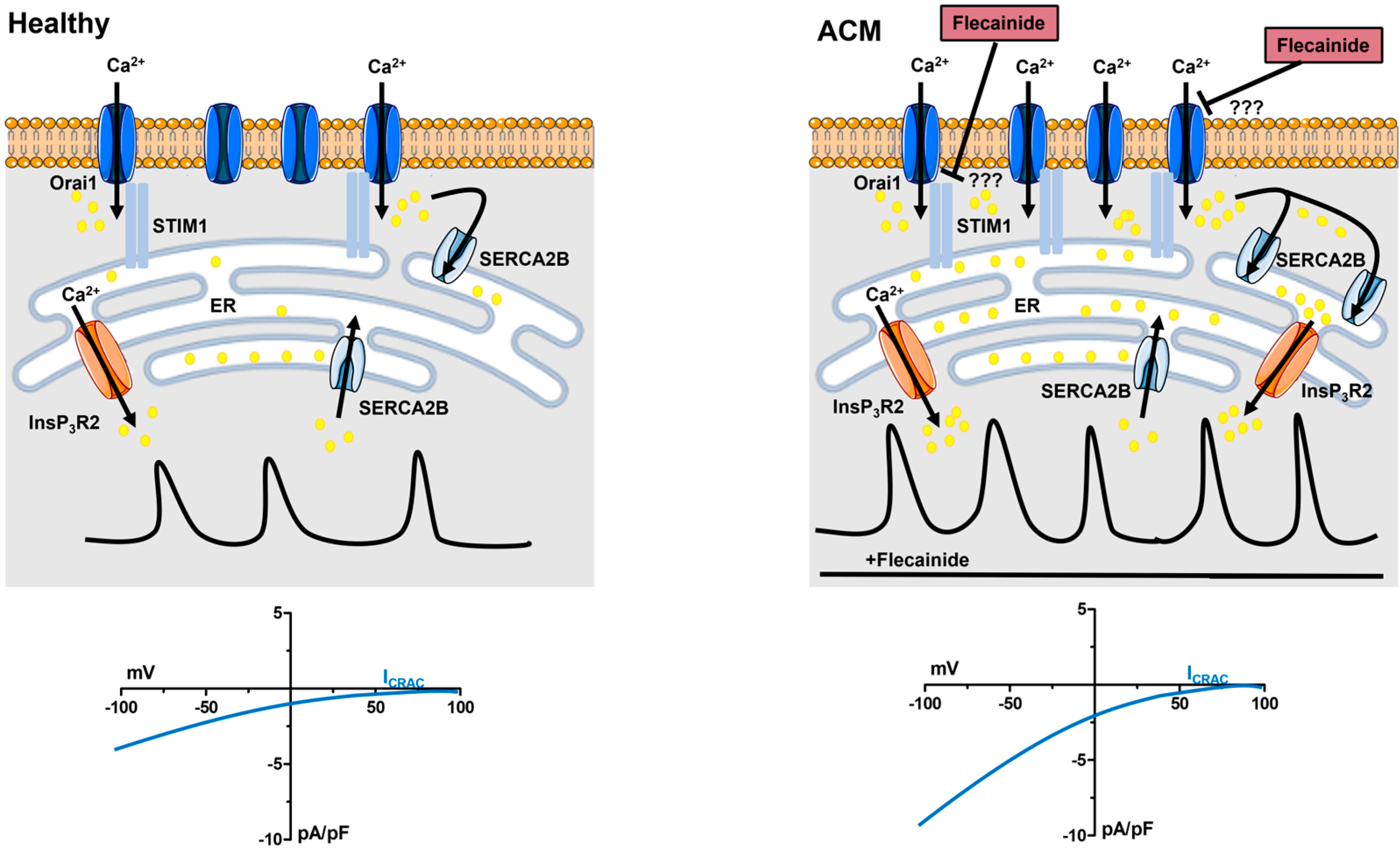
6. Flecainide Inhibits SOCE and Prevents Ca2+-Dependent Fibro-Adipogenic Differentiation in ACM C-hMSCs

A recent investigation carried out by our laboratory has shown that intracellular Ca2+ signalling drives C-hMSC proliferation by stimulating extracellular signal-regulated kinase (ERK) phosphorylation [53]. In addition, this study confirmed that a functional SOCE is expressed in C-hMSCs and is sensitive to InsP3-induced depletion of the ER Ca2+ store [53]. Therefore, based upon the evidence provided by the Pompilio group that ACM C-hMSCs are more likely to undergo fibro-adipogenic differentiation as compared to C-hMSCs [13,14], we established a collaborative endeavour to understand whether spontaneous Ca2+ oscillations were dysregulated in ACM C-hMSCs. Then, we evaluated whether SOCE could be pharmacologically targeted to prevent aberrant lipid/fibrotic accumulation in the ACM heart [4].

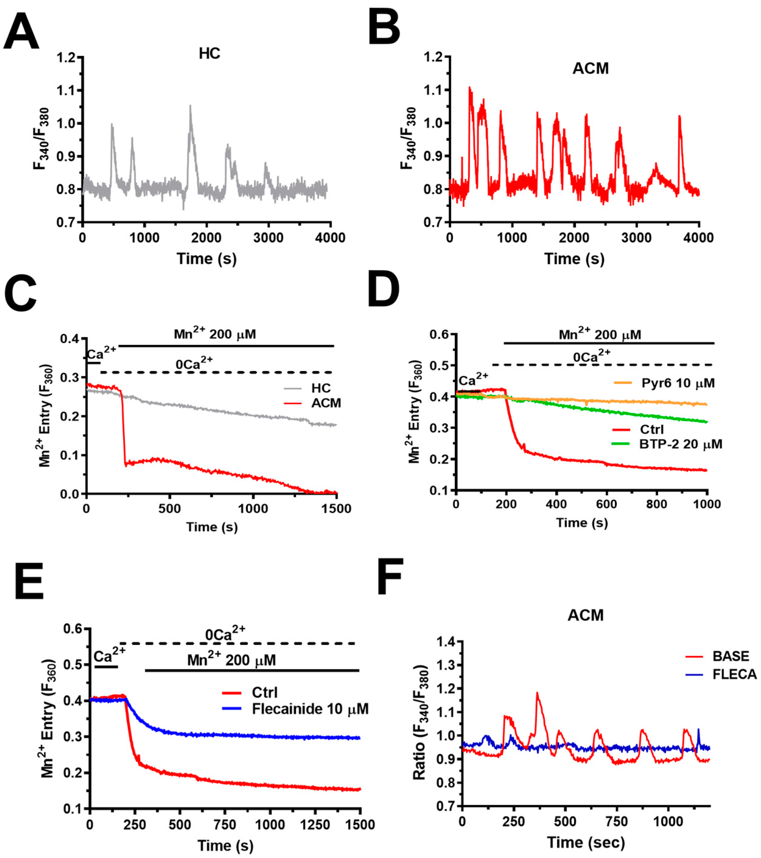
6.1. Intracellular Ca2+ Oscillations Are Up-Regulated in ACM C-hMSCs

Single-cell imaging of the Ca2+-sensitive fluorophore, Fura-2, represents a widespread technique to analyse intracellular Ca2+ oscillations in tens to hundreds of non-excitable cells [62,63,64,98,99,100], including hMSCs [54,55,56]. This approach led to the observation that spontaneous Ca2+ oscillations in ACM C-hMSCs were significantly more robust than C-hMSCs in terms of percentage of oscillating cells, amplitude, and frequency (~2.2 mHz vs. ~1.2 mHz) of the single Ca2+ transients (Figure 4A,B) [4]. C-hMSCs are not amenable to genetic manipulation of the Ca2+ handling machinery [53]. However, a pharmacological approach confirmed that spontaneous Ca2+ oscillations in ACM C-hMSCs were driven by the Gq-protein-coupled P2Y1 receptors, triggered by rhythmic InsP3-induced ER Ca2+ release, and maintained over time by SOCE [4]. Conversely, RyR2 was not expressed, whereas voltage-gated Ca2+ entry did not support spontaneous Ca2+ oscillations [4]. Interestingly, the Mn2+ quenching assay, which is widely employed to monitor basal Ca2+ entry in non-excitable cells [70,75,101], revealed that SOCE was constitutively activated in C-hMSCs and was significantly enhanced in ACM C-hMSCs (Figure 4C,D) [4]. Constitutive SOCE regulates resting [Ca2+]i and maintains [Ca2+]ER, thereby finely tuning the magnitude and duration of InsP3-induced ER Ca2+ release [70,73,74,75,101,102]. In accordance, basal [Ca2+]i and InsP3-induced ER Ca2+ release were both up-regulated in ACM C-hMSCs [4]. These findings were supported by the molecular evidence that the expression of STIM1 and InsP3R2 was enhanced at both transcript and protein levels [4]. In addition, SERCA2B, which sequesters cytosolic Ca2+ into the ER lumen in non-excitable cells, was also up-regulated in ACM C-hMSCs [4]. Intriguingly, intracellular Ca2+ spiking was enhanced in C-hMSCs transduced with lentiviral particles containing a short hairpin against PKP2, which caused an increase in the expression levels of STIM1 and SERCA2B proteins [4].
These data support a model according to which (Figure 3): (1) STIM1 protein, the sensor of [Ca2+]ER that is activated in response to large falls in [Ca2+]ER occurring during autocrine/paracrine ATP signalling, is up-regulated in ACM C-hMSCs; (2) this leads to the recruitment of more Orai1 hexamers in the plasma membrane and therefore increases constitutive SOCE in ACM C-hMSCs; (3) the larger influx of Ca2+ causes a remarkable increase in [Ca2+]ER, also because of SERCA2B up-regulation, in ACM C-hMSCs; (4) the enhanced ER Ca2+ loading, on the one hand, sensitises InsP3Rs to open at a higher frequency; on the other hand, it increases the availability of free intraluminal Ca2+ that can be released during each Ca2+ spike. In this context, it is worth clarifying that, although the SOCE signal driving the spontaneous Ca2+ oscillations in C-hMSCs has been termed constitutive [4], it is stimulated by ATP released in either an autocrine or paracrine manner via P2Y1 receptor activation. The term “constitutive” reflects the lack of any agonist in the perfusate when intracellular Ca2+ oscillations and basal Ca2+ entry are monitored.

6.2. Intracellular Ca2+ Oscillations in ACM C-hMSCs: Is There Any Role for Lysosomal Ca2+ Mobilization?

It is worth noticing that ER Ca2+ release through InsP3Rs in C-hMSCs could also be activated by lysosomal Ca2+ release via two pore channels (TPCs) [53]. TPC1 and TPC2 are activated by the Ca2+-mobilizing second messenger, nicotinic acid adenine dinucleotide phosphate (NAADP) [103,104], and are both expressed in C-hMSCs, in which they trigger the intracellular Ca2+ oscillations by which foetal bovine serum stimulates proliferation [53]. Transmission electron microscopy revealed that lysosomal vesicles can establish quasi-synaptic membrane contact sites with the juxtaposed ER cisternae in C-hMSCs [53]. Herein, NAADP-induced lysosomal Ca2+ release via TPCs may activate InsP3Rs via the CICR process [53]. This signalling pathway may trigger long-lasting oscillations in [Ca2+]i not only in C-hMSCs but also in many other cell types [62,64,105,106]. In addition, NAADP-induced lysosomal Ca2+ release may contribute to cardiac hypertrophy induced by β-adrenergic stimulation [107,108] and ischemia-reperfusion injury [109]. Therefore, future investigations will have to assess whether NAADP and TPCs play any role in the spontaneous Ca2+ oscillations arising in C-hMSCs and whether their contribution, if any, is increased in ACM. Intriguingly, SOCE has also been shown to support lysosomal Ca2+ refilling, such that the increase in constitutive SOCE has the potential to increase lysosomal Ca2+ release in ACM C-hMSCs [110].

6.3. Intracellular Ca2+ Oscillations Drive Fibro-Adipogenic Differentiation in ACM C-hMSCs

Unexpectedly, we found that spontaneous Ca2+ oscillations in ACM C-hMSCs showed an increase in amplitude and frequency (up to 8 mHz) during adipogenic differentiation [4]. This finding was surprising since, both in BM- and AD-derived MSCs, the Ca2+ spiking activity decreases when they are committed to an adipogenic phenotype [51,56]. The pharmacological blockade of the Ca2+ signalling machinery that sustains the accelerated Ca2+ spikes, i.e., SOCE and InsP3Rs, prevented ACM C-hMSCs from differentiating into either adipocytes or fibroblasts [4]. Therefore, it takes an increase in the amplitude and frequency of spontaneous Ca2+ oscillations to induce ACM C-hMSCs to undergo fibro-adipogenic differentiation. The multifunctional Ca2+/CaM-dependent protein kinase II (CaMKII) represents the most suitable decoder of intracellular Ca2+ oscillations not only in the brain but also in the heart [111,112,113]. CaMKII is a multimer of eight to twelve subunits that are present in four different isoforms (α, β, γ, and δ), each encoded by a distinct gene, arranged in two-stacked hexameric rings [114]. CaMKIIδ is the major isoform in the heart and exists as different spliced variants, e.g., CaMKIIδ2/B, CaMKIIδ2/C, and CaMKIIδ2/C [111]. Briefly, in response to an increase in [Ca2+]i, the Ca2+/CaM complex can bind to two neighbouring subunits, thereby enabling CaMKII to autophosphorylate on threonine 287 (Thr287). Autophosphorylation traps Ca2+/CaM and prolongs CaMKII activity even when the [Ca2+]i returns to the baseline. Low-frequency Ca2+ transients are less effective than high-frequency Ca2+ transients to evoke autophosphorylation and Ca2+-independent activity since the Ca2+/CaM complex can dissociate from the holoenzyme between two consecutive Ca2+ spikes [115]. We found that CaMKIIδ and CaMKIIγ were the predominant CaMKII isoforms in C-hMSCs; however, there was no difference in CaMKII protein expression between C-hMSCs and ACM C-hMSCs. However, CaMKII protein in ACM C-hMSCs was hyperphosphorylated as compared to healthy cells, which is consistent with their greater eagerness to generate Ca2+ oscillations [4]. In agreement with this hypothesis, the pharmacological blockade of CaMKII activity with KN93 prevented fibro-adipogenic differentiation in ACM C-hMSCs [4]. Therefore, CaMKII is likely to translate the spontaneous Ca2+ oscillations that occur at a higher frequency in ACM C-hMSCs into a transcriptional programme that redirects their differentiation fate to fibroblasts or adipocytes. One could rightly notice that a frequency of 8 mHz might be too slow to enable the accumulation of Ca2+/CaM-bound CaMKII subunits sufficient for autophosphorylation and autonomous CaMKII activity [116]. Nevertheless, CaMKII does not only integrate the information encoded in the oscillation frequency but also in the length of the single Ca2+ pulses; an increase in the individual Ca2+ spike duration decreases the frequency threshold for CaMKII activation and autonomous activity [115]. The long duration of individual Ca2+ spikes in ACM C-hMSCs, ranging from ~200 s up to ~300 s (Figure 4B), enables coincident binding of CaM on neighbouring subunits and of Thr287 autophosphorylation even at 8 mHz [115,116]. For instance, the slow Ca2+ oscillations evoked by fertilisation in mouse oocytes were able to induce long-lasting oscillations in CaMKII activity that were crucial to resuming embryonic development [117]. The mechanism by which CaMKII stimulates the fibro-adipogenic differentiation of ACM-hMSCs is not clear [4]. However, CaMKII promoted adipogenesis in porcine BM-MSCs by increasing the expression of two adipogenic transcription factors, such as CCAAT/enhancer binding protein α (C/EBPα) and peroxisome proliferator activated receptor γ (PPARγ), by stimulating the PI3K/Akt-Fox01 signalling pathway [118].

6.4. Flecainide Abolishes Intracellular Ca2+ Oscillations and Fibro-Adipogenic Differentiation by Targeting SOCE

SOCE is either down-regulated or up-regulated in a growing number of disorders, including severe combined immunodeficiency [119], neurodegenerative diseases [120], genetic myopathies [121], pulmonary arterial hypertension [122], and cancer [123]. Preliminary evidence indicated that, even though Orai1 is weakly expressed in adult ventricular cardiomyocytes from humans and mice, its expression increases and leads to SOCE up-regulation in cardiac hypertrophy and heart failure [124]. Therefore, the pharmacological manipulation of SOCE with selective agonists or blockers of Orai1-mediated Ca2+ entry represents a promising therapeutic approach to rescue aberrant Ca2+-dependent processes in multiple cell types [65,122,125,126,127,128,129,130,131]. The evidence that flecainide may physically interact with many pro-arrhythmic ion channels led our group to hypothesise that it could also inhibit constitutive SOCE in ACM C-hMSCs. Consistently, single-cell Ca2+ imaging experiments carried out in our laboratory showed that 10 µM flecainide blocked constitutive SOCE (Figure 4E) and thereby inhibited InsP3-induced ER Ca2+ release in these cells [4]. As expected, based upon these findings, flecainide also suppressed spontaneous Ca2+ oscillations (Figure 4F) and interfered with ACM C-hMSC fibro-adipogenic differentiation [4]. Of note, ACM C-hMSC cells do not express NaV1.5; therefore, the inhibitory effect of flecainide is not related to the inhibition of voltage-gated inward Na+ currents [4]. In this view, it must be pointed out that Nav1.5-mediated Na+ currents could favour intracellular Ca2+ oscillations only by switching the NCX into the reverse (i.e., Ca2+ in, Na+ out) mode [132,133]. However, the pharmacological blockade of the reverse-mode NCX activity did not affect the spontaneous Ca2+ oscillations in ACM C-hMSCs [4]. It turns out that flecainide-induced SOCE inhibition underlies the suppression of both the unsolicited Ca2+ spikes and fibro-adipogenic differentiation that take place in C-hMSCs expanded from ACM patients. The mechanisms by which flecainide inhibits constitutive SOCE remain to be elucidated (Figure 3). SOCE in ACM C-hMSCs is also sensitive to two established inhibitors of this Ca2+-entry pathway, namely the pyrazole compounds Pyr6 and BTP-2 (YM-58483) [7,123,134,135,136,137]. These drugs are known to selectively block Orai1-mediated SOCE [7,123,134,135,136,137]; however, they could also affect Orai2 [138,139]. Nevertheless, Orai2 is a negative modulator of Orai1 [76,77,78]. Therefore, the direct inhibition of Orai2 is predicted to enhance, not inhibit, SOCE. These observations support the view that Pyr6, BTP-2, and flecainide interfere with extracellular Ca2+ entry through Orai1.
The mechanism by which Pyr6 inhibits the ICRAC and SOCE is still unclear, although it is unlikely to prevent the STIM-dependent Orai1 channel activation process [136]. BTP-2, in turn, could target a binding site that is located on the extracellular side of the Orai1 channel pore when administered at low micromolar doses [140], as throughout our investigations [4,53]. This hypothesis is supported by the evidence that BTP-2 does not block the ICRAC when intracellularly applied [141] and does not prevent the physical interaction between STIM1 and Orai1 [142], as suggested for another established SOCE inhibitor, i.e., 2-Aminoethyldiphenyl Borate [143]. Future studies are mandatory to unveil how flecainide blocks constitutive SOCE in ACM C-hMSCs. Nevertheless, the evidence presented in [4] further supports the emerging view that this pleiotropic drug targets multiple signalling pathways that are involved in ACM pathogenesis: (1) NaV1.5 and (2) RyR2 in cardiomyocytes, thereby rescuing the spontaneous electrophysiological events that lead to tachycardia and ventricular fibrillation; (3) SOCE in C-MSCs, thereby alleviating the aberrant remodelling of cardiac structure that predisposes the heart to generate arrhythmic events. Interestingly, we have previously shown that the β-blocker propranolol inhibits constitutive SOCE and reduces the vasculogenic activity of circulating endothelial colony forming cells in infantile hemangioma (IH) both in vitro and in children affected by IH, thereby resulting in a therapeutic benefit in IH patients [144,145]. The hypothesis that propranolol effectively targets ACM by inhibiting both β2-adrenergic receptors in sinoatrial fibres and cardiac myocytes and SOCE in C-MSCs is certainly worth of further investigation.
Notably, several Orai1 inhibitors have already entered Clinical Trials for the treatment of multiple diseases, including psoriasis (CM2489 from CalciMedica), severe acute pancreatitis (CM4620 from CalciMedica) (https://clinicaltrials.gov/ct2/show/NCT03401190 (accessed on 30 May 2023)), critical COVID-19 pneumonia (Auxora, from CalciMedica) (https://clinicaltrials.gov/ct2/show/NCT04661540 (accessed on 30 May 2023)), mild asthma (RP3128 from Rizen Pharmaceuticals) (https://clinicaltrials.gov/ct2/show/NCT02958982 (accessed on 30 May 2023)), and non-Hodgkin’s lymphoma (RP4010, also from Rizen Pharmaceuticals) (https://clinicaltrials.gov/ct2/show/NCT03119467 (accessed on 30 May 2023)) [134]. These studies confirmed that SOCE inhibitors can be administered orally, are safe, and do not induce serious adverse effects in human subjects [134]. In line with this evidence, several Food and Drug Administration (FDA)-approved drugs were recently found to inhibit both the ICRAC and SOCE [146]: teriflunomide, leflunomide, roflumilast, tolvaptan, and lansoprazole. Therefore, targeting SOCE represents a promising therapeutic avenue for many diseases that still lack a valid therapeutic option, including ACM.

7. Future Directions

The mechanism by which flecainide inhibits SOCE requires further investigation. Likewise, the molecular determinants that increase constitutive SOCE may go beyond STIM1 overexpression and include the derangement of other regulatory factors, such as an increase in STIM1 expression or a reduction in Orai2 or Orai3 protein levels. Nevertheless, these preliminary findings strongly hint at flecainide as a pleiotropic drug that owes its efficacy to its ability to target multiple pro-arrhythmic signalling pathways in ACM. In vivo investigations will have to assess whether it does interfere with cardiac fibrofatty remodelling in transgenic mouse models of ACM. It will also be imperative to assess whether flecainide does interfere with CaMKII hyperactivation in ACM C-MSCs and, if so, to untangle the signalling pathways that are recruited by CaMKII to stimulate fibro-adipogenic differentiation. In accordance with this, Maione et al. did not assess whether inhibiting SOCE or InsP3Rs reduced CaMKII hyperphosphorylation in ACM C-hMSCs. Addressing this issue is mandatory to provide a clear-cut landscape of the molecular mechanisms by which an increase in constitutive SOCE promotes C-hMSC fibro-adipogenic differentiation in ACM hearts. Currently, we cannot rule out the possibility that CaMKII in ACM-hMSCs is hyperphosphorylated because of a decrease in the expression and/or activity of protein phosphatases 1 and 2A, which dephosphorylate CaMKII when [Ca2+]i returns to the baseline [147,148]. This would in turn lead to SOCE hyperactivation and ER Ca2+ overload since it has long been known that CaMKII is able to stimulate SOCE [149,150,151]. However, there is no doubt that the pharmacological blockade of SOCE inhibits both spontaneous Ca2+ oscillations and fibro-adipogenic differentiation in ACM C-hMSCs [4]. In this view, the evidence that flecainide suppresses fibro-adipogenic differentiation paves the way for future investigations assessing whether other FDA-approved SOCE inhibitors could also be used to treat ACM by preventing fibro-fatty remodelling in C-MSCs. Finally, it could also be evaluated whether flecainide is also effective to target other disorders that are associated with SOCE up-regulation, such as severe acute pancreatitis, chronic inflammation, and cancer.

8. Conclusions

Flecainide is a class Ic AAD that proved to be effective in the management of heart rate by targeting NaV1.5, which mediates the fast inward Na+ current, and RyR2, which mediates aberrant SR Ca2+ release. Flecainide is emerging as a promising therapeutic tool to treat ACM, which involves a complex remodelling of the Ca2+-handling machinery in ventricular cardiomyocytes. In addition, studies conducted on C-MSCs isolated from ACM patients and transgenic mouse models of ACM revealed that intracellular Ca2+ dynamics is also dysregulated in the stromal compartment of the heart. SOCE, which represents the main intracellular Ca2+ entry pathway in non-excitable cells, is constitutively activated in ACM C-hMSCs and causes Ca2+ overload in the InsP3-sensitive ER Ca2+ reservoir. This results in an increase in both the amplitude and frequency of spontaneous Ca2+ oscillations that finely tune MSC fate, thereby favouring fibro-adipogenic differentiation in ACM C-hMSCs. Therefore, SOCE up-regulation in C-hMSCs could play a pathogenic role in ACM by stimulating the cardiac structural remodelling that increases the incidence of arrhythmic events. Intriguingly, flecainide, when applied at doses known to inhibit Nav1.2 and RyR2 channels, inhibited constitutive SOCE, thereby suppressing the spontaneous Ca2+ oscillations that occur during fibro-adipogenic differentiation and preventing ACM C-hMSC differentiation into fibroblasts or adipocytes. These pieces of evidence provide the first proof-of-concept that flecainide is also effective against SOCE and indicate that SOCE could be targeted for therapeutic purposes in ACM.

Author Contributions

Conceptualization, F.M.; writing—original draft preparation, F.M.; writing—review and editing, V.B., T.S., P.F., G.S. and R.B.-R. All authors have read and agreed to the published version of the manuscript.

Funding

This research has been supported by the EU Horizon 2020 FETOPEN-2018–2020 Program under Grant Agreement N. 828984, LION-HEARTED (F.M.); the Italian Ministry of Education, University, and Research (MIUR): Dipartimenti di Eccellenza Program (2018–2022)—Dept. of Biology and Biotechnology “L. Spallanzani,” University of Pavia (F.M.); and Fondo Ricerca Giovani from the University of Pavia (F.M.). R.B.-R. is supported by the National Council of Science and Technology (CONACYT), Identification Number: CVU121216.

Informed Consent Statement

Not applicable.

Data Availability Statement

Not applicable.

Conflicts of Interest

The authors declare no conflict of interest.

References

  1. Austin, K.M.; Trembley, M.A.; Chandler, S.F.; Sanders, S.P.; Saffitz, J.E.; Abrams, D.J.; Pu, W.T. Molecular mechanisms of arrhythmogenic cardiomyopathy. Nat. Rev. Cardiol. 2019, 16, 519–537. [Google Scholar] [CrossRef]
  2. Corrado, D.; Link, M.S.; Calkins, H. Arrhythmogenic Right Ventricular Cardiomyopathy. N. Engl. J. Med. 2017, 376, 61–72. [Google Scholar] [CrossRef]
  3. Ermakov, S.; Scheinman, M. Arrhythmogenic Right Ventricular Cardiomyopathy–Antiarrhythmic Therapy. Arrhythmia Electrophysiol. Rev. 2015, 4, 86–89. [Google Scholar] [CrossRef] [Green Version]
  4. Maione, A.S.; Faris, P.; Iengo, L.; Catto, V.; Bisonni, L.; Lodola, F.; Negri, S.; Casella, M.; Guarino, A.; Polvani, G.; et al. Ca2+ dysregulation in cardiac stromal cells sustains fibro-adipose remodeling in Arrhythmogenic Cardiomyopathy and can be modulated by flecainide. J. Transl. Med. 2022, 20, 522. [Google Scholar] [CrossRef]
  5. Emrich, S.M.; Yoast, R.E.; Trebak, M. Physiological Functions of CRAC Channels. Annu. Rev. Physiol. 2021, 84, 355–379. [Google Scholar] [CrossRef]
  6. Moccia, F.; Brunetti, V.; Perna, A.; Guerra, G.; Soda, T.; Berra-Romani, R. The Molecular Heterogeneity of Store-Operated Ca2+ Entry in Vascular Endothelial Cells: The Different roles of Orai1 and TRPC1/TRPC4 Channels in the Transition from Ca2+-Selective to Non-Selective Cation Currents. Int. J. Mol. Sci. 2023, 24, 3259. [Google Scholar] [CrossRef]
  7. Prakriya, M.; Lewis, R.S. Store-Operated Calcium Channels. Physiol. Rev. 2015, 95, 1383–1436. [Google Scholar] [CrossRef] [PubMed] [Green Version]
  8. Jiang, L.H.; Mousawi, F.; Yang, X.; Roger, S. ATP-induced Ca2+-signalling mechanisms in the regulation of mesenchymal stem cell migration. Cell. Mol. Life Sci. 2017, 74, 3697–3710. [Google Scholar] [CrossRef] [PubMed] [Green Version]
  9. Wang, L.; Roger, S.; Yang, X.B.; Jiang, L.H. Role of the store-operated Ca2+ channel in ATP-induced Ca2+ signalling in mesenchymal stem cells and regulation of cell functions. Front. Biosci. (Landmark Ed.) 2021, 26, 1737–1745. [Google Scholar] [CrossRef] [PubMed]
  10. Ahamad, N.; Singh, B.B. Calcium channels and their role in regenerative medicine. World J. Stem Cells 2021, 13, 260–280. [Google Scholar] [CrossRef]
  11. James, C.A.; Jongbloed, J.D.H.; Hershberger, R.E.; Morales, A.; Judge, D.P.; Syrris, P.; Pilichou, K.; Domingo, A.M.; Murray, B.; Cadrin-Tourigny, J.; et al. International Evidence Based Reappraisal of Genes Associated With Arrhythmogenic Right Ventricular Cardiomyopathy Using the Clinical Genome Resource Framework. Circ. Genom. Precis. Med. 2021, 14, e003273. [Google Scholar] [CrossRef] [PubMed]
  12. Garcia-Gras, E.; Lombardi, R.; Giocondo, M.J.; Willerson, J.T.; Schneider, M.D.; Khoury, D.S.; Marian, A.J. Suppression of canonical Wnt/beta-catenin signaling by nuclear plakoglobin recapitulates phenotype of arrhythmogenic right ventricular cardiomyopathy. J. Clin. Investig. 2006, 116, 2012–2021. [Google Scholar] [CrossRef] [PubMed] [Green Version]
  13. Sommariva, E.; Brambilla, S.; Carbucicchio, C.; Gambini, E.; Meraviglia, V.; Dello Russo, A.; Farina, F.M.; Casella, M.; Catto, V.; Pontone, G.; et al. Cardiac mesenchymal stromal cells are a source of adipocytes in arrhythmogenic cardiomyopathy. Eur. Heart J. 2016, 37, 1835–1846. [Google Scholar] [CrossRef] [Green Version]
  14. Maione, A.S.; Stadiotti, I.; Pilato, C.A.; Perrucci, G.L.; Saverio, V.; Catto, V.; Vettor, G.; Casella, M.; Guarino, A.; Polvani, G.; et al. Excess TGF-beta1 Drives Cardiac Mesenchymal Stromal Cells to a Pro-Fibrotic Commitment in Arrhythmogenic Cardiomyopathy. Int. J. Mol. Sci. 2021, 22, 2673. [Google Scholar] [CrossRef] [PubMed]
  15. Brown, R.D.; Ambler, S.K.; Mitchell, M.D.; Long, C.S. The cardiac fibroblast: Therapeutic target in myocardial remodeling and failure. Annu. Rev. Pharmacol. Toxicol. 2005, 45, 657–687. [Google Scholar] [CrossRef] [PubMed]
  16. Camelliti, P.; Borg, T.K.; Kohl, P. Structural and functional characterisation of cardiac fibroblasts. Cardiovasc. Res. 2005, 65, 40–51. [Google Scholar] [CrossRef] [Green Version]
  17. Corrado, D.; Basso, C.; Judge, D.P. Arrhythmogenic Cardiomyopathy. Circ. Res. 2017, 121, 784–802. [Google Scholar] [CrossRef]
  18. Gaine, S.P.; Calkins, H. Antiarrhythmic Drug Therapy in Arrhythmogenic Right Ventricular Cardiomyopathy. Biomedicines 2023, 11, 1213. [Google Scholar] [CrossRef]
  19. Rolland, T.; Badenco, N.; Maupain, C.; Duthoit, G.; Waintraub, X.; Laredo, M.; Himbert, C.; Frank, R.; Hidden-Lucet, F.; Gandjbakhch, E. Safety and efficacy of flecainide associated with beta-blockers in arrhythmogenic right ventricular cardiomyopathy. EP Eur. 2022, 24, 278–284. [Google Scholar] [CrossRef]
  20. Lavalle, C.; Trivigno, S.; Vetta, G.; Magnocavallo, M.; Mariani, M.V.; Santini, L.; Forleo, G.B.; Grimaldi, M.; Badagliacca, R.; Lanata, L.; et al. Flecainide in Ventricular Arrhythmias: From Old Myths to New Perspectives. J. Clin. Med. 2021, 10, 3696. [Google Scholar] [CrossRef]
  21. Stevens, T.L.; Wallace, M.J.; Refaey, M.E.; Roberts, J.D.; Koenig, S.N.; Mohler, P.J. Arrhythmogenic Cardiomyopathy: Molecular Insights for Improved Therapeutic Design. J. Cardiovasc. Dev. Dis. 2020, 7, 21. [Google Scholar] [CrossRef]
  22. Salvage, S.C.; Chandrasekharan, K.H.; Jeevaratnam, K.; Dulhunty, A.F.; Thompson, A.J.; Jackson, A.P.; Huang, C.L. Multiple targets for flecainide action: Implications for cardiac arrhythmogenesis. Br. J. Pharmacol. 2018, 175, 1260–1278. [Google Scholar] [CrossRef] [PubMed] [Green Version]
  23. Salvage, S.C.; Huang, C.L.; Fraser, J.A.; Dulhunty, A.F. How does flecainide impact RyR2 channel function? J. Gen. Physiol. 2022, 154. [Google Scholar] [CrossRef]
  24. Andrikopoulos, G.K.; Pastromas, S.; Tzeis, S. Flecainide: Current status and perspectives in arrhythmia management. World J. Cardiol. 2015, 7, 76–85. [Google Scholar] [CrossRef]
  25. Benitah, J.P.; Gomez, A.M. Is the Debate on the Flecainide Action on the RYR2 in CPVT Closed? Circ. Res. 2021, 128, 332–334. [Google Scholar] [CrossRef] [PubMed]
  26. Aliot, E.; Capucci, A.; Crijns, H.J.; Goette, A.; Tamargo, J. Twenty-five years in the making: Flecainide is safe and effective for the management of atrial fibrillation. Europace 2011, 13, 161–173. [Google Scholar] [CrossRef]
  27. Nitta, J.; Sunami, A.; Marumo, F.; Hiraoka, M. States and sites of actions of flecainide on guinea-pig cardiac sodium channels. Eur. J. Pharmacol. 1992, 214, 191–197. [Google Scholar] [CrossRef]
  28. Horvath, B.; Hezso, T.; Kiss, D.; Kistamas, K.; Magyar, J.; Nanasi, P.P.; Banyasz, T. Late Sodium Current Inhibitors as Potential Antiarrhythmic Agents. Front. Pharmacol. 2020, 11, 413. [Google Scholar] [CrossRef] [Green Version]
  29. Wang, G.K.; Russell, C.; Wang, S.Y. State-dependent block of wild-type and inactivation-deficient Na+ channels by flecainide. J. Gen. Physiol. 2003, 122, 365–374. [Google Scholar] [CrossRef]
  30. Belardinelli, L.; Liu, G.; Smith-Maxwell, C.; Wang, W.Q.; El-Bizri, N.; Hirakawa, R.; Karpinski, S.; Li, C.H.; Hu, L.; Li, X.J.; et al. A novel, potent, and selective inhibitor of cardiac late sodium current suppresses experimental arrhythmias. J. Pharmacol. Exp. Ther. 2013, 344, 23–32. [Google Scholar] [CrossRef]
  31. Melgari, D.; Zhang, Y.; El Harchi, A.; Dempsey, C.E.; Hancox, J.C. Molecular basis of hERG potassium channel blockade by the class Ic antiarrhythmic flecainide. J. Mol. Cell. Cardiol. 2015, 86, 42–53. [Google Scholar] [CrossRef] [Green Version]
  32. Kryshtal, D.O.; Blackwell, D.J.; Egly, C.L.; Smith, A.N.; Batiste, S.M.; Johnston, J.N.; Laver, D.R.; Knollmann, B.C. RYR2 Channel Inhibition Is the Principal Mechanism of Flecainide Action in CPVT. Circ. Res. 2021, 128, 321–331. [Google Scholar] [CrossRef]
  33. Bannister, M.L.; MacLeod, K.T.; George, C.H. Moving in the right direction: Elucidating the mechanisms of interaction between flecainide and the cardiac ryanodine receptor. Br. J. Pharmacol. 2022, 179, 2558–2563. [Google Scholar] [CrossRef]
  34. Hwang, H.S.; Hasdemir, C.; Laver, D.; Mehra, D.; Turhan, K.; Faggioni, M.; Yin, H.; Knollmann, B.C. Inhibition of cardiac Ca2+ release channels (RyR2) determines efficacy of class I antiarrhythmic drugs in catecholaminergic polymorphic ventricular tachycardia. Circ. Arrhythmia Electrophysiol. 2011, 4, 128–135. [Google Scholar] [CrossRef] [PubMed] [Green Version]
  35. Echt, D.S.; Liebson, P.R.; Mitchell, L.B.; Peters, R.W.; Obias-Manno, D.; Barker, A.H.; Arensberg, D.; Baker, A.; Friedman, L.; Greene, H.L.; et al. Mortality and morbidity in patients receiving encainide, flecainide, or placebo. The Cardiac Arrhythmia Suppression Trial. N. Engl. J. Med. 1991, 324, 781–788. [Google Scholar] [CrossRef] [PubMed]
  36. Pantlin, P.G.; Bober, R.M.; Bernard, M.L.; Khatib, S.; Polin, G.M.; Rogers, P.A.; Morin, D.P. Class 1C antiarrhythmic drugs in atrial fibrillation and coronary artery disease. J. Cardiovasc. Electrophysiol. 2020, 31, 607–611. [Google Scholar] [CrossRef] [PubMed] [Green Version]
  37. Ashraf, H.; Ko, N.K.; Ladia, V.; Agasthi, P.; Prendiville, T.; O’Herlihy, F.; Pujari, S.H.; Mulpuru, S.K.; Scott, L.; Sorajja, D. Use of Flecainide in Stable Coronary Artery Disease: An Analysis of Its Safety in Both Nonobstructive and Obstructive Coronary Artery Disease. Am. J. Cardiovasc. Drugs 2021, 21, 563–572. [Google Scholar] [CrossRef]
  38. Turturiello, D.; Cappato, R. The many NOs to the use of Class IC antiarrhythmics: Weren’t the guidelines too strict? Eur. Heart J. Suppl. 2022, 24, I47–I53. [Google Scholar] [CrossRef]
  39. Ermakov, S.; Gerstenfeld, E.P.; Svetlichnaya, Y.; Scheinman, M.M. Use of flecainide in combination antiarrhythmic therapy in patients with arrhythmogenic right ventricular cardiomyopathy. Heart Rhythm. 2017, 14, 564–569. [Google Scholar] [CrossRef]
  40. Towbin, J.A.; McKenna, W.J.; Abrams, D.J.; Ackerman, M.J.; Calkins, H.; Darrieux, F.C.C.; Daubert, J.P.; de Chillou, C.; DePasquale, E.C.; Desai, M.Y.; et al. 2019 HRS expert consensus statement on evaluation, risk stratification, and management of arrhythmogenic cardiomyopathy: Executive summary. Heart Rhythm. 2019, 16, e373–e407. [Google Scholar] [CrossRef] [Green Version]
  41. Moccia, F.; Lodola, F.; Stadiotti, I.; Pilato, C.A.; Bellin, M.; Carugo, S.; Pompilio, G.; Sommariva, E.; Maione, A.S. Calcium as a Key Player in Arrhythmogenic Cardiomyopathy: Adhesion Disorder or Intracellular Alteration? Int. J. Mol. Sci. 2019, 20, 3986. [Google Scholar] [CrossRef] [PubMed] [Green Version]
  42. Cerrone, M.; Montnach, J.; Lin, X.; Zhao, Y.T.; Zhang, M.; Agullo-Pascual, E.; Leo-Macias, A.; Alvarado, F.J.; Dolgalev, I.; Karathanos, T.V.; et al. Plakophilin-2 is required for transcription of genes that control calcium cycling and cardiac rhythm. Nat. Commun. 2017, 8, 106. [Google Scholar] [CrossRef] [PubMed] [Green Version]
  43. Eisner, D.A.; Caldwell, J.L.; Kistamas, K.; Trafford, A.W. Calcium and Excitation-Contraction Coupling in the Heart. Circ. Res. 2017, 121, 181–195. [Google Scholar] [CrossRef]
  44. Kim, J.C.; Perez-Hernandez, M.; Alvarado, F.J.; Maurya, S.R.; Montnach, J.; Yin, Y.; Zhang, M.; Lin, X.; Vasquez, C.; Heguy, A.; et al. Disruption of Ca2+i Homeostasis and Connexin 43 Hemichannel Function in the Right Ventricle Precedes Overt Arrhythmogenic Cardiomyopathy in Plakophilin-2-Deficient Mice. Circulation 2019, 140, 1015–1030. [Google Scholar] [CrossRef]
  45. van Opbergen, C.J.M.; Bagwan, N.; Maurya, S.R.; Kim, J.C.; Smith, A.N.; Blackwell, D.J.; Johnston, J.N.; Knollmann, B.C.; Cerrone, M.; Lundby, A.; et al. Exercise Causes Arrhythmogenic Remodeling of Intracellular Calcium Dynamics in Plakophilin-2-Deficient Hearts. Circulation 2022, 145, 1480–1496. [Google Scholar] [CrossRef]
  46. Chelko, S.P.; Keceli, G.; Carpi, A.; Doti, N.; Agrimi, J.; Asimaki, A.; Beti, C.B.; Miyamoto, M.; Amat-Codina, N.; Bedja, D.; et al. Exercise triggers CAPN1-mediated AIF truncation, inducing myocyte cell death in arrhythmogenic cardiomyopathy. Sci. Transl. Med. 2021, 13, eabf0891. [Google Scholar] [CrossRef] [PubMed]
  47. Priori, S.G.; Chen, S.R. Inherited dysfunction of sarcoplasmic reticulum Ca2+ handling and arrhythmogenesis. Circ. Res. 2011, 108, 871–883. [Google Scholar] [CrossRef]
  48. Venetucci, L.; Denegri, M.; Napolitano, C.; Priori, S.G. Inherited calcium channelopathies in the pathophysiology of arrhythmias. Nat. Rev. Cardiol. 2012, 9, 561–575. [Google Scholar] [CrossRef]
  49. Moreau, A.; Reisqs, J.B.; Delanoe-Ayari, H.; Pierre, M.; Janin, A.; Deliniere, A.; Bessiere, F.; Meli, A.C.; Charrabi, A.; Lafont, E.; et al. Deciphering DSC2 arrhythmogenic cardiomyopathy electrical instability: From ion channels to ECG and tailored drug therapy. Clin. Transl. Med. 2021, 11, e319. [Google Scholar] [CrossRef]
  50. Forostyak, O.; Forostyak, S.; Kortus, S.; Sykova, E.; Verkhratsky, A.; Dayanithi, G. Physiology of Ca2+ signalling in stem cells of different origins and differentiation stages. Cell Calcium 2016, 59, 57–66. [Google Scholar] [CrossRef]
  51. Torre, E.C.; Bicer, M.; Cottrell, G.S.; Widera, D.; Tamagnini, F. Time-Dependent Reduction of Calcium Oscillations in Adipose-Derived Stem Cells Differentiating towards Adipogenic and Osteogenic Lineage. Biomolecules 2021, 11, 1400. [Google Scholar] [CrossRef] [PubMed]
  52. Moccia, F.; Ruffinatti, F.A.; Zuccolo, E. Intracellular Ca2+ Signals to Reconstruct A Broken Heart: Still A Theoretical Approach? Curr. Drug Targets 2015, 16, 793–815. [Google Scholar] [CrossRef] [PubMed]
  53. Faris, P.; Casali, C.; Negri, S.; Iengo, L.; Biggiogera, M.; Maione, A.S.; Moccia, F. Nicotinic Acid Adenine Dinucleotide Phosphate Induces Intracellular Ca2+ Signalling and Stimulates Proliferation in Human Cardiac Mesenchymal Stromal Cells. Front. Cell Dev. Biol. 2022, 10, 874043. [Google Scholar] [CrossRef] [PubMed]
  54. Kawano, S.; Shoji, S.; Ichinose, S.; Yamagata, K.; Tagami, M.; Hiraoka, M. Characterization of Ca2+ signaling pathways in human mesenchymal stem cells. Cell Calcium 2002, 32, 165–174. [Google Scholar] [CrossRef] [PubMed]
  55. Kawano, S.; Otsu, K.; Shoji, S.; Yamagata, K.; Hiraoka, M. Ca2+ oscillations regulated by Na+-Ca2+ exchanger and plasma membrane Ca2+ pump induce fluctuations of membrane currents and potentials in human mesenchymal stem cells. Cell Calcium 2003, 34, 145–156. [Google Scholar] [CrossRef] [PubMed]
  56. Kawano, S.; Otsu, K.; Kuruma, A.; Shoji, S.; Yanagida, E.; Muto, Y.; Yoshikawa, F.; Hirayama, Y.; Mikoshiba, K.; Furuichi, T. ATP autocrine/paracrine signaling induces calcium oscillations and NFAT activation in human mesenchymal stem cells. Cell Calcium 2006, 39, 313–324. [Google Scholar] [CrossRef]
  57. Berridge, M.J. Inositol trisphosphate and calcium oscillations. Biochem. Soc. Symp. 2007, 74, 1–7. [Google Scholar] [CrossRef]
  58. Berridge, M.J. Inositol trisphosphate and calcium signalling mechanisms. Biochim. Biophys. Acta (BBA)-Mol. Cell Res. 2009, 1793, 933–940. [Google Scholar] [CrossRef] [Green Version]
  59. Miyakawa, T.; Maeda, A.; Yamazawa, T.; Hirose, K.; Kurosaki, T.; Iino, M. Encoding of Ca2+ signals by differential expression of IP3 receptor subtypes. EMBO J. 1999, 18, 1303–1308. [Google Scholar] [CrossRef]
  60. Dai, J.M.; Kuo, K.H.; Leo, J.M.; van Breemen, C.; Lee, C.H. Mechanism of ACh-induced asynchronous calcium waves and tonic contraction in porcine tracheal muscle bundle. Am. J. Physiol. Lung Cell. Mol. Physiol. 2006, 290, L459–L469. [Google Scholar] [CrossRef] [Green Version]
  61. Di Capite, J.; Ng, S.W.; Parekh, A.B. Decoding of cytoplasmic Ca2+ oscillations through the spatial signature drives gene expression. Curr. Biol. 2009, 19, 853–858. [Google Scholar] [CrossRef] [PubMed] [Green Version]
  62. Zuccolo, E.; Kheder, D.A.; Lim, D.; Perna, A.; Nezza, F.D.; Botta, L.; Scarpellino, G.; Negri, S.; Martinotti, S.; Soda, T.; et al. Glutamate triggers intracellular Ca2+ oscillations and nitric oxide release by inducing NAADP- and InsP3 -dependent Ca2+ release in mouse brain endothelial cells. J. Cell. Physiol. 2019, 234, 3538–3554. [Google Scholar] [CrossRef] [PubMed]
  63. Balducci, V.; Faris, P.; Balbi, C.; Costa, A.; Negri, S.; Rosti, V.; Bollini, S.; Moccia, F. The human amniotic fluid stem cell secretome triggers intracellular Ca2+ oscillations, NF-kappaB nuclear translocation and tube formation in human endothelial colony-forming cells. J. Cell. Mol. Med. 2021, 25, 8074–8086. [Google Scholar] [CrossRef] [PubMed]
  64. Berra-Romani, R.; Faris, P.; Pellavio, G.; Orgiu, M.; Negri, S.; Forcaia, G.; Var-Gaz-Guadarrama, V.; Garcia-Carrasco, M.; Botta, L.; Sancini, G.; et al. Histamine induces intracellular Ca2+ oscillations and nitric oxide release in endothelial cells from brain microvascular circulation. J. Cell. Physiol. 2020, 235, 1515–1530. [Google Scholar] [CrossRef] [PubMed]
  65. Moccia, F.; Dragoni, S.; Lodola, F.; Bonetti, E.; Bottino, C.; Guerra, G.; Laforenza, U.; Rosti, V.; Tanzi, F. Store-dependent Ca2+ entry in endothelial progenitor cells as a perspective tool to enhance cell-based therapy and adverse tumour vascularization. Curr. Med. Chem. 2012, 19, 5802–5818. [Google Scholar] [CrossRef] [PubMed]
  66. Thillaiappan, N.B.; Chavda, A.P.; Tovey, S.C.; Prole, D.L.; Taylor, C.W. Ca2+ signals initiate at immobile IP3 receptors adjacent to ER-plasma membrane junctions. Nat. Commun. 2017, 8, 1505. [Google Scholar] [CrossRef] [Green Version]
  67. Taylor, C.W.; Machaca, K. IP3 receptors and store-operated Ca2+ entry: A license to fill. Curr. Opin. Cell Biol. 2018, 57, 1–7. [Google Scholar] [CrossRef]
  68. Emrich, S.M.; Yoast, R.E.; Xin, P.; Arige, V.; Wagner, L.E.; Hempel, N.; Gill, D.L.; Sneyd, J.; Yule, D.I.; Trebak, M. Omnitemporal choreographies of all five STIM/Orai and IP3Rs underlie the complexity of mammalian Ca2+ signaling. Cell Rep. 2021, 34, 108760. [Google Scholar] [CrossRef]
  69. Lewis, R.S. Store-Operated Calcium Channels: From Function to Structure and Back Again. Cold Spring Harb. Perspect. Biol. 2020, 12, a035055. [Google Scholar] [CrossRef]
  70. Brandman, O.; Liou, J.; Park, W.S.; Meyer, T. STIM2 is a feedback regulator that stabilizes basal cytosolic and endoplasmic reticulum Ca2+ levels. Cell 2007, 131, 1327–1339. [Google Scholar] [CrossRef] [Green Version]
  71. Ahmad, M.; Ong, H.L.; Saadi, H.; Son, G.Y.; Shokatian, Z.; Terry, L.E.; Trebak, M.; Yule, D.I.; Ambudkar, I. Functional communication between IP3R and STIM2 at subthreshold stimuli is a critical checkpoint for initiation of SOCE. Proc. Natl. Acad. Sci. USA 2022, 119, e2114928118. [Google Scholar] [CrossRef]
  72. Subedi, K.P.; Ong, H.L.; Son, G.Y.; Liu, X.; Ambudkar, I.S. STIM2 Induces Activated Conformation of STIM1 to Control Orai1 Function in ER-PM Junctions. Cell Rep. 2018, 23, 522–534. [Google Scholar] [CrossRef] [PubMed] [Green Version]
  73. Mignen, O.; Constantin, B.; Potier-Cartereau, M.; Penna, A.; Gautier, M.; Gueguinou, M.; Renaudineau, Y.; Shoji, K.F.; Felix, R.; Bayet, E.; et al. Constitutive calcium entry and cancer: Updated views and insights. Eur. Biophys. J. 2017, 46, 395–413. [Google Scholar] [CrossRef] [PubMed]
  74. Moccia, F.; Zuccolo, E.; Soda, T.; Tanzi, F.; Guerra, G.; Mapelli, L.; Lodola, F.; D’Angelo, E. Stim and Orai proteins in neuronal Ca2+ signaling and excitability. Front. Cell. Neurosci. 2015, 9, 153. [Google Scholar] [CrossRef] [Green Version]
  75. Zuccolo, E.; Laforenza, U.; Negri, S.; Botta, L.; Berra-Romani, R.; Faris, P.; Scarpellino, G.; Forcaia, G.; Pellavio, G.; Sancini, G.; et al. Muscarinic M5 receptors trigger acetylcholine-induced Ca2+ signals and nitric oxide release in human brain microvascular endothelial cells. J. Cell. Physiol. 2019, 234, 4540–4562. [Google Scholar] [CrossRef]
  76. Yoast, R.E.; Emrich, S.M.; Zhang, X.; Xin, P.; Johnson, M.T.; Fike, A.J.; Walter, V.; Hempel, N.; Yule, D.I.; Sneyd, J.; et al. The native ORAI channel trio underlies the diversity of Ca2+ signaling events. Nat. Commun. 2020, 11, 2444. [Google Scholar] [CrossRef] [PubMed]
  77. Vaeth, M.; Yang, J.; Yamashita, M.; Zee, I.; Eckstein, M.; Knosp, C.; Kaufmann, U.; Karoly Jani, P.; Lacruz, R.S.; Flockerzi, V.; et al. ORAI2 modulates store-operated calcium entry and T cell-mediated immunity. Nat. Commun. 2017, 8, 14714. [Google Scholar] [CrossRef] [PubMed] [Green Version]
  78. Eckstein, M.; Vaeth, M.; Aulestia, F.J.; Costiniti, V.; Kassam, S.N.; Bromage, T.G.; Pedersen, P.; Issekutz, T.; Idaghdour, Y.; Moursi, A.M.; et al. Differential regulation of Ca2+ influx by ORAI channels mediates enamel mineralization. Sci. Signal. 2019, 12, eaav4663. [Google Scholar] [CrossRef]
  79. Lee, S.H.; Park, Y.; Song, M.; Srikanth, S.; Kim, S.; Kang, M.K.; Gwack, Y.; Park, N.H.; Kim, R.H.; Shin, K.H. Orai1 mediates osteogenic differentiation via BMP signaling pathway in bone marrow mesenchymal stem cells. Biochem. Biophys. Res. Commun. 2016, 473, 1309–1314. [Google Scholar] [CrossRef] [Green Version]
  80. Ahamad, N.; Sun, Y.; Nascimento Da Conceicao, V.; Xavier Paul Ezhilan, C.R.D.; Natarajan, M.; Singh, B.B. Differential activation of Ca2+ influx channels modulate stem cell potency, their proliferation/viability and tissue regeneration. NPJ Regen. Med. 2021, 6, 67. [Google Scholar] [CrossRef]
  81. Ahamad, N.; Sun, Y.; Singh, B.B. Increasing cytosolic Ca2+ levels restore cell proliferation and stem cell potency in aged MSCs. Stem Cell Res. 2021, 56, 102560. [Google Scholar] [CrossRef]
  82. Peng, H.; Hao, Y.; Mousawi, F.; Roger, S.; Li, J.; Sim, J.A.; Ponnambalam, S.; Yang, X.; Jiang, L.H. Purinergic and Store-Operated Ca2+ Signaling Mechanisms in Mesenchymal Stem Cells and Their Roles in ATP-Induced Stimulation of Cell Migration. Stem Cells 2016, 34, 2102–2114. [Google Scholar] [CrossRef] [PubMed] [Green Version]
  83. Kim, T.J.; Seong, J.; Ouyang, M.; Sun, J.; Lu, S.; Hong, J.P.; Wang, N.; Wang, Y. Substrate rigidity regulates Ca2+ oscillation via RhoA pathway in stem cells. J. Cell. Physiol. 2009, 218, 285–293. [Google Scholar] [CrossRef] [Green Version]
  84. Tao, R.; Sun, H.Y.; Lau, C.P.; Tse, H.F.; Lee, H.C.; Li, G.R. Cyclic ADP ribose is a novel regulator of intracellular Ca2+ oscillations in human bone marrow mesenchymal stem cells. J. Cell. Mol. Med. 2011, 15, 2684–2696. [Google Scholar] [CrossRef] [Green Version]
  85. Sauer, H.; Sharifpanah, F.; Hatry, M.; Steffen, P.; Bartsch, C.; Heller, R.; Padmasekar, M.; Howaldt, H.P.; Bein, G.; Wartenberg, M. NOS inhibition synchronizes calcium oscillations in human adipose tissue-derived mesenchymal stem cells by increasing gap-junctional coupling. J. Cell. Physiol. 2011, 226, 1642–1650. [Google Scholar] [CrossRef]
  86. Mestril, S.; Kim, R.; Hinman, S.S.; Gomez, S.M.; Allbritton, N.L. Stem/Proliferative and Differentiated Cells within Primary Murine Colonic Epithelium Display Distinct Intracellular Free Ca2+ Signal Codes. Adv. Healthc. Mater. 2021, 10, e2101318. [Google Scholar] [CrossRef]
  87. Okada, H.; Okabe, K.; Tanaka, S. Finely-Tuned Calcium Oscillations in Osteoclast Differentiation and Bone Resorption. Int. J. Mol. Sci. 2020, 22, 180. [Google Scholar] [CrossRef]
  88. Tyser, R.C.; Miranda, A.M.; Chen, C.M.; Davidson, S.M.; Srinivas, S.; Riley, P.R. Calcium handling precedes cardiac differentiation to initiate the first heartbeat. eLife 2016, 5, e17113. [Google Scholar] [CrossRef] [PubMed]
  89. Pchelintseva, E.; Djamgoz, M.B.A. Mesenchymal stem cell differentiation: Control by calcium-activated potassium channels. J. Cell. Physiol. 2018, 233, 3755–3768. [Google Scholar] [CrossRef] [PubMed]
  90. Titushkin, I.; Sun, S.; Shin, J.; Cho, M. Physicochemical control of adult stem cell differentiation: Shedding light on potential molecular mechanisms. J. Biomed. Biotechnol. 2010, 2010, 743476. [Google Scholar] [CrossRef] [PubMed] [Green Version]
  91. Sun, S.; Liu, Y.; Lipsky, S.; Cho, M. Physical manipulation of calcium oscillations facilitates osteodifferentiation of human mesenchymal stem cells. FASEB J. 2007, 21, 1472–1480. [Google Scholar] [CrossRef] [PubMed] [Green Version]
  92. Yu, Y.L.; Chou, R.H.; Chen, L.T.; Shyu, W.C.; Hsieh, S.C.; Wu, C.S.; Zeng, H.J.; Yeh, S.P.; Yang, D.M.; Hung, S.C.; et al. EZH2 regulates neuronal differentiation of mesenchymal stem cells through PIP5K1C-dependent calcium signaling. J. Biol. Chem. 2011, 286, 9657–9667. [Google Scholar] [CrossRef] [PubMed] [Green Version]
  93. Lepski, G.; Jannes, C.E.; Maciaczyk, J.; Papazoglou, A.; Mehlhorn, A.T.; Kaiser, S.; Teixeira, M.J.; Marie, S.K.; Bischofberger, J.; Nikkhah, G. Limited Ca2+ and PKA-pathway dependent neurogenic differentiation of human adult mesenchymal stem cells as compared to fetal neuronal stem cells. Exp. Cell Res. 2010, 316, 216–231. [Google Scholar] [CrossRef] [PubMed]
  94. Mozolewski, P.; Jeziorek, M.; Schuster, C.M.; Bading, H.; Frost, B.; Dobrowolski, R. The role of nuclear Ca2+ in maintaining neuronal homeostasis and brain health. J. Cell Sci. 2021, 134. [Google Scholar] [CrossRef]
  95. Hanna, H.; Andre, F.M.; Mir, L.M. Electrical control of calcium oscillations in mesenchymal stem cells using microsecond pulsed electric fields. Stem Cell Res. Ther. 2017, 8, 91. [Google Scholar] [CrossRef] [Green Version]
  96. Thrivikraman, G.; Madras, G.; Basu, B. Electrically driven intracellular and extracellular nanomanipulators evoke neurogenic/cardiomyogenic differentiation in human mesenchymal stem cells. Biomaterials 2016, 77, 26–43. [Google Scholar] [CrossRef]
  97. Masoumi, N.; Ghollasi, M.; Raheleh, H.; Eftekhari, E.; Ghiasi, M. Carbachol, along with calcium, indicates new strategy in neural differentiation of human adipose tissue-derived mesenchymal stem cells in vitro. Regen. Ther. 2023, 23, 60–66. [Google Scholar] [CrossRef]
  98. Samanta, K.; Mirams, G.R.; Parekh, A.B. Sequential forward and reverse transport of the Na+ Ca2+ exchanger generates Ca2+ oscillations within mitochondria. Nat. Commun. 2018, 9, 156. [Google Scholar] [CrossRef] [Green Version]
  99. Berra-Romani, R.; Raqeeb, A.; Torres-Jácome, J.; Guzman-Silva, A.; Guerra, G.; Tanzi, F.; Moccia, F. The mechanism of injury-induced intracellular calcium concentration oscillations in the endothelium of excised rat aorta. J. Vasc. Res. 2012, 49, 65–76. [Google Scholar] [CrossRef] [Green Version]
  100. Scorza, S.I.; Milano, S.; Saponara, I.; Certini, M.; De Zio, R.; Mola, M.G.; Procino, G.; Carmosino, M.; Moccia, F.; Svelto, M.; et al. TRPML1-Induced Lysosomal Ca2+ Signals Activate AQP2 Translocation and Water Flux in Renal Collecting Duct Cells. Int. J. Mol. Sci. 2023, 24, 1647. [Google Scholar] [CrossRef]
  101. Zuccolo, E.; Laforenza, U.; Ferulli, F.; Pellavio, G.; Scarpellino, G.; Tanzi, M.; Turin, I.; Faris, P.; Lucariello, A.; Maestri, M.; et al. Stim and Orai mediate constitutive Ca2+ entry and control endoplasmic reticulum Ca2+ refilling in primary cultures of colorectal carcinoma cells. Oncotarget 2018, 9, 31098–31119. [Google Scholar] [CrossRef] [Green Version]
  102. Faris, P.; Rumolo, A.; Tapella, L.; Tanzi, M.; Metallo, A.; Conca, F.; Negri, S.; Lefkimmiatis, K.; Pedrazzoli, P.; Lim, D.; et al. Store-Operated Ca2+ Entry Is Up-Regulated in Tumour-Infiltrating Lymphocytes from Metastatic Colorectal Cancer Patients. Cancers 2022, 14, 3312. [Google Scholar] [CrossRef]
  103. Moccia, F.; Negri, S.; Faris, P.; Perna, A.; De Luca, A.; Soda, T.; Romani, R.B.; Guerra, G. Targeting Endolysosomal Two-Pore Channels to Treat Cardiovascular Disorders in the Novel COronaVIrus Disease 2019. Front. Physiol. 2021, 12, 629119. [Google Scholar] [CrossRef]
  104. Negri, S.; Faris, P.; Moccia, F. Endolysosomal Ca2+ signaling in cardiovascular health and disease. Int. Rev. Cell Mol. Biol. 2021, 363, 203–269. [Google Scholar] [CrossRef]
  105. Kilpatrick, B.S.; Eden, E.R.; Schapira, A.H.; Futter, C.E.; Patel, S. Direct mobilisation of lysosomal Ca2+ triggers complex Ca2+ signals. J. Cell Sci. 2013, 126, 60–66. [Google Scholar] [CrossRef] [PubMed] [Green Version]
  106. Moccia, F.; Zuccolo, E.; Di Nezza, F.; Pellavio, G.; Faris, P.S.; Negri, S.; De Luca, A.; Laforenza, U.; Ambrosone, L.; Rosti, V.; et al. Nicotinic acid adenine dinucleotide phosphate activates two-pore channel TPC1 to mediate lysosomal Ca2+ release in endothelial colony-forming cells. J. Cell. Physiol. 2021, 236, 688–705. [Google Scholar] [CrossRef]
  107. Gul, R.; Park, D.R.; Shawl, A.I.; Im, S.Y.; Nam, T.S.; Lee, S.H.; Ko, J.K.; Jang, K.Y.; Kim, D.; Kim, U.H. Nicotinic Acid Adenine Dinucleotide Phosphate (NAADP) and Cyclic ADP-Ribose (cADPR) Mediate Ca2+ Signaling in Cardiac Hypertrophy Induced by beta-Adrenergic Stimulation. PLoS ONE 2016, 11, e0149125. [Google Scholar] [CrossRef]
  108. Capel, R.A.; Bolton, E.L.; Lin, W.K.; Aston, D.; Wang, Y.; Liu, W.; Wang, X.; Burton, R.A.; Bloor-Young, D.; Shade, K.T.; et al. Two-pore Channels (TPC2s) and Nicotinic Acid Adenine Dinucleotide Phosphate (NAADP) at Lysosomal-Sarcoplasmic Reticular Junctions Contribute to Acute and Chronic beta-Adrenoceptor Signaling in the Heart. J. Biol. Chem. 2015, 290, 30087–30098. [Google Scholar] [CrossRef] [PubMed] [Green Version]
  109. Davidson, S.M.; Foote, K.; Kunuthur, S.; Gosain, R.; Tan, N.; Tyser, R.; Zhao, Y.J.; Graeff, R.; Ganesan, A.; Duchen, M.R.; et al. Inhibition of NAADP signalling on reperfusion protects the heart by preventing lethal calcium oscillations via two-pore channel 1 and opening of the mitochondrial permeability transition pore. Cardiovasc. Res. 2015, 108, 357–366. [Google Scholar] [CrossRef] [Green Version]
  110. Sbano, L.; Bonora, M.; Marchi, S.; Baldassari, F.; Medina, D.L.; Ballabio, A.; Giorgi, C.; Pinton, P. TFEB-mediated increase in peripheral lysosomes regulates store-operated calcium entry. Sci. Rep. 2017, 7, 40797. [Google Scholar] [CrossRef]
  111. Reyes Gaido, O.E.; Nkashama, L.J.; Schole, K.L.; Wang, Q.; Umapathi, P.; Mesubi, O.O.; Konstantinidis, K.; Luczak, E.D.; Anderson, M.E. CaMKII as a Therapeutic Target in Cardiovascular Disease. Annu. Rev. Pharmacol. Toxicol. 2023, 63, 249–272. [Google Scholar] [CrossRef] [PubMed]
  112. Smedler, E.; Uhlen, P. Frequency decoding of calcium oscillations. Biochim. Biophys. Acta (BBA)-Gen. Subj. 2014, 1840, 964–969. [Google Scholar] [CrossRef] [PubMed] [Green Version]
  113. Parekh, A.B. Decoding cytosolic Ca2+ oscillations. Trends Biochem. Sci. 2011, 36, 78–87. [Google Scholar] [CrossRef]
  114. Rostas, J.A.P.; Skelding, K.A. Calcium/Calmodulin-Stimulated Protein Kinase II (CaMKII): Different Functional Outcomes from Activation, Depending on the Cellular Microenvironment. Cells 2023, 12, 401. [Google Scholar] [CrossRef]
  115. De Koninck, P.; Schulman, H. Sensitivity of CaM kinase II to the frequency of Ca2+ oscillations. Science 1998, 279, 227–230. [Google Scholar] [CrossRef] [Green Version]
  116. Dupont, G.; Houart, G.; De Koninck, P. Sensitivity of CaM kinase II to the frequency of Ca2+ oscillations: A simple model. Cell Calcium 2003, 34, 485–497. [Google Scholar] [CrossRef]
  117. Markoulaki, S.; Matson, S.; Ducibella, T. Fertilization stimulates long-lasting oscillations of CaMKII activity in mouse eggs. Dev. Biol. 2004, 272, 15–25. [Google Scholar] [CrossRef] [Green Version]
  118. Zhang, F.; Ye, J.; Meng, Y.; Ai, W.; Su, H.; Zheng, J.; Liu, F.; Zhu, X.; Wang, L.; Gao, P.; et al. Calcium Supplementation Enhanced Adipogenesis and Improved Glucose Homeostasis Through Activation of Camkii and PI3K/Akt Signaling Pathway in Porcine Bone Marrow Mesenchymal Stem Cells (pBMSCs) and Mice Fed High Fat Diet (HFD). Cell. Physiol. Biochem. 2018, 51, 154–172. [Google Scholar] [CrossRef]
  119. Trebak, M.; Kinet, J.P. Calcium signalling in T cells. Nat. Rev. Immunol. 2019, 19, 154–169. [Google Scholar] [CrossRef]
  120. Abjorsbraten, K.S.; Skaaraas, G.; Cunen, C.; Bjornstad, D.M.; Binder, K.M.G.; Bojarskaite, L.; Jensen, V.; Nilsson, L.N.G.; Rao, S.B.; Tang, W.; et al. Impaired astrocytic Ca2+ signaling in awake-behaving Alzheimer’s disease transgenic mice. eLife 2022, 11, e75055. [Google Scholar] [CrossRef]
  121. Protasi, F.; Girolami, B.; Roccabianca, S.; Rossi, D. Store-operated calcium entry: From physiology to tubular aggregate myopathy. Curr. Opin. Pharmacol. 2023, 68, 102347. [Google Scholar] [CrossRef]
  122. Masson, B.; Le Ribeuz, H.; Sabourin, J.; Laubry, L.; Woodhouse, E.; Foster, R.; Ruchon, Y.; Dutheil, M.; Boet, A.; Ghigna, M.R.; et al. Orai1 Inhibitors as Potential Treatments for Pulmonary Arterial Hypertension. Circ. Res. 2022, 131, e102–e119. [Google Scholar] [CrossRef] [PubMed]
  123. Moccia, F.; Zuccolo, E.; Poletto, V.; Turin, I.; Guerra, G.; Pedrazzoli, P.; Rosti, V.; Porta, C.; Montagna, D. Targeting Stim and Orai Proteins as an Alternative Approach in Anticancer Therapy. Curr. Med. Chem. 2016, 23, 3450–3480. [Google Scholar] [CrossRef] [PubMed]
  124. Sabourin, J.; Beauvais, A.; Luo, R.; Montani, D.; Benitah, J.P.; Masson, B.; Antigny, F. The SOCE Machinery: An Unbalanced Knowledge between Left and Right Ventricular Pathophysiology. Cells 2022, 11, 3282. [Google Scholar] [CrossRef]
  125. Sabourin, J.; Bartoli, F.; Antigny, F.; Gomez, A.M.; Benitah, J.P. Transient Receptor Potential Canonical (TRPC)/Orai1-dependent Store-operated Ca2+ Channels: NEW TARGETS OF ALDOSTERONE IN CARDIOMYOCYTES. J. Biol. Chem. 2016, 291, 13394–13409. [Google Scholar] [CrossRef] [Green Version]
  126. Riva, B.; Pessolano, E.; Quaglia, E.; Cordero-Sanchez, C.; Bhela, I.P.; Topf, A.; Serafini, M.; Cox, D.; Harris, E.; Garibaldi, M.; et al. STIM1 and ORAI1 mutations leading to tubular aggregate myopathies are sensitive to the Store-operated Ca2+-entry modulators CIC-37 and CIC-39. Cell Calcium 2022, 105, 102605. [Google Scholar] [CrossRef]
  127. Luo, R.; Gomez, A.M.; Benitah, J.P.; Sabourin, J. Targeting Orai1-Mediated Store-Operated Ca2+ Entry in Heart Failure. Front. Cell Dev. Biol. 2020, 8, 586109. [Google Scholar] [CrossRef]
  128. Ma, G.; Wen, S.; Huang, Y.; Zhou, Y. The STIM-Orai Pathway: Light-Operated Ca2+ Entry Through Engineered CRAC Channels. Adv. Exp. Med. Biol. 2017, 993, 117–138. [Google Scholar] [CrossRef]
  129. Azimi, I.; Stevenson, R.J.; Zhang, X.; Meizoso-Huesca, A.; Xin, P.; Johnson, M.; Flanagan, J.U.; Chalmers, S.B.; Yoast, R.E.; Kapure, J.S.; et al. A new selective pharmacological enhancer of the Orai1 Ca2+ channel reveals roles for Orai1 in smooth and skeletal muscle functions. ACS Pharmacol. Transl. Sci. 2020, 3, 135–147. [Google Scholar] [CrossRef] [Green Version]
  130. Moccia, F.; Dragoni, S.; Poletto, V.; Rosti, V.; Tanzi, F.; Ganini, C.; Porta, C. Orai1 and Transient Receptor Potential Channels as novel molecular targets to impair tumor neovascularisation in renal cell carcinoma and other malignancies. Anti-Cancer Agents Med. Chem. 2014, 14, 296–312. [Google Scholar]
  131. Moccia, F.; Bonetti, E.; Dragoni, S.; Fontana, J.; Lodola, F.; Romani, R.B.; Laforenza, U.; Rosti, V.; Tanzi, F. Hematopoietic progenitor and stem cells circulate by surfing on intracellular Ca2+ waves: A novel target for cell-based therapy and anti-cancer treatment? Curr. Signal Transduct. Ther. 2012, 7, 161–176. [Google Scholar]
  132. Andrikopoulos, P.; Baba, A.; Matsuda, T.; Djamgoz, M.B.; Yaqoob, M.M.; Eccles, S.A. Ca2+ influx through reverse mode Na+/ Ca2+ exchange is critical for vascular endothelial growth factor-mediated extracellular signal-regulated kinase (ERK) 1/2 activation and angiogenic functions of human endothelial cells. J. Biol. Chem. 2011, 286, 37919–37931. [Google Scholar] [CrossRef] [Green Version]
  133. Andrikopoulos, P.; Fraser, S.P.; Patterson, L.; Ahmad, Z.; Burcu, H.; Ottaviani, D.; Diss, J.K.; Box, C.; Eccles, S.A.; Djamgoz, M.B. Angiogenic functions of voltage-gated Na+ Channels in human endothelial cells: Modulation of vascular endothelial growth factor (VEGF) signaling. J. Biol. Chem. 2011, 286, 16846–16860. [Google Scholar] [CrossRef] [Green Version]
  134. Stauderman, K.A. CRAC channels as targets for drug discovery and development. Cell Calcium 2018, 74, 147–159. [Google Scholar] [CrossRef]
  135. Tian, C.; Du, L.; Zhou, Y.; Li, M. Store-operated CRAC channel inhibitors: Opportunities and challenges. Future Med. Chem. 2016, 8, 817–832. [Google Scholar] [CrossRef] [PubMed] [Green Version]
  136. Schleifer, H.; Doleschal, B.; Lichtenegger, M.; Oppenrieder, R.; Derler, I.; Frischauf, I.; Glasnov, T.N.; Kappe, C.O.; Romanin, C.; Groschner, K. Novel pyrazole compounds for pharmacological discrimination between receptor-operated and store-operated Ca2+ entry pathways. Br. J. Pharmacol. 2012, 167, 1712–1722. [Google Scholar] [CrossRef] [Green Version]
  137. Zhang, X.; Xin, P.; Yoast, R.E.; Emrich, S.M.; Johnson, M.T.; Pathak, T.; Benson, J.C.; Azimi, I.; Gill, D.L.; Monteith, G.R.; et al. Distinct pharmacological profiles of ORAI1, ORAI2, and ORAI3 channels. Cell Calcium 2020, 91, 102281. [Google Scholar] [CrossRef]
  138. Chauvet, S.; Jarvis, L.; Chevallet, M.; Shrestha, N.; Groschner, K.; Bouron, A. Pharmacological Characterization of the Native Store-Operated Calcium Channels of Cortical Neurons from Embryonic Mouse Brain. Front. Pharmacol. 2016, 7, 486. [Google Scholar] [CrossRef] [PubMed] [Green Version]
  139. Zuccolo, E.; Lim, D.; Kheder, D.A.; Perna, A.; Catarsi, P.; Botta, L.; Rosti, V.; Riboni, L.; Sancini, G.; Tanzi, F.; et al. Acetylcholine induces intracellular Ca2+ oscillations and nitric oxide release in mouse brain endothelial cells. Cell Calcium 2017, 66, 33–47. [Google Scholar] [CrossRef] [PubMed]
  140. Takezawa, R.; Cheng, H.; Beck, A.; Ishikawa, J.; Launay, P.; Kubota, H.; Kinet, J.P.; Fleig, A.; Yamada, T.; Penner, R. A pyrazole derivative potently inhibits lymphocyte Ca2+ influx and cytokine production by facilitating transient receptor potential melastatin 4 channel activity. Mol. Pharmacol. 2006, 69, 1413–1420. [Google Scholar] [CrossRef]
  141. Zitt, C.; Strauss, B.; Schwarz, E.C.; Spaeth, N.; Rast, G.; Hatzelmann, A.; Hoth, M. Potent inhibition of Ca2+ release-activated Ca2+ channels and T-lymphocyte activation by the pyrazole derivative BTP2. J. Biol. Chem. 2004, 279, 12427–12437. [Google Scholar] [CrossRef] [Green Version]
  142. Jairaman, A.; Prakriya, M. Molecular pharmacology of store-operated CRAC channels. Channels 2013, 7, 402–414. [Google Scholar] [CrossRef] [Green Version]
  143. DeHaven, W.I.; Smyth, J.T.; Boyles, R.R.; Bird, G.S.; Putney, J.W., Jr. Complex actions of 2-aminoethyldiphenyl borate on store-operated calcium entry. J. Biol. Chem. 2008, 283, 19265–19273. [Google Scholar] [CrossRef] [PubMed] [Green Version]
  144. Zuccolo, E.; Bottino, C.; Diofano, F.; Poletto, V.; Codazzi, A.C.; Mannarino, S.; Campanelli, R.; Fois, G.; Marseglia, G.L.; Guerra, G.; et al. Constitutive Store-Operated Ca2+ Entry Leads to Enhanced Nitric Oxide Production and Proliferation in Infantile Hemangioma-Derived Endothelial Colony-Forming Cells. Stem Cells Dev. 2016, 25, 301–319. [Google Scholar] [CrossRef] [PubMed]
  145. Campanelli, R.; Codazzi, A.C.; Poletto, V.; Abba, C.; Catarsi, P.; Fois, G.; Avanzini, M.A.; Brazzelli, V.; Tzialla, C.; De Silvestri, A.; et al. Kinetic and Angiogenic Activity of Circulating Endothelial Colony Forming Cells in Patients with Infantile Haemangioma Receiving Propranolol. Thromb. Haemost. 2019, 119, 274–284. [Google Scholar] [CrossRef] [PubMed]
  146. Rahman, S.; Rahman, T. Unveiling some FDA-approved drugs as inhibitors of the store-operated Ca2+ entry pathway. Sci. Rep. 2017, 7, 12881. [Google Scholar] [CrossRef] [PubMed] [Green Version]
  147. Strack, S.; Barban, M.A.; Wadzinski, B.E.; Colbran, R.J. Differential inactivation of postsynaptic density-associated and soluble Ca2+/calmodulin-dependent protein kinase II by protein phosphatases 1 and 2A. J. Neurochem. 1997, 68, 2119–2128. [Google Scholar] [CrossRef] [Green Version]
  148. Shioda, N.; Fukunaga, K. Physiological and Pathological Roles of CaMKII-PP1 Signaling in the Brain. Int. J. Mol. Sci. 2017, 19, 20. [Google Scholar] [CrossRef] [Green Version]
  149. Aromolaran, A.A.; Blatter, L.A. Modulation of intracellular Ca2+ release and capacitative Ca2+ entry by CaMKII inhibitors in bovine vascular endothelial cells. Am. J. Physiol. Cell Physiol. 2005, 289, C1426–C1436. [Google Scholar] [CrossRef] [Green Version]
  150. Li, S.; Xue, J.; Sun, Z.; Liu, T.; Zhang, L.; Wang, L.; You, H.; Fan, Z.; Zheng, Y.; Luo, D. CaMKII Potentiates Store-Operated Ca2+ Entry Through Enhancing STIM1 Aggregation and Interaction with Orai1. Cell. Physiol. Biochem. 2018, 46, 1042–1054. [Google Scholar] [CrossRef]
  151. Ji, Y.; Guo, X.; Zhang, Z.; Huang, Z.; Zhu, J.; Chen, Q.H.; Gui, L. CaMKIIdelta meditates phenylephrine induced cardiomyocyte hypertrophy through store-operated Ca2+ entry. Cardiovasc. Pathol. 2017, 27, 9–17. [Google Scholar] [CrossRef] [PubMed]
Figure 1. Flecainide inhibits NaV1.5 and RyR2. Flecainide blocks NaV1.5 channels, thereby blocking the fast inward Na+ current (INa) in atrial and ventricular cardiomyocytes with a half-maximum inhibitory concentration (IC50) of 345 μM. By doing so, flecainide reduces the velocity of AP conduction at multiple levels: the right atrium, the atrioventricular node, and the His-Purkinje system. This, in turn, renders the heart less susceptible to VT. Flecainide-induced inhibition of INaL is also able to increase the threshold for delayed afterdepolarization (DAD) generation in response to aberrant RyR2-mediated SR Ca2+ release in catecholaminergic polymorphic ventricular tachycardia (CPVT). Flecainide binds to the extracellular side of the Nav1.5 channel pore when it opens in response to membrane depolarization and attenuates extracellular Na+ entry into the cytoplasm. Subsequent closing of the inactivating gate (i.e., cytosolic III–IV linker) traps flecainide within the channel pore such that flecainide-induced Nav1.5 inhibition increases by accelerating pulsing frequency and is, therefore, use-dependent. Under these conditions, the IC50 drops to 7.4 μM [22]. Flecainide can also block INaL, which results from a gain-of-function mutation of NaV1.5, thereby reducing the AP duration (APD) in Purkinje fibres and effectively treating LQTS3. The IC50 of flecainide-induced inhibition of wild-type INaL was significantly lower as compared to inactivation-deficient INaL i.e., 0.61 µM vs. 365 µM [29]. RyR2 represents an additional molecular target for flecainide. Flecainide may directly inhibit RyR2, thereby normalising aberrant SR Ca2+ release in CPVT. In addition, by inhibiting INa, flecainide may also reduce the intracellular Na+ concentration ([Na+]i), thereby favouring cytosolic Ca2+ efflux through the Na+/Ca2+ exchanger (NCX) and thus reducing Ca2+ sequestration into the SR. This indirectly reduces RyR2-mediated Ca2+ release in CPVT because of a reduction in SR free Ca2+ levels.
Jcm 12 05295 g001
Figure 2. Flecainide inhibits voltage-gated KV channels. At concentrations lower than 10 µM (IC50 = 1.5 µM), flecainide inhibits Kv11.1, which mediates the rapid delayed rectifier K+ current (IKR), also known as hERG, thereby increasing APD in atrial and ventricular cardiomyocytes. Docking simulations indicate that flecainide targets IKR by accessing the Kv11.1 channel cavity from the cytosol and thereafter interacting with a binding site that is located low in the channel pore [31].
Jcm 12 05295 g002
Figure 3. Remodelling of the Ca2+ handling machinery in ACM C-hMSCs. In healthy C-MSCs (C-hMSCs), constitutive SOCE is mediated by the interaction between STIM1 and Orai1, thereby replenishing the ER with Ca2+ in a SERCA2B-dependent manner. Periodic ER Ca2+ release through InsP3Rs, including InsP3R2, leads to spontaneous Ca2+ oscillations. The basal ICRAC in C-hMSCs has yet to be measured but is likely to display the archetypal biophysical features of the ICRAC originally described in mast cells, including the inwardly-rectifying current-to-voltage relationship and a reversal potential more positive than +60 mV. In ACM C-hMSCs, constitutive SOCE is enhanced due to STIM1 protein overexpression. This is likely to be reflected in an increase in the ICRAC (to be experimentally demonstrated). The increase in constitutive SOCE is associated with the up-regulation of SERCA2B and InsP3R2 proteins. This complex remodelling of the Ca2+ handling machinery causes an increase in the amplitude and frequency of spontaneous Ca2+ oscillations in ACM C-hSMCs. Flecainide suppresses these repetitive Ca2+ spikes by blocking constitutive SOCE. The mechanism by which flecainide inhibits SOCE is still unclear (as denoted by ???): it could either directly plug the Orai1 channel pore or prevent the physical association between STIM1 and Orai1.
Jcm 12 05295 g003
Figure 4. Spontaneous Ca2+ oscillations and constitutive SOCE are up-regulated in ACM C-hMSCs. (A), representative spontaneous Ca2+ oscillations in a C-hMSC loaded with the Ca2+-sensitive fluorophore, Fura-2. HC: healthy control. (B), representative spontaneous Ca2+ oscillations in an ACM C-hMSC loaded with the Ca2+-sensitive fluorophore, Fura-2. Note that the amplitude and frequency of repetitive Ca2+ spikes are enhanced as compared to C-hMSCs. ACM: arrhythmogenic cardiomyopathy. (C), the Mn2+-quenching assay technique revealed that the rate of constitutive Ca2+ entry was larger in ACM C-hSMCs. Most Ca2+-permeable channels, including Orai1, are permeable to Mn2+. In these experiments, extracellular Ca2+ is replaced by Mn2+ and EGTA is added to the extracellular solution to buffer any remaining Ca2+ trace. In the presence of Ca2+-permeable channels that are constitutively open on the plasma membrane, extracellular Mn2+ rapidly diffuses within the cytosol and quenches Fura-2 fluorescence at 360 nm, which is the isosbestic point for this ratiometric Ca2+ indicator. The rate of Mn2+-induced Fura-2 fluorescence quenching is thus indicative of the rate of basal Ca2+ entry. HC: healthy control. ACM: arrhythmogenic cardiomyopathy. (D), the Mn2+-quenching assay technique revealed that the enhanced constitutive SOCE in ACM C-hMSCs was inhibited by the selective Orai1 inhibitors, Pyr6 (10 µM) and BTP-2 (20 µM). (E), constitutive SOCE in ACM C-hMSCs was significantly (p < 0.05) reduced by flecainide (10 µM). Ctrl: control. (F), 10 µM flecainide suppressed spontaneous Ca2+ oscillations in ACM C-hMSCs. Base: oscillations recorded in the absence of flecainide. Fleca: oscillations recorded in the presence of 10 µM flecainide. Modified from [16] (https://creativecommons.org/licenses/by/4.0/ (accessed on 10 June 2023)).
Jcm 12 05295 g004
Disclaimer/Publisher’s Note: The statements, opinions and data contained in all publications are solely those of the individual author(s) and contributor(s) and not of MDPI and/or the editor(s). MDPI and/or the editor(s) disclaim responsibility for any injury to people or property resulting from any ideas, methods, instructions or products referred to in the content.

