Article

Impact of Comorbidities and Previous Surgery on Mid-Term Results of Revision Total Knee Arthroplasty for Periprosthetic Joint Infection

1
Department of Orthopaedic Surgery, University Hospital of Heidelberg, Schlierbacher Landstrasse 200a, 69118 Heidelberg, Germany
2
Institute of Medical Biometry and Informatics, University of Heidelberg, Im Neuenheimer Feld 305, 69120 Heidelberg, Germany
3
Adult Hip and Knee Service, Nuffield Orthopaedic Centre, Oxford University Hospitals NHS Foundation Trust, Windmill Road, Headington, Oxford OX3 7LD, UK
4
Orthopaedic Centre Paulinenhilfe, Diakonie-Klinikum Stuttgart, Rosenbergstraße 38, 70176 Stuttgart, Germany
*
Author to whom correspondence should be addressed.
J. Clin. Med. 2023, 12(17), 5542; https://doi.org/10.3390/jcm12175542
Submission received: 11 June 2023 / Revised: 13 August 2023 / Accepted: 23 August 2023 / Published: 25 August 2023
(This article belongs to the Special Issue Knee Replacement Surgery: Latest Advances and Prospects)

Abstract

:
(1) Background: In the treatment of periprosthetic joint infection (PJI), the individual host status and previous surgical procedures appear to have a relevant influence on success rates and clinical outcome of knee revision surgery. Current data about the predictive value are limited in this subgroup of patients. (2) Methods: Retrospectively, 107 patients (109 knees) undergoing two-stage exchange knee arthroplasty for PJI using a rotating-hinge design with at least two years follow-up. The cumulative incidence (CI) for different endpoints was estimated with death as competing risk. Univariate and multivariate analyses for potential predictive factors were performed. Patient-related outcome measures (PROMs) for clinical outcome were evaluated. (3) Results: At 8 years, the CI of any revision was 29.6%, and of any reoperation was 38.9%. Significant predictors for risk of re-revision were the Charlson Comorbidity Index (CCI) and the number of previous surgical procedures prior to explanation of the infected implant. The functional and clinical outcome demonstrated acceptable results in the present cohort with a high comorbidity level. (4) Conclusions: A compromised host status and multiple previous surgical procedures were identified as negative predictors for re-revision knee surgery in the treatment of PJI. Reinfection remained the major reason for re-revision. Overall mortality was high.

1. Introduction

Regarding the aging population and steadily increasing numbers of primary total joint procedures, the complexity of cases and number of (re-)revision total knee procedures will substantially rise [1,2]. Estimated projections predict a sevenfold increase from 2005 to 2030 [3]. Periprosthetic joint infection (PJI) is a devastating complication and a major reason for (re-)revision surgery. While infection occurs in 1–2% of cases after primary total knee arthroplasty [4,5,6,7,8], higher infection rates exceeding 10% [9,10] are reported for revision TKA. Revisions for infection carry an even higher risk of subsequent re-revision than revision for aseptic reasons [11]. From the patients’ perspective, PJI is associated with an increased morbidity and mortality [12,13] as well as pain, poor functional outcomes, and reduced quality of life [14,15,16]. In the treatment of PJI, the orthopaedic surgeon must consider both patient and surgical factors and a multi-disciplinary setting is required. The two-stage approach remains the gold standard with the highest success rates [5,17] and variable implant survival rates [18,19,20].
Recent registry data have demonstrated that the early risk for re-revision is substantial within the first two years following revision surgery [21]. It has been shown that the host status of the patient must be considered as a relevant prognostic factor. There are several reports on implant survival and revision rates for various implant types in the treatment PJI following knee arthroplasty [17,22]. However, many available studies lack crucial information about medical comorbidities, the number of previous operations and details of the revision procedures. Several studies do not report on overall reoperation rates, which is crucial from the patients’ perspective. There are very few studies that have demonstrated a predictive value of these factors for patient reported outcome [23,24,25,26].
The purpose of the current study was to report on the clinical and radiological results, as well as mid-term survivorship of two-stage revision knee arthroplasty performed at a tertiary referral centre for periprosthetic joint infection using a rotating-hinge implant design. Furthermore, potential predictive factors for re-revision surgery were explored.

2. Patients and Methods
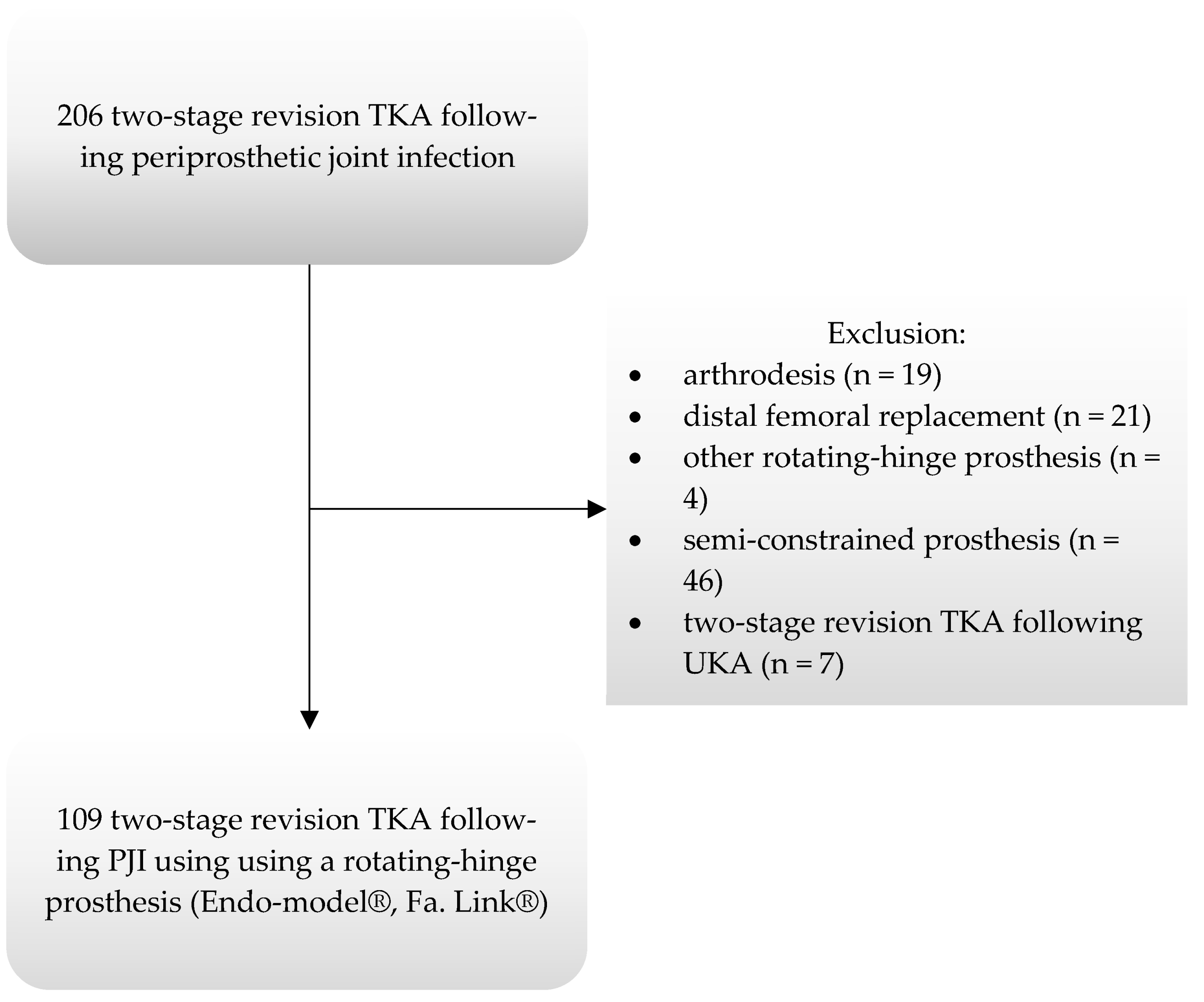
2.1. Study Cohort

In the present retrospective cohort study, we reviewed all patients who had undergone two-stage revision TKA for PJI at our institution between January 2001 and December 2015 using a rotating-hinge TKA at time of re-implantation. The institutional review board approved all procedures (S-152/2017) and the study was conducted in accordance with the Helsinki Declaration of 1975, as revised in 2013 [27]. Informed consent was obtained from all participating patients prior to inclusion in the study.
Patients were prospectively monitored following the index surgery with the institutions’ joint replacement registry, but the study design was set up retrospectively. Exclusion criteria were defined as cases involving the use of a distal femoral and/or proximal tibial replacement, oncologic indications for primary TKA, or patients who had received therapy with debridement and implant retention (DAIR) or reimplantation using non- or semi-constrained implants. For the diagnosis of PJI the patients’ medical history, physical examination, laboratory and radiographic findings, and joint fluid aspiration results were evaluated [28]. Of 206 consecutive patients revised for periprosthetic joint infection in the study period, 107 patients (109 knees) fulfilled the inclusion criteria (Figure 1). Patient demographics, medical comorbidities, patient- and surgery-related factors, and implant details were collected and are presented in Table 1 and Table 2.

2.2. Revision Protocol

All procedures were carried out using a two-stage protocol. First-stage treatment included implant and cement removal, radical debridement of infected and necrotic soft tissue and bone, and insertion of an antibiotic polymethylmethacrylate (PMMA) spacer. Intraoperative fluid and tissue samples were collected in all cases and sent for microbiological (+/− histological) analysis. After a period of 6 weeks of antibiotic therapy and an antibiotic-free interval of 2 weeks, a clinical reassessment was performed including a joint aspiration and a laboratory control. In patients with a positive culture or clinical/laboratory signs of a persisting infection, a re-debridement with a spacer exchange was performed (22 cases; 21%). In all other patients, the re-implantation was planned directly after the confirmation of a negative aspiration. Standardized preoperative planning of the prosthesis size and position was performed in all patients on calibrated radiographs. At time of re-implantation, a fully cemented rotating-hinge implant system was used. At our institution, the Endo-model® rotating-hinge implant system (Link®, Waldemar Link GMBH and Co., Hamburg, Germany) was used, which is characterized by a metal hinge, which allows flexion-extension and axial rotations and thus reduces the forces acting on the prosthesis anchorage. Cases performed after 2011 utilised the modular version (Endo-Model® SL®, Link®, Waldemar Link GMBH and Co., Hamburg, Germany) of the implant, which allowed for individual determination of stem length. The majority of patients (90%) received the complete two-stage exchange procedure at our institution according to the revision protocol for PJI, whereas 10% had the first stage explantation at an external institution. The reconstruction was performed using fully cemented fixation of diaphyseal stems and meta-epiphyseal components of the implant. In 55 cases (51%), tibial augments were used. In 5 procedures, metaphyseal cones were utilized for femoral reconstruction. Patella resurfacing was performed in 19 patients (17%).
Antibiotic treatment for both first-stage treatment and second-stage treatment was administered for 6 weeks postoperative. According to sensitivity testing, penicillins and cephalosporines for intravenous treatment and penicillins, lincomycins, and fluoroquinolones for oral treatment were used. For multi-resistant bacteria, glycopeptides, carbapenems, and oxazolidonines were administered.

2.3. Clinical Follow-Up and Radiographic Assessment

Clinical and radiographic follow-up examinations were recommended in regular intervals at 6 weeks, 12 weeks, 1 year, 3 years, 5 years, and then every 5 years thereafter. At the most recent follow-up, the following patient related outcome measures (PROMs) were assessed: American Knee Society Score (AKSS) [29], Oxford Knee Score (OKS) [30], and Visual analogue scale (VAS) [31]. Patients who were unable to attend clinical follow-up were contacted via telephone or via mail to complete the questionnaires. In deceased patients, information on further revision procedures or complications between the last clinical follow-up and death was obtained using information from their general practitioner and hospital records.
Radiographs were examined by two independent experienced orthopaedic surgeons (C.M., D.S.), blinded to outcome. The first postoperative and subsequent serial anteroposterior and lateral radiographs of the knee were examined to determine subsidence, radiolucent lines, osteolysis, heterotopic ossifications, and signs of loosening. The threshold for subsidence was defined as >2 mm on serial radiographs. Radiolucent lines were defined as progressive lines of >1 mm adjacent to the implant-cement or cement-bone interface. Osteolysis was defined as progressive areas of bone resorption or endosteal erosion. Implant loosening was diagnosed if radiolucent lines of over 2 mm were present around the entire implant or if implant migration by >2 mm or a change in implant axis >3° was observed.

2.4. Statistical Analysis

Following exploratory data analysis, implant survival for the endpoints revision (defined as removal or exchange of at least one of the components), reinfection, aseptic loosening, and reoperation was estimated, calculating the cumulative incidence of revision with 95% confidence intervals (CI) with death as competing risk. Survival was calculated up to 8 years, with a minimum of 23 knees still being at risk. Univariate analysis using a Cox proportional hazards regression model was performed to screen predictive risk factors for revision. Subsequently, a Cox proportional hazards regression model was used for multivariate analysis, including age at operation, BMI, Charlson Comorbidity Index (CCI), and number of previous operations before explantation as continuous independent variables. We considered p-values of <0.05 to be statistically significant. SPSS Version 24.0 (IBM SPSS Statistics, IBM, Armonk, NY, USA) and GraphPad Prism Version 6.0 (GraphPad Software, San Diego, CA, USA) were used to record and analyse the collected data.

3. Results

A total of 109 two-stage exchange arthroplasties for periprosthetic joint infection using a rotating-hinge-knee in 107 patients were included. Mean follow-up was 58 months (range: 0–188 months); patient demographics as well surgical and implant-related factors are summarized in Table 1. At the most recent follow-up, 52 of 109 knees (48%) were available for clinical assessment. Of those, 37 patients underwent clinical and radiological examination, whereas 15 patients had a standardized phone interview at final follow-up. A total of 27 patients (25%) died without need for further revision surgery. Four patients were lost to follow-up (3%) (Figure 2).

3.1. Survival Analysis

26 knees (24%) underwent a re-revision procedure. In three patients (11.5%), revision was performed for aseptic loosening, two of those for aseptic loosening of the femoral component and one for the tibial component. One revision (3.8%) was performed for mechanical failure of the locking mechanism and another one for a periprosthetic fracture of the femur. Three revisions (11.5%) were performed at external institutions for unknown reasons. Eighteen knees (69.2%) were revised for recurrent deep infection (Table 3). The mean time to reinfection after reimplantation was 33 months (range, 1 to 121 months) and 42% of re-infections occurred within the first year following index surgery. The cumulative incidence of the endpoint revision for any reason with death as a competing risk was 29.6% (95% CI: 19.3% to 39.9%) at 8 years (Figure 3). The cumulative incidence of the endpoint revision for recurrent infection was 20.6% (95% CI: 11.3% to 29.9%) at 8 years (Figure 4). The cumulative incidence of the endpoint revision for aseptic loosening was 5.6% (95% CI: 0% to 12.3%) at 8 years (Figure 5).
Additionally, nine re-operations (8.2%) with implant retention were performed for open reduction and internal fixation (ORIF) (three knees), wound healing issues/postoperative haematoma (four knees), and patellar maltracking (two knees) (Table 3). The cumulative incidence of the endpoint all reoperations was 38.9% (95% CI: 28.2% to 49.6%) at 8 years (Figure 6).

3.2. Mortality

In total, 33 patients (30.2%) were deceased at the most recent follow-up. Of those, 27 patients died without need for further re-revision surgery, whereas six patients underwent prior re-revision. The mortality rate estimated with the Kaplan–Meier analysis was 9.2% (95% CI: 4.2% to 15.9%) at 2 years and 21.0% (95% CI: 13.9% to 31.7%) at 5 years (Figure 7).

3.3. Regression Analysis

Multivariate analysis identified CCI (HR: 1.200 [95% CI: 1.001–1.437]; p = 0.049) and number of previous operations before explantation (HR: 1.146 [95% CI: 1.012–1.298]; p = 0.032) as significant predictive factors for re-revision (Table 4). Sex, age, BMI, preoperative positive cultures, use of modular implants, use of augments or patella resurfacing, perioperative bloodloss, operative time, and ASA score did not show any significant influence on the risk for re-revision surgery in the present cohort.

3.4. Microorganisms Causing Reinfection

In 77% of patients, a pathogen organism could be isolated at the time of index surgery. In cases with recurrent infection (n = 18), a positive culture of a pathogen microorganism could be isolated in 13 cases (72%). In 5 cases (28%), decisive clinical and radiological signs of PJI led to revisions surgery. In 5 of 13 patients (38%) with positive cultures, the same pathogen leading to reinfection was detected and classified as a persistent infection. Microorganisms for persistent infection were Enterococcus faecalis (two knees), Staphylococcus aureus (one knee), Staphylococcus caprae (one knee), and Candida parapsilosis (one knee).

3.5. Clinical Outcome

At the most recent follow-up, the mean postoperative knee flexion was 93° (SD: 17°, range: 40° to 130°). The mean Knee Society score was 71 points (SD: 21 points, range: 26 to 97 points); 43% of the patients showed excellent outcome (AKSS > 80) and 37% showed poor outcome (AKSS < 60). The mean Knee society functional score was 38 points (SD: 27 points, range: 0 to 100 points). Clinical evaluation according to the OKS demonstrated very good results in 12% of the patients (48–40 points), good results in 23% (39–30 points), fair in 34% (29–20 points), and poor in 31% (less than 20 points), showing a mean OKS of 26 points (SD: 10 points, range: 8 to 47 points). Patients’ self-assessment based on the VAS (0–10) for pain revealed a median score of 4 (range 0 to 10).

3.6. Radiographic Findings

In total, 37 patients were available for radiological assessment. Of those, three knees (8%) showed radiographic evidence of implant loosening, one for the femoral component and two for the entire implant. At the most recent follow-up, these impending revisions were not revised due to limited overall health condition without medical clearance for surgery. Of the remaining patients, progressive radiolucent lines were detected in 21 cases (62%). For the femoral component, epiphyseal radiolucent lines were observed in one patient, epimetadiaphyseal radiolucent lines in three patients, and epimetadiaphyseal radiolucent lines in one patient. For the tibial component, epiphyseal radiolucent lines were observed in one patient, epimetadiaphyseal radiolucent lines in four patients, and epimetadiaphyseal radiolucent lines in two patients. For both components, epimetaphyseal radiolucent lines were observed in six patients and epimetadiaphyseal radiolucent lines in three patients. Overall, none of those patients showed clinical signs of loosening or instability.
Heterotopic ossification was seen in three patients, leading to a functional ankylosis in one patient. Radiographically, two patients showed patellar maltracking. No signs of osteolysis were detected.

4. Discussion

In the present study, we evaluated the clinical and radiological mid-term results of two-stage exchange arthroplasty for periprosthetic joint infection using a cemented rotating-hinge TKA (Endo-model®, Fa. Link®) in a retrospective cohort of patients with a high level of comorbidity as well as multiple previous revisions. The cumulative incidence for the endpoint “any revision surgery” was 29.6% at 8 years (95% CI: 19.3% to 39.9%) and for the endpoint “any reoperation” the cumulative incidence was 38.9% at 8 years (95% CI: 28.2% to 49.6%). The main cause for re-revision surgery was recurrent periprosthetic joint infection with a cumulative incidence for the endpoint “reinfection” of 20.6% at 8 years (95% CI: 11.3% to 29.9%). In comparison, the revision rate for aseptic failure with 5.6% at 8 years (95% CI: 0% to 12.3%) was significantly lower. Using regression analysis, the study identified both the number of previous operations as well as the comorbidity level (as defined by the Charlson Comorbidity Index) as significant predictive factors regarding the risk for further re-revision surgery. Implant revision rates were comparable to previous reports. However, it is also important to consider the most relevant endpoint from the patients’ perspective, i.e., all reoperations, that accounted for approximately 10% additional surgical re-interventions. It is also worth noting that one third of patients had a poor functional outcome. The present data is hence helpful to counsel patients prior to revision for PJI about the probability of future surgeries and the substantial risk of limited joint function.
The eradication of PJI is challenging and requires a multidisciplinary treatment setting, including orthopaedic surgeons, microbiologists, and internal medicine specialists. Regarding the surgical procedure, two-stage exchange arthroplasty is still considered the gold standard in the treatment of PJI [5,17]. Nevertheless, neglecting studies with low patient numbers and/or short-term follow-up, the reinfection rates of previous studies range between 7% and 36% [19,20,23,24,32,33,34,35]. The available literature is difficult to compare because of variable outcome parameters and a substantial variation in definition of success and failure in heterogeneous study cohorts. According to our findings, previous operations and previous revision surgery both showed a statistically significant and clinically highly relevant influence on the re-revision rate. Our results show that every previous operation before explantation increases the risk for re-revision by 15%. Sabry et al. demonstrated a high number of 105 patients (33.4%) with failure to eradicate PJI in their study of 314 patients following two-stage exchange arthroplasty for PJI [24]. In their study, patients with recurrent infection showed an increased number of previous surgeries [24]. Similarly, Pelt et al. found that patients with more than four surgeries between primary TKA and explantation of the prosthesis due to treatment of PJI appear to have a higher risk of failure [32]. Furthermore, in both studies the need for plastic coverage procedures showed a greater risk for re-revision [24,32]. In contrast to these results, Haleem et al. could demonstrate a relatively high 10-year-survivorship of 85% for the endpoint “reinfection” [19]. As 91% of the implants used in their study were posterior-stabilized prosthesis, it can be assumed that these patients had less ligamentary and/or bony defects due to fewer prior operations [19].
Moreover, with an increase of previous revisions, the success rate for PJI is decreasing [36]. While patients without prior revision surgery undergoing treatment for PJI showed a success rate of 89%, patients with at least one revision surgery in prehistory showed a lower success rate of 73% [36]. Taking previous septic revision into consideration, the risk of re-revision rises even more. Mortazavi et al. demonstrated an overall survival rate for re-revision of 72.9% at 90 months in a study of 475 patients (499 knees) following revision surgery [10]. When only patients following two-stage exchange arthroplasty for septic modes were considered, the survival rate dropped to 52.4% at 90 months [23]. These observations demonstrate that previous revision surgery leading to bony and soft tissue deficiencies are associated with higher failure rates and risk for re-revision after treatment of PJI.
Another important factor for a successful treatment of PJI is the patients’ general health status. Concerning risk factors for revision surgery following primary TKA, there are some studies demonstrating that sociodemographic factors such as male sex, smoking, or an elevated BMI have been described as risk factors for revision surgery, while age was not associated with a higher risk for revision [4,37,38,39,40,41]. Furthermore, the influence of comorbidities was examined. While a history of diabetes or rheumatoid arthritis could be confirmed as risk factors for revision surgery [8,39,42,43], high blood pressure or cardiovascular diseases showed no increased impact on the rate of revision surgery [44,45]. Although these factors were mainly described in cohorts of patients following primary TKA, some studies could identify positive tendencies for these factors as predictors for re-revision surgery. Watts et al. were able to demonstrate that patients with a BMI > 40 kg/m2 had a 4.45-fold and 4.86-fold risk for subsequent revision and reinfection, respectively, compared with a matched, non-obese cohort [46]. In general, the data for comorbidities for recurrent revision after PJI treatment is very limited. Even though particular comorbidities are associated with a higher severity for PJI, from our perspective the influence is multifactorial. The present study demonstrates that an increase in CCI by one point leads to an increased risk for re-revision by 15% and an increased risk for reinfection by 28%. Similarly, Cochran et al. have demonstrated that a higher CCI is associated with a higher risk for reinfection in patients receiving a primary TKA [5]. In their study, the adjusted risk of reinfection was 48% higher in patients with a CCI of 5 and more, compared to patients with a CCI of 0 [5]. Furthermore, Mortazavi et al. showed that patients with a CCI > 3 were 2.48 times more likely to develop reinfection following revision TKA for any reason compared to patients with a CCI of 0 to 2 [10].
The distribution of isolated pathogens in our study, with a high number of staphylococceal infections, predominantly S. epidermidis, compares well to previous data in the literature [23,26,47,48,49]. In 23% of our cases, no pathogen could be isolated; this is attributed to antibiotic use prior to sample taking. Similar to Malekzadeh et al. and Kim et al. [50,51], there were no differences in the outcome regarding re-revision (p = 0.952) or reinfection (p = 0.744) between patients with positive or negative cultures. In a large study cohort by Zmistowski et al., it was demonstrated that recurrent PJI were mainly caused by a different pathogen, whereas in only 29 of 92 patients (31.5%) the same pathogen could be identified, which was classified as a persistent PJI [52]. Similar to these results, our results demonstrate a persistence of PJI in 38%. Therefore, we assume PJI failure is predominately related to a secondary infection instead of failed surgical treatment. This may be related to a generally poor medical condition concerning soft tissue and comorbidities.
The functional outcome after revision TKA is associated with inferior results when compared to primary TKA [53,54,55] due to the presence of bony defects and poor soft tissue [56]. Studies report similar functional results between aseptic and septic study cohorts if local tissue damage is comparable [57,58,59,60]. Compared to existing data, the functional and clinical scores in our study demonstrate similar results except for the KSFS [57,60,61]. This could be explained by the substantial comorbidities and high number of previous surgeries leading to impaired soft tissue condition in our study group. Furthermore, the use of a constraint prosthesis must be interpreted as another relevant factor for functional outcome. While Konrads et al. showed a mean OKS of 33.9 in 52 patients after PJI [59], Bejon et al. and Matar et al. demonstrated lower mean scores of 27 and 26, respectively, in patients with a compromised host status [36,62]. These results match the mean OKS of 26 in the present study.
As a tertiary referral centre, the majority of our patients presented numerous medical comorbidities as well as several prior surgical procedures, leading to a poor local bone/tissue stock. Moreover, in the current study, only patients with rotating-hinge implants were used to limit potential bias as a result of different implant designs. In this context, the present results, with a cumulative incidence for the endpoint “any revision surgery” of 29.6% at 8 years (95% CI: 19.3% to 39.9%) and for the endpoint “reinfection” of 20.6% at 8 years (95% CI: 11.3% to 29.9%), are on an acceptable level when compared to previously reported numbers.
The study has several limitations, including the retrospective study design as well as a missing concluded guideline for the surgical and medical treatment for infection. Highly variable patient-related factors, prior treatments, and surgical procedures must be considered as confounders. Additionally, the decision of the treatment strategy was made by different orthopedic surgeons with different experiences in knee arthroplasty revision surgery and a potential preoperative selection.

5. Conclusions

In conclusion, two-stage exchange arthroplasty for periprosthetic joint infection using a cemented rotating-hinge TKA outlined acceptable mid-term results regarding clinical function as well as survival rates with the endpoint “re-revision surgery” and “reinfection”. Recurrent periprosthetic joint infection was the major reason for subsequent revision surgery whereas aseptic loosening appeared of minor importance in the chosen competing risk analysis. A high number of previous operations as well as a high Charlson Comorbidity Index were clinically relevant predictive factors regarding the risk for further revision surgery. Mortality at 5 years was high in the present cohort. Patients with multiple comorbidities and a high number of previous operations need to be informed about the increased risks of PJI treatment with regard to re-revision risk, suboptimal in functional outcome, and mortality risk.

Author Contributions

Conceptualization, K.-A.K., C.M. and T.W.; Methodology, K.-A.K. and C.M.; Validation, K.-A.K. and D.M.S.; Formal Analysis, K.-A.K., T.B., C.M. and T.W.; Investigation, K.-A.K., D.M.S., C.M. and T.W.; Data Curation, K.-A.K.; Writing—Original Draft Preparation, K.-A.K., C.M. and T.W.; Writing—Review & Editing, K.-A.K., F.W., B.L., A.A., C.M. and T.W.; Visualization, K.-A.K.; Supervision, T.W.; Project Administration, K.-A.K. and T.W. All authors have read and agreed to the published version of the manuscript.

Funding

This research received no external funding.

Institutional Review Board Statement

The study was conducted in accordance with the Declaration of Helsinki, and approved by the Institutional Review Board of the University of Heidelberg (S-152/2017).

Informed Consent Statement

Informed consent was obtained from all subjects involved in the study.

Data Availability Statement

The datasets used and analyzed during the current study are available from the corresponding author on reasonable request.

Conflicts of Interest

Merle reports grants, honorial speaking and support for attending meeting and/or travel from Zimmer Biomet outside the submitted work. Additionally, he reports consulting fees, honorial speaking and support for attending meeting and/or travel for Medacta. Koch, Spranz, Westhauser, Bruckner, Lehner, Alvand, and Walker have nothing to disclose.

References

  1. Patel, A.; Pavlou, G.; Mujica-Mota, R.E.; Toms, A.D. The epidemiology of revision total knee and hip arthroplasty in England and Wales: A comparative analysis with projections for the United States. A study using the National Joint Registry dataset. Bone Jt. J. 2015, 97, 1076–1081. [Google Scholar] [CrossRef]
  2. Inacio, M.C.S.; Paxton, E.W.; Graves, S.E.; Namba, R.S.; Nemes, S. Projected increase in total knee arthroplasty in the United States—An alternative projection model. Osteoarthr. Cartil. 2017, 25, 1797–1803. [Google Scholar] [CrossRef] [PubMed]
  3. Kurtz, S.; Ong, K.; Lau, E.; Mowat, F.; Halpern, M. Projections of primary and revision hip and knee arthroplasty in the United States from 2005 to 2030. J. Bone Jt. Surg. Am. Vol. 2007, 89, 780–785. [Google Scholar] [CrossRef]
  4. Kurtz, S.M.; Ong, K.L.; Lau, E.; Bozic, K.J.; Berry, D.; Parvizi, J. Prosthetic joint infection risk after TKA in the Medicare population. Clin. Orthop. Relat. Res. 2010, 468, 52–56. [Google Scholar] [CrossRef]
  5. Cochran, A.R.; Ong, K.L.; Lau, E.; Mont, M.A.; Malkani, A.L. Risk of Reinfection After Treatment of Infected Total Knee Arthroplasty. J. Arthroplast. 2016, 31, 156–161. [Google Scholar] [CrossRef] [PubMed]
  6. Pulido, L.; Ghanem, E.; Joshi, A.; Purtill, J.J.; Parvizi, J. Periprosthetic joint infection: The incidence, timing, and predisposing factors. Clin. Orthop. Relat. Res. 2008, 466, 1710–1715. [Google Scholar] [CrossRef]
  7. Jamsen, E.; Huhtala, H.; Puolakka, T.; Moilanen, T. Risk factors for infection after knee arthroplasty. A register-based analysis of 43,149 cases. J. Bone Jt. Surg. Am. Vol. 2009, 91, 38–47. [Google Scholar] [CrossRef]
  8. Lenguerrand, E.; Whitehouse, M.R.; Beswick, A.D.; Kunutsor, S.K.; Foguet, P.; Porter, M.; Blom, A.W. Risk factors associated with revision for prosthetic joint infection following knee replacement: An observational cohort study from England and Wales. Lancet Infect. Dis. 2019, 19, 589–600. [Google Scholar] [CrossRef]
  9. Suarez, J.; Griffin, W.; Springer, B.; Fehring, T.; Mason, J.B.; Odum, S. Why do revision knee arthroplasties fail? J. Arthroplast. 2008, 23 (Suppl. S1), 99–103. [Google Scholar] [CrossRef]
  10. Mortazavi, S.M.; Molligan, J.; Austin, M.S.; Purtill, J.J.; Hozack, W.J.; Parvizi, J. Failure following revision total knee arthroplasty: Infection is the major cause. Int. Orthop. 2011, 35, 1157–1164. [Google Scholar] [CrossRef]
  11. Dai, W.L.; Lin, Z.M.; Shi, Z.J.; Wang, J. Outcomes Following Revision Total Knee Arthroplasty Septic versus Aseptic Failure: A National Propensity-Score-Matched Comparison. J. Knee Surg. 2020, 34, 1227–1236. [Google Scholar] [CrossRef] [PubMed]
  12. Lum, Z.C.; Natsuhara, K.M.; Shelton, T.J.; Giordani, M.; Pereira, G.C.; Meehan, J.P. Mortality during Total Knee Periprosthetic Joint Infection. J. Arthroplast. 2018, 33, 3783–3788. [Google Scholar] [CrossRef]
  13. Boddapati, V.; Fu, M.C.; Mayman, D.J.; Su, E.P.; Sculco, P.K.; McLawhorn, A.S. Revision Total Knee Arthroplasty for Periprosthetic Joint Infection Is Associated with Increased Postoperative Morbidity and Mortality Relative to Noninfectious Revisions. J. Arthroplast. 2018, 33, 521–526. [Google Scholar] [CrossRef] [PubMed]
  14. Cahill, J.L.; Shadbolt, B.; Scarvell, J.M.; Smith, P.N. Quality of life after infection in total joint replacement. J. Orthop. Surg. 2008, 16, 58–65. [Google Scholar] [CrossRef] [PubMed]
  15. Kunutsor, S.K.; Beswick, A.D.; Peters, T.J.; Gooberman-Hill, R.; Whitehouse, M.R.; Blom, A.W.; Moore, A.J. Health Care Needs and Support for Patients Undergoing Treatment for Prosthetic Joint Infection following Hip or Knee Arthroplasty: A Systematic Review. PLoS ONE 2017, 12, e0169068. [Google Scholar] [CrossRef] [PubMed]
  16. Moore, A.J.; Blom, A.W.; Whitehouse, M.R.; Gooberman-Hill, R. Deep prosthetic joint infection: A qualitative study of the impact on patients and their experiences of revision surgery. BMJ Open 2015, 5, e009495. [Google Scholar] [CrossRef]
  17. Kunutsor, S.K.; Whitehouse, M.R.; Lenguerrand, E.; Blom, A.W.; Beswick, A.D. Re-Infection Outcomes Following One-and Two-Stage Surgical Revision of Infected Knee Prosthesis: A Systematic Review and Meta-Analysis. PLoS ONE 2016, 11, e0151537. [Google Scholar] [CrossRef]
  18. Petis, S.M.; Perry, K.I.; Mabry, T.M.; Hanssen, A.D.; Berry, D.J.; Abdel, M.P. Two-Stage Exchange Protocol for Periprosthetic Joint Infection Following Total Knee Arthroplasty in 245 Knees without Prior Treatment for Infection. J. Bone Jt. Surg. Am. Vol. 2019, 101, 239–249. [Google Scholar] [CrossRef]
  19. Haleem, A.A.; Berry, D.J.; Hanssen, A.D. Mid-term to long-term followup of two-stage reimplantation for infected total knee arthroplasty. Clin. Orthop. Relat. Res. 2004, 35–39. [Google Scholar] [CrossRef]
  20. Mahmud, T.; Lyons, M.C.; Naudie, D.D.; Macdonald, S.J.; McCalden, R.W. Assessing the gold standard: A review of 253 two-stage revisions for infected TKA. Clin. Orthop. Relat. Res. 2012, 470, 2730–2736. [Google Scholar] [CrossRef]
  21. Grimberg, A.; Jansson, V.; Melsheimer, O.; Steinbrück, A. Endoprothesenregister Deutschland [EPRD]—Jahresbericht 2019; German Arthroplasty Registry EPRD: Berlin, Germany, 2019. [Google Scholar]
  22. Jamsen, E.; Stogiannidis, I.; Malmivaara, A.; Pajamaki, J.; Puolakka, T.; Konttinen, Y.T. Outcome of prosthesis exchange for infected knee arthroplasty: The effect of treatment approach. Acta Orthop. 2009, 80, 67–77. [Google Scholar] [CrossRef] [PubMed]
  23. Mortazavi, S.M.; Schwartzenberger, J.; Austin, M.S.; Purtill, J.J.; Parvizi, J. Revision total knee arthroplasty infection: Incidence and predictors. Clin. Orthop. Relat. Res. 2010, 468, 2052–2059. [Google Scholar] [CrossRef] [PubMed]
  24. Sabry, F.Y.; Buller, L.; Ahmed, S.; Klika, A.K.; Barsoum, W.K. Preoperative prediction of failure following two-stage revision for knee prosthetic joint infections. J. Arthroplast. 2014, 29, 115–121. [Google Scholar] [CrossRef]
  25. Ma, C.Y.; Lu, Y.D.; Bell, K.L.; Wang, J.W.; Ko, J.Y.; Wang, C.J.; Kuo, F.C. Predictors of Treatment Failure After 2-Stage Reimplantation for Infected Total Knee Arthroplasty: A 2- to 10-Year Follow-Up. J. Arthroplast. 2018, 33, 2234–2239. [Google Scholar] [CrossRef] [PubMed]
  26. Blom, A.W.; Brown, J.; Taylor, A.H.; Pattison, G.; Whitehouse, S.; Bannister, G.C. Infection after total knee arthroplasty. J. Bone Jt. Surg. Br. Vol. 2004, 86, 688–691. [Google Scholar] [CrossRef]
  27. World Medical Association. World Medical Association Declaration of Helsinki: Ethical principles for medical research involving human subjects. JAMA 2013, 310, 2191–2194. [Google Scholar] [CrossRef]
  28. Parvizi, J.; Gehrke, T.; Chen, A.F. Proceedings of the International Consensus on Periprosthetic Joint Infection. Bone Jt. J. 2013, 95, 1450–1452. [Google Scholar] [CrossRef]
  29. Insall, J.N.; Dorr, L.D.; Scott, R.D.; Scott, W.N. Rationale of the Knee Society clinical rating system. Clin. Orthop. Relat. Res. 1989, 248, 13–14. [Google Scholar] [CrossRef]
  30. Murray, D.W.; Fitzpatrick, R.; Rogers, K.; Pandit, H.; Beard, D.J.; Carr, A.J.; Dawson, J. The use of the Oxford hip and knee scores. J. Bone Jt. Surg. Br. Vol. 2007, 89, 1010–1014. [Google Scholar] [CrossRef]
  31. Rolfson, O.; Eresian Chenok, K.; Bohm, E.; Lübbeke, A.; Denissen, G.; Dunn, J.; Lyman, S.; Franklin, P.; Dunbar, M.; Overgaard, S.; et al. Patient-reported outcome measures in arthroplasty registries. Acta Orthop. 2016, 87 (Suppl. S1), 3–8. [Google Scholar] [CrossRef]
  32. Pelt, C.E.; Grijalva, R.; Anderson, L.; Anderson, M.B.; Erickson, J.; Peters, C.L. Two-Stage Revision TKA Is Associated with High Complication and Failure Rates. Adv. Orthop. 2014, 2014, 659047. [Google Scholar] [CrossRef] [PubMed]
  33. Kurd, M.F.; Ghanem, E.; Steinbrecher, J.; Parvizi, J. Two-stage exchange knee arthroplasty: Does resistance of the infecting organism influence the outcome? Clin. Orthop. Relat. Res. 2010, 468, 2060–2066. [Google Scholar] [CrossRef] [PubMed]
  34. Cha, M.S.; Cho, S.H.; Kim, D.H.; Yoon, H.K.; Cho, H.S.; Lee, D.Y.; Lee, S.H.; Hwang, S.C. Two-Stage Total Knee Arthroplasty for Prosthetic Joint Infection. Knee Surg. Relat. Res. 2015, 27, 82–89. [Google Scholar] [CrossRef] [PubMed]
  35. Kubista, B.; Hartzler, R.U.; Wood, C.M.; Osmon, D.R.; Hanssen, A.D.; Lewallen, D.G. Reinfection after two-stage revision for periprosthetic infection of total knee arthroplasty. Int. Orthop. 2012, 36, 65–71. [Google Scholar] [CrossRef]
  36. Bejon, P.; Berendt, A.; Atkins, B.L.; Green, N.; Parry, H.; Masters, S.; McLardy-Smith, P.; Gundle, R.; Byren, I. Two-stage revision for prosthetic joint infection: Predictors of outcome and the role of reimplantation microbiology. J. Antimicrob. Chemother. 2010, 65, 569–575. [Google Scholar] [CrossRef]
  37. Namba, R.S.; Inacio, M.C.; Paxton, E.W. Risk factors associated with deep surgical site infections after primary total knee arthroplasty: An analysis of 56,216 knees. J. Bone Jt. Surg. Am. Vol. 2013, 95, 775–782. [Google Scholar] [CrossRef]
  38. Song, K.H.; Kim, E.S.; Kim, Y.K.; Jin, H.Y.; Jeong, S.Y.; Kwak, Y.G.; Cho, Y.K.; Sung, J.; Lee, Y.S.; Oh, H.B.; et al. Differences in the risk factors for surgical site infection between total hip arthroplasty and total knee arthroplasty in the Korean Nosocomial Infections Surveillance System (KONIS). Infect. Control Hosp. Epidemiol. 2012, 33, 1086–1093. [Google Scholar] [CrossRef]
  39. Dowsey, M.M.; Choong, P.F. Obese diabetic patients are at substantial risk for deep infection after primary TKA. Clin. Orthop. Relat. Res. 2009, 467, 1577–1581. [Google Scholar] [CrossRef]
  40. Durand, F.; Berthelot, P.; Cazorla, C.; Farizon, F.; Lucht, F. Smoking is a risk factor of organ/space surgical site infection in orthopaedic surgery with implant materials. Int. Orthop. 2013, 37, 723–727. [Google Scholar] [CrossRef]
  41. Malinzak, R.A.; Ritter, M.A.; Berend, M.E.; Meding, J.B.; Olberding, E.M.; Davis, K.E. Morbidly obese, diabetic, younger, and unilateral joint arthroplasty patients have elevated total joint arthroplasty infection rates. J. Arthroplast. 2009, 24, 84–88. [Google Scholar] [CrossRef]
  42. Jamsen, E.; Nevalainen, P.; Eskelinen, A.; Huotari, K.; Kalliovalkama, J.; Moilanen, T. Obesity, diabetes, and preoperative hyperglycemia as predictors of periprosthetic joint infection: A single-center analysis of 7181 primary hip and knee replacements for osteoarthritis. J. Bone Jt. Surg. Am. Vol. 2012, 94, e101. [Google Scholar] [CrossRef] [PubMed]
  43. Ravi, B.; Croxford, R.; Hollands, S.; Paterson, J.M.; Bogoch, E.; Kreder, H.; Hawker, G.A. Increased risk of complications following total joint arthroplasty in patients with rheumatoid arthritis. Arthritis Rheumatol. 2014, 66, 254–263. [Google Scholar] [CrossRef]
  44. Bozic, K.J.; Lau, E.; Kurtz, S.; Ong, K.; Berry, D.J. Patient-related risk factors for postoperative mortality and periprosthetic joint infection in medicare patients undergoing TKA. Clin. Orthop. Relat. Res. 2012, 470, 130–137. [Google Scholar] [CrossRef] [PubMed]
  45. Dowsey, M.M.; Choong, P.F. Obesity is a major risk factor for prosthetic infection after primary hip arthroplasty. Clin. Orthop. Relat. Res. 2008, 466, 153–158. [Google Scholar] [CrossRef]
  46. Watts, C.D.; Wagner, E.R.; Houdek, M.T.; Osmon, D.R.; Hanssen, A.D.; Lewallen, D.G.; Mabry, T.M. Morbid obesity: A significant risk factor for failure of two-stage revision total knee arthroplasty for infection. J. Bone Jt. Surg. Am. Vol. 2014, 96, e154. [Google Scholar] [CrossRef]
  47. Lindberg-Larsen, M.; Pitter, F.T.; Voldstedlund, M.; Schroder, H.M.; Bagger, J. Microbiological diagnosis in revision of infected knee arthroplasties in Denmark. Infect. Dis. 2017, 49, 824–830. [Google Scholar] [CrossRef]
  48. Nickinson, R.S.; Board, T.N.; Gambhir, A.K.; Porter, M.L.; Kay, P.R. The microbiology of the infected knee arthroplasty. Int. Orthop. 2010, 34, 505–510. [Google Scholar] [CrossRef]
  49. Aggarwal, V.K.; Bakhshi, H.; Ecker, N.U.; Parvizi, J.; Gehrke, T.; Kendoff, D. Organism profile in periprosthetic joint infection: Pathogens differ at two arthroplasty infection referral centers in Europe and in the United States. J. Knee Surg. 2014, 27, 399–406. [Google Scholar] [CrossRef]
  50. Malekzadeh, D.; Osmon, D.R.; Lahr, B.D.; Hanssen, A.D.; Berbari, E.F. Prior use of antimicrobial therapy is a risk factor for culture-negative prosthetic joint infection. Clin. Orthop. Relat. Res. 2010, 468, 2039–2045. [Google Scholar] [CrossRef]
  51. Kim, Y.H.; Kulkarni, S.S.; Park, J.W.; Kim, J.S.; Oh, H.K.; Rastogi, D. Comparison of infection control rates and clinical outcomes in culture-positive and culture-negative infected total-knee arthroplasty. J. Orthop. 2015, 12 (Suppl. S1), S37–S43. [Google Scholar] [CrossRef]
  52. Zmistowski, B.; Tetreault, M.W.; Alijanipour, P.; Chen, A.F.; Della Valle, C.J.; Parvizi, J. Recurrent periprosthetic joint infection: Persistent or new infection? J. Arthroplast. 2013, 28, 1486–1489. [Google Scholar] [CrossRef] [PubMed]
  53. Saleh, K.J.; Dykes, D.C.; Tweedie, R.L.; Mohamed, K.; Ravichandran, A.; Saleh, R.M.; Gioe, T.J.; Heck, D.A. Functional outcome after total knee arthroplasty revision: A meta-analysis. J. Arthroplast. 2002, 17, 967–977. [Google Scholar] [CrossRef] [PubMed]
  54. Hanssen, A.D.; Rand, J.A. A comparison of primary and revision total knee arthroplasty using the kinematic stabilizer prosthesis. J. Bone Jt. Surg. Am. Vol. 1988, 70, 491–499. [Google Scholar] [CrossRef]
  55. Baker, P.; Cowling, P.; Kurtz, S.; Jameson, S.; Gregg, P.; Deehan, D. Reason for revision influences early patient outcomes after aseptic knee revision. Clin. Orthop. Relat. Res. 2012, 470, 2244–2252. [Google Scholar] [CrossRef]
  56. Sharkey, P.F.; Hozack, W.J.; Rothman, R.H.; Shastri, S.; Jacoby, S.M. Insall Award paper. Why are total knee arthroplasties failing today? Clin. Orthop. Relat. Res. 2002, 7–13. [Google Scholar] [CrossRef]
  57. Patil, N.; Lee, K.; Huddleston, J.I.; Harris, A.H.; Goodman, S.B. Aseptic versus septic revision total knee arthroplasty: Patient satisfaction, outcome and quality of life improvement. Knee 2010, 17, 200–203. [Google Scholar] [CrossRef]
  58. Rajgopal, A.; Vasdev, A.; Gupta, H.; Dahiya, V. Revision total knee arthroplasty for septic versus aseptic failure. J. Orthop. Surg. 2013, 21, 285–289. [Google Scholar] [CrossRef] [PubMed]
  59. Konrads, C.; Franz, A.; Hoberg, M.; Rudert, M. Similar Outcomes of Two-Stage Revisions for Infection and One-Stage Revisions for Aseptic Revisions of Knee Endoprostheses. J. Knee Surg. 2019, 32, 897–899. [Google Scholar] [CrossRef]
  60. Ro, D.H.; Kim, J.K.; Kim, S.; Han, H.S.; Lee, M.C. Periprosthetic Joint Infection Does Not Preclude Good Outcomes after a Revision Total Knee Arthroplasty: A 7-Year Follow-Up Study of 144 Retrospective Cases. BioMed Res. Int. 2018, 2018, 2582140. [Google Scholar] [CrossRef]
  61. Van Kempen, R.W.; Schimmel, J.J.; van Hellemondt, G.G.; Vandenneucker, H.; Wymenga, A.B. Reason for revision TKA predicts clinical outcome: Prospective evaluation of 150 consecutive patients with 2-years followup. Clin. Orthop. Relat. Res. 2013, 471, 2296–2302. [Google Scholar] [CrossRef]
  62. Matar, H.E.; Stritch, P.; Emms, N. Higher failure rate of two-stage revision for infected knee arthroplasties in significantly compromised (host-C) patients. Knee Surg. Sports Traumatol. Arthrosc. 2019, 27, 2206–2210. [Google Scholar] [CrossRef] [PubMed]
Figure 1. Study cohort.
Jcm 12 05542 g001
Figure 2. Distribution of knees at most recent follow-up.
Jcm 12 05542 g002
Figure 3. The cumulative incidence of the endpoint revision for any reason with death as a competing risk was 29.6% (95% CI: 19.3% to 39.9%) at 8 years.
Jcm 12 05542 g003
Figure 4. The cumulative incidence of the endpoint revision for recurrent infection with death as a competing risk was 20.6% (95% CI: 11.3% to 29.9%) at 8 years.
Jcm 12 05542 g004
Figure 5. The cumulative incidence of the endpoint revision for aseptic loosening with death as a competing risk was 5.6% (95% CI: 0% to 12.3%) at 8 years.
Jcm 12 05542 g005
Figure 6. The cumulative incidence of the endpoint all reoperations was 38.9% (95% CI: 28.2% to 49.6%) at 8 years.
Jcm 12 05542 g006
Figure 7. The mortality rate estimated with the Kaplan–Meier analysis was 9.2% (95% CI: 4.2% to 15.9%) at 2 years and 21.0% (95% CI: 13.9% to 31.7%) at 5 years.
Jcm 12 05542 g007
Table 1. Patient demographics, medical comorbidities, patient- and surgery-related factors, and implant details.
Demographics
     Number of knees109 (100%)
     Age (mean, SD), in years69.61 (9.35)
     Gender n (%)
       Male55 (50.5%)
       Female54 (49.5%)
     Body mass index (mean, SD)30.96 (5.62)
Comorbidity Scores
     ASA score (%)
       ASA I2 (1.8%)
       ASA II27 (24.8%)
       ASA III75 (68.8%)
       ASA IV5 (4.6%)
     Charlson Comorbidity Index
       020 (18.3%)
       1–236 (33.0%)
       3–426 (23.9%)
       527 (24.8%)
Surgical-related factors
     Two-stage revision
       Complete n (%)98 (90%)
       Reimplantation n (%)11 (10%)
     Pathogen isolation at index surgery
       Gram-positive70 (64%)
           Staphylococci51 (47%)
              Staphylococcus aureus 14 (13%)
              Methicillin-resistant Staphylococcus aureus1 (1%)
              Coagulase-negative staphylococci/Staphylococcus epidermidis27 (25%)
              Methicillin-resistant Staphylococcus epidermidis2 (2%)
       Streptococci6 (6%)
       Enterococci4 (4%)
       Other gram-positive9 (8%)
       Gram-negative8 (7%)
       Fungus2 (2%)
       Poly-microbial4 (4%)
       No organism growth25 (23%)
     Blood loss (mean, SD), (ml)1339 (915)
     Operating time (mean, SD), (minutes)188 (58)
     Previous operations (mean, SD)4.94 (2.92)
Previous revision procedure
       Yes62 (56.9%)
           Aseptic23 (21.1%)
           Septic39 (35.8%)
       No47 (43.1%)
     Surgical procedures between explantation and reimplantation
       Yes22 (20.2%)
       No87 (79.8%)
Implant-related factors
     Generation of RHK
       Modular n (%)55 (50.5%)
       Non-modular n (%)54 (49.5%)
     Retropatellar replacement
       With n (%)19 (17.4%)
       Without n (%)90 (82.6%)
     Tibial/Femoral Augments
       With n (%)55 (50.5%)
       Without n (%)54 (49.5%)
Table 2. Comorbidities in study cohort according to Charlson Comorbidity Index.
ComorbidityTotal n = 109 (100%)
Myocardial infarction13 (12%)
Congestive heart failure34 (31%)
Peripheral vascular disease5 (5%)
Cerebrovascular accident or transient ischemic attacks12 (11%)
Dementia0 (0%)
Chronic obstructive disease26 (24%)
Connective tissue disease11 (10%)
Peptic ulcer disease55 (50%)
Liver disease7 (6%)
     mild6 (5%)
     moderate to severe1 (1%)
Diabetes mellitus34 (31%)
     uncomplicated26 (24%)
     End-organ damage8 (7%)
Hemiplegia3 (3%)
Moderate to severe chronic kidney disease40 (37%)
Solid tumour4 (4%)
Leukemia0 (0%)
Lymphoma0 (0%)
Metastasic tumour1 (1%)
AIDS0 (0%)
Table 3. Causes of re-revisions and reoperations.
Number of Knees
Re-Revisions26
     Recurrent deep infection18
     Aseptic loosening3
        Femoral component2
        Tibial component1
     Implant failure1
     Periprosthetic fracture1
     Revision for unknown reason3
Reoperations (excluding reasons leading to revision)9
     Open reduction and internal fixation (ORIF)3
     Wound healing/postoperative haematoma4
     Patellar maltracking2
Table 4. Multivariate Cox regression model for the likelihood of any revision dependent on specific risk factors.
Risk FactorHazard Ratio (95% CI)p-Value
Age at operation1.010 (0.953–1.070)0.733
BMI0.993 (0.917–1.076)0.869
Charlson Comorbidity Index1.200 (1.001–1.437)0.049
Previous operations1.146 (1.012–1.298)0.032
Disclaimer/Publisher’s Note: The statements, opinions and data contained in all publications are solely those of the individual author(s) and contributor(s) and not of MDPI and/or the editor(s). MDPI and/or the editor(s) disclaim responsibility for any injury to people or property resulting from any ideas, methods, instructions or products referred to in the content.

