We appreciate the comments of Schimmelbusch et al. [1] on our article [2].
- (1)
- We apologize for the misunderstanding. Figures 3 and 4 do not represent enhanced depth imaging (EDI) OCT acquisition of optic disc drusen (ODD) and peripapillary hyperreflective ovoid mass-like structures (PHOMSs). The purpose of those images was only to represent how they appear in OCT scans, and they were not used for testing OCT sensitivity to detect ODDs and PHOMSs. In our study we acquired both EDI and non-EDI images to recognize both ODDs and PHOMSs, as shown in Figure 1 and Figure 2, but only EDI images were analyzed according to ODDs consortium protocols [3]. Following the example of other similar studies [4], we decided to publish non-EDI figures, but we forgot to highlight that the selected images were not in EDI. However, to avoid misunderstandings, a detailed description of the acquiring protocols of OCT images could be useful.
- (2)
- The authors confuted the role of ultrasound (US) in ODD detection. To support their opinion, they cited a study that reported a low sensitivity rate in ODD detection with US [5]. Actually, according to the cited article, the low sensitivity rate was present only when the exam was performed through the lens (axial scan). On the contrary, when the lens was avoided (parabulbar scan), the sensitivity rate was 100%, and parabulbar US scan was chosen as the exam of choice in ODD detection [2,5]. In addition, other contemporary similar studies that analyzed OCT sensitivity in ODD detection chose US as the baseline exam in ODD recognition. Ref. [6] Malmqvist et al. also reported that the absence of ODD on OCT does not entirely rule out the diagnosis; other modalities are needed, among which include B-scan US [4]. For this reason, to detect ODD in our study, a parabulbar exam with US was performed, as happens whenever other kinds of optic nerve diseases are suspected, avoiding axial scans [7,8].
- (3)
- We agree with the authors that PHOMSs can be found in several conditions and should not be considered as ODD markers, but, as PHOMSs are considered possible markers of ODD by several authors [3,9,10], we decided to perform a US-OCT comparison, both including and excluding PHOMSs, in attempt to achieve the maximum sensitivity value in the OCT detection of ODDs. Unfortunately, in each scenario, OCT did not achieve US standards in ODD evaluation. Therefore, considering the debate in the literature around the role of PHOMSs in ODD detection [3,9,10], we demonstrated that OCT is not a valid alternative to the ODD detection method even if PHOMSs are included. The higher US sensitivity could be related to the presence of calcium deposits that are very well detected with US, but not with OCT.
We are confident that the results of our study are reliable and well documented. We thank the authors again for their comment on our article because they provided us with the opportunity to better explain some of the aspects of our work. Obviously, a multimodal approach including US, Fundus autofluorescence (FAF), and EDI-OCT could improve the strength of the diagnosis of optic nerve head drusen.
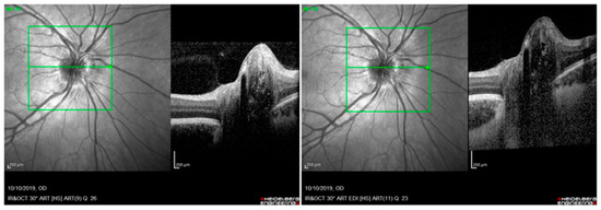
Figure 1.
Optical coherence tomography (OCT): optic disc drusen (ODD), visible as hypo-reflective structures with a total or partial hyperreflective margin, as described by the ODD studies consortium. On the left: ODD without enhanced depth imaging (EDI) acquisition modality; on the right: ODD using EDI acquisition modality. Both images are acquired at the same level of the optic disc (green arrow inside the green box).

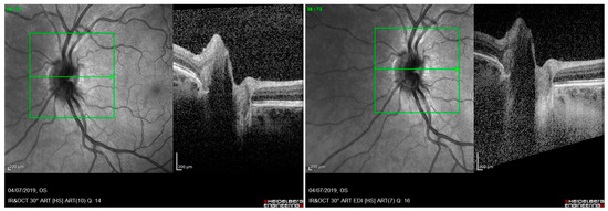
Figure 2.
Optical coherence tomography (OCT): hyperreflective peripapillary structure similar to an ovoid mass peripapillary hyperreflective ovoid mass structure (PHOMS). On the left: PHOMS without using enhanced depth imaging (EDI) acquisition modality; on the right: PHOMS using EDI acquisition modality. Both images are acquired at the same level of the optic disc (green arrow inside the green box).

Conflicts of Interest
The authors declare no conflict of interest.
References
- Schimmelbusch, K.J.; Collison, F.T. Comment on Rosa et al. Optic Nerve Drusen Evaluation: A Comparison between Ultrasound and OCT. J. Clin. Med. 2022, 11, 3715. J. Clin. Med. 2023, 12, 5607. [Google Scholar] [CrossRef]
- Rosa, N.; De Bernardo, M.; Abbinante, G.; Vecchio, G.; Cione, F.; Capasso, L. Optic Nerve Drusen Evaluation: A Comparison between Ultrasound and OCT. J. Clin. Med. 2022, 11, 3715. [Google Scholar] [CrossRef] [PubMed]
- Malmqvist, L.; Bursztyn, L.; Costello, F.; Digre, K.; Fraser, J.A.; Fraser, C.; Katz, B.; Lawlor, M.; Petzold, A.; Sibony, P.; et al. The optic disc drusen studies consortium recommendations for diagnosis of optic disc drusen using optical coherence tomography. J. Neuroophthalmol. 2018, 38, 299–307. [Google Scholar] [CrossRef] [PubMed]
- Caramoy, A.; Engel, L.; Koch, K.R.; Kirchhof, B.; Cursiefen, C.; Heindl, L.M. Multiple imaging modalities for the detection of optic nerve head drusen: Is echography still mandatory? Acta Ophthalmol. 2017, 95, 320–323. [Google Scholar] [CrossRef] [PubMed]
- Rajagopal, R.; Mitchell, E.; Sylvester, C.; Lope, L.A.; Nischal, K.K. Detection of Optic Disc Drusen in Children Using Ultrasound through the Lens and Avoiding the Lens-Point of Care Ultrasound Technique of Evaluation Revisited. J. Clin. Med. 2019, 8, 1449. [Google Scholar] [CrossRef] [PubMed]
- Guo, X.; Wu, Y.; Wu, Y.; Liu, H.; Ming, S.; Cui, H.; Fan, K.; Li, S.; Lei, B. Detection of superficial and buried optic disc drusen with swept-source optical coherence tomography. BMC Ophthalmol. 2022, 22, 219. [Google Scholar] [CrossRef] [PubMed]
- Rosa, N.; De Bernardo, M.; Di Stasi, M.; Cione, F.; Capaldo, I. A-Scan Ultrasonographic Evaluation of Patients with Idiopathic Intracranial Hypertension: Comparison of Optic Nerves. J. Clin. Med. 2022, 11, 6153. [Google Scholar] [CrossRef] [PubMed]
- Rosa, N.; Cennamo, G.; De Bernardo, M. Editorial: Ocular ultrasonography and optical coherence tomography in the optic nerve disease. Front. Med. 2023, 10, 1161123. [Google Scholar] [CrossRef] [PubMed]
- Flores-Rodríguez, P.; Gili, P.; Martín-Ríos, M.D. Sensitivity and specificity of time-domain and spectral-domain optical coherence tomography in differentiating optic nerve head drusen and optic disc oedema. Ophthalmic. Physiol. Opt. 2012, 32, 213–221. [Google Scholar] [CrossRef] [PubMed]
- Traber, G.L.; Weber, K.P.; Sabah, M.; Keane, P.A.; Plant, G.T. Enhanced depth imaging optical coherence tomography of optic nerve head drusen: A comparison of cases with and without visual field loss. Ophthalmology 2017, 124, 66–73. [Google Scholar] [CrossRef] [PubMed]
Disclaimer/Publisher’s Note: The statements, opinions and data contained in all publications are solely those of the individual author(s) and contributor(s) and not of MDPI and/or the editor(s). MDPI and/or the editor(s) disclaim responsibility for any injury to people or property resulting from any ideas, methods, instructions or products referred to in the content. |
© 2023 by the authors. Licensee MDPI, Basel, Switzerland. This article is an open access article distributed under the terms and conditions of the Creative Commons Attribution (CC BY) license (https://creativecommons.org/licenses/by/4.0/).