Article

A Comparison of Immersive vs. Non-Immersive Virtual Reality Exercises for the Upper Limb: A Functional Near-Infrared Spectroscopy Pilot Study with Healthy Participants

1
Department of Chronic and Degenerative Diseases, Faculty of Health Sciences (FGW), Potsdam University, 14476 Potsdam, Germany
2
Department of Neurology, Otto-von-Guericke University, 39120 Magdeburg, Germany
3
Faculty of Informatics, Otto-von-Guericke University, 39106 Magdeburg, Germany
4
Edith-Stein Fachklinik, 76887 Bad Bergzabern, Germany
5
University Medical Center of the Johannes Gutenberg University Mainz, 55131 Mainz, Germany
*
Author to whom correspondence should be addressed.
These authors contributed equally to this work.
J. Clin. Med. 2023, 12(18), 5781; https://doi.org/10.3390/jcm12185781
Submission received: 19 July 2023 / Revised: 21 August 2023 / Accepted: 28 August 2023 / Published: 5 September 2023
(This article belongs to the Section Clinical Neurology)

Abstract

:
Functional near-infrared spectroscopy (fNIRS) allows for a reliable assessment of oxygenated blood flow in relevant brain regions. Recent advancements in immersive virtual reality (VR)-based technology have generated many new possibilities for its application, such as in stroke rehabilitation. In this study, we asked whether there is a difference in oxygenated hemoglobin (HbO2) within brain motor areas during hand/arm movements between immersive and non-immersive VR settings. Ten healthy young participants (24.3 ± 3.7, three females) were tested using a specially developed VR paradigm, called “bus riding”, whereby participants used their hand to steer a moving bus. Both immersive and non-immersive conditions stimulated brain regions controlling hand movements, namely motor cortex, but no significant differences in HbO2 could be found between the two conditions in any of the relevant brain regions. These results are to be interpreted with caution, as only ten participants were included in the study.

1. Introduction

Virtual reality (VR)-based platforms are used nowadays in many settings [1], including the restoration of upper limb function. This application is particularly common in the research on rehabilitation of stroke survivors, whose upper limb movement is impaired in about half of all patients and, therefore, requires intensive practice usually supervised by a physiotherapist. Here, several studies reported beneficial effects of additional practice in VR [2,3], related to both functional outcomes [4] and neuroplasticity [5] in stroke patients.
A wide variety of VR systems are available on the market—from those expensive and customizable to the low-cost ones, using either immersive or non-immersive VR technology, sensor- or marker-based, with or without controllers. In recent years, immersive VR systems—usually containing a head-mounted display (HMD)—have been developed and produced at ever increasing pace. One of the obvious advantages is the optional three-dimensional (3D) environment, which offers a more realistic experience to users. Prior to the advent of technological advancements enabling 3D systems, VR settings predominantly consisted of 2D environments, usually via computer/laptop screens or projectors, known as non-immersive VR. Since the emergence of the 3D technology, many studies have sought to examine disparities between them (for a review, see [6]). However, others reported that in 3D systems, users become more engaged in the environment, being active participants, rather than just being passive observers, which brings the possibility of explorative interaction with the environment [7], but also better memory performance [8] and perception [9]. In addition, immersive designs in 3D can produce greater psycho-physiological responses in healthy users [10] as well as greater improvements in recovery of stroke patients [11], compared to non-immersive ones. Nevertheless, it remains unknown if physiological signatures, as observed by functional near-infrared spectroscopy (fNIRS), differ between the two modalities (3D vs. 2D)—which may be of great relevance for the recovery of stroke survivors.
Functional near-infrared spectroscopy (fNIRS) is suitable for the investigation of the effects of movement on cerebral oxygenation and hemodynamics—it is able to quantify changes in the concentration of oxygenated hemoglobin (oxyHb) and deoxygenated hemoglobin (deoxyHb) non-invasively [12]. A number of previous studies used fNIRS to assess various upper limb movements and conditions, such as simple hand movements [13], hand-grip strength [14], skill development [15], focal upper limb dystonia [16], stroke rehabilitation [3,17], cortical reorganization [18] and so on. However, to the best of our knowledge, up to now there are no studies which have combined VR and fNIRS to track upper limb movements. Moreover, no study so far has compared upper limb movements in 2D and 3D using fNIRS.
Therefore, for this study, we developed a VR-based paradigm to evaluate the functionality of the upper limb. Our primary inquiry revolves around the potential dissimilarities in activation, as observed by fNIRS, within the corresponding brain regions between:
(a)
The immersive VR and the non-immersive desktop (nVR) conditions;
(b)
Both of these conditions compared to the baseline (B) condition.
Considering the findings of previous studies, we hypothesized increased activation in relevant brain areas—primarily motor cortex and neighboring areas—in both VR and nVR conditions compared to B. Regarding possible differences between the immersive (VR) and non-immersive (nVR) conditions, based on the existing literature, no clear hypothesis could be formulated.

2. Materials and Methods

2.1. Study Design and Participants
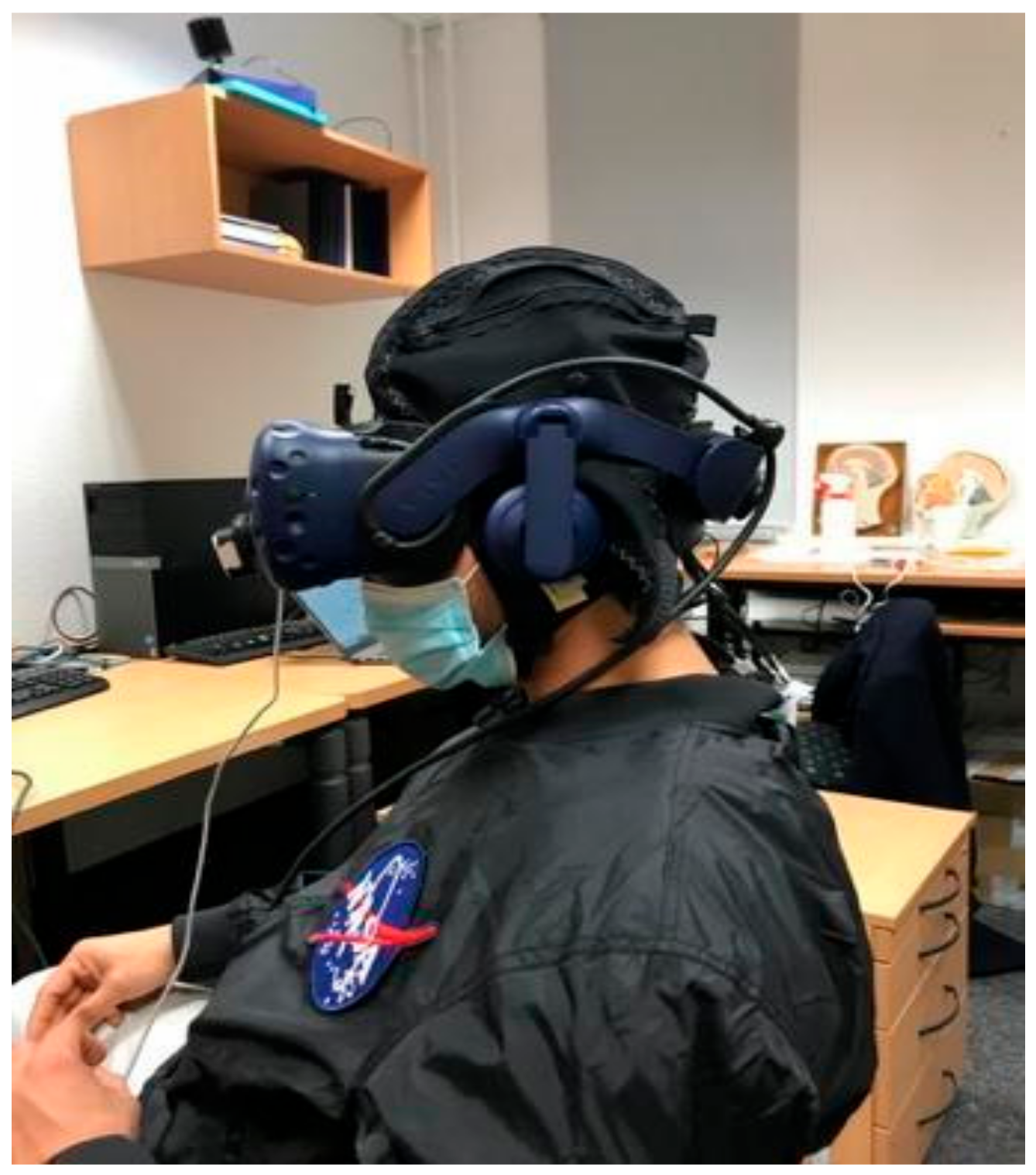
This study was organized as cross-sectional, with one group and two conditions (immersive and non-immersive), in accordance with the declaration of Helsinki. Ten healthy young participants (24.3 ± 3.7 years old, three females) were included in this proof-of-concept study.

2.2. VR Setup

All measurements took place in a quiet testing room with closed and blinded windows with the lights off, in order to reduce any possible sound and light interference.
First, the whole procedure was explained to the participant, followed by one familiarization trial with each paradigm, both in immersive (HMD) and non-immersive settings (on the laptop screen). Next, the participant was asked to sit in the chair in front of the table on which the laptop (Dell, Round Rock, Texas, US) with screen size of 61 cm, full HD resolution) was placed at a distance of about one meter from the participant’s head. Then, the complete fNIRS system (see below for detailed description) was mounted on the participant’s head and tested for signal quality. Following this, the participant was instructed to don the HMD (HTC-Vive Pro, Taoyuan City, Taiwan) on their head while ensuring optimal adjustments for utmost comfort. On the front side of the HMD, the Leap Motion (Leap Motion Company, San Francisco, CA, USA) controller was placed so that hand movements could be captured within the virtual environment. Once the entire setup was ready, the paradigms were played one after the other, in the same order for each participant. They were informed verbally when to begin and stop performing the task. Upon completion of paradigms in the immersive condition, the HMD was removed from participant’s head and the Leap Motion controller placed on the table facing upwards. This setup allowed for the continued detection and projection of hand movements onto the laptop screen. With these preparations complete, the testing of paradigms in the non-immersive condition commenced (Figure 1).

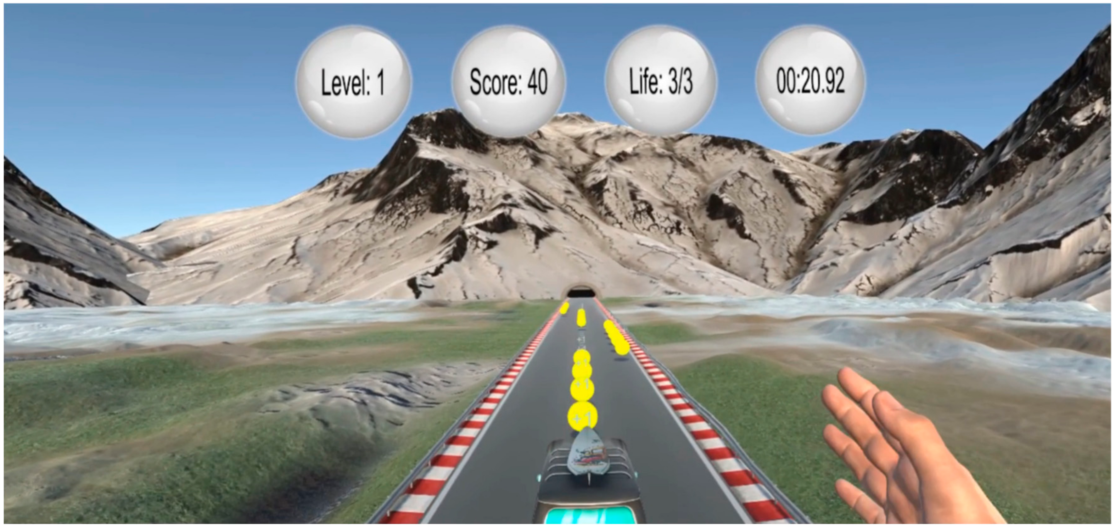
2.3. VR Paradigm—Bus Riding

A specially developed paradigm for this study, called bus riding, was used in order to stimulate hand movement. In this paradigm, participants used their hand to steer a moving bus, as shown in Figure 2. By moving the right hand, be it the wrist or fingers, in the metacarpophalangeal joints, right and left, the bus responsively adjusts its position while transitioning between lanes. The participant’s hand remains perpendicular to the lane and hand movements are in palmar- and dorsi-flexion. In addition, participants should avoid obstacles and collect coins placed on the road. The more the game progresses, the higher the driving speed, and thus the difficulty level, becomes. Steering the bus completely off the road leads to re-initiation of the game and return to the starting position. During each run, the goal was to collect a maximum number of points.
Participants were allowed to play in both immersive and non-immersive settings for three runs per 30 s, with 30 s of passive break between runs, whereby the hand was placed on the table. Additional baseline recordings were taken immediately before the first and after the last run, in both settings.

2.4. Data Acquisition

Hemodynamic brain signals were obtained using a NIRSport 2 system (NIRx Medical Technologies, Glen Head, NY, USA) in accordance with the protocol used by a previous study [19]. The system was equipped with seven detectors and eight light source optodes emitting at 760 and 850 nm. The electrodes were separated by an inter-optode distance of 30 mm and covered sensorimotor brain areas such as premotor and supplementary motor cortex (PMC–SMA), primary motor cortex (M1), somatosensory association cortex (SAC), and inferior parietal cortex (IPC) on both hemispheres.
Cortical hemodynamic changes were recorded at a frequency of 10.17 Hz in 22 channels. The fNIRS optodes were placed according to the 10–20 EEG system [20] on top of an fNIRS cap.

2.5. fNIRS Preprocessing and Analysis

Hemodynamic time series were analyzed using Satori (Brain Innovation BV, Maastricht, The Netherlands). Raw wavelength output from the fNIRS system was trimmed by 10 s at the beginning and 10 s at the end of each run, and then converted into optical densities through application of the modified Beer–Lambert law [21]. Motion artefacts were removed through temporal derivative distribution repair (TDDR) [22] and monotonic spike removal. Other artefacts (breathing, Mayer waves, heartbeat, low-frequency drifts) were addressed through linear detrending, low-pass filtering (Butterworth, 0.4 Hz) and high-pass filtering (Butterworth, 0.01 Hz) [23]. Finally, the optical densities were converted into concentration data and normalized into z-scores.
The task related brain activation between immersive (VR) and non-immersive (nVR) was analyzed with a GLM-based short channel regression, using the one of the eight short-separation channels that was closest to the long-separation channel. As the fNIRS signal is contaminated by the back-reflections of the skin and the skull, it was proposed to use these short source-detector separation optodes as regressors, as they are sensitive to the blood flow in the superficial layers of the human brain [24]. This should improve the signal across long separation fNIRS channels, as regional blood flow can differ substantially across the entire PFC. To control for multiple comparisons during channel wise t-tests, the false discovery rate (FDR) correction (q < 0.05) [25] was applied. The resulting t-map was displayed with Satori and used for further interpretation of the results. For all tests, a p-value of <0.05 was considered significant.

3. Results

Complete datasets for all three conditions (VR, nVR, B) from ten healthy young participants were obtained and analyzed in this study (age 24.3 ± 3.7, all right-handed, three females, all university students).

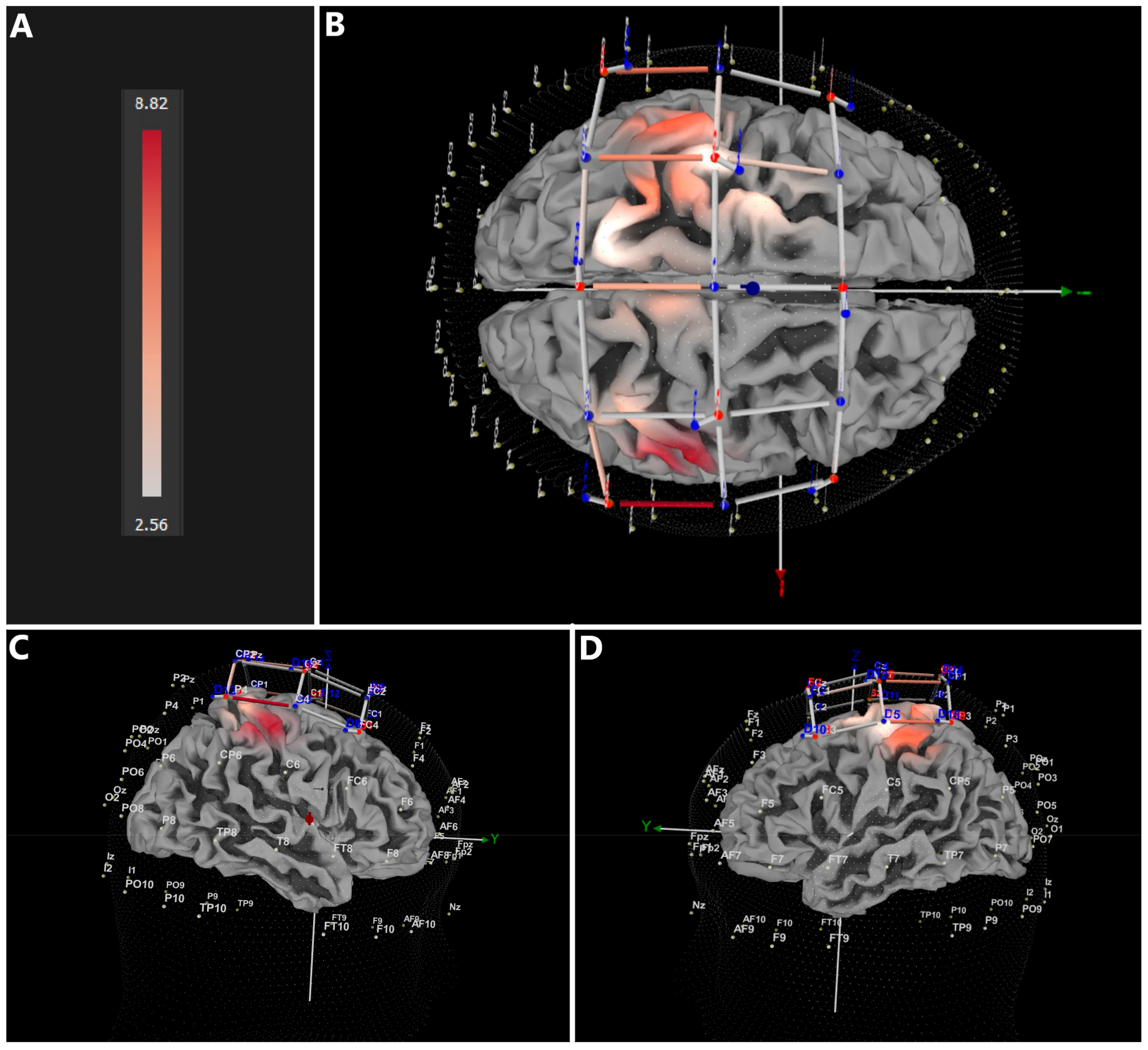
3.1. Immersive (VR) vs. Baseline (B)

As depicted in Figure 3 and listed in Table 1, the VR condition (VR) induced significantly more increase (CI = 3.755 − 8.820) in oxy-hemoglobin (HbO2) compared to the baseline condition (B). The most affected brain regions corresponded with primary motor cortex (M1), but also premotor and supplementary motor cortices. Moreover, the most activated topographical location in M1 corresponded well with regions responsible for processing arm- and hand-related information.

3.2. Non-Immersive (nVR) vs. Baseline (B)

Similar to the immersive VR condition, participants revealed significantly larger increase (FDR-corrected) in HbO2 in the non-immersive (nVR) compared to the B condition (CI = 3.802–9.796) (Figure 4 and Table 1). The affected regions corresponded to those found in the immersive VR condition.

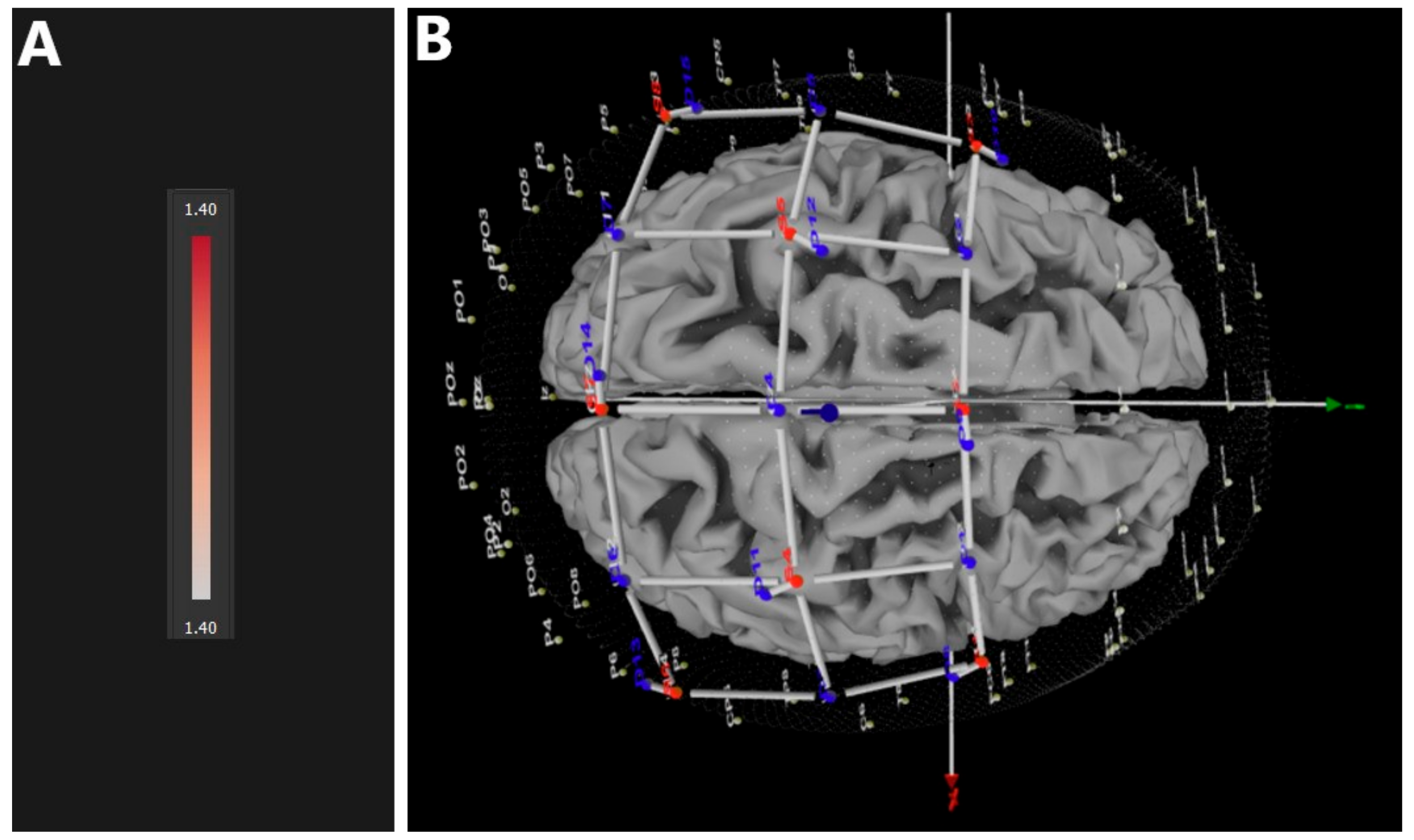
3.3. Immersive (VR) vs. Non-Immersive (nVR)

In contrast to comparisons of both VR and nVR conditions with baseline, no significant difference in HbO2 could be found between immersive VR and non-immersive (nVR) conditions in any of the relevant brain areas (Figure 5). No behavioral differences between the two conditions were found.

4. Discussion

This study confirmed the hypothesized effects of both immersive VR and non-immersive (nVR) conditions of a specially developed paradigm for inducing activation in brain regions controlling hand movements, namely increased HbO2 in the motor cortex and neighboring brain regions relative to the baseline (B). No significant differences could be found between VR and nVR conditions in HbO2 in any of the relevant brain regions. Thus, this study was the first one to determine such effects for all three conditions (VR, nVR, B).
In accordance with earlier studies, we found that fNIRS can be used to detect an increase in HbO2 in corresponding brain regions as a result of upper limb movement. Previous studies used fNIRS for various applications related to upper limb movement—including various single hand tasks [13], movement intention recognition for brain–computer interfaces [26,27,28], performance of spatial tasks using upper limbs [29], etc. Moreover, numerous studies are available on fNIRS application in stroke patients suffering from upper limb paresis—such as in assessment of unilateral vs. bilateral upper limb training [30], determination of effects of active and passive upper limb training modes [3], assessment of sensory-motor cortex reorganization following upper limb paresis due to stroke [18], and so on. However, none of these studies combined fNIRS and specially developed paradigms in VR to investigate changes in HbO2 resulting from upper limb movement.
Brain regions in which an increase in HbO2 was detected in this study are in agreement with the literature. Hand and wrist motion leads to an increase in HbO2 in the motor cortex and neighboring regions in the contralateral hemisphere [31,32]. A bilateral hemispheric increase in HbO2 in response to right upper limb movement is already a known phenomenon, especially when motor imagery is involved [33]. Movements in the wrist joint in certain directions can also elicit bilateral hemispheric responses; in addition, wrist joint movements in left and right directions cause increases in HbO2 in more brain regions than upward and downward movements [32]. In addition, the VR paradigm might have led to the activation of the observation network of the brain, which differs from the pure movement network [34].
VR-based applications have gained a significant momentum in recent years—rather than being passive observers, users are allowed to engage in various VR environments as active participants, which has been exploited by gaming, sports, medicine, education, military, and also research [7]. It was reported that user satisfaction is generally higher in VR compared to other methodologies, which might lead to higher learning rates and skill improvement [7]. An earlier study reported no difference between VR and desktop conditions in memory performance of younger adults, unlike in older adults, who performed better in the desktop condition [35]. A recent review reported that immersive VR can be considered beneficial for rehabilitation of stroke patients suffering from upper limb paresis [36]. Other studies support the efficacy of fully immersive VR over non-immersive VR for improving 3D perception and evaluation of cognitive functions [8,9], but also for enhancing the sense of presence and emotional response [10]. A recent meta-analysis on stroke patients reported beneficial effects of both immersive and non-immersive VR-based rehabilitation protocols, with immersive using head-mounted displays (HMDs) inducing the largest improvements in upper extremity function, as assessed by the Fugl-Meyer test [6]. Using EEG, it was found that learning in immersive VR leads to better knowledge transfer compared to conventional learning [37]. Despite these behavioral differences in some previous studies, we could not find brain activity differences between the VR and nVR condition in the present study. Although we cannot exclude the possibility that with a larger sample and with other methods for assessing brain activity (e.g., fMRI), differences between VR and nVR could have been observed, the present null finding speaks against large differences in brain activity induced by VR as opposed to nVR conditions. Note that no previous study has assessed changes in blood oxygenation (HbO2), as measured by fNIRS, resulting from upper limb movement in VR and nVR conditions—this precludes the direct comparability of our results. Additionally, it prompts the need for future studies using a larger sample of participants, to confirm or contradict our findings. Nevertheless, the results of our study provide novel insights into this important topic, which can be of relevance not only in health but also in various clinical conditions, such as stroke.
Despite providing significant results, our study contains some limitations. This particularly relates to the sample size, since only ten participants were included. However, considering the proof-of-concept nature of this study and its novelty, the idea was to take care of the resources and still assess the feasibility, including the specially designed paradigm coupled with the latest generation state-of-the-art fNIRS system. Even with the small number of participants, we obtained significant results when comparing both active conditions (VR and nVR) to the baseline condition; still, we could not detect any significant differences between the two active conditions, which might be—at least partially—due to a low number of participants. Thus, additional studies on larger samples of participants are needed. Moreover, we could not find any behavioral differences between the two active conditions (VR and nVR)—perhaps an assessment of elderly participants using this paradigm would lead to behavioral differences as well. Additionally, although we applied the 10–20 standardized system, participants’ heads vary in size and shape, therefore we cannot determine the exact neuroanatomical hemodynamic localization—therefore, future studies with more accurate methodologies are required.
In conclusion, results of this study brought two important findings: (I) compared to baseline, upper limb movements in both VR and nVR conditions lead to a significant increase in HbO2 in corresponding motor cortex and neighboring brain regions, and (II) no significant difference in HbO2 between the VR and nVR conditions could be found. These results are to be interpreted with caution, as only ten participants were included and fNIRS signal could not be obtained from very precise locations. Nevertheless, these results can be considered beneficial for all future studies using both immersive and non-immersive VR-based technology, both in healthy participants and various patient groups suffering from brain damage.

Author Contributions

Conceptualization, M.D. and N.G.M.; methodology, M.D., O.M., A.D., S.K. and N.G.M.; formal analysis, M.D. and S.K.; investigation, M.D., O.M. and A.D.; writing—original draft preparation, M.D., O.M. and N.G.M.; writing—review and editing, A.D., S.K., C.H. and B.B.; funding acquisition, N.G.M. All authors have read and agreed to the published version of the manuscript.

Funding

This research was funded by the Deutsche Forschungsgemeinschaft (DFG, German Research Foundation)—Projektnummer 491466077.

Institutional Review Board Statement

The study was conducted in accordance with the Declaration of Helsinki. Ethical review and approval were waived for this study due to the feasibility nature of this study, using healthy young participants.

Informed Consent Statement

Informed consent was obtained from all subjects involved in the study.

Data Availability Statement

All data are stored at the FGW—Potsdam University and can only be obtained under special permission.

Acknowledgments

Special acknowledgement to Andreas Schwaab and Ben Fishman from Mastricht University for their help with the data analysis.

Conflicts of Interest

The authors declare no conflict of interest.

References

  1. Slater, M.; Sanchez-Vives, M.V. Enhancing Our Lives with Immersive Virtual Reality. Front. Robot. AI 2016, 3, 74. [Google Scholar] [CrossRef]
  2. Mekbib, D.B.; Han, J.; Zhang, L.; Fang, S.; Jiang, H.; Zhu, J.; Roe, A.W.; Xu, D. Virtual reality therapy for upper limb rehabilitation in patients with stroke: A meta-analysis of randomized clinical trials. Brain Inj. 2020, 34, 456–465. [Google Scholar] [CrossRef] [PubMed]
  3. Zheng, J.; Shi, P.; Fan, M.; Liang, S.; Li, S.; Yu, H. Effects of passive and active training modes of upper-limb rehabilitation robot on cortical activation: A functional near-infrared spectroscopy study. NeuroReport 2021, 32, 479–488. [Google Scholar] [CrossRef] [PubMed]
  4. Kiper, P.; Szczudlik, A.; Agostini, M.; Opara, J.; Nowobilski, R.; Ventura, L.; Tonin, P.; Turolla, A. Virtual Reality for Upper Limb Rehabilitation in Subacute and Chronic Stroke: A Randomized Controlled Trial. Arch. Phys. Med. Rehabil. 2018, 99, 834–842.e4. [Google Scholar] [CrossRef]
  5. Hao, J.; Xie, H.; Harp, K.; Chen, Z.; Siu, K.-C. Effects of Virtual Reality Intervention on Neural Plasticity in Stroke Rehabilitation: A Systematic Review. Arch. Phys. Med. Rehabil. 2022, 103, 523–541. [Google Scholar] [CrossRef]
  6. Hao, J.; He, Z.; Yu, X.; Remis, A. Comparison of Immersive and Non-Immersive Virtual Reality for Upper Extremity Functional Recovery in Patients with Stroke: A Systematic Review and Network Meta-Analysis; Springer: Berlin/Heidelberg, Germany, 2023. [Google Scholar] [CrossRef]
  7. Checa, D.; Bustillo, A. A review of immersive virtual reality serious games to enhance learning and training. Multimed. Tools Appl. 2020, 79, 5501–5527. [Google Scholar] [CrossRef]
  8. Ventura, S.; Brivio, E.; Riva, G.; Baños, R.M. Immersive Versus Non-immersive Experience: Exploring the Feasibility of Memory Assessment Through 360° Technology. Front. Psychol. 2019, 10, 2509. [Google Scholar] [CrossRef]
  9. Paes, D.; Irizarry, J.; Pujoni, D. An evidence of cognitive benefits from immersive design review: Comparing three-dimensional perception and presence between immersive and non-immersive virtual environments. Autom. Constr. 2021, 130, 103849. [Google Scholar] [CrossRef]
  10. Pallavicini, F.; Pepe, A.; Minissi, M.E. Gaming in Virtual Reality: What Changes in Terms of Usability, Emotional Response and Sense of Presence Compared to Non-Immersive Video Games? Simul. Gaming 2019, 50, 136–159. [Google Scholar] [CrossRef]
  11. Jin, M.; Pei, J.; Bai, Z.; Zhang, J.; He, T.; Xu, X.; Zhu, F.; Yu, D.; Zhang, Z. Effects of virtual reality in improving upper extremity function after stroke: A systematic review and meta-analysis of randomized controlled trials. Clin. Rehabil. 2022, 36, 573–596. [Google Scholar] [CrossRef]
  12. Herold, F.; Wiegel, P.; Scholkmann, F.; Müller, N.G. Applications of Functional Near-Infrared Spectroscopy (fNIRS) Neuroimaging in Exercise–Cognition Science: A Systematic, Methodology-Focused Review. J. Clin. Med. 2018, 7, 466. [Google Scholar] [CrossRef] [PubMed]
  13. Kurkin, S.; Badarin, A.; Grubov, V.; Maksimenko, V.; Hramov, A. The oxygen saturation in the primary motor cortex during a single hand movement: Functional near-infrared spectroscopy (fNIRS) study. Eur. Phys. J. Plus 2021, 136, 548. [Google Scholar] [CrossRef]
  14. Herold, F.; Behrendt, T.; Törpel, A.; Hamacher, D.; Müller, N.G.; Schega, L. Cortical hemodynamics as a function of handgrip strength and cognitive performance: A cross-sectional fNIRS study in younger adults. BMC Neurosci. 2021, 22, 10. [Google Scholar] [CrossRef] [PubMed]
  15. Nishiyori, R.; Harris, M.; Baur, K.; Meehan, S. Changes in cortical hemodynamics with the emergence of skilled motor ability in infants: An fNIRS study. Brain Res. 2021, 1772, 147666. [Google Scholar] [CrossRef]
  16. Prôa, R.; Balardin, J.; de Faria, D.D.; Paulo, A.M.; Sato, J.R.; Baltazar, C.A.; Borges, V.; Silva, S.M.C.A.; Ferraz, H.B.; Aguiar, P.d.C. Motor Cortex Activation During Writing in Focal Upper-Limb Dystonia: An fNIRS Study. Neurorehabilit. Neural Repair 2021, 35, 729–737. [Google Scholar] [CrossRef]
  17. Arun, K.M.; Smitha, K.A.; Sylaja, P.N.; Kesavadas, C. Identifying Resting-State Functional Connectivity Changes in the Motor Cortex Using fNIRS During Recovery from Stroke. Brain Topogr. 2020, 33, 710–719. [Google Scholar] [CrossRef]
  18. Delorme, M.; Vergotte, G.; Perrey, S.; Froger, J.; Laffont, I. Time course of sensorimotor cortex reorganization during upper extremity task accompanying motor recovery early after stroke: An fNIRS study. Restor. Neurol. Neurosci. 2019, 37, 207–218. [Google Scholar] [CrossRef]
  19. Seidel-Marzi, O.; Hähner, S.; Ragert, P.; Carius, D. Task-Related Hemodynamic Response Alterations During Slacklining: An fNIRS Study in Advanced Slackliners. Front. Neuroergonom. 2021, 2, 4. [Google Scholar] [CrossRef]
  20. Jurcak, V.; Tsuzuki, D.; Dan, I. 10/20, 10/10, and 10/5 systems revisited: Their validity as relative head-surface-based positioning systems. NeuroImage 2007, 34, 1600–1611. [Google Scholar] [CrossRef]
  21. Huppert, T.J.; Diamond, S.G.; Franceschini, M.A.; Boas, D.A. HomER: A review of time-series analysis methods for near-infrared spectroscopy of the brain. Appl. Opt. 2009, 48, D280. [Google Scholar] [CrossRef]
  22. Fishburn, F.A.; Ludlum, R.S.; Vaidya, C.J.; Medvedev, A.V. Temporal Derivative Distribution Repair (TDDR): A motion correction method for fNIRS. NeuroImage 2019, 184, 171–179. [Google Scholar] [CrossRef] [PubMed]
  23. Pinti, P.; Scholkmann, F.; Hamilton, A.; Burgess, P.; Tachtsidis, I. Current Status and Issues Regarding Pre-processing of fNIRS Neuroimaging Data: An Investigation of Diverse Signal Filtering Methods Within a General Linear Model Framework. Front. Hum. Neurosci. 2019, 12, 505. [Google Scholar] [CrossRef] [PubMed]
  24. Gagnon, L.; Cooper, R.J.; Yücel, M.A.; Perdue, K.L.; Greve, D.N.; Boas, D.A. Short separation channel location impacts the performance of short channel regression in NIRS. NeuroImage 2012, 59, 2518–2528. [Google Scholar] [CrossRef] [PubMed]
  25. Singh, A.K.; Dan, I. Exploring the false discovery rate in multichannel NIRS. NeuroImage 2006, 33, 542–549. [Google Scholar] [CrossRef]
  26. Li, C.; Xu, Y.; He, L.; Zhu, Y.; Kuang, S.; Sun, L. Research on fNIRS Recognition Method of Upper Limb Movement Intention. Electronics 2021, 10, 1239. [Google Scholar] [CrossRef]
  27. Sattar, N.Y.; Kausar, Z.; Usama, S.A.; Farooq, U.; Shah, M.F.; Muhammad, S.; Khan, R.; Badran, M. fNIRS-Based Upper Limb Motion Intention Recognition Using an Artificial Neural Network for Transhumeral Amputees. Sensors 2022, 22, 726. [Google Scholar] [CrossRef]
  28. Rea, M.; Rana, M.; Lugato, N.; Terekhin, P.; Gizzi, L.; Brötz, D.; Fallgatter, A.; Birbaumer, N.; Sitaram, R.; Caria, A. Lower Limb Movement Preparation in Chronic Stroke. Neurorehabilit. Neural Repair 2014, 28, 564–575. [Google Scholar] [CrossRef]
  29. Morioka, S.; Yamada, M.; Komori, T. Frontal Lobe Activity during the Performance of Spatial Tasks: fNIRS Study. J. Phys. Ther. Sci. 2008, 20, 135–139. [Google Scholar] [CrossRef]
  30. Xu, G.; Huo, C.; Yin, J.; Li, W.; Xie, H.; Li, X.; Li, Z.; Wang, Y.; Wang, D. Effective brain network analysis in unilateral and bilateral upper limb exercise training in subjects with stroke. Med. Phys. 2022, 49, 3333–3346. [Google Scholar] [CrossRef]
  31. Abtahi, M.; Amiri, A.M.; Byrd, D.; Mankodiya, K. Hand Motion Detection in fNIRS Neuroimaging Data. Healthcare 2017, 5, 20. [Google Scholar] [CrossRef]
  32. Jalalvandi, M.; Alam, N.R.; Sharini, H. Optical Imaging of Brain Motor Cortex Activation During Wrist Movement Using Functional Near-Infrared Spectroscopy (fNIRS). Arch. Neurosci. 2019, 6. [Google Scholar] [CrossRef]
  33. Batula, A.M.; Mark, J.A.; Kim, Y.E.; Ayaz, H. Comparison of Brain Activation during Motor Imagery and Motor Movement Using fNIRS. Comput. Intell. Neurosci. 2017, 2017, 5491296. [Google Scholar] [CrossRef] [PubMed]
  34. Khaksari, K.; Smith, E.G.; Miguel, H.O.; Zeytinoglu, S.; Fox, N.; Gandjbakhche, A.H. An fNIRS Study of Brain Lateralization During Observation and Execution of a Fine Motor Task. Front. Hum. Neurosci. 2022, 15, 798870. [Google Scholar] [CrossRef]
  35. Plechatá, A.; Sahula, V.; Fayette, D.; Fajnerová, I. Age-Related Differences with Immersive and Non-immersive Virtual Reality in Memory Assessment. Front. Psychol. 2019, 10, 1330. [Google Scholar] [CrossRef]
  36. Patsaki, I.; Dimitriadi, N.; Despoti, A.; Tzoumi, D.; Leventakis, N.; Roussou, G.; Papathanasiou, A.; Nanas, S.; Karatzanos, E. The effectiveness of immersive virtual reality in physical recovery of stroke patients: A systematic review. Front. Syst. Neurosci. 2022, 16, 880447. [Google Scholar] [CrossRef] [PubMed]
  37. Baceviciute, S.; Terkildsen, T.; Makransky, G. Remediating learning from non-immersive to immersive media: Using EEG to investigate the effects of environmental embeddedness on reading in Virtual Reality. Comput. Educ. 2021, 164, 104122. [Google Scholar] [CrossRef]
Figure 1. Participant wearing HTC-Vive and fNIRS (covered with a dark cap).
Jcm 12 05781 g001
Figure 2. VR-based bus riding paradigm.
Jcm 12 05781 g002
Figure 3. Brain regions showing significant FDR-corrected differences between immersive VR and baseline conditions; (A) CI heatmap, (B) top view, (C) lateral right view, (D) lateral left view.
Jcm 12 05781 g003
Figure 4. Brain regions showing significant FDR-corrected differences between non-immersive (nVR) and baseline conditions; (A) CI heatmap, (B) top view, (C) lateral right view, (D) lateral left view.
Jcm 12 05781 g004
Figure 5. FDR-corrected brain map showing no differences between immersive (VR) and non-immersive (nVR) conditions. (A) CI heatmap, (B) top view.
Jcm 12 05781 g005
Table 1. Confidence intervals (CI) of the difference for comparisons showing significant differences (VR vs. B and nVR vs. B) in oxy-hemoglobin (HbO2).
ComparisonConfidence Interval (CI) of the Difference in HbO2
MinMax
VR vs. B2.5608.820
nVR vs. B3.8029.796
Disclaimer/Publisher’s Note: The statements, opinions and data contained in all publications are solely those of the individual author(s) and contributor(s) and not of MDPI and/or the editor(s). MDPI and/or the editor(s) disclaim responsibility for any injury to people or property resulting from any ideas, methods, instructions or products referred to in the content.

