Article

Ventilator Acquired Pneumonia in COVID-19 ICU Patients: A Retrospective Cohort Study during Pandemia in France

1
Intensive Care Unit, Timone University Hospital APHM, 13005 Marseille, France
2
Marseille Immunopole, School of Medicine La Timone Medical Campus, Aix-Marseille University, 13005 Marseille, France
3
EA 3279 CEReSS Health Service Research and Quality of Life Center, School of Medicine La Timone Medical Campus, Aix-Marseille University, 13005 Marseille, France
4
Support Unit for Clinical Research and Economic Evaluation, Department of Clinical Research and Innovation, Assistance Publique—Hôpitaux de Marseille, 13005 Marseille, France
5
Marseille Private Hospital, Beauregard Hospital, 13005 Marseille, France
6
INSERM, C2VN, School of Medicine La Timone Medical Campus, Aix-Marseille University, 13005 Marseille, France
*
Author to whom correspondence should be addressed.
J. Clin. Med. 2023, 12(2), 421; https://doi.org/10.3390/jcm12020421
Submission received: 12 October 2022 / Revised: 20 December 2022 / Accepted: 23 December 2022 / Published: 4 January 2023
(This article belongs to the Section Intensive Care)

Abstract

:
Describe the characteristics of ventilation-acquired pneumonia (VAP) and potential risk factors in critically ill SARS-CoV-2 patients admitted in three French public hospitals during the first year of the COVID-19 pandemic. We conducted a monocentric retrospective study in seven Marseille intensive care units (ICUs) aiming to describe VAP characteristics and identify their risk factors. VAP patients were compared to a non-VAP control group. From March to November 2020, 161 patients admitted for viral-induced acute respiratory failure (ARF) requiring invasive mechanical ventilation (IMV) were included. This cohort was categorized in two groups according to the development or not of a VAP during their stay in ICU. 82 patients (51%) developed ventilation-acquired pneumonia. Most of them were men (77%) and 55% had hypertension. In the VAP population, 31 out of 82 patients (38%) had received dexamethasone and 47% were administered antibiotic course prior to ICU admission. An amount of 88% of respiratory infections were late VAPs with a median delay of 10 days from the onset of IMV. Gram negative bacteria were responsible for 62% of VAPs with Pseudomonas spp. being the most documented bacteria. Less than a third of the ICU-acquired infections were due to multidrug resistant (MDR) bacteria mainly displaying AmpC cephalosporin hyper production resistance phenotype. Multivariate analysis revealed that early Dexamethasone administration in ICU, male sex, older age and ROX score were risk factors for VAP whereas pre-ICU antimicrobial treatment and higher IGS 2 were protective factors. VAP is a frequent ICU-related complication affecting half of patients infected with SARS-CoV-2 and requiring IMV. It was responsible for increased morbidity due to a longer ICU and hospital stay. VAP risk factors included demographic factors such as age and sex. Dexamethasone was associated with a threefold greater risk of developing VAP during ICU stay. These results need to be comforted by large multi-centric studies before questioning the only available and effective treatment against SARS-CoV-2 in ICU patients.

1. Introduction

The COVID-19 pandemic has, since late December 2019, created a global health crisis, infected millions of people and caused a significant number of casualties [1]. Thanks to broad anti-viral management care including large-scale vaccination policies, Severe Acute Respiratory Syndrome Coronavirus 2-related (SARS-CoV-2) hospitalizations have decreased but its mutation-prone genome coupled with concerns about duration of vaccine effectiveness explain why it remains a public health concern to this day [2,3,4,5]. In 2020, France faced two waves of infection, the first in spring and the second in autumn, with a large number of patients admitted to the hospital and the intensive care units (ICU). This patient overflow saturated public hospital wards and critical care units, which in turn forced a rapid structural and labor force reorganization in order to maintain adequate health services. SARS-CoV-2-related symptoms vary from mild with fever, coughing, headaches and myalgia, to severe with life-threatening hypoxic respiratory failure [6]. In critically ill adult patients, severe acute respiratory syndrome (SARS) is the main cause of organ failure and the need for invasive mechanical ventilation (IMV) is often required [7,8]. The main complication of prolonged intubation is ventilator-acquired pneumonia (VAP). Increasing IMV duration and hospital length of stay [9,10], VAPs are responsible for significant morbidity and an estimated VAP-attributable mortality rates between 5–13% [11]. Although the incidence and mortality rates have decreased with the development of preventive strategies, VAP remains the most common cause of nosocomial infection in ICUs. Current challenges in the management of VAP include the lack of a gold standard for diagnosis, the absence of effective preventive strategies and the rise in microbial resistance [12]. SARS-CoV-2 patients are particularly at risk of developing VAP; indeed, several studies have shown that the average duration of mechanical ventilation is longer in COVID-19 Acute Respiratory Distress Syndrome (ARDS) compared to “classical” ARDS [13,14]. However, the incidence of nosocomial pneumonia in this population remains unclear with numbers ranging between 23% to 50% [15,16]. Risk factors for VAP have thoroughly been studied in the ICU population but there is little data regarding the COVID-19 ICU population. Dexamethasone has emerged as a cornerstone treatment for oxygen-requiring SARS-CoV-2 patients as it is the first drug having consistently shown decreased mortality [17]. On the other hand, corticosteroids have been associated with immunosuppression and subsequent superinfections in non-COVID ICU patients [18]. The impact of wide-use systemic corticosteroid therapy and immunomodulating drugs on VAP in a COVID-19 setting has yet to be fully understood. It stands to reason that further exploring the characteristics of VAP in a COVID-19 setting is the first step to reducing its incidence and improving the outcome of mechanically ventilated patients.

2. Materials and Methods

2.1. Setting and Study Design

This study was conducted to analyze the incidence and risk factors for VAP in COVID-19 patients during the first year of the pandemic in Marseille, France.
A retrospective analysis of prospectively collected data of 248 adult patients with severe COVID-19 admitted to seven ICUs of three tertiary public hospitals between March 2020 and November 2020 was performed. A number of 87 patients who were never exposed to invasive mechanical ventilation were excluded, leaving 161 mechanically ventilated patients left as our final cohort. We then distinguished 82 patients who developed at least one VAP during their ICU stay and proceeded to describe the incidence and characteristics of this group. The remaining 79 patients were used as a control group (Figure 1).
The participating centers shared comparable preventive care regarding hospital acquired infections including VAP. No selective digestive decontamination was used and stress ulcer prophylaxis was applied when indicated.
All consecutive patients with a SARS-CoV-2 infection diagnosis and admitted to the participating ICUs during the study period were included. Exclusion criteria encompassed age <18 years, admission to ICU for other reasons than COVID-19, and no exposure to invasive mechanical ventilation.

2.2. Diagnosis and Definitions

The COVID-19 infection diagnostis was obtained through polymerase chain reaction (PCR) performed on nasal or tracheo-bronchial swabs at admission. Nucleic acids were extracted using the EZ1 Virus Mini Kit v2.0 (Qiagen®, Courtaboeuf, France) and the two RT-PCR assays were carried out using the LightCycler Multiplex RNA Virus Master kit (Roche Diagnostics®, Mannheim, Germany) as previously described.
In a few cases with negative PCR swabs contrasting with strong clinical, radiological and anamnestic evidence, SARS-CoV-2 infection diagnosis was still diagnosed.
Regarding VAP in this study, the diagnosis was obtained with the combination of two criteria according to the latest European center for disease control’s definition for VAP [19]: (1) clinical, biological and radiological signs, leading the practitioner to suspect an infection and prescribe an antibiotic treatment, and (2) a positive respiratory microbiological isolate.
Infections were considered ICU-acquired infections if they occurred ≥48 h from ICU admission. VAP was classified into early versus late whether it occurred <5 days or ≥5 days from the start of mechanical ventilation.

2.3. Collected Data

The following patient data were collected at admission: age, sex, body mass index (BMI), comorbidities including diabetes, hypertension, cardiovascular disease, metabolic syndrome, active smoking, chronic lung disease, chronic kidney disease and immunocompromised status. Patient characteristics at ICU admission included severity scores, PaO2/FiO2 ratio and biological inflammation markers. Three critical patient severity scores were collected and are listed as follows: “Index de Gravité Simplifié 2” (IGS 2), Charlson and ROX. These complementary scores describe patient gravity, estimating predicted in-hospital mortality for the first two using patient characteristics within the first 24 h of ICU admission and patient comorbidities respectively. The ROX score is a tool predicting high-flow nasal cannula failure and the need for mechanical ventilation.
New prognostic biomarkers including neutrophil to lymphocyte ratio (NLR) and Systemic immune-inflammation index (SII) have been studied in recent years as potential tools for sepsis management [20] as well as COVID-19 prognosis [21] and were also collected for analysis.
Infected patients within the first 48 h of ICU admission were also reported.
We also described infectious-related therapies used before and during early ICU stay such as corticosteroids, immunomodulating drugs (anti IL-6, anti IL-1, ruxolitinib, Lopinavir/Ritonavir), hydroxychloroquine and antibiotics.
ICU patient management, complications and outcomes were collected. Respiratory support such as high-flow nasal cannula (HFNC) and non-invasive ventilation (NIV) use, prone positioning, use of neuromuscular blockers (NMB), the need for Extracorporeal membrane oxygenation (ECMO) as well as high-dose corticosteroid therapy were reported. Other organ failures and support systems such as septic shock and norepinephrine use, acute renal failure and renal replacement therapy (RRT) were documented. Median mechanical ventilation duration, hospital and ICU length of stay (LOS) and mortality rates were also reported.

2.4. Microbiological Investigations

The bacteriologic diagnosis was obtained by quantitative culture using a positive threshold of 105 and 102 Colony Forming Units (CFU/mL) for endotracheal aspirates and BAL fluids, respectively. Cultures were considered as polymicrobial if >2 microorganisms grew. Isolates were identified using Matrix-assisted laser desorption ionization-time of flight mass spectrometry (MALDI-TOF MS) with a log score >1.9 as previously described. Antibiotic susceptibility testing was performed using disk diffusion method according to the European Committee on Antimicrobial Susceptibility Testing (EUCAST) and minimum inhibitory concentrations (MICs) were obtained using the E-test method (Biomérieux, Marcy l’Etoile, France).
Among enterobacteria, extended spectrum beta-lactamase producers (ESBL) were identified when a synergy between ceftriaxone and clavulanate was observed. Other enterobacteria resistant to ceftriaxone were considered as AmpC-hyper producers (CASEH). We also screened for methicillin-resistant Staphylococcus aureus (MRSA) and Stenotrophomonas maltophilia resistant isolates.
Herpesviridae (Herpes simplex virus, cytomegalovirus and Epstein-Barr virus) were reported but were considered to be reactivations and not considered a component of VAP. Viral reactivations (HSV and CMV) and median Ct SARS-CoV-2 viral loads and duration of PCR positivity were also documented.
We also screened for evidence of invasive pulmonary aspergillosis (IPA), as described in COVID-19 patients [22].

2.5. Statistical Analysis

Descriptive statistics were produced for demographic, clinical, and laboratory characteristics of patients. Mean, median and interquartile range [IQR] are reported for continuous variables, and numbers and percentages are reported for categorical variables. Groups were compared with unpaired t-test or Mann-Whitney nonparametric tests, according to data distribution, for continuous variables and with chi-square test (or Fisher exact test when appropriate) for categorical variables.
The most relevant risk factors associated with VAP were identified by univariate logistic regression analysis. Variables with a p-value of less than 0.2 in univariate analysis were included in the final multivariable logistic regression model. Odds ratios (ORs) and 95% CIs are presented. SPSS version 20.0 software (IBM, Armonk, NY, USA) was used for all statistical analyses. A p-value of < 0.05 was regarded as statistically significant.

3. Results

3.1. Patient Characteristics

82/161 (51%) patients developed at least one ventilation-acquired pneumonia during ICU stay.
VAP patient clinical characteristics are summarized in Table 1. Median age was significantly slightly higher in this population at 65 years compared to 62 years in the non-VAP group (p = 0.02). An amount of 83% were men and the average BMI was 27.7 kg/m2 (25.3–32.1) with 32% of obese patients. An amount of 91% of VAP patients displayed at least one comorbidity, the most frequent being high blood pressure and diabetes, present in 45/82 patients (55%) and 25/82 patients (30%) respectively. Comorbidities were similar in VAP and non-VAP groups. Active smoking was rare in our study, reported at 6%.
Table 1 also reported patient status at ICU admission. Within the three severity scores, the ROX predictor scored significantly worse in the ventilation-acquired pneumoniae cohort compared to the control group (3.7 vs. 5.8; p = 0.012). Biological markers such as ferritin, fibrinogen, D-dimer levels were similar in both groups; however, C-reactive protein serum levels were higher in VAP patients (185 mg/L vs. 98 mg/L; p = 0.03). Both NLR and SII biological parameters were significantly higher in the VAP group (p = 0.016). A total of 13/82 patients were infected within the first 48 h of ICU admission (16%), with an equal percentage found in the non-VAP group. Percentage of CT scan lung lesions at ICU admission were also statistically more important in VAP patients with an average of 37% of damaged lung compared to 27% in the control group (p = 0.038). Delay between conventional hospitalization and ICU admission was short, with patients being transferred to the ICU at day 0 in the VAP group versus day 1 in non-VAP patients; however, this difference was not statistically significant.

3.2. Infectious Disease Management

Table 2 summarizes antimicrobial courses administered before and during ICU stay. Half of VAP patients (47%) had received antibiotics before ICU admission, significantly less than non-VAP patients (65%; p = 0.024), with a median treatment duration of 3 days before ICU (IQR, 2-5). Hydroxychloroquine use was similar in both groups, prescribed in about 30% of patients in pre-ICU and early ICU settings. Rate of antibiotic in the first 2 days of ICU stay reached 83% in VAP patients, with broad spectrum antibiotics predom- inantly used (i.e., 3rd generation cephalosporins, fluoroquinolones, β -lactam and β -lactam inhibitors, carbapenems, aminosides, anti-methicillin-resistant Staphylococcus aureus [MRSA]). Dexamethasone 6 mg per day was used in 31/82 (38%) VAP patients and 20/79 (25%) non-VAP patients (p = 0.09) with a global incidence of 31% when analyzing our 161 ventilated patients. Immunomodulatory drugs were administered in 33% of VAP patients and in 21% of non-VAP patients (p = 0.1).

3.3. Patient Management, Complications and Outcome during ICU Stay

Information about patient respiratory management and other ICU specific therapies as well as complications are detailed in Table 3. Noninvasive respiratory support consisted in high-flow nasal cannula and noninvasive ventilation, used respectively in 63% and 28% of VAP patients, respectively. There was no difference in the usage rate of use of these treatments between the two groups. Over 90% of patients, regardless of the group, required neuromuscular blockers and 13/82 VAP patients (16%) required extra corporeal membrane oxygenation (ECMO) support. Nitric oxyde was used twice as often in the VAP group than in the control group (p = 0.004). High-dose methylprednisolone treatment was statistically associated with VAPs (p = 0.006). Although norepinephrine was more frequently used in VAP patients (p = 0.001), there was no difference in incidence of septic shock or acute renal failure between both groups. Pneumothorax and cardiac rhythm disorders were also more frequently reported in VAP patients. Duration of COVID-19 PCR positivity was 13 days in the VAP group and 11 days in the non-VAP group (p = 0.2). ICU LOS, hospital LOS and days under mechanical ventilation were all significantly longer in VAP patients (p < 0.001). A 25% mortality rate was reported and did not significantly differ between groups.

3.4. VAP Characteristics and Bacterial Documentation

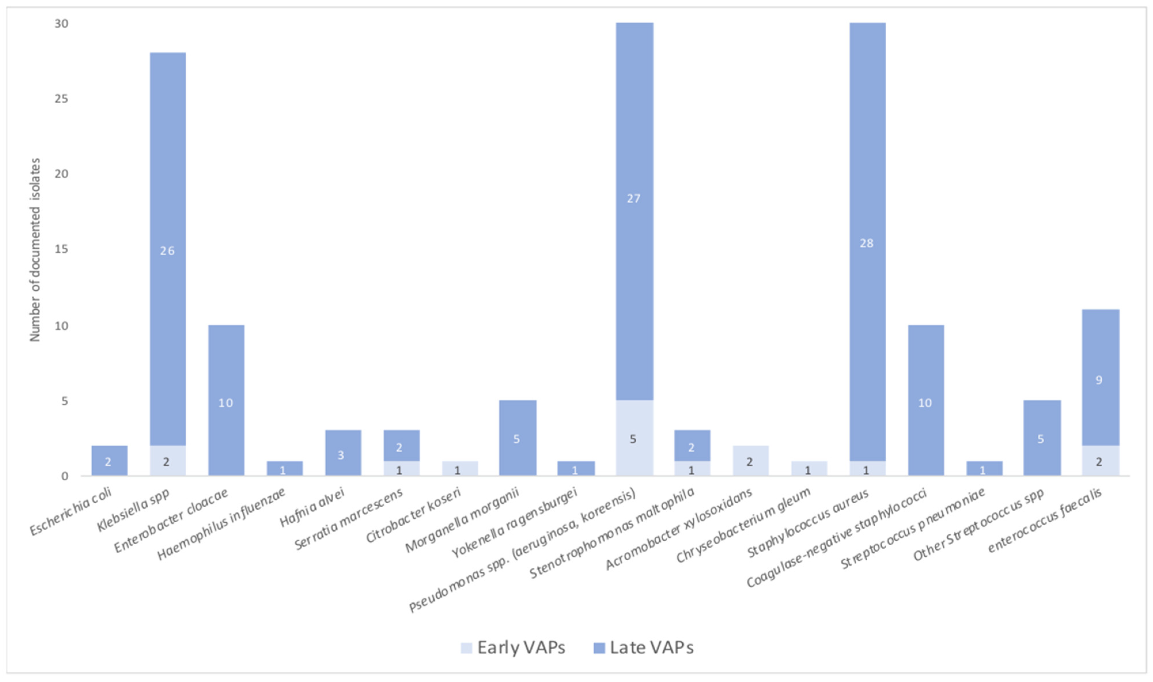
The type and incidence of each bacterium responsible for VAPs is detailed in Table 4 and Figure 2. These infections were categorized into early and late VAPs, on whether they occurred before or after the 5th day of mechanical ventilation. An amount of 72 patients (88%) developed late VAPs with a median delay of 10 days after intubation (IQR; 7–17). A total of 31% of VAPs were due to multidrug resistant bacteria (MDR) with no significant difference between early and late infections. VAP associated blood stream infections (BSI) were exclusively documented in late VAPs (20/72), with 33% caused by MDR bacteria.
Regardless of the time to onset of VAP, three organisms were responsible for 60% of infections: Klebsiella spp., Pseudomonas spp. and Staphylococcus aureus. Gram negative species were predominantly involved in 62% of VAPs. When analyzing the different types of bacteria between both VAP groups, we noticed that Staphylococcus aureus was mainly documented in late VAPs with an incidence of 22% vs. only 6% in early VAPs, however this difference was not statistically significant (p = 0.08). Most of the strains were Methicillin-sensitive Staphylococcus aureus (MSSA) while only 5/29 isolates displayed a Methicillin-resistant phenotype (MRSA), the latter all being reported in late VAPs. There were no differences between both groups regarding other bacterial species involved in VAPs except from Acromobacter xylosoxidans which was mainly found in early VAPs (p = 0.005). There were no recorded cases of invasive pulmonary aspergillosis in this study.

3.5. Antibiotic Resistance Mechanisms

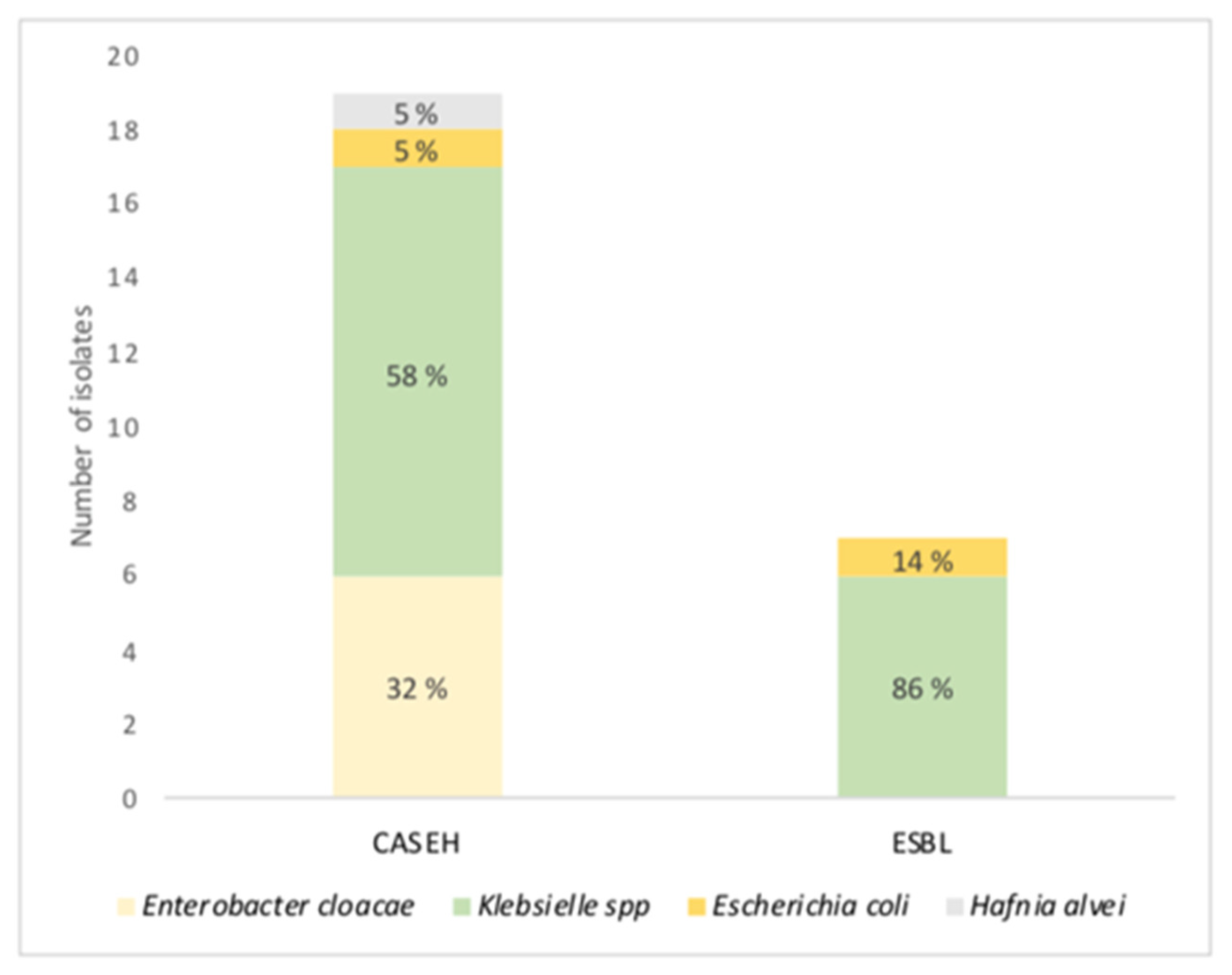
Finally, we analyzed the different types of antibiotic resistance mechanisms among VAP and BSI MDR infections (Figure 3). These infections regarded 26% of patients who experienced at least one infection during their ICU stay and in 17% of our ventilated cohort. The dominant resistance mechanisms were cephalosporinase hyper-production (CASEH), Ceftazidime resistant Pseudomonas aeruginosa (CAZ I/R PA) and extended-spectrum beta-lactamase (ESBL) with respective incidences of 41%, 24% and 15%. MRSA was documented in 6/46 isolates. Early VAPs and VAP-free blood stream infections accounted for 4/46 of BMR infections (9%) in ventilated patients, whilst late VAPs accounted for 67%.
Klebsiella spp. (pneumoniae, aerogenes) was the main strain displaying CASEH and ESBL resistance mechanisms with incidences of 58% and 86%, respectively. Other enterobacteriaceae species involved included Enterobacter cloacae, Escherichia coli and Hafnia alvei.

3.6. Multivariate Analysis

In this multivariate analysis (Table 5), we singled out clinically relevant factors that had a univariate p score < 0.2 in order to identify potential VAP risk factors. Age and male sex were associated to VAP (p = 0.014 and 0.018, respectively). IGS 2 and ROX scores at admission were associated to VAP with paradoxically higher IGS 2 scores having a protective effect. Dexamethasone was significantly associated with VAP incidence OR 3.2 (1.01–10), p = 0.046. In univariate analysis, antimicrobial treatment prior to ICU admission was strongly correlated with lower VAP incidence, thus constituting a protective fac-tor [p = 0.009; OR = 0.23 (0.07–0.7)]. Neutrophil to lymphocyte ratio was no longer associated to VAP in multivariate analysis.

4. Discussion

4.1. Ventilation-Acquired Pneumonia Characteristics

The studied population was mainly composed by comorbid males (83%) with high blood pressure and diabetes being the two most described comorbidities. This reinforces the already common knowledge that SARS-CoV-2 is most virulent in these types of patients [23]. Time between ICU admission to intubation was very short with a median intubation date at day 1 reflecting the critical level of respiratory failure these patients faced.
Our work provides a detailed description of VAP microbiological distribution. Infections occurred at day 3 (IQR, 3–3) for early VAP and day 9 (6–14) for late VAPs. The proportion between early and late onset VAPs in this study, with a majority of late VAPs (88%) is consistent with other reports. Most infections were caused by Gram negative bacteria (62%) with Pseudomonas aeruginosa being the most documented bacteria (21%). These numbers are similar to the ones published by Grasselli et al. [24]. Staphylococcus aureus was mostly found in late VAPs (p = 0.08) contrasting with the distribution of Gram-positive bacteria in VAP settings that are usually found in the early stages. Staphylococcus aureus has demonstrated the ability to interact and infect different cell strains, while becoming increasingly resistant to antibiotic therapy and a reservoir of bacteria that can make the infection difficult to treat [25]. However, this could simply be explained by a lack of power from low early VAP incidence.
Another key finding with potentially significant clinical implications is that MDR bacteria caused about one third of VAPs. The most common antibiotic resistance mechanism being AmpC cephalosporin hyperproduction (CASEH) found in 90% of the cases in either Klebsiella or Enterobacter spp. MRSA strains represented 17% of all Staphylococcus aureus VAPs, remaining low and consistent with literature. This high proportion of MDR infections could be explained by a large amount of patients (47%) receiving broad spectrum antibiotics prior to ICU admission, including 3rd generation cephalosporins. Additionally, the influx of critically ill patients during the pandemic may have influenced the quality of critical care provided by overloading medical staff, potentially increasing the risk of ventilation-associated lower respiratory tract infections (VA-LRTI) by MDR bacteria. Indeed, previous studies reported that contact isolation measures, especially inappropriate glove use, could increase the transmission of MDR bacteria [26].

4.2. Ventilation-Acquired Pneumonia Risk Factors

Age and sex where the two demographic characteristics associated with VAPs in multivariate analysis. Age has widely been recognized as a risk factor for nosocomial infection and VAP in COVID and non-COVID settings. Various mechanisms such as decreased immune function, frequent chronic illnesses and malnutrition explain why older patients are more likely to develop hospital-acquired infections such as VAPs [24,27]. Male gender has been associated with VAP in non-COVID patients [28] but our study is the first to report this risk factor in a COVID cohort. However, the SARS-CoV-2 itself could be a confounding factor as we know that this virus is more virulent in men and responsible for higher ICU admission and mortality rates compared to women [29].
The ROX score, recently developed, has been used during the COVID-19 pandemic by certain teams to predict the need for invasive mechanical ventilation after high-flow nasal cannula failure [30]. This clinical score (SpO2/FiO2/RR) was the only score associated to VAP incidence and may reflect that the level of acute respiratory failure before intubation could be associated to the development of VA-LRTI.
The role of dexamethasone’s impact on VAP was a key question in this study. This treatment at a dose of 6 mg per day during 10 days is a cornerstone treatment for severe cases of COVID-19 infection, as it is the only treatment, being associated with a significant lower in-ICU mortality [17]. However, the impact of this treatment on VAP incidence in SARS-CoV-2 infected patients is still up for debate with contrasting literature on the subject. Reys et al. revealed in a prospective study that dexamethasone was a risk factor for ICU-acquired respiratory tract infections whilst Gragueb-Chatti et al. found no relation between VAP or BSI and dexamethasone [31,32]. In our study, the multivariate analysis revealed that dexamethasone was associated with higher VAP incidence with an odds ratio of 3.2 (1.01–10). Other corticosteroid treatments were used such as high-dose methylprednisolone which had a higher incidence in the VAP group. However, this treatment was mainly started after the diagnosis of VAP making its causality as a potential risk factor difficult to judge. Dexamethasone’s purpose is to mitigate inflammation-related organ injuries. ICU patients with low biological markers at admission could be a sub-group of patients in which dexamethasone favors bacterial super infections rather than treating inflammation-induced lesions. Further prospective studies are needed to confirm this potential unintended effect. In this situation, alveolar injury marker can be useful for the diagnosis of VAP. Recently Angiopoietin-2 (Ang-2) and soluble intercellular adhesion molecule-1 (ICAM-1), were identified as markers of pulmonary endothelial injury in COVID-19 ARDS [33].
The multivariate analysis revealed that antibiotic treatment administered prior to ICU admission was associated with a reduction in VAP incidence (see Table 5). Based on our definition of VAP, high antibiotic exposure prior to bacterial samplings might have caused falsely negative results leading to unaccounted non-documented VAPs. Respiratory bacterial co-infection rates during standard hospitalization in COVID-19 were low, with 755 reported in literature [34,35]. The impact of inadequate probabilistic antibiotic treatment on the emergence of antibiotic resistance has been an ongoing and growing global health issue [12]. Coupling this to the low evidence level of results obtained from our retrospective study, it seems unreasonable to advocate for pre-emptive antibiotic treatment before ICU admission with the objective of reducing VAP incidence in COVID-19. On the other hand, recent ICU health care-associated pneumonia (HCAP) recommendations stated that systemic prophylactic antibiotics, through selective digestive decontamination, may help reduce HCAP incidence [36]. Future prospective studies are needed to clarify the use and timing of prophylactic antibiotics. Recently, multivalent fucose derivative has been described potential broad spectrum ant biofilm agent, although its place in adjunctive treatment of VAP needs to be clarified [37].
Neutrophil to lymphocyte ratio at ICU admission was associated to VAP in the univariate analysis but no longer in the multivariate analysis. NLR has been used in these last years as a marker for endothelial dysfunction, a mechanism particularly present in COVID-19 pathogenesis. Different studies found associations between this biological marker and poor clinical outcome, with Jimeno et al. demonstrating a relation between peak NLR and death. However, NLR lacks specificity and was mostly described in prognostication of sepsis and oncological diseases making it difficult to use on a practical basis. Disease-specific thresholds may increase the reliability of this marker and could be a focus point for future studies.

4.3. Incidence of Ventilation-Acquired Pneumonia

In this retrospective study, we analyzed the epidemiologic and etiological factors influencing the development of VAP in a cohort of critically ill COVID-19 patients. The incidence of VAP in this study was 51% as reported in literature with incidences ranging between 44% to 64% [38,39]. This contrasts with VAP occurrence in non-COVID ICU settings which seem to have lower incidence rates (13–29%) [16,28]. One of the recurrent explanations mentioned in literature is the longer duration of mechanical ventilation in SARS-CoV-2-related ARDS compared to other types of ARDS. In our study, the median duration of mechanical ventilation was 18 days (IQR, 8.2–33) which is slightly longer than other reports (13–15 days). This could be due to the fact that this study analyzes the earliest period of the COVID-19 pandemic, a time in which intensivists were encouraged to intubate patients at early stages of respiratory failure. Besides, the tropism of SARS-CoV-2 for nervous tissue and its association with confusion agitation and encephalopathy [40], may lead to a more difficult to weaning from mechanical ventilation. On a cellular level, one of the strongest predictors of nosocomial infection in critically ill patients is impaired immune cell function. Patients with COVID-19 experience a complex dysregulation of their immune function with features of both hyper-inflammatory activation and organ damage as well as impaired antimicrobial functions. Notably, damage to the alveolar membrane, although not specific to COVID-19, may facilitate invasion of bacterial species [41], similarly to influenza infection. However, when comparing VAP incidences in a COVID versus influenza ICU population, these remained significantly higher in the COVID-19 cohort (50% vs. 30%) [42]. A reason for this could be widespread thrombosis which distinguishes the pulmonary pathophysiology of COVID-19, from that of equally severe influenza virus infection. These lesions might promote local immunity alteration, bacterial colonization, and further lung infection. However, further studies are required to confirm this hypothesis.

4.4. Limitations

The retrospective design of our study could lead to an interpretative bias. The low incidence of dexamethasone is not representative of an up-to-date ICU COVID-19 cohort and weakens the generalization of our results. Blood stream infections were mainly associated with VAP (80%), thus the analysis of their association with dexamethasone was difficult.

5. Conclusions

In this retrospective study, we analyzed the characteristics and risk factors involved in ventilation-acquired pneumonia in a COVID-19 cohort within three French public hospitals from March to November 2020. After adjusting for confounding factors, the main risk factors for ventilation-acquired pneumonia were dexamethasone, older age and male gender whereas antibiotic treatment prior to ICU admission was a protective factor. As dexamethasone has been the only evidence-based treatment that has shown decreased mortality in severe cases of COVID-19, future robust multicentric studies are needed before questioning the risk-benefit ratio of this treatment.

Author Contributions

J.M. and J.B. contributed to the conception and design of the study, had full access to all the data in the study and take responsibility for the integrity of the data and the accuracy of the data analysis. J.B., J.M., J.C., M.B. and D.T. contributed to the acquisition of data. J.M., M.B. and J.B. contributed to the analysis and interpretation of the data. J.M. and M.B. contributed to the statistical analyses. All authors participated in manuscript writing, revision and approval for final submission. All authors have read and agreed to the published version of the manuscript.

Funding

This research received no external funding.

Institutional Review Board Statement

This study was approved by the local Ethics Commission (2020-53) and the French Society of Anesthesia and Critical Care (00010254-2020-262). According to French law, the informed consent was not required due to the design of the study.

Informed Consent Statement

According to French law, a written informed consent from patients was not necessary, owing to the retrospective nature of the study, but the patients had the possibility to withdraw their health data.

Data Availability Statement

The data presented in this study are available on request from the corresponding author.

Conflicts of Interest

The authors declare no conflict of interest.

References

  1. COVID-19 Map. Johns Hopkins Coronavirus Resource Center. Available online: https://coronavirus.jhu.edu/map.html (accessed on 30 July 2022).
  2. Fiolet, T.; Kherabi, Y.; MacDonald, C.J.; Ghosn, J.; Peiffer-Smadja, N. Comparing COVID-19 vaccines for their characteristics, efficacy and effectiveness against SARS-CoV-2 and variants of concern: A narrative review. Clin. Microbiol. Infect. 2022, 28, 202–221. [Google Scholar] [CrossRef]
  3. Tregoning, J.S.; Flight, K.E.; Higham, S.L.; Wang, Z.; Pierce, B.F. Progress of the COVID-19 vaccine effort: Viruses, vaccines and variants versus efficacy, effectiveness and escape. Nat. Rev. Immunol. 2021, 21, 626–636. [Google Scholar] [CrossRef]
  4. European Centre for Disease Prevention and Control. SARS-CoV-2 Variants of Concern as of 28 July 2022. Available online: https://www.ecdc.europa.eu/en/covid-19/variants-concern (accessed on 30 July 2022).
  5. Tracking SARS-CoV-2 Variants. Available online: https://www.who.int/activities/tracking-SARS-CoV-2-variants (accessed on 30 July 2022).
  6. Umakanthan, S.; Sahu, P.; Ranade, A.V.; Bukelo, M.M.; Rao, J.S.; Lf, A.-M.; Dahal, S.; Kumar, H.; Kv, D. Origin, transmission, diagnosis and management of coronavirus disease 2019 (COVID-19). Postgrad. Med. J. 2020, 1. [Google Scholar] [CrossRef]
  7. Le Terrier, C.; Suh, N.; Wozniak, H.; Boroli, F.; Giudicelli-Bailly, A.; Sangla, F.; Legouis, D.; Bendjelid, K.; Quintard, H.; Pugin, J. Delayed intubation is associated with mortality in patients with severe COVID-19: A single-centre observational study in Switzerland. Anaesth. Crit. Care Pain Med. 2022, 41. [Google Scholar] [CrossRef] [PubMed]
  8. Grasselli, G.; Zangrillo, A.; Zanella, A.; Antonelli, M.; Cabrini, L.; Castelli, A.; Cereda, D.; Coluccello, A.; Foti, G.; Fumagalli, R.; et al. Baseline Characteristics and Outcomes of 1591 Patients Infected with SARS-CoV-2 Admitted to ICUs of the Lombardy Region, Italy. JAMA 2020, 323, 1574–1581, Erratum in JAMA 2021, 325, 2120. [Google Scholar] [CrossRef] [PubMed] [Green Version]
  9. Martin-Loeches, I.; Rodriguez, A.H.; Torres, A. New guidelines for hospital-acquired pneumonia/ventilator-associated pneumonia: USA vs. Europe. Curr. Opin. Crit. Care 2018, 24, 347–352. [Google Scholar] [CrossRef] [PubMed]
  10. Richardson, S.; Hirsch, J.S.; Narasimhan, M.; Crawford, J.M.; McGinn, T.; Davidson, K.W. the Northwell COVID-19 Research Consortium. Presenting Characteristics, Comorbidities, and Outcomes among 5700 Patients Hospitalized with COVID-19 in the New York City Area. JAMA 2020, 323, 2052–2059. [Google Scholar] [CrossRef]
  11. Melsen, W.G.; Rovers, M.M.; Groenwold, R.; Bergmans, D.C.; Camus, C.; Bauer, T.T.; Hanisch, E.; Klarin, B.; Koeman, M.; A Krueger, W.; et al. Attributable mortality of ventilator-associated pneumonia: A meta-analysis of individual patient data from randomised prevention studies. Lancet Infect. Dis. 2013, 13, 665–671. [Google Scholar] [CrossRef]
  12. Laxminarayan, R.; Duse, A.; Wattal, C.; Zaidi, A.K.M.; Wertheim, H.F.L.; Sumpradit, N.; Vlieghe, E.; Hara, G.L.; Gould, I.M.; Goossens, H.; et al. Antibiotic resistance—The need for global solutions. Lancet Infect. Dis. 2013, 13, 1057–1098. [Google Scholar] [CrossRef] [Green Version]
  13. Gamberini, L.; Tonetti, T.; Spadaro, S.; Zani, G.; Mazzoli, C.A.; Capozzi, C.; Giampalma, E.; Reggiani, M.L.B.; Bertellini, E.; Castelli, A.; et al. Factors influencing liberation from mechanical ventilation in coronavirus disease 2019: Multicenter observational study in fifteen Italian ICUs. J. Intensive Care 2020, 8, 80, Erratum in J. Intensive Care 2020, 8, 96. [Google Scholar] [CrossRef]
  14. Schenck, E.J.; Hoffman, K.; Goyal, P.; Choi, J.; Torres, L.; Rajwani, K.; Tam, C.W.; Ivascu, N.; Martinez, F.J.; Berlin, D.A. Respiratory Mechanics and Gas Exchange in COVID-19–associated Respiratory Failure. Ann. Am. Thorac. Soc. 2020, 17, 1158–1161. [Google Scholar] [CrossRef] [PubMed]
  15. Zhou, F.; Yu, T.; Du, R.; Fan, G.; Liu, Y.; Liu, Z.; Xiang, J.; Wang, Y.; Song, B.; Gu, X.; et al. Clinical course and risk factors for mortality of adult inpatients with COVID-19 in Wuhan, China: A retrospective cohort study. Lancet 2020, 395, 1054–1062. [Google Scholar] [CrossRef] [PubMed]
  16. Maes, M.; Higginson, E.; Pereira-Dias, J.; Curran, M.D.; Parmar, S.; Khokhar, F.; Cuchet-Lourenço, D.; Lux, J.; Sharma-Hajela, S.; Ravenhill, B.; et al. Ventilator-associated pneumonia in critically ill patients with COVID-19. Crit. Care 2021, 25, 25, Erratum in Crit. Care 2021, 25, 130. [Google Scholar] [CrossRef] [PubMed]
  17. The RECOVERY Collaborative Group. Dexamethasone in Hospitalized Patients with COVID-19. N. Engl. J. Med. 2021, 384, 693–704. [Google Scholar] [CrossRef]
  18. Moreno, G.; Rodríguez, A.; Reyes, L.F.; Gomez, J.; Sole-Violan, J.; Díaz, E.; Bodí, M.; Trefler, S.; Guardiola, J.; on behalf of the GETGAG Study Group; et al. Corticosteroid treatment in critically ill patients with severe influenza pneumonia: A propensity score matching study. Care Med. 2018, 44, 1470–1482. [Google Scholar] [CrossRef]
  19. Plachouras, D.; Lepape, A.; Suetens, C. ECDC definitions and methods for the surveillance of healthcare-associated infections in intensive care units. Intensive Care Med. 2018, 44, 2216–2218. [Google Scholar] [CrossRef] [Green Version]
  20. de Jager, C.P.; van Wijk, P.T.; Mathoera, R.B.; de Jongh-Leuvenink, J.; van der Poll, T.; Wever, P.C. Lymphocytopenia and neutrophil-lymphocyte count ratio predict bacteremia better than conventional infection markers in an emergency care unit. Crit. Care 2010, 14, R192. [Google Scholar] [CrossRef] [Green Version]
  21. Predictive Value of Systemic Immune-Inflammation index and Neutrophil-to-Lymphocyte Ratio in Patients with Severe COVID-19. Clin. Appl. Thromb. Hemost. 2022, 28, 10760296221111391. [CrossRef]
  22. van Arkel, A.L.E.; Rijpstra, T.A.; Belderbos, H.N.A.; van Wijngaarden, P.; Verweij, P.E.; Bentvelsen, R.G. COVID-19-associated Pulmonary Aspergillosis. Am. J. Respir. Crit. Care Med. 2020, 202, 132–135. [Google Scholar] [CrossRef]
  23. Dessie, Z.G.; Zewotir, T. Mortality-related risk factors of COVID-19: A systematic review and meta-analysis of 42 studies and 423,117 patients. BMC Infect. Dis. 2021, 21, 855. [Google Scholar] [CrossRef]
  24. Grasselli, G.; Scaravilli, V.; Mangioni, D.; Scudeller, L.; Alagna, L.; Bartoletti, M.; Bellani, G.; Biagioni, E.; Bonfanti, P.; Bottino, N.; et al. Hospital-Acquired Infections in Critically Ill Patients with COVID-19. Chest 2021, 160, 454–465. [Google Scholar] [CrossRef] [PubMed]
  25. Pang, X.C.; Zhang, H.X.; Zhang, Z.; Rinkiko, S.; Cui, Y.M.; Zhu, Y.Z. The Two-Way Switch Role of ACE2 in the Treatment of Novel Coronavirus Pneumonia and Underlying Comorbidities. Molecules 2021, 26, 142. [Google Scholar] [CrossRef] [PubMed]
  26. Harris, A.D.; Pineles, L.; Belton, B.; Johnson, J.K.; Shardell, M.; Loeb, M.; Newhouse, R.; Dembry, L.; Braun, B.; Perencevich, E.; et al. Universal Glove and Gown Use and Acquisition of Antibiotic-Resistant Bacteria in the ICU. JAMA 2013, 310, 1571–1580. [Google Scholar] [CrossRef] [PubMed]
  27. Wu, D.; Wu, C.; Zhang, S.; Zhong, Y. Risk Factors of Ventilator-Associated Pneumonia in Critically III Patients. Front. Pharmacol. 2019, 10, 482. [Google Scholar] [CrossRef] [Green Version]
  28. Forel, J.-M.; Voillet, F.; Pulina, D.; Gacouin, A.; Perrin, G.; Barrau, K.; Jaber, S.; Arnal, J.-M.; Fathallah, M.; Auquier, P.; et al. Ventilator-associated pneumonia and ICU mortality in severe ARDS patients ventilated according to a lung-protective strategy. Crit. Care 2012, 16, R65. [Google Scholar] [CrossRef] [PubMed] [Green Version]
  29. Peckham, H.; de Gruijter, N.M.; Raine, C.; Radziszewska, A.; Ciurtin, C.; Wedderburn, L.R.; Rosser, E.C.; Webb, K.; Deakin, C.T. Male sex identified by global COVID-19 meta-analysis as a risk factor for death and ITU admission. Nat. Commun. 2020, 11, 1–10. [Google Scholar] [CrossRef]
  30. Roca, O.; Caralt, B.; Messika, J.; Samper, M.; Sztrymf, B.; Hernández, G.; García-De-Acilu, M.; Frat, J.-P.; Masclans, J.R.; Ricard, J.-D. An Index Combining Respiratory Rate and Oxygenation to Predict Outcome of Nasal High-Flow Therapy. Am. J. Respir. Crit. Care Med. 2019, 199, 1368–1376. [Google Scholar] [CrossRef]
  31. Reyes, L.F.; Rodriguez, A.; Bastidas, A.; Parra-Tanoux, D.; Fuentes, Y.V.; García-Gallo, E.; Moreno, G.; Ospina-Tascon, G.; Hernandez, G.; Silva, E.; et al. Dexamethasone as risk-factor for ICU-acquired respiratory tract infections in severe COVID-19. J. Crit. Care 2022, 69. [Google Scholar] [CrossRef] [PubMed]
  32. Gragueb-Chatti, I.; Lopez, A.; Hamidi, D.; Guervilly, C.; Loundou, A.; Daviet, F.; Cassir, N.; Papazian, L.; Forel, J.-M.; Leone, M.; et al. Impact of dexamethasone on the incidence of ventilator-associated pneumonia and blood stream infections in COVID-19 patients requiring invasive mechanical ventilation: A multicenter retrospective study. Ann. Intensive Care 2021, 11, 1–10. [Google Scholar] [CrossRef]
  33. Granata, G.; Stracquadanio, S.; Consoli, G.M.L.; Cafiso, V.; Stefani, S.; Geraci, C. Synthesis of a calix[4]arene derivative exposing multiple units of fucose and preliminary investigation as a potential broad-spectrum antibiofilm agent. Carbohydr. Res. 2019, 476, 60–64. [Google Scholar] [CrossRef]
  34. Rawson, T.M.; Moore, L.S.P.; Zhu, N.; Ranganathan, N.; Skolimowska, K.; Gilchrist, M.; Satta, G.; Cooke, G.; Holmes, A.H. Bacterial and Fungal Coinfection in Individuals With Coronavirus: A Rapid Review To Support COVID-19 Antimicrobial Prescribing. Clin. Infect. Dis. 2020, 71, 2459–2468. [Google Scholar] [CrossRef]
  35. Hughes, S.; Troise, O.; Donaldson, H.; Mughal, N.; Moore, L.S.P. Bacterial and fungal coinfection among hospitalized patients with COVID-19: A retrospective cohort study in a UK secondary-care setting. Clin. Microbiol. Infect. 2020, 26, 1395–1399. [Google Scholar] [CrossRef]
  36. Leone, M.; Bouadma, L.; Bouhemad, B.; Brissaud, O.; Dauger, S.; Gibot, S.; Hraiech, S.; Jung, B.; Kipnis, E.; Launey, Y.; et al. Pneumonies associées aux soins de réanimation. Anesthésie Réanimation 2018, 4, 421–441. [Google Scholar] [CrossRef]
  37. Jimeno, S.; Ventura, P.S.; Castellano, J.M.; García-Adasme, S.I.; Miranda, M.; Touza, P.; Lllana, I.; López-Escobar, A. Prognostic implications of neutrophil-lymphocyte ratio in COVID-19. Eur. J. Clin. Investig. 2020, 51. [Google Scholar] [CrossRef]
  38. Pickens, C.O.; Gao, C.A.; Cuttica, M.J.; Smith, S.B.; Pesce, L.L.; Grant, R.A.; Kang, M.; Morales-Nebreda, L.; Bavishi, A.A.; Arnold, J.M.; et al. Bacterial Superinfection Pneumonia in Patients Mechanically Ventilated for COVID-19 Pneumonia. Am. J. Respir. Crit. Care Med. 2021, 204, 921–932. [Google Scholar] [CrossRef] [PubMed]
  39. Razazi, K.; Arrestier, R.; Haudebourg, A.F.; Benelli, B.; Carteaux, G.; Decousser, J.-W.; Fourati, S.; Woerther, P.L.; Schlemmer, F.; Charles-Nelson, A.; et al. Risks of ventilator-associated pneumonia and invasive pulmonary aspergillosis in patients with viral acute respiratory distress syndrome related or not to Coronavirus 19 disease. Crit. Care 2020, 24, 699, Erratum in Crit. Care 2021, 25, 118. [Google Scholar] [CrossRef]
  40. Helms, J.; Kremer, S.; Merdji, H.; Clere-Jehl, R.; Schenck, M.; Kummerlen, C.; Collange, O.; Boulay, C.; Fafi-Kremer, S.; Ohana, M.; et al. Neurologic Features in Severe SARS-CoV-2 Infection. N. Engl. J. Med. 2020, 382, 2268–2270. [Google Scholar] [CrossRef] [PubMed]
  41. Mason, R.J. Pathogenesis of COVID-19 from a cell biology perspective. Eur. Respir. J. 2020, 55, 2000607. [Google Scholar] [CrossRef] [PubMed] [Green Version]
  42. Rouzé, A.; Martin-Loeches, I.; Povoa, P.; Makris, D.; Artigas, A.; Bouchereau, M.; Lambiotte, F.; Metzelard, M.; Cuchet, P.; Geronimi, C.B.; et al. Relationship between SARS-CoV-2 infection and the incidence of ventilator-associated lower respiratory tract infections: A European multicenter cohort study. Intensive Care Med. 2021, 47, 188–198. [Google Scholar] [CrossRef]
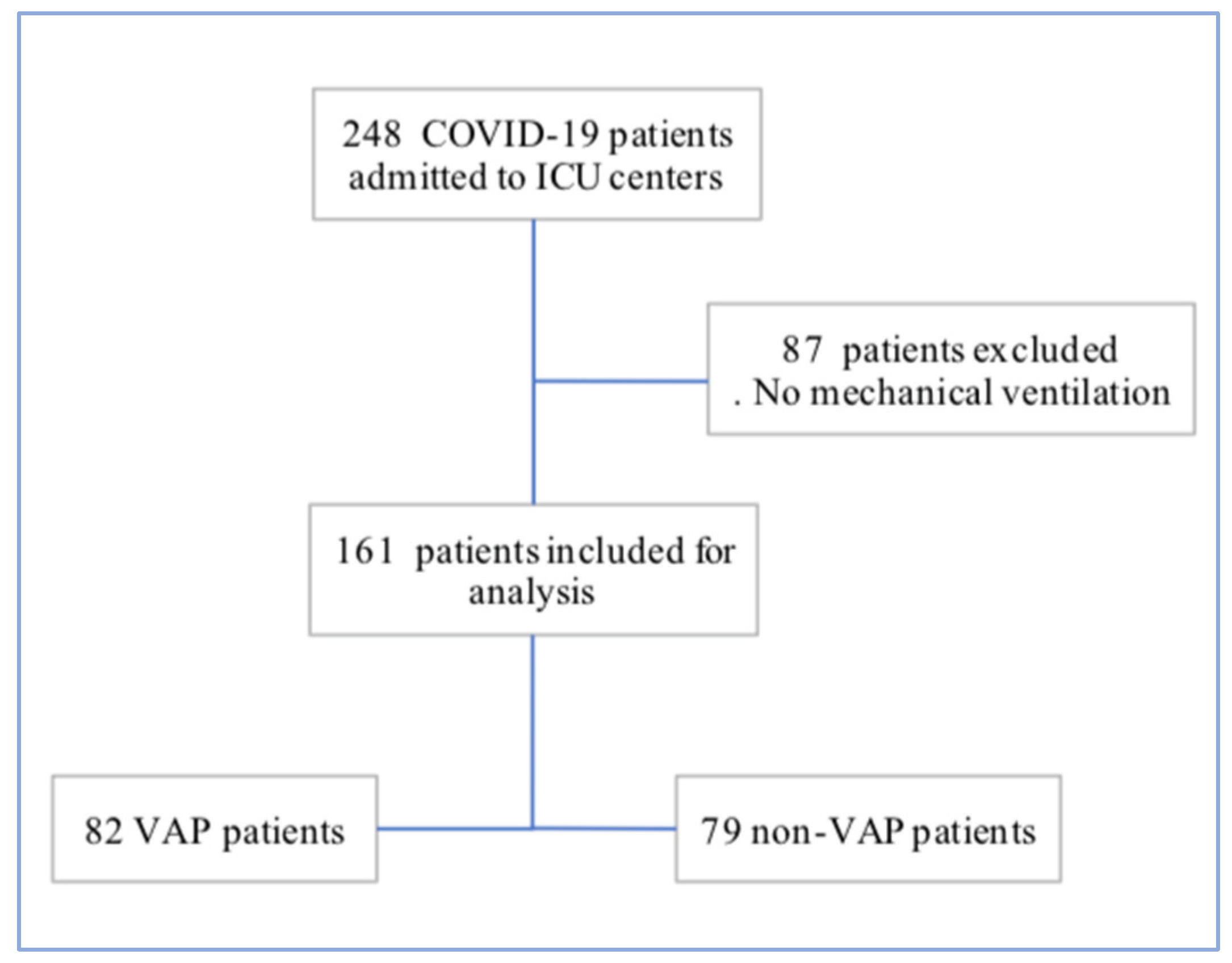
Figure 1. Study Flow chart.
Jcm 12 00421 g001
Figure 2. Ventilation-acquired pneumonia bacterial documentation.
Jcm 12 00421 g002
Figure 3. Antibiotic resistance mechanisms according to type of infection and bacteria.
Jcm 12 00421 g003aJcm 12 00421 g003b
Table 1. Patient clinical and biological characteristics.
VariableAll Patients VAP PatientsNon VAP Patientsp-ValueOR (95% CI)
N = 161N = 82N = 79
(51%)(49%)
Age—YEAR, median63(57–72)65(58–73)62(54–69)0.021.03 (1.00–1.06)
Sex—Male124(77)68(82.9)56(70.9)0.069
BMI kg/m2, median28(25–32)27.7(25.3–32.1)28(25.8–33.8)0.510.98 (0.93–1.03)
 18–24.9 31(19)16(23.2)15(21.4)0.8
 25–29.952(32)27(39.1)25(35.7)0.67
 30–4049(30)23(33.3)26(37.1)0.63
 >407(4)3(3.9)4(5.3)0.71
Comorbidities147(91.3)75(91.5)72(91.1)
 Hypertension88(54)45(54.9)43(54.4)0.951.01 (0.54–1.89)
 Diabetes58(36)25(30.5)33(41.8)0.130.61 (0.32–1.17)
 Cardiovascular disease31(19)19(23.2)12(15.2)0.21.68 (0.75–3.74)
  Coronary disease20(12.4)11(13.4)9(11.4)0.69
  Chronic heart failure3(19)1(1.2)2(2.5)0.61
  Occlusive arteriopathy4(2.5)3(3.7)1(1.3)0.62
 Metabolic syndrome36(22.4)16(19.5)20(25.3)0.37
 Current smoking 6(3.7)5(6.1)1(1.3)0.145.06 (0.57–44.3)
 Chronic lung disease39(24)20(24.4)19(24.1)0.961.01 (0.49–2.09)
  Obstructive sleep apnea21(13)11(13.4)10(12.7)0.88
 Chronic kidney disease12(7.5)4(4.9)8(10.1)0.210.45 (0.13–1.57)
 Immunocompromised *20(12.4)11(13.4)9(11.4)0.691.2 (0.47–3.08)
 Chronic treatment103(64)52(63.4)51(64.6)0.880.95 (0.5–1.81)
Status at ICU admission
 Delay between hospitalisation and ICU, d0(1–3)0(0–1.25)1(0–4)0.12
 IGS 235(28–43)34(30–43)35(27–43)0.86
 CHARLSON3(1–4)3(2–4)2(1–4)0.1
 SOFA4(3–7)4(3–7)4(2–7)0.43
 ROX4.6(3–9.3)3.7(3–7)5.8(3–10.1)0.012
 PaO2/FiO2 ratio (mmHg)131(96–180)132.5(91.5–179)130(100–181.5)0.420.99 (0.99–1)
 Maximum Temperature (°C)38.3(37.5–39)38.3(37.6–38.7)38.4(37.3–39)0.70.93 (0.67–1.31)
 Neutrophil to lymphocyte ratio 7.3(5.1–13)8.3(5.5–15.6)6(4.6–9.2)0.016
 Systemic immune-inflammation index (SII)1610(971–3270)2113(1116–3900)1281(834–2054)0.0161.11 (1.01–1.2)
 Ferritin (µg/L)1427(1013–2345)1241(796–1611)2273(976–4141)0.19
 Fibrinogen (g/L)6.6(5.9–7.7)6.7(5.9–8)6.6(5.7–7.3)0.59
 D-dimer (µg/L)1.5(1–4)1.5(0.8–4)1.6(1.1–2.9)0.9
 Procalcitonin (ng/mL)0.3(0.19–0.58)0.3(0.2–0.5)0.3(0.2–1.3)0.49
 C-reactive protein (mg/dL)170(91.6–227)185.9(120–251)98.7(83.6–184)0.030.89 (0.64–1.23)
 Infection within 48 h of ICU admission26(16.1)13(15.9)13(16.5)0.91
 CT scan lung lesion (%)31.2 37.3 27.8 0.038
Data throughout are presented as No. (%) of the included patients or median (interquartile range). *: includes solid tumors, active hematological malignancy, solid organ transplant, HIV, immunosupressive treatment.
Table 2. Anti-infective therapies.
VariableAll PatientsVAP PatientsNon VAP Patientsp-ValueOR (95% CI)
N = 161N = 82N = 79
(51%)(49%)
Early ICU management
Antibiotic treatment within 48 h of ICU admission137(85.6)68(82.9)69(88.5)0.32
Corticosteroids
 Dexamethasone 6 mg51(31.7)31(37.8)20(25.3)0.091.8 (0.9–3.5)
 Median duration of dexamethasone 6 mg10(7–10)10(7–10)10(6–10)0.22
Immunomodulators44(27.3)27(32.9)17(21.5)0.1
 Anakinra (anti IL-1)19(11.8)10(12.2)9(11.4)0.87
 Tocilizumab (anti IL-6)4(2.5)1(1.2)3(3.8)0.31
 Ruxolitinib (Jakavi)19(11.8)12(14.6)7(8.9)0.26
 Lopinavir/Ritonavir18(11.2)13(15.9)5(6.3)0.063
Hydroxychloroquine107(66.5)50(61)57(72)0.13
Pre ICU management
 Antibiotic treatment90(56.3)39(47.6)51(65.4)0.0240.48 (0.25–0.91)
 3rd generation cephalosporin60(37.2)25(30)35(44)0.069
 Azithromycin60(37.2)24(29)36(45.5)0.035
 Penicillin A +/− Clavulanic acid20(12.4)9(10.9)11(13.9)0.57
Piperacillin—Tazobactam11(6.8)6(7.3)5(6.3)0.8
 Duration of pre ICU antibiotic treatment, d (IQR)4(2–5)3(2–5)4(2–5.5)0.16
 Hydroxychloroquine52(32)23(28)29(36.7)0.24
 Dexamethasone 6 mg8(5)3(3)5(6.3)0.48
Table 3. ICU management and outcome.
VariableAll PatientsVAP PatientsNon VAP Patientsp-ValueOR (95% CI)
N = 161N = 82N = 79
(51%)(49%)
Respiratory management
 Treated with High-flow nasal cannula (HFNC)105(65.2)52(63.4)53(67.1)0.620.85 (0.44–1.6)
 Treated with Non-invasive ventilation (NIV)43(26.7)23(28)20(25.3)0.691.15 (0.57–2.3)
 Neuromuscular blockers (NMB)152(94.4)79(96.3)73(92.4)0.32
 Prone position132(82)72(87.8)60(75.9)0.05
 Nitric oxyde (NO)52(32)35(42.6)17(21.5)0.004
 Extracorporeal membrane oxygenation (ECMO)24(14.9)13(15.9)11(13.9)0.73
 Solumedrol 2 mg/kg21(13)17(20.7)4(5.1)0.0064.9 (1.5–15.3)
ICU complications and outcome
 Norephinephrin > 1 mg/h82(50.9)52(63.4)30(37.9)0.001
 Septic shock50(31)30(36.6)20(25.3)0.12
 Blood stream infections40(25.5)32(80)8(20)< 0.001
 Acute renal failure76(47.5)43(52.4)33(42.3)0.2
 Renal replacement therapy 24(14.9)13(15.9)11(13.9)0.73
 Pneumothorax11(6.9)10(12.2)1(1.3)0.006
 Rythm disorders39(24.2)30(36.6)9(11.4)< 0.001
 Conduction disorders14(8.7)10(12.2)4(5.1)0.108
 HSV-1 reactivation19(11.8)13(15.9)6(7.6)0.104
 CMV reactivation18(11.2)13(15.9)5(6.3)0.055
 SARS-CoV 2 viral loads (Ct/mL)28.9(24.9–31.7)29.7(24.8–32)28(24.9–31)0.381.03 (0.96–1.10)
 Duration of PCR positivity, d12(6.5–17)13(8–17)11(5.7–17)0.21.02 (0.98–1.07)
 Delay between ICU and IMV0(0–1.5)0(0–2)1(0–1)0.2
 IMV duration, d18(8.2–33)31.5(15–47.2)10(5–18.2)< 0.001
 Length of stay—ICU, d24.5(15–43)39.5(21–57)17(8–25)< 0.001
 Length of stay—Hospital, d35(21–50)43(30–62.5)24(14–37.5)< 0.001
 Death during ICU stay41(25.5)21(25.6)20(25.3)0.96
Table 4. Ventilation-acquired pneumonia characteristics and bacterial documentation.
VariableAll VAPsEarly VAPsLate VAPsp-Value
N = 82N = 10N = 72
Average delay of VAPs, d9(6–14)3(3–3)10(7–17)< 0.001
Polymicrobial VAPs (> 2 isolates)15(18)2(20)13(18)1
Multi-drug resistant (MDR) VAPs26(31.7)2(20)24(33)0.49
VAP associated blood stream infections (BSI)20(24)-20(27.7)
 VAP associated MDR BSIs8(33) 8(33)
Documented microbiological isolatesN = 148N = 16N = 132
  Gram negative bacteria (%)(62)(81)(59)
  Fermenters
Escherichia coli2 2(2.7)1
Klebsiella spp.28(18.7)2(12)26(19)0.87
Enterobacter cloacae10(6.7) 10(7)0.45
Haemophilus influenzae1 1(0.7)1
Hafnia alvei3 3(2.2)1
Serratia marcescens3 1(6)2(2.7)0.8
Citrobacter koseri1 1(6) 0.24
Morganella morganii5 5(3.7)0.87
Yokenella ragensburgei1 1(0.7)1
  Non fermenters
Pseudomonas spp. (aeruginosa, koreensis)32(21.4)5(31)27(20)0.67
Stenotrophomonas maltophila3 1(10)2(2.7)0.8
Acromobacter xylosoxidans2 2(12) 0.005
Chryseobacterium gleum1 1(6) 0.24
Gram positive bacteria (%)(38)(19)(41)
Staphylococcus aureus29(20)1(6)29(22)0.08
 Methicillin-sensitive Staphylococcus aureus24 1 23 0.68
 Methicillin-resistant Staphylococcus aureus5 5 0.76
 Coagulase-negative staphylococci10(6.7) 10(7)0.45
Streptococcus pneumoniae1 1(0.7)1
 Other Streptococcus spp.5 5(3.7)1
Enterococcus faecalis11(7.4)2(12)9(6.7)0.87
Table 5. Multivariate risk factors associated with VAPs.
VariableVAP PatientsNon VAP PatientsOR (95% CI)Multivariate
N = 82N = 79Adjusted
(51%)(49%)p-Value
Age—YEAR, median65(58–73)62(54–69)1.08 (1.01–1.15)0.014
Sex—Male34(30–43)35(27–43)3.99 (1.26–12.6)0.018
IGS 234(30–43)35(27–43)0.94 (0.89–0.99)0.037
CHARLSON3(2–4)2(1–4)1.19 (0.79–1.78)0.39
ROX3.7(3–7)5.8(3–10.1)0.85 (0.75–0.97)0.015
Dexamethasone 6 mg31(37.8)20(25.3)3.2 (1.01–10)0.046
Immunomodulatory therapy27(32.9)17(21.5)2.73 (0.84–8.8)0.092
Antibiotic course prior to ICU39(47.6)51(65.4)0.23 (0.07–0.7)0.009
Neutrophil Lymphocyte ratio8.3(5.5–15.6)6(4.6–9.2)0.99 (0.95–1.03)0.82
Duration of PCR positivity, d13(8–17)11(5.7–17)1.02 (0.96–1.08)0.41
Delay between hospitalization and ICU, d0(0–1.25)1(0–4)0.96 (0.79–1.16)0.7
Delay between ICU and IMV, d0(0–2)1(0–1)1.18 (0.92–1.51)0.18
Disclaimer/Publisher’s Note: The statements, opinions and data contained in all publications are solely those of the individual author(s) and contributor(s) and not of MDPI and/or the editor(s). MDPI and/or the editor(s) disclaim responsibility for any injury to people or property resulting from any ideas, methods, instructions or products referred to in the content.