Article

Superior Mesenteric Artery Injury during Robotic Radical Nephrectomy: Scenarios and Management Strategies

1
The Catherine and Joseph Aresty Department of Urology, USC Institute of Urology, Keck School of Medicine, University of Southern California, Los Angeles, CA 90033, USA
2
Department of Urology, Guy’s and St Thomas NHS Foundation Trust, London SE1 9RT, UK
3
Department of Urology, Hackensack Meridian School of Medicine, Hackensack, NJ 07601, USA
*
Author to whom correspondence should be addressed.
J. Clin. Med. 2023, 12(2), 427; https://doi.org/10.3390/jcm12020427
Submission received: 19 December 2022 / Revised: 30 December 2022 / Accepted: 3 January 2023 / Published: 5 January 2023
(This article belongs to the Section General Surgery)

Abstract

:
Injury to the superior mesenteric artery (SMA) is a rare, underreported, and potentially devastating complication. This study aims to propose a systematic workup to describe how to prevent and manage SMA injury in a standardized stepwise manner. Three different instances of intraoperative injury to the SMA are described in an accompanying video. All three occurred when the SMA was misidentified as the left renal artery during left robotic radical nephrectomy. In the first case, the SMA was mistakenly identified as the renal artery, but after further dissection, the real renal artery was identified and SMA injury was prevented. In the second case, the SMA was clipped and the real left renal artery was subsequently identified, requiring clip removal. In the third case, the SMA was clipped and completely transected, requiring prompt repair by vascular surgery with a successful outcome. This study aims to propose a systematic workup to describe how to prevent and manage SMA injury in a standardized stepwise manner. The proper anatomic recognition of the SMA may prevent its injury. Intraoperative SMA injury should be promptly identified and repaired to avoid its devastating consequences.

1. Introduction

Robotic-assisted radical nephrectomy has become an increasingly popular option for the treatment of renal masses [1,2,3]. Robotic nephrectomy is more commonly performed via a transperitoneal approach due to anatomical familiarity and adequate working space [4]. The retroperitoneal approach represents a valid alternative, especially for posterior-laterally located renal masses, as it allows direct access to the renal artery without the need for colon mobilization [4].
There are several advantages of robotic technology to renal surgery, including an enhanced degree of freedom, three-dimensional visualization, higher magnification, surgeon’s ergonomics, the use of fluorescence imaging, and the elimination of tremors leading to a widespread utilization to perform more complex surgeries [5,6,7,8,9,10]. However, there is a potential risk for catastrophic complications during complex robotic renal surgery due to the close proximity of vital vascular structures [11,12,13,14,15].
Injury to the Superior Mesenteric Artery (SMA) is a rare, underreported, and potentially devastating complication. It can occur in patients with large left renal tumors, bulky lymphadenopathy, or in the setting of re-do surgery with significant retroperitoneal and intraabdominal scarring, which may distort the vascular anatomy. In most cases, inadvertent injury occurs due to misidentifying the SMA as the left renal artery (LRA) [16,17].
Failure to recognize and promptly repair an SMA injury may result in ischemic bowel and mortality in up to 50% of the cases [11,18]. Therefore, it is imperative that surgeons are able to recognize and manage this potentially life-threatening complication.
This study describes three different scenarios of injury to the SMA when misidentified as the LRA during left robotic radical nephrectomy with an accompanying video. A systematic workup is presented to describe how to prevent and manage SMA injury in a standardized stepwise manner.

2. Materials and Methods

Robotic surgeons from high-volume centers were asked to contribute video content on intraoperative SMA injury. Videos were anonymized, centers deidentified, and dates or times of surgery removed. Surgeons consented for their video material to be used in this study and accompanying video. Patients consented to the video recording of their surgical procedures for education and publication purposes. Three patients underwent left robotic-assisted radical nephrectomy for renal tumors. A transperitoneal approach was performed in two patients, while a retroperitoneal approach was used in one patient.

3. Results

The SMA can be injured by either clipping or transection, which could be either partial or complete. The management strategy varies according to the mechanism of injury. Moreover, the common aim is the prompt identification, recognition, and restoration of the anatomical disruption for proper blood supply.

3.1. Case #1: Timely Recognition of SMA Misidentification

The left renal hilum is dissected through a retroperitoneal approach. Suspicion was raised when the left renal vein (LRV) was identified posterior to the renal artery. After further dissection, the true left renal artery was identified. Therefore, with timely recognition, injury to the SMA was avoided.

3.2. Case #2: SMA Clipping

Through a transperitoneal approach, the left renal hilum is dissected (Figure 1a). Following further dissection, the apparent left renal artery is identified and clipped. Then, the left renal vein is identified and clipped (Figure 1b). Later, a dilated left renal vein is seen, which raises suspicion for a patent left renal artery, indicating arterial inflow to the kidney (Figure 1c). To manage this adverse event, a bulldog is placed proximally. Using a Harmonic Scalpel, the arterial clip is then removed (Figure 1d). The tip of the harmonic scalpel must be placed carefully to avoid SMA injury. Later, the venous clip is then removed in a similar fashion (Figure 1e). Next, the true renal artery is identified and clipped; following inspection of the SMA, renal vein and artery are examined. The left renal vein has collapsed, which indicates no inflow to the kidney. After confirmation and inspection of the vascular structures, the renal vein is once again clipped (Figure 1f). The error was recognized within 5 min after the SMA clip was placed. The patient was discharged on the postoperative day 2 without further complications.

3.3. Case #3: SMA Clipping and Complete Transection

Through a transperitoneal approach, the left renal hilum was dissected. Significantly distorted anatomy was encountered due to the large left-sided tumor. The SMA, which was located just superior to the left renal vein, was incorrectly identified as the left renal artery, ligated with Hem-o-lok clips, and transected. The left renal vein was then clipped and transected in a similar fashion. Continuing further dissection, an artery arising from the aorta was identified, i.e., the true renal artery. Immediately upon recognition (Figure 2), vascular surgeon assistance was requested immediately. A repair was performed by vascular surgery. A later clinical evaluation of the patient was negative for bowel ischemia, and the patient recovered.

4. Systematic Workup Algorithm Management of SMA Injury

4.1. SMA Clipping

In cases where the SMA is clipped with Hem-o-lok®, the first recommended step is to remove the clip with a harmonic scalpel, which allows for a precise division of the clip, decreasing the risk of damaging the underlying vessel [19]. Therefore, it is advised that the tip of the harmonic scalpel must be placed carefully to avoid SMA injury (Figure 3). Vascular surgery should be promptly informed in case intervention is needed. In addition, continuous wave doppler ultrasound is used to evaluate the integrity of the artery blood flow [20]. In cases where there is a diminished or turbulent blood flow with evidence of thrombosis, an open thrombectomy can be performed [17] (Figure 4).

4.2. SMA Transection

If the SMA is transected, vascular surgery should be immediately consulted, and the repair can be performed using several options: (1) end-to-end re-anastomosis, (2) GORE-TEX® Vascular Graft, (3) end-to-end SMA to renal artery stump anastomosis, or (4) splenic-to-SMA transposition [16,17,21,22,23,24]. Surgeons should choose the option with which they are more comfortable and best suits the type of vascular injury (Figure 4).
Of note, it is critical to highlight when to suspect that the SMA can be misidentified as the LRA during a left radical nephrectomy (Table 1). Therefore, it is recommended to follow the artery in its full trajectory to confirm that it enters the kidney (Video S1—Supplementary Materials).

5. Discussion

The SMA arises from the abdominal aorta between the celiac trunk and the renal arteries. It provides blood supply to the small and large bowel up to splenic flexure. The SMA provides multiple branches encompassing the inferior pancreaticoduodenal (IPD), middle colic, right colic, jejunal, ileal, and ileocolic artery. It is important to note that communicating arteries exist between the superior and inferior mesenteric arteries (IMA), allowing for multiple pathways of blood supply to the bowel. The major anastomotic network between distal branches of the SMA and IMA is known as the marginal artery of Drummond, while a proximal anastomosis between the middle colic artery (SMA branch) and the left colic artery (IMA branch) is known as the arc of Riolan or meandering artery [25,26].
In the classical anatomical relation, the LRV crosses anterior to the abdominal aorta and posterior to the SMA in the crotch of the angle between the SMA and the aorta, the renal artery courses posterior to the LRV, while the SMA drapes over the LRV and can potentially compress the LRV, which is known as the Nutcracker Syndrome (Figure 5). Given the anatomical relationship between the SMA, renal artery, renal vein, and kidney’s location in relation to the abdominal aorta, it is the proximal portion of the SMA that could be more commonly misidentified as the LRA, especially in cases where left renal tumors and/or bulky lymphadenopathy may distort the vascular anatomy increasing the risk of intraoperative vascular injury even for experienced surgeons [11,14,15,16,17]. Therefore, the full identification of the aorta is important in preventing this complication.
A thorough understanding of this surgical anatomy and common variations in renal vascularity is of utmost importance before embarking on kidney surgery [13].
The classical presentation of a single renal artery emerging laterally from the abdominal aorta is seen in only 25% of cases due to great anatomical variability of the renal artery [11], while a retro-aortic left renal vein and circumaortic left renal vein are vascular anomalies that could be encountered in 3% and 3.5% of the cases, respectively [20,27].
Thus, the steep learning curve in minimally invasive surgery and identifying the course of the renal vessels and their relations with SMA and the aorta are essential factors in the appropriate management and prevention of complications during kidney surgery [11,28].
The location of this type of injury determines the severity of ischemia and is a strong predictor of mortality in models based on traumatic vascular injury [29]; SMA injuries are stratified based on the Fullen classification, which is determined by the anatomic segment of the SMA that is involved [30] (Table 2) (Figure 6). This classification is based on the great number of collateral vessels emerging from the SMA; the level at which the injury occurred can affect the outcome in both bowel ischemia and mortality. Zone I injuries affect the trunk proximal to the first major branch, leading to a greater degree of ischemia, with mortality reported up to 76% [29]. Theoretically, this may be due to the presence of effective collateral circulation in more distal zones. It is important to take into consideration several anatomical variations that have been reported on Drummond’s marginal artery that could jeopardize the collateral circulation at any level or zone of injury predisposing to bowel ischemia, such as an inconsistent right marginal artery, an absent or tortuous marginal artery at the splenic flexure or Griffith’s point, and the absence of the artery at the sigmoid colon in 65%, 7%, and 9%, respectively [26]. All SMA injuries presented in this study are classified as zone I (Table 2) (Video S1—Supplementary Materials).
Another critical parameter for the successful management of SMA injury is time. This injury inevitably leads to bowel ischemia. Unfortunately, bowel ischemia may not appear until several hours following injury. If not recognized, bowel infarction and necrosis can occur (Figure 7). Severe abdominal pain, metabolic acidosis with elevated anion-gap, and elevated serum lactate may be found due to bowel ischemia [25]. Therefore, it is imperative to perform a delayed inspection for bowel viability and an abdominal CT angiogram in cases where the SMA is injured [31].
Currently, the majority of data reported on this topic is derived from small case series or case reports [11,16,17,24,29], and there are no universally applicable guidelines for assessing, reporting, and managing this daunting intraoperative injury [32,33,34,35,36].

6. Conclusions

The proper anatomic recognition of the SMA may prevent its injury. Intraoperative SMA injury should be promptly identified and repaired to avoid its devastating consequences.

Supplementary Materials

The following supporting information can be downloaded at: https://www.mdpi.com/article/10.3390/jcm12020427/s1. Video S1: SMA Injury during Robotic Radical Nephrectomy: Scenarios and Management Strategies.

Author Contributions

Conceptualization and methodology designed by A.S.S., L.G.M., A.L.R., L.C.P., E.F., G.E.C., B.C., M.S., I.G., and R.S.; investigation, A.S.S., L.G.M., A.L.R., L.C.P., J.P., and R.S.; figures, video, and material design, A.S.S., L.G.M., A.L.R., and L.C.P.; writing—original draft preparation, A.S.S., L.G.M., A.L.R., L.C.P., J.P., E.F., G.E.C., and R.S.; writing—review and editing, A.S.S., L.G.M., A.L.R., L.C.P., J.P., E.F., G.E.C., and R.S.; visualization, A.S.S., B.C., M.S., I.G., and R.S.; supervision, I.G. and R.S. All authors have read and agreed to the published version of the manuscript.

Funding

This research received no external funding.

Institutional Review Board Statement

IRB approval was not required for a retrospective review of surgical videos as they were completely anonymized, the centers were de-identified, and the dates or times of surgery were removed.

Informed Consent Statement

Surgeons consent for their video material to be used in this study and accompanying video. Patients consented to the video recording of their surgical procedures for education and publication purposes.

Data Availability Statement

Not applicable.

Conflicts of Interest

The authors declare no conflict of interest.

References

  1. Campbell, S.; Uzzo, R.G.; Allaf, M.E.; Bass, E.B.; Cadeddu, J.A.; Chang, A.; Clark, P.E.; Davis, B.J.; Derweesh, I.H.; Giambarresi, L. Renal mass and localized renal cancer: AUA guideline. J. Urol. 2017, 198, 520–529. [Google Scholar] [CrossRef] [PubMed] [Green Version]
  2. Ljungberg, B.; Bensalah, K.; Canfield, S.; Dabestani, S.; Hofmann, F.; Hora, M.; Kuczyk, M.A.; Lam, T.; Marconi, L.; Merseburger, A.S. EAU guidelines on renal cell carcinoma: 2014 update. Eur. Urol. 2015, 67, 913–924. [Google Scholar] [CrossRef] [PubMed]
  3. Cacciamani, G.E.; Gill, K.; Gill, I.S. Robotic versus open urological oncological surgery: Study protocol of a systematic review and meta-analysis. BMJ Open 2020, 10, e036609. [Google Scholar] [CrossRef] [PubMed] [Green Version]
  4. Carbonara, U.; Crocerossa, F.; Campi, R.; Veccia, A.; Cacciamani, G.E.; Amparore, D.; Checcucci, E.; Loizzo, D.; Pecoraro, A.; Marchioni, M. Retroperitoneal Robot-assisted Partial Nephrectomy: A Systematic Review and Pooled Analysis of Comparative Outcomes. Eur. Urol. Open Sci. 2022, 40, 27–37. [Google Scholar] [CrossRef] [PubMed]
  5. Murphy, D.; Challacombe, B.; Khan, M.; Dasgupta, P. Robotic technology in urology. Postgrad. Med. J. 2006, 82, 743–747. [Google Scholar] [CrossRef] [PubMed] [Green Version]
  6. Brassetti, A.; Cacciamani, G.E.; Mari, A.; Garisto, J.D.; Bertolo, R.; Sundaram, C.P.; Derweesh, I.; Bindayi, A.; Dasgupta, P.; Porter, J.; et al. On-Clamp vs. Off-Clamp Robot-Assisted Partial Nephrectomy for cT2 Renal Tumors: Retrospective Propensity-Score-Matched Multicenter Outcome Analysis. Cancers 2022, 14, 4431. [Google Scholar] [CrossRef] [PubMed]
  7. Cacciamani, G.E.; Sebben, M.; Tafuri, A.; Nassiri, N.; Cocci, A.; Russo, G.I.; Hung, A.; de Castro Abreu, A.L.; Gill, I.S.; Artibani, W. Consulting ‘Dr. Google’ for minimally invasive urological oncological surgeries: A contemporary web-based trend analysis. Int. J. Med. Robot 2021, 17, e2250. [Google Scholar] [CrossRef]
  8. Cacciamani, G.E.; Medina, L.G.; Gill, T.S.; Mendelsohn, A.; Husain, F.; Bhardwaj, L.; Artibani, W.; Sotelo, R.; Gill, I.S. Impact of renal hilar control on outcomes of robotic partial nephrectomy: Systematic review and cumulative meta-analysis. Eur. Urol. Focus 2019, 5, 619–635. [Google Scholar] [CrossRef] [Green Version]
  9. Cacciamani, G.E.; Shakir, A.; Tafuri, A.; Gill, K.; Han, J.; Ahmadi, N.; Hueber, P.A.; Gallucci, M.; Simone, G.; Campi, R.; et al. Best practices in near-infrared fluorescence imaging with indocyanine green (NIRF/ICG)-guided robotic urologic surgery: A systematic review-based expert consensus. World J. Urol. 2020, 38, 883–896. [Google Scholar] [CrossRef]
  10. Puliatti, S.; Eissa, A.; Checcucci, E.; Piazza, P.; Amato, M.; Ferretti, S.; Scarcella, S.; Rivas, J.G.; Taratkin, M.; Marenco, J.; et al. New imaging technologies for robotic kidney cancer surgery. Asian J. Urol. 2022, 9, 253–262. [Google Scholar] [CrossRef]
  11. Mayor, N.; Sapre, N.; Sandford, B.; Challacombe, B. Superior Mesenteric Artery Injury During Robot-assisted Laparoscopic Nephrectomy: A Robotic Nightmare. Eur. Urol. Open Sci. 2022, 38, 44–48. [Google Scholar] [CrossRef] [PubMed]
  12. Veccia, A.; Dell’oglio, P.; Antonelli, A.; Minervini, A.; Simone, G.; Challacombe, B.; Perdonà, S.; Porter, J.; Zhang, C.; Capitanio, U.; et al. Robotic partial nephrectomy versus radical nephrectomy in elderly patients with large renal masses. Minerva Urol. Nefrol. 2020, 72, 99–108. [Google Scholar] [CrossRef] [PubMed] [Green Version]
  13. Abreu, A.L.; Medina, L.G.; Chopra, S.; Gill, K.; Cacciamani, G.E.; Azhar, R.A.; Ashrafi, A.; Winter, M.; Fay, C.; Weaver, F.; et al. Robotic Renal Artery Aneurysm Repair. Eur. Urol. 2020, 78, 87–96. [Google Scholar] [CrossRef] [PubMed]
  14. Cacciamani, G.E.; Gill, T.; Medina, L.; Ashrafi, A.; Winter, M.; Sotelo, R.; Artibani, W.; Gill, I.S. Impact of host factors on robotic partial nephrectomy outcomes: Comprehensive systematic review and meta-analysis. J. Urol. 2018, 200, 716–730. [Google Scholar] [CrossRef] [PubMed]
  15. Cacciamani, G.E.; Medina, L.G.; Gill, T.; Abreu, A.; Sotelo, R.; Artibani, W.; Gill, I.S. Impact of surgical factors on robotic partial nephrectomy outcomes: Comprehensive systematic review and meta-analysis. J. Urol. 2018, 200, 258–274. [Google Scholar] [CrossRef]
  16. Zhang, L.; Zhang, X.; Chang, P.; Yang, J.; Zheng, D.; Zhang, D.; Wen, S.; Jing, S. A Novel Approach for Repairing Superior Mesenteric Artery Injury During Left Nephrectomy—6-year Follow-up. Urology 2020, 144, 241–244. [Google Scholar] [CrossRef]
  17. Blunt Jr, L.W.; Matsumura, J.; Carter, M.F.; Gonzalez, C.M.; Smith, N.D. Repair of superior mesenteric artery ligation during left nephrectomy with a native renal vein patch. Urology 2004, 64, 377–378. [Google Scholar] [CrossRef]
  18. Paul, J.S.; Webb, T.P.; Aprahamian, C.; Weigelt, J.A. Intraabdominal vascular injury: Are we getting any better? J. Trauma Acute Care Surg. 2010, 69, 1393–1397. [Google Scholar] [CrossRef]
  19. Ramani, A.P.; Ryndin, I.; Veetil, R.T.; Han, H.; Hendlin, K.; Monga, M. Novel technique for removal of misdirected laparoscopic weck clips. Urology 2007, 70, 168–169. [Google Scholar] [CrossRef]
  20. Aichroth, J.; Fox, T. Retroaortic Left Renal Vein. J. Diagn. Med. Sonogr. 2013, 29, 11–14. [Google Scholar] [CrossRef]
  21. Kumar, S.; Navariya, S.C.; Bhirud, D.P.; Ranjan, S.K.; Mittal, A.; Mammen, K.J. Revascularisation of iatrogenic superior mesenteric artery injury by end to end anastomosis during robot assisted nephrectomy. Int. J. Surg. Case Rep. 2019, 63, 40–43. [Google Scholar] [CrossRef] [PubMed]
  22. Bourland, W.A.; Kispert, J.F.; Hyde, G.L.; Kazmers, A. Trauma to the proximal superior mesenteric artery: A case report and review of the literature. J. Vasc. Surg. 1992, 15, 669–674. [Google Scholar] [CrossRef] [PubMed]
  23. Afonso, C.; Pereira, J.; Eufrásio, P.; Constantino, J.; Rebelo, P. Splenomesenteric bypass as revascularisation technique after iatrogenic injury of the superior mesenteric artery during radical nephrectomy: A case report. Int. J. Surg. Case Rep. 2019, 60, 34–37. [Google Scholar] [CrossRef]
  24. Kumar, S.; Mandal, A.K.; Acharya, N.; Thingnam, S.K.; Bhalla, V.; Singh, S.K. Superior mesenteric artery injury during en bloc excision of a massive left adrenal tumor. Urol. Int. 2007, 78, 182–184. [Google Scholar] [CrossRef]
  25. Shaikh, H.; Wehrle, C.J.; Khorasani-Zadeh, A. Anatomy, Abdomen and Pelvis, Superior Mesenteric Artery; StatPearls Publishing: Treasure Island, FL, USA, 2018. [Google Scholar]
  26. Mann, M.R.; Kawzowicz, M.; Komosa, A.J.; Sherer, Y.M.; Łazarz, D.P.; Loukas, M.; Tubbs, R.S.; Pasternak, A. The marginal artery of Drummond revisited: A systematic review. Transl. Res. Anat. 2021, 24, 100118. [Google Scholar] [CrossRef]
  27. Hostiuc, S.; Rusu, M.C.; Negoi, I.; Dorobanțu, B.; Grigoriu, M. Anatomical variants of renal veins: A meta-analysis of prevalence. Sci. Rep. 2019, 9, 1–15. [Google Scholar] [CrossRef] [PubMed] [Green Version]
  28. Nevoux, P.; Zini, L.; Villers, A.; Boleslawski, E.; Nunes, B.; Zerbib, P. Celiac axis and superior mesenteric artery: Danger zone for left nephrectomy. J. Endourol. 2008, 22, 2571–2574. [Google Scholar] [CrossRef]
  29. Asensio, J.A.; Britt, L.; Borzotta, A.; Peitzman, A.; Miller, F.B.; Mackersie, R.C.; Pasquale, M.D.; Pachter, H.L.; Hoyt, D.B.; Rodriguez, J.L. Multiinstitutional experience with the management of superior mesenteric artery injuries. J. Am. Coll. Surg. 2001, 193, 354–365. [Google Scholar] [CrossRef]
  30. Fullen, W.D.; Hunt, J.; Altemeier, W.A. The clinical spectrum of penetrating injury to the superior mesenteric arterial circulation. J. Trauma Acute Care Surg. 1972, 12, 656–664. [Google Scholar] [CrossRef]
  31. Melmer, P.D.; Clatterbuck, B.; Parker, V.; Castater, C.A.; Klingensmith, N.J.; Ramos, C.R.; Busby, S.; Hurst, S.D.; Koganti, D.; Williams, K.N. Superior Mesenteric Artery and Vein Injuries: Operative Strategies and Outcomes. Vasc. Endovasc. Surg. 2022, 56, 40–48. [Google Scholar] [CrossRef]
  32. Sayegh, A.S.; Eppler, M.; Ballon, J.; Hemal, S.; Goldenberg, M.; Sotelo, R.; Cacciamani, G.E. Strategies for Improving the Standardization of Perioperative Adverse Events in Surgery and Anesthesiology:“The Long Road from Assessment to Collection, Grading and Reporting”. J. Clin. Med. 2022, 11, 5115. [Google Scholar] [CrossRef] [PubMed]
  33. Eppler, M.; Sayegh, A.S.; Goldenberg, M.; Sholklapper, T.; Hemal, S.; Cacciamani, G.E. If You Know Them, You Avoid Them: The Imperative Need to Improve the Narrative Regarding Perioperative Adverse Events. J. Clin. Med. 2022, 11, 4978. [Google Scholar] [CrossRef] [PubMed]
  34. Cacciamani, G.E.; Sholklapper, T.; Dell-Kuster, S.; Biyani, S.C.; Francis, N.; Kaafarani, H.M.; Desai, M.; Gill, I.; Collaboration, I.G.S. Standardizing The Intraoperative Adverse Events Assessment to Create a Positive Culture of Reporting Errors in Surgery and Anesthesiology. Ann. Surg. 2022, 276, e75–e76. [Google Scholar] [CrossRef] [PubMed]
  35. Cacciamani, G.; Sholklapper, T.; Dell-Kuster, S.; Biyani, C.; Francis, N.; Kaafarani, H.; Desai, M.; Sotelo, R.; Gill, I. Assessing, grading, and reporting intraoperative adverse events during and after surgery. Br. J. Surg. 2021, 109, 301–302. [Google Scholar]
  36. Cacciamani, G.E. Intraoperative adverse events grading tools and their role in honest and accurate reporting of surgical outcomes. Surgery 2022, 172, 1035–1036. [Google Scholar] [CrossRef] [PubMed]
Figure 1. SMA clipping after misidentification as the left renal artery with prompt recognition and management. (a): left renal hilum is dissected through a transperineal approach. (b): the apparent LRA and LRV are clipped. (c): SMA clipped and dilated LRV indicates patent inflow to the kidney. (d): a bulldog clamp is placed proximally, and a Harmonic Scalpel is used to remove the clip. (e): vascular structures are identified and examined, the true LRA is identified posterior and lateral to LRV, and the SMA is identified anteriorly to the LRV. (f): the true LRA is identified and clipped, and the LRV is clipped and collapsed, which indicates no inflow to the kidney. SMA: superior mesenteric artery; LRV: left renal vein; LRA: left renal artery.
Jcm 12 00427 g001
Figure 2. SMA complete transection after misidentification as the left renal artery. SMA: superior mesenteric artery; LRV: left renal vein; LRA: left renal artery.
Jcm 12 00427 g002
Figure 3. Tip of the harmonic scalpel placed carefully for a precise division of the clip to avoid SMA injury.
Jcm 12 00427 g003
Figure 4. Systematic workup algorithm management of SMA injury.
Jcm 12 00427 g004
Figure 5. Classic anatomical relationship between the aorta, left renal vein, and SMA. RRA: right renal artery; RRV = right renal vein; IVC: inferior vena cava; SMA: superior mesenteric artery; LRV: left renal vein; IPA: inferior phrenic artery; PA: phrenic artery.
Jcm 12 00427 g005
Figure 6. Fullen Anatomic Classification of Superior Mesenteric Artery. A: Aorta; SMA: Superior mesenteric artery; IPD: Inferior pancreaticoduodenal artery; R. Col: Right colic artery.
Jcm 12 00427 g006
Figure 7. Bowel ischemia following SMA injury during robotic radical nephrectomy. SMA: superior mesenteric artery; LRA: left renal artery; LRV: left renal vein.
Jcm 12 00427 g007
Table 1. When to suspect SMA.
When to Suspect SMA?
An artery is identified anterior to the renal vein
An artery has an atypical orientation (i.e., transverse)
An artery is medial to the abdominal aorta
More than one large artery encountered, not previously seen on CT
The abdominal aorta has not been fully identified
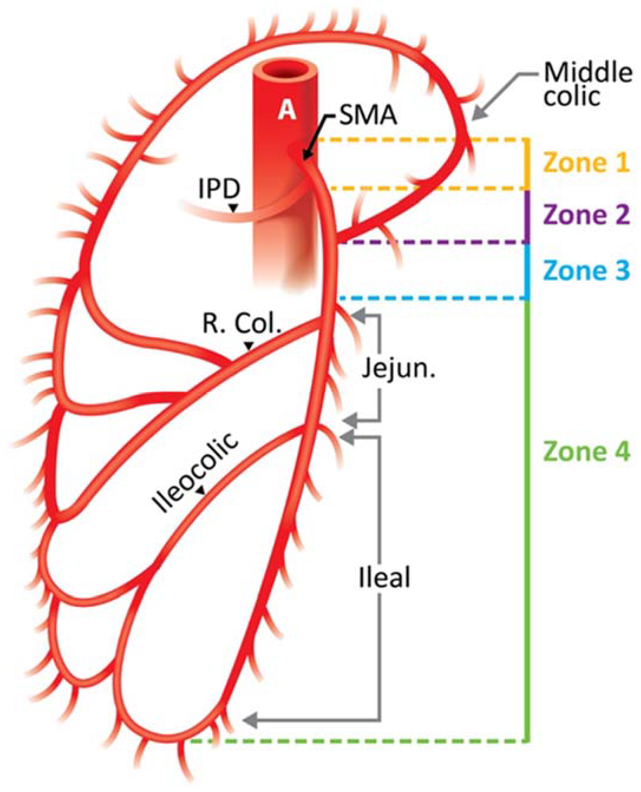
Table 2. Fullen Anatomic Classification of Superior Mesenteric Artery [30].
ZoneSegment of the SMA InvolvedGradeIschemic CategoryBowel Segment Affected
ITrunk proximal to first major branchIMaximalJejunum, ileum, right colon
IITrunk between pancreaticoduodenal and middle colicIIModerateMajor segment, small bowel and/or right colon
IIITrunk distal to middle colicIIIMinimalMinor segment or segments, small bowel, or right colon
IVSegmental branches, jejunal, ileal, or colicIVNoneNo ischemic bowel
Disclaimer/Publisher’s Note: The statements, opinions and data contained in all publications are solely those of the individual author(s) and contributor(s) and not of MDPI and/or the editor(s). MDPI and/or the editor(s) disclaim responsibility for any injury to people or property resulting from any ideas, methods, instructions or products referred to in the content.

