Review

FAPI PET/CT in Diagnostic and Treatment Management of Colorectal Cancer: Review of Current Research Status

Department of Nuclear Medicine, The First Hospital of China Medical University, Shenyang 110001, China
*
Authors to whom correspondence should be addressed.
These authors contributed equally to this work.
J. Clin. Med. 2023, 12(2), 577; https://doi.org/10.3390/jcm12020577
Submission received: 9 December 2022 / Revised: 27 December 2022 / Accepted: 9 January 2023 / Published: 11 January 2023
(This article belongs to the Section Nuclear Medicine & Radiology)

Abstract

:
FAPI PET/CT is a novel imaging tool targeting fibroblast activation protein (FAP), with high tumor uptake rate and low background noise. Therefore, the appearance of FAPI PET/CT provides a good tumor-to-background ratio between tumor and non-tumor tissues, which is beneficial to staging, tumor description and detection. Colorectal cancer has the biological characteristics of high expression of FAP, which provides the foundation for targeted FAP imaging. FAPI PET/CT may have a potential role in changing the staging and re-staging of colorectal cancer, monitoring recurrence and treatment management, and improving the prognosis of patients. This review will summarize the application status of FAPI PET/CT in colorectal cancer and provide directions for further application research.

1. Introduction

Colorectal cancer (CRC) is a malignant tumor with a high incidence and mortality around the world [1], and about 20% of patients have metastasis at the first diagnosis, which seriously endangers human health. Early diagnosis and definite staging can help to prolong the life expectancy of more colorectal cancer patients and reduce unnecessary treatment [2,3]. The application of positron emission tomography can obtain multiple biomarkers in vivo, providing visual evidence for disease management and treatment strategy for colorectal cancer [4,5,6].
As a membrane anchored peptidase highly expressed in cancer-associated fibroblasts (CAFs) in more than 90% of epithelial tumors, fibroblast activating protein (FAP) can lead to the progression of different cancers. The uptake of various tracers targeting FAP-labeled fibroblast-activating protein inhibitors (FAPI) is high in most tumors but low in normal tissues, such as in the brain and abdomen, resulting in a good tumor-to-background ratio (TBR) and good tumor delineation. Compared to FDG PET/CT, FAPI PET/CT has more advantages in detecting primary and metastatic lesions. In addition, targeting FAP imaging may break new ground with its applications, such as non-invasive tumor characterization, tumor staging and tumor treatment effect monitoring [7]. This article reviews the current research status of FAPI PET/CT in diagnosis and management of colorectal cancer (Table 1).

2. Fibroblast Activation Protein (FAP) and FAPI

FAP is an atypical type II transmembrane serine protease with dipeptidyl peptidase (DPP) and endopeptidase activities [8]. FAP is highly expressed on activated fibroblasts, and fibroblasts undergo dimerization and glycosylation when activated [9]. In adult tissues, activated FAP almost only exists in the stroma of wound healing, fibrosis, and malignant tumors [10]. FAP participates in epithelial–mesenchymal transition (EMT) in malignant tumors, promotes the invasion and migration of tumor cells, and ultimately promotes tumor metastasis [11], which indicates that CAFs play an important role in the development of tumors. Compared with normal fibroblasts, the expression of FAP in CAFs has relative specificity. In histopathological studies, FAP-positive CAFs have been found in more than 90% of epithelial tumors, so FAP has become a therapeutic and imaging target for various malignant tumors [12,13,14]. The accuracy and effectiveness of PET probe diagnosis and treatment depend on targeting specificity and tumor-specific uptake [15].
By using chelators to connect FAP inhibitors (FAPI) or FAP antibodies to radionuclides and then performing PET/CT imaging, PET/MR imaging or SPECT imaging, the imaging research of malignant tumors targeting FAP was carried out [16,17,18]. The imaging agent may be used to determine the clinical stages and monitor the recurrence of cancer and may be used as a biomarker or patient selection tool in clinical trials to predict clinical treatment response.

3. Colorectal Cancer and CAFs

Colorectal cancer accounts for about 10% of cancer and cancer-related deaths diagnosed worldwide each year [1,19]. Colorectal cancer is a disease influenced by many factors, such as genetics, the environment and diet. Most colorectal cancer is caused by genetic or epigenetic changes in stem cells or stem cell-like cells, which inactivate tumor suppressor genes and activate oncogenes, and aberrant foci gradually develop into adenoma and cancer [20,21,22].
CRC is a complex tumor which consists of tumor stroma or tumor microenvironments composed of other cells and non-cellular components in addition to tumor cells. Cellular components include immune cells at different activation stages, vascular cells and mesenchymal cells mainly composed of fibroblasts [23,24,25]. In the early stages of CRC, CAFs are activated, accumulate, and exert a strong response to promote the proliferation of connective tissue. Over 90% of the total tumor masses are composed of CAFs and their fibrosis gradually develops into cancer through co-transformation with stem cells. The CAFs are key components of tumor stroma, which can support immune suppression microenvironment; promote the growth, invasion and angiogenesis of colorectal cancer cells; and are closely related to the poor prognosis of patients [26,27,28,29]. Studies have shown that the removal of tumor stroma can improve delivery of therapy or systemically applied radiation and enhance the immune response of the body [30]. Therefore, CAFs play an important role in the occurrence and development of CRC. Because there are many CAFs in the extracellular matrix of colorectal cancer and CAFs already exist in the early stage of colorectal cancer metastasis, it is possible to detect the expression of CAFs in colorectal cancer via special methods so as to determine the tumor size and metastasis of colorectal cancer and provide powerful evidence for determining the early clinical staging, re-staging, and monitoring recurrence of colorectal cancer. In colorectal cancer, FAP is characterized by over-expression in tumor cells and tumor stroma, and its expression level is a sign of the early development of CRC. Increased expression level of FAP is related to the poor prognosis, high grade and invasion of CRC [31,32,33]. Furthermore, FAP is a promising therapeutic target for colorectal cancer. It is reported that colorectal cancer can be treated by 90Y-FAPI46 [34]. Therefore, FAP represents a highly interesting target for CRC diagnosis and treatment applications, FAPI PET/CT targeting FAP can provide imaging basis for CRC staging, re-staging, and monitoring recurrence.

4. FAPI PET/CT Imaging

4.1. FAPI PET/CT Imaging in Normal Biological Organs

Good TBR is an important factor that affects image quality, and it is also an important prerequisite for disease diagnosis. Previous studies have reported the biodistribution of 68Ga-labeled FAPI imaging agents in various normal organs in human body imaging. The results showed that the FAPI tracer rapidly reached its stable physiological biodistribution. Its uptake could be detected as early as 10 min in normal organs, primary tumors and metastatic foci, and the visible image could be maintained for 3 h. The uptake of imaging agent in the liver decreased over time (SUVmax at 10 min vs. SUVmax at 3.3 h: 7.4 vs. 5.0). In contrast, tracer uptake in tumor tissues is very fast, and retention amount is higher than in normal organs (SUVmax at 10 min vs. SUVmax at 3.3 h: 15.5 vs. 13.4). Catherine Meyer et al. observed the highest TBR in bone marrow, with a ratio of 31 at 3.3 h. The ratio of tumor to muscle was 10.7 at 10 min and more than doubled to 22.8 at 3.3 h. At 3.3 h, the next highest TBRs were observed in the heart (19.1), spleen (18.9), and liver (16.8) [35], indicating that the maximum TBR in the lesion steadily increases as background activity decreases. Radioactive tracers in non-target organs are washed out imperceptibly and quickly removed, thus reducing the radiation load in normal organs. Stable clearance from radioactive blood pool and a rapid accumulation of activity were observed in the bladder, and the activity doubled at 60 min, which indicated that the kidney cleared quickly and produced high contrast images [36].
The uptake of FAPI is low in normal tissues. Giesel et al. reported that 68Ga-FAPI uptake was lower than 18F-FDG uptake in brain parenchyma (68Ga-FAPI, 0.09 vs. 18F-FDG, 10.72), oral mucosa (68Ga-FAPI, 2.04 vs. 18F-FDG, 3.33), parotid gland (68Ga-FAPI, 1.71 vs. 18F-FDG, 2.04), myocardial (68Ga-FAPI, 1.50 vs. 18F-FDG, 3.27), blood pool (68Ga-FAPI, 1.81 vs. 18F-FDG, 2.34), liver (68Ga-FAPI, 1.42 vs. 18F-FDG, 3.10) and pancreas (68Ga-FAPI, 1.82 vs. 18F-FDG, 1.99), spleen (68Ga-FAPI, 1.33 vs. 18F-FDG, 2.60), and kidney cortex (68Ga-FAPI, 2.20 vs. 18F-FDG, 2.80) [37]. Because the uptake of FAPI by normal brain and liver tissue is obviously lower than that of FDG, FAPI creates better conditions for imaging brain and liver tumors. Therefore, FAPI may be better at detecting metastatic lesions of malignant tumors (such as brain metastases and liver metastases), and the detection effect of FAPI may be superior to that of 18F-FDG. For the gastrointestinal tract, FAPI did not show obvious absorption (measured in colon transversum: mean SUVmax 1.40 vs. 2.05). When gastrointestinal tumors occur, good contrast is more conducive to the description of malignant tumors [37,38].
Because of its low intake and high contrast in normal tissues, FAPI PET/CT can show the pathological areas characterized by fibrosis well. FAPI PET/CT may have unique advantages in tumor or non-tumor lesions with fibrosis occurring in brain parenchyma, heart, spleen, liver and pancreas tissue, but further studies are necessary to prove it.

4.2. FAPI PET/CT Features of Primary and Metastatic Colorectal Cancer

According to a relevant study [39], the SUVmax, SUVmean and FAP immunohistochemistry scores of 68GA-FAPI-46 were higher in various cancer tissues than in adjacent non-cancer tissues: the SUVmax (7.7 vs. 1.6); the SUVmean (6.2 vs. 1.0); the FAP immunohistochemistry score (2.8 vs. 0.9). The cancer types with the highest uptake rates were pancreas, stomach, colon, and uterus. FAP immunohistochemistry score was positively correlated with 68Ga-FAPI-46 SUVmax (r = 0.781 [95% CI, 0.376–0.936], p < 0.001) and SUVmean (r = 0.783 [95% CI, 0.379–0.936], p < 0.001) in cancer tissues [39]. In addition, studies have shown that the expression level of FAP in CRC is positively correlated with the consensus molecular subtype 4 (CMS4) of treatment resistance and poor prognosis factor, and FAPI PET/CT is valuable for comprehensive evaluation of CMS4 in CRC [40]. Therefore, 68Ga-FAPI-46 shows high metabolic uptake in tumor tissues, which can reflect the expression quantity of FAP in tumor lesions and provides a new means and method for non-invasive imaging and targeted FAP therapy of solid tumors. In a study on 68Ga-FAPI uptake of 28 different cancers, it was found that colorectal cancer had intermediate FAPI uptake, and SUVmax was between 6 and 12 [41]. A 56-year-old sigmoid adenocarcinoma patient (ybT4bN0Mo) underwent 68Ga-FAPI-46 PET/CT imaging before operation, and immunohistochemical staining was performed after operation. The results showed that the area corresponding to the resected tumor showed intense uptake of 68Ga-FAPI-46 (SUVmax, 15.9; SUVmean, 12.8), which was consistent with the results of immunohistochemistry and shows that FAP is strongly expressed in the stroma inside and around the lesions [39] (Figure 1).
The liver is the primary target organ for colorectal cancer metastasis. Because of its low background degree of FAPI, the liver has a good TBR, which may be helpful to detect liver metastases [38]. The existence of liver metastases is very important for the treatment plan and survival rate of colorectal cancer [42,43]. As early as 1994, FAPI was recognized as a potential molecular probe target, and the TBR can reach 21:1, which is more favorable for the detection of occult liver metastases. At that time, antibody 131I-mAb-F19 was used to detect liver metastasis of colorectal cancer less than 1 cm [44]. This means that tumor mesenchymal cells with high expression of FAP exist in early liver metastases of colorectal cancer and can be detected by FAPI PET/CT, which may make up for the lack of obvious FDG uptake caused by the small number of tumor cells in early liver metastasis. In the study of monitoring liver metastasis of colorectal cancer, it was found that 68Ga-FAPI PET/CT could detect more true positive patients (12 cases vs. 10 cases) than 18F-FDG PET/CT, and their sensitivities to liver metastasis were 96.6% and 70.8%, respectively [45]. Clemens Kratochwil et al. used PET/CT to detect high FAPI uptake of less than 1 cm in the liver and lung, and the diagnosis of metastatic colon cancer was confirmed [41]. A study of 28 cases of malignant tumors in the lower digestive tract showed for the first time that FAPI showed high uptake and good TBR in primary and metastatic colorectal cancer lesions, which further changed the clinical stage of TNM in related patients and improved target volume delineation [46]. For the lymph node metastasis of colorectal cancer, the present research shows that there are many stroma in the lymph nodes and corresponding lymph node metastases (LNM) of CRC, which indicates that FAPI PET/CT may improve the identification of lymph node metastases of CRC [47].
68Ga-FAPI PET/CT showed intermediate 68Ga-FAPI uptake in primary colorectal cancer lesions (SUVmax, 8.6). The SUVmax and SUVmean of FAPI in metastatic lesions were 7.95 (±3.49) and 3.96 (±1.92), respectively. At the same time, the activity of background and normal organs was very low (muscle, SUVmax:1.60 and SUVmean: 1.11; blood pool, SUVmax:1.85 and SUVmean: 1.21; normal liver parenchyma: SUVmax: 1.46 and SUVmean: 0.84), resulting in a TBR greater than 3, which was statistically significant compared to tumor activity (p ≤ 0.002 each) [46]. In this study, FAPI PET/CT showed a good application prospect in most primary and metastatic colorectal cancer (especially lymph node and liver metastasis). Compared to FDG PET/CT, the SUVmax of FAPI was higher, which was more beneficial to the discovery of lesions (Table 2).

4.3. FAPI vs. FDG Imaging in Colorectal Cancer

At present, 18F-FDG PET/CT is widely used in the disease management and treatment strategy of colorectal cancer to monitor metastasis [48,49,50]. Glucose metabolism is a key factor in the development and progress of cancer, and the uptake of 18F-FDG is related to glucose metabolism [51]. However, with the development of targeted medicine and the increase of knowledge about the biology of colorectal cancer, it has been found that tumor heterogeneity leads to more aggressive tumor growth and negatively affects treatment response and patient survival [52]. Meanwhile, the existence of tumor heterogeneity may affect the glucose consumption and the uptake of 18F-FDG [53]. In addition, 18F-FDG PET/CT also has certain limitations. Glucose metabolism is characteristic of many diseases, resulting in poor specificity and organ physiological distribution of 18F-FDG (eg. in the liver), resulting in low TBR of the image, which affects the diagnosis. Because colorectal cancer is prone to liver metastasis and metastatic lesions may be heterogeneous, FDG may be restricted in detecting colorectal cancer metastatic lesions, especially liver metastasis. The emergence of FAPI PET/CT may remedy the defect of FDG PET/CT. The uptake of FAPI PET/CT in normal brain, liver and oral mucosa is low and has a better diagnostic effect and lower background noise when monitoring primary and/or metastatic lesions [54,55,56,57]. FAPI PET/CT may provide superior visualization for colorectal, liver, and breast cancers, among others.
68Ga-FAPI PET/CT is a potential examination tool for disease management and treatment strategies of cancer. Compared to FDG, FAPI PET was more accurate for the overall staging and restaging, and FAPI PET altered clinical management in more than 20% of patients [58]. In the study of primary and recurrent gastrointestinal tumors, the detection rate of 68Ga-FAPI PET/CT for primary tumors was higher than that of 18F-FDG PET/CT, and the detection rates were 100% and 53% respectively (11 cases of gastric cancers and 9 cases of colorectal cancers). In addition, 68Ga-FAPI PET/CT showed that the tumor outline was clearer and the contrast of tumor background was higher. It was found that the lesions of primary colorectal cancer showed high uptake in 18F-FDG PET/CT and 68Ga-FAPI PET/CT, but the semi-quantitative parameter SUVmax was found in the analysis. The SUVmax of 68Ga-FAPI in these primary lesions was higher than that of 18F-FDG (15.9 vs. 7.9). In the assessment of recurrence, 68Ga-FAPI PET/CT had the higher true positive rates than that of 18F-FDG PET/CT (100% vs. 44%). On the contrary, the false positive rates of 68Ga-FAPI and 18F-FDG PET/CT for recurrence were different (11% vs. 2%). As a result, the specificity of 68Ga-FAPI was lower than that of 18F-FDG for patients in whom recurrence was detected (67% vs. 94%). In the evaluation of lymph node metastasis and organ metastasis of colorectal cancer, 68Ga-FAPI PET/CT detected more suspicious lymph nodes and organs than 18F-FDG PET/CT, and the uptake of 68Ga-FAPI was higher than 18F-FDG, showing high sensitivity (79% vs. 54%, respectively), while the specificity of 68Ga-FAPI PET/CT is not higher than that of 18F-FDG PET/CT (82% vs. 89%) [59] (Figure 2). 68Ga-FAPI PET/CT is obviously superior to 18F-FDG PET/CT in the display and image quality of peritoneal metastases of colorectal cancer (Table 3), and the average SUVmax value is obviously higher [60]. Moreover, in most studies, FAPI PET/CT is superior to FDG PET/CT in sensitivity and specificity for primary and metastatic colorectal cancer (especially lymph nodes and liver metastases), but the number of related studies and included patients is still relatively small. Further research will further understand the diagnostic performance of FAPI PET/CT for better clinical application.
A 23-year-old female patient, 68Ga-FAPI-04 PET/CT showed that 68Ga-FAPI-04 was intensely taken up by lesion in the sigmoid colon, the lymph nodes in the retroperitoneum and the left supraclavicular fossa, and the pelvic abdominal peritoneum. Pathologically, it was confirmed as signet ring cell carcinoma of the sigmoid colon with peritoneal metastasis. The lesions were delineated more clearly by 68Ga-FAPI-04 scan, and more lesions were detected comparing to 18F-FDG scan [61]. Due to liver physiological distribution of 18F-FDG, 18F-FDG PET/CT may not be able to detect metastatic tumors or small metastatic tumors with low FDG uptake.
For the detection of colorectal cancer recurrence, compared to FDG, 68Ga-FAPI-04 PET/CT was found to have advantages in detecting recurrent signet ring cell colon cancer. A 36-year-old woman after surgery for left colon cancer, 18F-FDG PET/CT showed mild heterogeneous FDG uptake in the left perirenal soft tissue density lesions, with an SUVmax of 2.2, and an intense FAPI uptake in 68Ga-FAPI-04 PET/CT with an SUVmax of 12.8 [62].
Previous studies have confirmed that FAPI PET/CT detected more suspicious lymph nodes and organs than FDG PET/CT, which was more sensitive for identifying metastasis and recurrence in colorectal cancer, especially lymph node metastasis and organ metastasis. Based on a small sample study, 68Ga-FAPI PET/CT changed tumor or radiation tumor management in approximately 73.3% (11/15) of patients with metastasis [46]. However, the number of patients included in most current studies was relatively small, and most of them were in the advanced clinical stage. For advanced patients, even if FAPI PET/CT shows more lesions, the clinical stage will not change, so the therapeutical plan will remain the same. In terms of re-staging, FAPI PET/CT has more advantage than 18F-FDG, which provides key evidence for changing the clinical management of patients. This indicates that FAPI PET/CT may be more suitable for monitoring recurrence and metastasis of colorectal cancer patients and evaluating the therapeutical results. Therefore, it may be more meaningful for FAPI PET/CT to guide therapeutical decision-making and patient management to include more patients with lower clinical stage or monitor patients with recurrent colorectal cancer in future studies [58]. In addition, the physiological uptake of 18F-FDG in the liver restricts the application of FDG PET/CT in the detection of liver metastasis of colorectal cancer [63]. Encouragingly, FAPI PET/CT could help to improve the depiction of liver metastasis as a result of the clear tumor delineation and high TBR.
Compared to FDG, FAPI PET/CT has advantages in detecting colorectal cancer metastasis, even in small metastatic lesions. However, the diagnostic performance of FAPI PET/CT may be exaggerated due to the deviation of patient distribution. Therefore, appropriate imaging agents should be selected according to the patient’s conditions. The combination of two imaging agents can provide better information for patients help guide follow-up clinical oncologic management.

5. Limitations of FAPI PET/CT Imaging

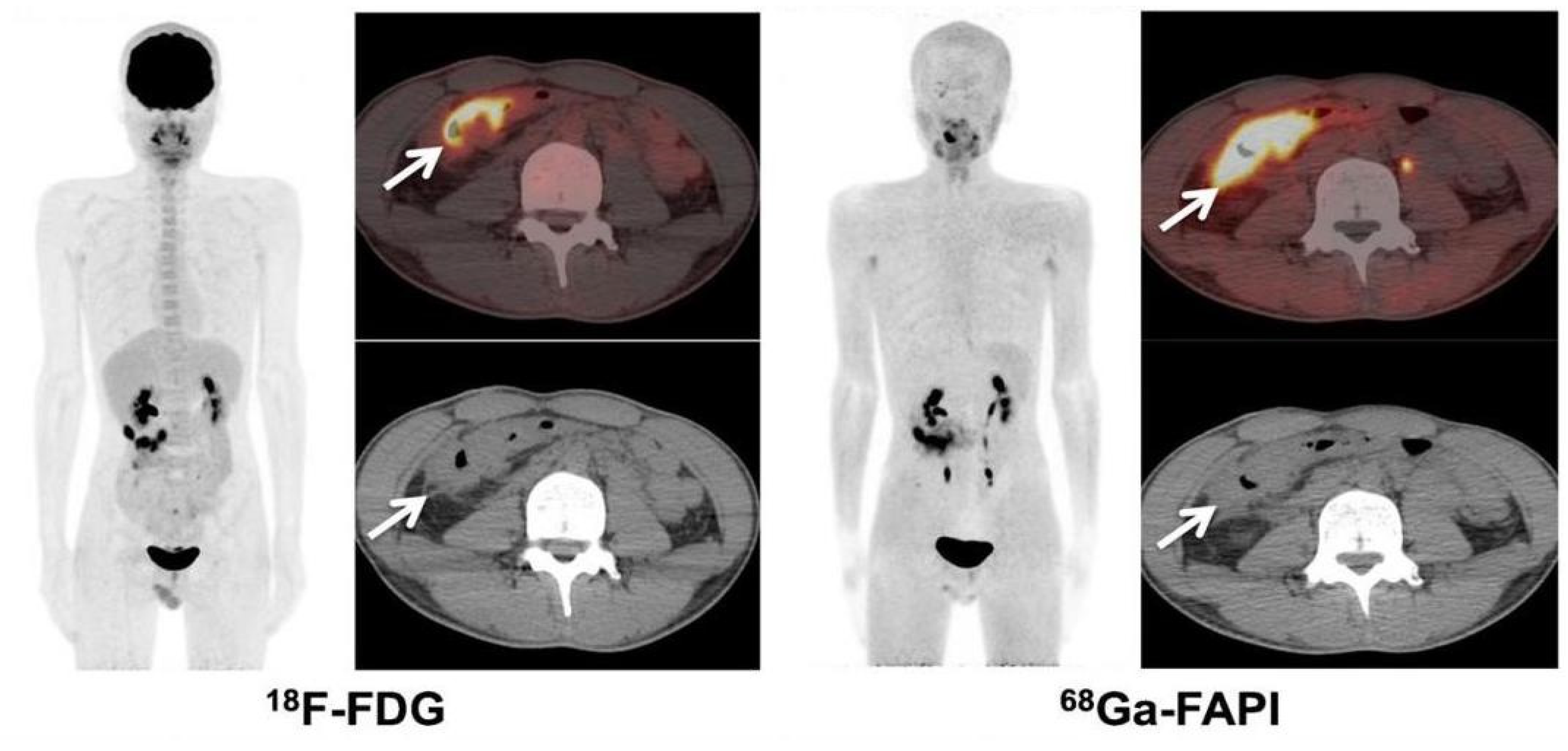
Research indicates that FAPI PET/CT may be a promising imaging method of improving the staging and treatment of colorectal cancer. Compared to FDG PET/CT, FAPI PET/CT has higher sensitivity in diagnosing the primary lesions of colorectal cancer and recurrence of colorectal cancer. However, it is also necessary to consider FAPI positive uptake due to inflammation-induced nonspecific fibrosis [64,65,66,67,68,69], which must be carefully distinguished. A recent study showed that FAP was also positive in Crohn’s disease [70]. A 28-year-old male with an 8-year history of Crohn’s disease underwent 68Ga-FAPI PET/CT imaging, which showed that there was an intense 68Ga-FAPI uptake in the diseased intestine (SUVmax, 14.1) [71]. However, in another patient with ulcerative colitis reported in this paper, there was only a phased increase in 18F-FDG intake consistent with the severity of the disease but no 68Ga-FAPI intake (Figure 3). This shows that we may obtain more and more valuable information on pathological tissue by combining different molecular probes, such as FDG, and analyzing the nature of pathological changes from multiple angles by integrating different information. Another 28-year-old woman with abdominal pain, intestinal dysfunction and weight loss for 3 months, 68Ga-FAPI PET/CT showed intense 68Ga-FAPI uptake in the transverse colon (SUVmax, 18.2) and a large number of abnormal lesions in the peritoneum as well as abnormal FAPI uptake (SUVmax, 10.6). Finally, this patient underwent colonic mucosa biopsy via endoscopy. The biopsy results showed granulomatous inflammation as well as acid-fast bacilli. The TSPOT.TB test (+). As a result, the patient suffers from intestinal tuberculosis [72]. These cases indicate that inflammatory enteritis may also be related to intense FAPI intake, which should be differentiated from colorectal cancer. Perhaps combined with the patient’s clinical symptoms and relevant medical history or relevant examination information, it can help us distinguish the nature of lesions with high FAPI uptake. As FAP is expressed on activated fibroblasts, when organ fibrosis occurs, the expression of FAP will increase and it will show high uptake in the subsequent FAPI PET/CT, which may be one of the reasons for false positive of FAPI PET/CT. Therefore, further exploring the FAPI PET/CT features of benign lesions characterized by fibrosis, combined with imaging information such as FDG PET/CT, may further improve the accuracy of FAPI PET/CT in the diagnosis of malignant tumors.

6. Discussion

Colorectal cancer is one of the main diseases that harm human health. Compared with FDG, FAPI PET/CT has advantages in detecting colorectal cancer metastasis, even in small metastatic lesions. However, the diagnostic performance of FAPI PET/CT may be exaggerated due to the deviation of patient distribution. However, FAPI PET/CT still provides a new perspective for the diagnosis and treatment of colorectal cancer.
Compared with FDG PET/CT, FAPI PET/CT has advantages in the diagnosis of primary tumors, various metastatic and recurrent lesions of colorectal cancer, providing important molecular imaging information for early diagnosis, accurate staging, guiding therapy, curative effect monitoring and recurrence evaluation. In addition, FAPI PET/CT may have special significance for delineating the target area of tumor radiotherapy. FDG PET mainly depends on the glucose metabolism of tumor cells to display the metabolic information of tumors and describes the shape of tumor by combining with the structural information of CT images. In solid tumors, apart from tumor cells, there is also a large amount of extracellular matrix. FAPI PET/CT can display the extracellular matrix by targeting fibroblasts in the tumor microenvironment, which may have special advantages in the morphological description of solid tumors. Therefore, the combination of FDG PET/CT and FAPI PET/CT image information may provide more useful information on tumor morphology and metabolism.
At present, the clinical studies on FAPI PET/CT are still in the early stage, and most of them are small samples lacking long-term follow-up data, and false positive results caused by inflammation may restrict the application of FAPI PET/CT. These problems also require systematic clinical research on a large number of patients. At present, immunohistochemical staining has been used to correlate FAP expression with FAPI uptake in lesions, trying to further clarify their internal relationship, but related research is still limited, especially on the difference in FAPI uptake between malignant and non-malignant diseases. At the same time, because of the low expression of FAP in normal tissues, the strategy of targeting FAP has obvious advantages in tumor treatment, which is worth further study.

7. Conclusions and Prospect

FAPI PET/CT, as a promising new PET imaging tool, may play an important role in tumor visualization and contribute to the diagnosis and staging of tumors. It is undeniable that FAPI PET/CT plays a unique role in the treatment of solid tumors, including colorectal cancer. Unfortunately, the exact mechanism and expression pattern of FAP in some diseases remain obscure, and the number of clinical studies on FAPI PET/CT is still small. Further research, including on its mechanism of action and more and larger-scale clinical studies, will help to clarify the true value of FAPI PET/CT in tumor and non-tumor diseases, which is of great significance to improving the diagnosis and treatment level. Although the research on FAPI PET/CT for colorectal cancer is still in the initial stage, we believe that with the deepening of research and the development of related technologies, FAPI PET/CT will be widely used in clinical applications.

Author Contributions

Writing—original draft preparation, Z.C. and S.W.; writing—review and editing, X.L. and Y.L.; investigation, S.X. and B.D. All authors have read and agreed to the published version of the manuscript.

Funding

This research was funded by National Natural Science Foundation of China, grant number 81971652, 82272043 and 82171987.

Institutional Review Board Statement

Not applicable.

Informed Consent Statement

Not applicable.

Data Availability Statement

Not applicable.

Conflicts of Interest

The authors declare no conflict of interest.

References

  1. Siegel, R.L.; Miller, K.D.; Fuchs, H.E.; Jemal, A. Cancer statistics, 2022. CA Cancer J. Clin. 2022, 72, 7–33. [Google Scholar] [CrossRef]
  2. Biller, L.H.; Schrag, D. Diagnosis and Treatment of Metastatic Colorectal Cancer: A Review. JAMA 2021, 325, 669–685. [Google Scholar] [CrossRef]
  3. Provenzale, D.; Ness, R.M.; Llor, X.; Weiss, J.M.; Abbadessa, B.; Cooper, G.; Early, D.S.; Friedman, M.; Giardiello, F.M.; Glaser, K.; et al. NCCN Guidelines Insights: Colorectal Cancer Screening, Version 2.2020. J. Natl. Compr. Cancer Netw. 2020, 18, 1312–1320. [Google Scholar] [CrossRef]
  4. Bae, S.U.; Won, K.S.; Song, B.I.; Jeong, W.K.; Baek, S.K.; Kim, H.W. Accuracy of F-18 FDG PET/CT with optimal cut-offs of maximum standardized uptake value according to size for diagnosis of regional lymph node metastasis in patients with rectal cancer. Cancer Imaging 2018, 18, 32. [Google Scholar] [CrossRef]
  5. Furtado, F.S.; Suarez-Weiss, K.E.; Vangelm, M.; Clark, J.W.; Cusack, J.C.; Hong, T.; Blaszkowsky, L.; Wo, J.; Striar, R.; Umutlu, L.; et al. Clinical impact of PET/MRI in oligometastatic colorectal cancer. Br. J. Cancer 2021, 125, 975–982. [Google Scholar] [CrossRef]
  6. Dagallier, C.; Avry, F.; Touchefeu, Y.; Buron, F.; Routier, S.; Chérel, M.; Arlicot, N. Development of PET Radioligands Targeting COX-2 for Colorectal Cancer Staging, a Review of in vitro and Preclinical Imaging Studies. Front. Med. 2021, 8, 675209. [Google Scholar] [CrossRef]
  7. Yang, T.; Ma, L.; Hou, H.; Gao, F.; Tao, W. FAPI PET/CT in the Diagnosis of Abdominal and Pelvic Tumors. Front. Oncol. 2021, 11, 797960. [Google Scholar] [CrossRef]
  8. Puré, E.; Blomberg, R. Pro-tumorigenic roles of fibroblast activation protein in cancer: Back to the basics. Oncogene 2018, 37, 4343–4357. [Google Scholar] [CrossRef]
  9. Lindner, T.; Giesel, F.L.; Kratochwil, C.; Serfling, S.E. Radioligands Targeting Fibroblast Activation Protein (FAP). Cancers 2021, 13, 5744. [Google Scholar] [CrossRef]
  10. Dendl, K.; Koerber, S.A.; Kratochwil, C.; Cardinale, J.; Finck, R.; Dabir, M.; Novruzov, E.; Watabe, T.; Kramer, V.; Choyke, P.L.; et al. FAP and FAPI-PET/CT in Malignant and Non-Malignant Diseases: A Perfect Symbiosis? Cancers 2021, 13, 4946. [Google Scholar] [CrossRef]
  11. Fitzgerald, A.A.; Weiner, L.M. The role of fibroblast activation protein in health and malignancy. Cancer Metastasis Rev. 2020, 39, 783–803. [Google Scholar] [CrossRef]
  12. Langbein, T.; Weber, W.A.; Eiber, M. Future of Theranostics: An Outlook on Precision Oncology in Nuclear Medicine. J. Nucl. Med. 2019, 60, 13S–19S. [Google Scholar] [CrossRef] [PubMed] [Green Version]
  13. Sollini, M.; Kirienko, M.; Gelardi, F.; Fiz, F.; Gozzi, N.; Chiti, A. State-of-the-art of FAPI-PET imaging: A systematic review and meta-analysis. Eur. J. Nucl. Med. Mol. Imaging 2021, 48, 4396–4414. [Google Scholar] [CrossRef] [PubMed]
  14. Baum, R.P.; Schuchardt, C.; Singh, A.; Chantadisai, M.; Robiller, F.C.; Zhang, J.; Mueller, D.; Eismant, A.; Almaguel, F.; Zboralski, D.; et al. Feasibility, Biodistribution, and Preliminary Dosimetry in Peptide-Targeted Radionuclide Therapy of Diverse Adenocarcinomas Using 177Lu-FAP-2286: First-in-Humans Results. J. Nucl. Med. 2022, 63, 415–423. [Google Scholar] [CrossRef]
  15. Calais, J. FAP: The Next Billion Dollar Nuclear Theranostics Target? J. Nucl. Med. 2020, 61, 163–165. [Google Scholar] [CrossRef]
  16. Huang, R.; Pu, Y.; Huang, S.; Yang, C.; Yang, F.; Pu, Y.; Li, J.; Chen, L.; Huang, Y. FAPI-PET/CT in Cancer Imaging: A Potential Novel Molecule of the Century. Front. Oncol. 2022, 12, 854658. [Google Scholar] [CrossRef]
  17. Lindner, T.; Loktev, A.; Altmann, A.; Giesel, F.; Kratochwil, C.; Debus, J.; Jäger, D.; Mier, W.; Haberkorn, U. Development of Quinoline-Based Theranostic Ligands for the Targeting of Fibroblast Activation Protein. J. Nucl. Med. 2018, 59, 1415–1422. [Google Scholar] [CrossRef] [Green Version]
  18. Loktev, A.; Lindner, T.; Mier, W.; Debus, J.; Altmann, A.; Jäger, D.; Giesel, F.; Kratochwil, C.; Barthe, P.; Roumestand, C.; et al. A Tumor-Imaging Method Targeting Cancer-Associated Fibroblasts. J. Nucl. Med. 2018, 59, 1423–1429. [Google Scholar] [CrossRef] [Green Version]
  19. Dekker, E.; Tanis, P.J.; Vleugels, J.L.A.; Kasi, P.M.; Wallace, M.B. Colorectal cancer. Lancet 2019, 394, 1467–1480. [Google Scholar] [CrossRef]
  20. Li, J.; Ma, X.; Chakravarti, D.; Shalapour, S.; DePinho, R.A. Genetic and biological hallmarks of colorectal cancer. Genes Dev. 2021, 35, 787–820. [Google Scholar] [CrossRef]
  21. Frank, M.H.; Wilson, B.J.; Gold, J.S.; Frank, N.Y. Clinical Implications of Colorectal Cancer Stem Cells in the Age of Single-Cell Omics and Targeted Therapies. Gastroenterology 2021, 160, 1947–1960. [Google Scholar] [CrossRef] [PubMed]
  22. De Angelis, M.L.; Francescangeli, F.; Zeuner, A.; Baiocchi, M. Colorectal Cancer Stem Cells: An Overview of Evolving Methods and Concepts. Cancers 2021, 13, 5910. [Google Scholar] [CrossRef] [PubMed]
  23. Mao, X.; Xu, J.; Wang, W.; Liang, C.; Hua, J.; Liu, J.; Zhang, B.; Meng, Q.; Yu, X.; Shi, S. Crosstalk between cancer-associated fibroblasts and immune cells in the tumor microenvironment: New findings and future perspectives. Mol. Cancer 2021, 20, 131. [Google Scholar] [CrossRef]
  24. Mhaidly, R.; Mechta-Grigoriou, F. Fibroblast heterogeneity in tumor micro-environment: Role in immunosuppression and new therapies. Semin. Immunol. 2020, 48, 101417. [Google Scholar] [CrossRef]
  25. Chen, Y.; McAndrews, K.M.; Kalluri, R. Clinical and therapeutic relevance of cancer-associated fibroblasts. Nat. Rev. Clin. Oncol 2021, 18, 792–804. [Google Scholar] [CrossRef]
  26. Ganguly, D.; Chandra, R.; Karalis, J.; Teke, M.; Aguilera, T.; Maddipati, R.; Wachsmann, M.B.; Ghersi, D.; Siravegna, G.; Zeh, H.J., 3rd; et al. Cancer-Associated Fibroblasts: Versatile Players in the Tumor Microenvironment. Cancers 2020, 12, 2652. [Google Scholar] [CrossRef] [PubMed]
  27. Zhang, N.; Ng, A.S.; Cai, S.; Li, Q.; Yang, L.; Kerr, D. Novel therapeutic strategies: Targeting epithelial-mesenchymal transition in colorectal cancer. Lancet Oncol. 2021, 22, e358–e368. [Google Scholar] [CrossRef]
  28. Siveke, J.T. Fibroblast-Activating Protein: Targeting the Roots of the Tumor Microenvironment. J. Nucl. Med. 2018, 59, 1412–1414. [Google Scholar] [CrossRef]
  29. Glabman, R.A.; Choyke, P.L.; Sato, N. Cancer-Associated Fibroblasts: Tumorigenicity and Targeting for Cancer Therapy. Cancers 2022, 14, 3906. [Google Scholar] [CrossRef]
  30. Barsoumian, H.B.; Ramapriyan, R.; Younes, A.I.; Caetano, M.S.; Menon, H.; Comeaux, N.I.; Cushman, T.R.; Schoenhals, J.E.; Cadena, A.P.; Reilly, T.P.; et al. Low-dose radiation treatment enhances systemic antitumor immune responses by overcoming the inhibitory stroma. J. Immunother. Cancer 2020, 8, e000537. [Google Scholar] [CrossRef]
  31. Miyashita, N.; Saito, A. Organ Specificity and Heterogeneity of Cancer-Associated Fibroblasts in Colorectal Cancer. Int. J. Mol. Sci. 2021, 22, 10973. [Google Scholar] [CrossRef] [PubMed]
  32. Fotsitzoudis, C.; Koulouridi, A.; Messaritakis, I.; Konstantinidis, T.; Gouvas, N.; Tsiaoussis, J.; Souglakos, J. Cancer-Associated Fibroblasts: The Origin, Biological Characteristics and Role in Cancer-A Glance on Colorectal Cancer. Cancers 2022, 14, 4394. [Google Scholar] [CrossRef] [PubMed]
  33. Sandberg, T.P.; Stuart, M.P.M.E.; Oosting, J.; Tollenaar, R.A.E.M.; Sier, C.F.M.; Mesker, W.E. Increased expression of cancer-associated fibroblast markers at the invasive front and its association with tumor-stroma ratio in colorectal cancer. BMC Cancer 2019, 19, 284. [Google Scholar] [CrossRef] [Green Version]
  34. Rathke, H.; Fuxius, S.; Giesel, F.L.; Lindner, T.; Debus, J.; Haberkorn, U.; Kratochwil, C. Two Tumors, One Target: Preliminary Experience With 90Y-FAPI Therapy in a Patient With Metastasized Breast and Colorectal Cancer. Clin. Nucl. Med. 2021, 46, 842–844. [Google Scholar] [CrossRef]
  35. Meyer, C.; Dahlbom, M.; Lindner, T.; Vauclin, S.; Mona, C.; Slavik, R.; Czernin, J.; Haberkorn, U.; Calais, J. Radiation Dosimetry and Biodistribution of 68Ga-FAPI-46 PET Imaging in Cancer Patients. J. Nucl. Med. 2020, 61, 1171–1177. [Google Scholar] [CrossRef]
  36. Ballal, S.; Yadav, M.P.; Moon, E.S.; Kramer, V.S.; Roesch, F.; Kumari, S.; Tripathi, M.; ArunRaj, S.T.; Sarswat, S.; Bal, C. Biodistribution, pharmacokinetics, dosimetry of [68Ga]Ga-DOTA.SA.FAPi, and the head-to-head comparison with [18F]F-FDG PET/CT in patients with various cancers. Eur. J. Nucl. Med. Mol. Imaging 2021, 48, 1915–1931. [Google Scholar] [CrossRef]
  37. Giesel, F.L.; Kratochwil, C.; Schlittenhardt, J.; Dendl, K.; Eiber, M.; Staudinger, F.; Kessler, L.; Fendler, W.P.; Lindner, T.; Koerber, S.A.; et al. Head-to-head intra-individual comparison of biodistribution and tumor uptake of 68Ga-FAPI and 18F-FDG PET/CT in cancer patients. Eur. J. Nucl. Med. Mol. Imaging 2021, 48, 4377–4385. [Google Scholar] [CrossRef]
  38. Giesel, F.L.; Kratochwil, C.; Lindner, T.; Marschalek, M.M.; Loktev, A.; Lehnert, W.; Debus, J.; Jäger, D.; Flechsig, P.; Altmann, A.; et al. 68Ga-FAPI PET/CT: Biodist.tribution and Preliminary Dosimetry Estimate of 2 DOTA-Containing FAP-Targeting Agents in Patients with Various Cancers. J. Nucl. Med. 2019, 60, 386–392. [Google Scholar] [CrossRef] [Green Version]
  39. Mona, C.E.; Benz, M.R.; Hikmat, F.; Grogan, T.R.; Lueckerath, K.; Razmaria, A.; Riahi, R.; Slavik, R.; Girgis, M.D.; Carlucci, G.; et al. Correlation of 68Ga-FAPi-46 PET Biodistribution with FAP Expression by Immunohistochemistry in Patients with Solid Cancers: Interim Analysis of a Prospective Translational Exploratory Study. J. Nucl. Med. 2022, 63, 1021–1026. [Google Scholar] [CrossRef]
  40. Strating, E.; Wassenaar, E.; Verhagen, M.; Rauwerdink, P.; van Schelven, S.; de Hingh, I.; Rinkes, I.B.; Boerma, D.; Witkamp, A.; Lacle, M.; et al. Fibroblast activation protein identifies Consensus Molecular Subtype 4 in colorectal cancer and allows its detection by 68Ga-FAPI-PET imaging. Br. J. Cancer 2022, 127, 145–155. [Google Scholar] [CrossRef]
  41. Kratochwil, C.; Flechsig, P.; Lindner, T.; Abderrahim, L.; Altmann, A.; Mier, W.; Adeberg, S.; Rathke, H.; Röhrich, M.; Winter, H.; et al. 68Ga FAPI PET/CT: Tracer Uptake in 28 Different Kinds of Cancer. J. Nucl. Med. 2019, 60, 801–805. [Google Scholar] [CrossRef] [Green Version]
  42. Wu, Y.; Yang, S.; Ma, J.; Chen, Z.; Song, G.; Rao, D.; Cheng, Y.; Huang, S.; Liu, Y.; Jiang, S.; et al. Spatiotemporal Immune Landscape of Colorectal Cancer Liver Metastasis at Single-Cell Level. Cancer Discov. 2022, 12, 134–153. [Google Scholar] [CrossRef]
  43. Zeng, X.; Ward, S.E.; Zhou, J.; Cheng, A.S.L. Liver Immune Microenvironment and Metastasis from Colorectal Cancer-Pathogenesis and Therapeutic Perspectives. Cancers 2021, 13, 2418. [Google Scholar] [CrossRef]
  44. Welt, S.; Divgi, C.R.; Scott, A.M.; Garin-Chesa, P.; Finn, R.D.; Graham, M.; Carswell, E.A.; Cohen, A.; Larson, S.M.; Old, L.J.; et al. Antibody targeting in metastatic colon cancer: A phase I study of monoclonal antibody F19 against a cell-surface protein of reactive tumor stromal fibroblasts. J. Clin. Oncol. 1994, 12, 1193–1203. [Google Scholar] [CrossRef]
  45. Şahin, E.; Elboğa, U.; Çelen, Y.Z.; Sever, Ö.N.; Çayırlı, Y.B.; Çimen, U. Comparison of 68Ga-DOTA-FAPI and 18FDG PET/CT imaging modalities in the detection of liver metastases in patients with gastrointestinal system cancer. Eur. J. Radiol. 2021, 142, 109867. [Google Scholar] [CrossRef]
  46. Koerber, S.A.; Staudinger, F.; Kratochwil, C.; Adeberg, S.; Haefner, M.F.; Ungerechts, G.; Rathke, H.; Winter, E.; Lindner, T.; Syed, M.; et al. The Role of 68Ga-FAPI PET/CT for Patients with Malignancies of the Lower Gastrointestinal Tract: First Clinical Experience. J. Nucl. Med. 2020, 61, 1331–1336. [Google Scholar] [CrossRef]
  47. Polack, M.; Hagenaars, S.C.; Couwenberg, A.; Kool, W.; Tollenaar, R.A.E.M.; Vogel, W.V.; Snaebjornsson, P.; Mesker, W.E. Characteristics of tumor stroma in regional lymph node metastases in colorectal cancer patients: A theoretical framework for future diagnostic imaging with FAPI PET/CT. Clin. Transl. Oncol. 2022, 24, 1776–1784. [Google Scholar] [CrossRef]
  48. Chen, S.H.; Miles, K.; Taylor, S.A.; Ganeshan, B.; Rodriquez, M.; Fraioli, F.; Wan, S.; Afaq, A.; Shortman, R.; Walls, D.; et al. FDG-PET/CT in colorectal cancer: Potential for vascular-metabolic imaging to provide markers of prognosis. Eur. J. Nucl. Med. Mol. Imaging 2021, 49, 371–384. [Google Scholar] [CrossRef]
  49. Liu, H.; Ye, Z.; Yang, T.; Xie, H.; Duan, T.; Li, M.; Wu, M.; Song, B. Predictive Value of Metabolic Parameters Derived From 18F-FDG PET/CT for Microsatellite Instability in Patients with Colorectal Carcinoma. Front. Immunol. 2021, 12, 724464. [Google Scholar] [CrossRef]
  50. van Helden, E.J.; Vacher, Y.J.L.; van Wieringen, W.N.; van Velden, F.H.P.; Verheul, H.M.W.; Hoekstra, O.S.; Boellaard, R.; Menke-van der Houven van Oordt, C.W. Radiomics analysis of pre-treatment [18F]FDG PET/CT for patients with metastatic colorectal cancer undergoing palliative systemic treatment. Eur. J. Nucl. Med. Mol. Imaging 2018, 45, 2307–2317. [Google Scholar] [CrossRef]
  51. Dendl, K.; Koerber, S.A.; Tamburini, K.; Mori, Y.; Cardinale, J.; Haberkorn, U.; Giesel, F.L. Advancement and Future Perspective of FAPI PET/CT In Gynecological Malignancies. Semin. Nucl. Med. 2022, 52, 628–634. [Google Scholar] [CrossRef]
  52. Zygulska, A.L.; Pierzchalski, P. Novel Diagnostic Biomarkers in Colorectal Cancer. Int. J. Mol. Sci. 2022, 23, 852. [Google Scholar] [CrossRef]
  53. Liu, X.; Zhang, Y.F.; Shi, Q.; Yang, Y.; Yao, B.H.; Wang, S.C.; Geng, G.Y. Prediction value of 18F-FDG PET/CT intratumor metabolic heterogeneity parameters for recurrence after radical surgery of stage II/III colorectal cancer. Front. Oncol. 2022, 12, 945939. [Google Scholar] [CrossRef]
  54. Dendl, K.; Koerber, S.A.; Finck, R.; Mokoala, K.M.G.; Staudinger, F.; Schillings, L.; Heger, U.; Röhrich, M.; Kratochwil, C.; Sathekge, M.; et al. 68Ga-FAPI-PET/CT in patients with various gynecological malignancies. Eur. J. Nucl. Med. Mol. Imaging 2021, 48, 4089–4100. [Google Scholar] [CrossRef] [PubMed]
  55. Kömek, H.; Can, C.; Güzel, Y.; Oruç, Z.; Gündoğan, C.; Yildirim, Ö.A.; Kaplan, İ.; Erdur, E.; Yıldırım, M.S.; Çakabay, B. 68Ga-FAPI-04 PET/CT, a new step in breast cancer imaging: A comparative pilot study with the 18F-FDG PET/CT. Ann. Nucl. Med. 2021, 35, 744–752. [Google Scholar] [CrossRef] [PubMed]
  56. Lan, L.; Liu, H.; Wang, Y.; Deng, J.; Peng, D.; Feng, Y.; Wang, L.; Chen, Y.; Qiu, L. The potential utility of [68Ga]Ga-DOTA-FAPI-04 as a novel broad-spectrum oncological and non-oncological imaging agent-comparison with [18F]FDG. Eur. J. Nucl. Med. Mol. Imaging 2022, 49, 963–979. [Google Scholar] [CrossRef] [PubMed]
  57. Hicks, R.J.; Roselt, P.J.; Kallur, K.G.; Tothill, R.W.; Mileshkin, L. FAPI PET/CT: Will It End the Hegemony of 18F-FDG in Oncology? J. Nucl. Med. 2021, 62, 296–302. [Google Scholar] [CrossRef]
  58. Qin, C.; Song, Y.; Gai, Y.; Ruan, W.; Liu, Q.; Liu, F.; Zheng, D.; Zhang, P.; Liu, H.; Zhang, T.; et al. Gallium-68-labeled fibroblast activation protein inhibitor PET in gastrointestinal cancer: Insights into diagnosis and management. Eur. J. Nucl. Med. Mol. Imaging 2022, 49, 4228–4240. [Google Scholar] [CrossRef]
  59. Pang, Y.; Zhao, L.; Luo, Z.; Hao, B.; Wu, H.; Lin, Q.; Sun, L.; Chen, H. Comparison of 68Ga-FAPI and 18F-FDG Uptake in Gastric, Duodenal, and Colorectal Cancers. Radiology 2021, 298, 393–402. [Google Scholar] [CrossRef]
  60. Elboga, U.; Sahin, E.; Kus, T.; Cayirli, Y.B.; Aktas, G.; Okuyan, M.; Cinkir, H.Y.; Teker, F.; Sever, O.N.; Aytekin, A.; et al. Comparison of 68Ga-FAPI PET/CT and 18FDG PET/CT Modalities in Gastrointestinal System Malignancies with Peritoneal Involvement. Mol. Imaging Biol. 2022, 24, 789–797. [Google Scholar] [CrossRef]
  61. Fu, L.; Hu, K.; Tang, G.; Wu, H.; Zhou, W. [68Ga]Ga-FAPI-04 PET/CT imaging in signet-ring cell carcinoma of sigmoid colon. Eur. J. Nucl. Med. Mol. Imaging 2021, 48, 1690–1691. [Google Scholar] [CrossRef]
  62. Güzel, Y.; Kaplan, İ.; Kepenek, F.; Söğütçü, N.; Kömek, H. Perirenal Infiltration of Signet Ring Cell Colon Carcinoma Shown by 68Ga-FAPI PET/CT. Clin. Nucl. Med. 2022, 47, e509–e512. [Google Scholar] [CrossRef] [PubMed]
  63. Kömek, H.; Can, C.; Kaplan, İ.; Gündoğan, C.; Kepenek, F.; Karaoglan, H.; Demirkıran, A.; Ebinç, S.; Güzel, Y.; Gündeş, E. Comparison of [68Ga]Ga-DOTA-FAPI-04 PET/CT and [18F]FDG PET/CT in colorectal cancer. Eur. J. Nucl. Med. Mol. Imaging 2022, 49, 3898–3909. [Google Scholar] [CrossRef] [PubMed]
  64. Chen, H.; Pang, Y.; Wu, J.; Zhao, L.; Hao, B.; Wu, J.; Wei, J.; Wu, S.; Zhao, L.; Luo, Z.; et al. Comparison of [68Ga]Ga-DOTA-FAPI-04 and [18F] FDG PET/CT for the diagnosis of primary and metastatic lesions in patients with various types of cancer. Eur. J. Nucl. Med. Mol. Imaging 2020, 47, 1820–1832. [Google Scholar] [CrossRef] [PubMed]
  65. Chen, H.; Zhao, L.; Ruan, D.; Pang, Y.; Hao, B.; Dai, Y.; Wu, X.; Guo, W.; Fan, C.; Wu, J.; et al. Usefulness of [68Ga]Ga-DOTA-FAPI-04 PET/CT in patients presenting with inconclusive [18F]FDG PET/CT findings. Eur. J. Nucl. Med. Mol. Imaging 2021, 48, 73–86. [Google Scholar] [CrossRef] [PubMed]
  66. Zhao, L.; Chen, S.; Lin, L.; Sun, L.; Wu, H.; Lin, Q.; Chen, H. [68Ga]Ga-DOTA-FAPI-04 improves tumor staging and monitors early response to chemoradiotherapy in a patient with esophageal cancer. Eur. J. Nucl. Med. Mol. Imaging 2020, 47, 3188–3189. [Google Scholar] [CrossRef]
  67. Varasteh, Z.; Mohanta, S.; Robu, S.; Braeuer, M.; Li, Y.; Omidvari, N.; Topping, G.; Sun, T.; Nekolla, S.G.; Richter, A.; et al. Molecular Imaging of Fibroblast Activity After Myocardial Infarction Using a 68Ga-Labeled Fibroblast Activation Protein Inhibitor, FAPI-04. J. Nucl. Med. 2019, 60, 1743–1749. [Google Scholar] [CrossRef]
  68. Schmidkonz, C.; Rauber, S.; Atzinger, A.; Agarwal, R.; Götz, T.I.; Soare, A.; Cordes, M.; Prante, O.; Bergmann, C.; Kleyer, A.; et al. Ramming, A. Disentangling inflammatory from fibrotic disease activity by fibroblast activation protein imaging. Ann. Rheum. Dis. 2020, 79, 1485–1491. [Google Scholar] [CrossRef]
  69. Luo, Y.; Pan, Q.; Yang, H.; Peng, L.; Zhang, W.; Li, F. Fibroblast Activation Protein-Targeted PET/CT with 68Ga-FAPI for Imaging IgG4-Related Disease: Comparison to 18F-FDG PET/CT. J. Nucl. Med. 2021, 62, 266–271. [Google Scholar] [CrossRef]
  70. Zidar, N.; Langner, C.; Jerala, M.; Boštjančič, E.; Drobne, D.; Tomažič, A. Pathology of Fibrosis in Crohn’s Disease-Contribution to Understanding Its Pathogenesis. Front. Med. 2020, 7, 167. [Google Scholar] [CrossRef]
  71. Luo, Y.; Pan, Q.; Xu, H.; Zhang, R.; Li, J.; Li, F. Active uptake of 68Ga-FAPI in Crohn’s disease but not in ulcerative colitis. Eur. J. Nucl. Med. Mol. Imaging 2020, 48, 1682–1683. [Google Scholar] [CrossRef] [PubMed]
  72. Zheng, J.; Lin, K.; Zheng, S.; Yao, S.; Miao, W. 68Ga-FAPI and 18F-PET/CT Images in Intestinal Tuberculosis. Clin. Nucl. Med. 2022, 47, 239. [Google Scholar] [CrossRef] [PubMed]
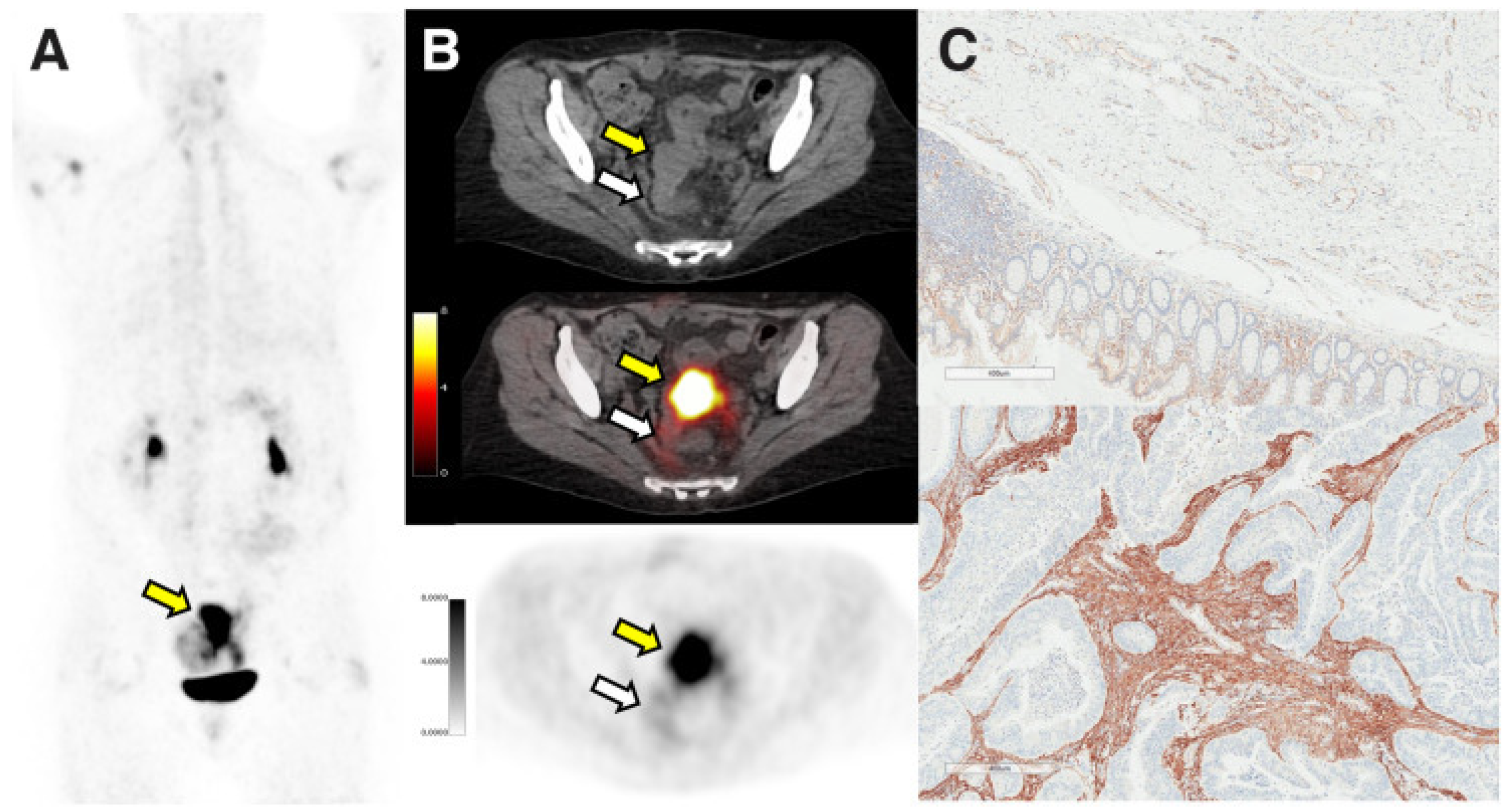
Figure 1. Figure shows the strong uptake of sigmoid adenocarcinoma in 68Ga-FAPi-46 PET/CT (SUVmax, 15.9; SUVmean, 12.8). The yellow arrow shows the adenocarcinoma of the sigmoid, and the white arrow shows the surgically removed normal area (PET maximumintensity projection (A), axial CT ((B), top), axial PET/CT [B, middle], and axial PET [B, bottom]). And immunohistochemistry shows that FAP demonstrated absent to weak expression in normal tissue ((C), top) and was strongly expressed in intratumoral and peritumoral stroma ((C), bottom). Adapted from Ref. [39].
Jcm 12 00577 g001
Figure 2. The uptake of 68GA-FAPI in the primary and metastatic lesions of colon cancer patients was higher than that of 18F-FDG, and the lesions were clearly shown. The black and white arrows show primary lesions of colon cancer. 18F-FDG PET/CT shows low to moderate metabolic activity in the primary colon and liver metastases ((A), left image: anterior maximum intensity projection image from 18F-FDG PET; right images: axial fused PET/CT images). 68Ga-FAPI PET/CT showed high uptake of primary lesions of colon and metastatic lesions of liver ((B), right image: anterior maximum intensity projection image from 68Ga-FAPI PET; left images: axial fused PET/CT images). Adapted from Ref. [59].
Jcm 12 00577 g002
Figure 3. 18F-FDG PET/CT showed hypermetabolism in the intestines around the anastomosis in the right upper quadrant of the abdomen (SUVmax 11.4). The involved bowels also had intense uptake of 68Ga-FAPI (SUVmax 14.1). The arrow shows high metabolic uptake of the intestinal tract around the anastomotic site after Crohn’s disease. Adapted from Ref. [71].
Jcm 12 00577 g003
Table 1. Summary of major studies investigating FAPI PET/CT in colorectal cancer.
AuthorYearTypeKey Message
Loktev
et al.
2018ArticleCancer-associated fibroblasts are abundant in colon cancer and can be imaged using molecular probes targeting FAP.
Kratochwil
et al.
2019ArticleFAPI PET/CT has intermediate strength uptake in colorectal cancer.
Koerber
et al.
2020Article68Ga-FAPI PET/CT can detect primary and metastatic tumors of the lower digestive tract, identify clinical stages and provide evidence for tumor management.
Rathke
et al.
2021CaseFAPI uptake was high in colorectal cancer-related metastases, and peritoneal metastases disappeared after treatment with 90Y-FAPI46.
Giesel
et al.
2021ArticleIn the background tissues of colon, the SUVmax of 68Ga-FAPI was significantly lower than that of 18F-FDG.
Şahin
et al.
2021Article68Ga-FAPI is superior to 18F-FDG in detecting liver metastasis of colorectal cancer.
Pang
et al.
2021Article68Ga-FAPI was superior to 18F-FDG in the detection of colorectal cancer primary and metastatic sites, showing higher uptake in most primary and metastatic sites.
Fu
et al.
2021Case68Ga-FAPI has strong uptake in sigmoid signet ring cell carcinoma and is more sensitive to primary lesions and peritoneal carcinomas than 18F-FDG.
Mona
et al.
2022ArticleFAPI PET/CT has strong uptake in colorectal cancer patients, and the intensity of FAPI uptake is related to the intensity of immunohistochemical staining of FAP.
Strating
et al.
2022ArticleFAPI PET/CT may have important potential value in evaluating CMS4 as an adverse prognostic factor for colorectal cancer.
Qin
et al.
2022Article68Ga-FAPI is of great significance in the accurate staging and clinical management of colorectal cancer.
Elboga
et al.
2022ArticleThe uptake level of FAPI in primary and metastatic lesions of colorectal cancer (especially liver metastasis) was significantly higher than that of FDG and had a better tumor-to-background ratio.
Güzel
et al.
2022Case68Ga-FAPI was superior to 18F-FDG in evaluating recurrent signet ring cell colon cancer.
Kömek
et al.
2022Article68Ga-FAPI has higher sensitivity and specificity in detecting primary lesions of colorectal cancer and lymph node and peritoneal metastasis.
Table 2. SUVmax of FAPI and FDG in colorectal cancer lesions.
AuthorYearLesion LocationFAPI #
SUVmax
FDG
SUVmax
Koerber
et al.
2020Primary lesions15.7-
Local relapse6.56-
Lymph node metastases8.33-
Liver metastases9.54-
Şahin
et al.
2021Primary lesions5.55.0
Pang
et al.
2021Primary lesions15.92.2
Lymph node metastases * 6.72.4
Liver metastases *9.71.7
Elboga
et al.
2022Primary lesions14.68.4
Lymph node metastases9.94.9
Peritoneal metastases10.73.1
Liver metastases12.25.0
Kömek
et al.
2022Primary lesions11.5418.93
Lymph node metastases3.62.25
Peritoneal metastases5.143.59
Liver metastases6.159.66
# FAPI: fibroblast-activating protein inhibitors. * The data included gastric, duodenal, and colorectal cancers.
Table 3. The sensitivity and specificity of FAPI and FDG in colorectal cancer lesions.
AuthorYearLesion LocationFAPI #
Sensitivity
FAPI #
Specificity
FDG
Sensitivity
FDG
Specificity
Şahin
et al.
2021Liver metastases96.6%-70.8%-
Pang
et al.
2021Lymph node metastases *79%82%54%89%
Kömek
et al.
2022Primary lesions100%100%100%85.3%
Lymph node metastases90%100%80%81.8%
Peritoneal metastases100%100%55%100%
# FAPI: fibroblast-activating protein inhibitors. * The data included gastric, duodenal, and colorectal cancers.
Disclaimer/Publisher’s Note: The statements, opinions and data contained in all publications are solely those of the individual author(s) and contributor(s) and not of MDPI and/or the editor(s). MDPI and/or the editor(s) disclaim responsibility for any injury to people or property resulting from any ideas, methods, instructions or products referred to in the content.