Article

The Negative Impact of Inflammation-Related Parameters in Prostate Cancer after Robot-Assisted Radical Prostatectomy: A Retrospective Multicenter Cohort Study in Japan (the MSUG94 Group)

1
Department of Urology, Gifu University Graduate School of Medicine, Gifu 501-1194, Japan
2
Department of Urology, Hiroshima City Hiroshima Citizens Hospital, Hiroshima 730-8518, Japan
3
Department of Urology, Yokohama City University, Yokohama 236-0004, Japan
4
Department of Nephro-Urologic Surgery and Andrology, Mie University Graduate School of Medicine, Tsu 514-8507, Japan
5
Department of Urology, University of Toyama, Toyama 930-0194, Japan
6
Department of Urology, Dokkyo Medical University Saitama Medical Center, Koshigaya 343-8555, Japan
7
Department of Urology, Tokyo Metropolitan Cancer and Infectious Diseases Center Komagome Hospital, Tokyo 113-8677, Japan
8
Department of Urology, Seirei Hamamatsu General Hospital, Hamamatsu 430-8558, Japan
9
Department of Urology, Toranomon Hospital, Tokyo 105-8470, Japan
10
Department of Surgery, Division of Urology, Kobe University Graduate School of Medicine, Kobe 650-0017, Japan
*
Author to whom correspondence should be addressed.
J. Clin. Med. 2023, 12(24), 7732; https://doi.org/10.3390/jcm12247732
Submission received: 15 November 2023 / Revised: 14 December 2023 / Accepted: 15 December 2023 / Published: 17 December 2023
(This article belongs to the Section Nephrology & Urology)

Abstract

:
Background and Objectives: We aimed to examine the relationship between the inflammation-related parameters, such as the neutrophil-to-lymphocyte ratio (NLR), and the pathological findings and biochemical recurrence (BCR) in patients with prostate cancer (PCa) undergoing robot-assisted radical prostatectomy (RARP). Materials and Methods: A retrospective multicenter cohort study of patients with PCa who underwent RARP at 10 institutes in Japan was conducted. This study enrolled 3195 patients. We focused on patients undergoing RARP who underwent the preoperative measurement of their inflammation-related parameters and who did not receive any neo- or adjuvant therapy. Data on the pre- and postoperative variables for the enrolled patients were obtained. The primary endpoint of this study was the association between BCR and the inflammation-related parameters after RARP. The secondary endpoint was the association between the inflammation-related parameters and the pathological diagnosis of PCa. Results: Data from 2429 patients with PCa who met the study’s eligibility criteria were analyzed. The median follow-up period was 25.1 months. The inflammation-related parameters were divided into two groups, and cutoff values were determined based on the receiver operating characteristics. There were no statistically significant differences in biochemical recurrence-free survival for any of the parameters. In the univariate analysis, the NLR was predictive of pathological T3 and lymphovascular invasion; however, there were no significant differences in the multivariate analysis. Conclusions: The inflammation-related parameters did not significantly affect the incidence of BCR, at least among patients with PCa who underwent RARP.

1. Introduction

The World Health Organization (WHO) reported that prostate cancer (PCa) is a common solid tumor in men; 1,414,259 new patients were diagnosed with PCa; and 375,304 deaths were PCa-related worldwide in 2020 [1]. Therefore, various surrogate markers and nomograms have been investigated to predict cancer progression and prognosis [2,3,4,5].
The relationships between inflammation-related parameters and tumorigenesis have been widely studied in various tumors [6,7,8,9,10]. In particular, PCa, which is a systemic, multifactorial disease, has been extensively investigated [11,12,13,14,15,16]. Inflammatory cells and mediators in the tumor microenvironment are known to mediate the inflammatory response and further interact with the tumor and peritumoral cells in an autocrine and/or paracrine manner [17]. The tumor microenvironment, which consists mainly of immune cells surrounding the cancer cells, has been suggested as a potential prognostic biomarker for PCa [18]. Therefore, inflammation-related parameters may be considered to contribute to the diagnosis and treatment of PCa as well as to the prognosis of the disease [18]. Lymphocyte-related markers of inflammation, such as C-reactive protein (CRP), the neutrophil-to-lymphocyte ratio (NLR), the platelet-to-lymphocyte ratio (PLR), the lymphocyte-to-monocyte ratio (LMR), and the systemic immune inflammation index (SII), are commonly used as markers of systemic inflammatory responses [1]. However, there is still no unified understanding of how inflammation-related parameters are related to PCa progression and oncological outcomes.
Therefore, we aimed to examine the relationship between the inflammation-related parameters and the pathological findings and BCR in patients with PCa who underwent robot-assisted RP.

2. Materials and Methods

2.1. Patient Population

This research was authorized by the Institutional Review Board of Gifu University (Approval No.: 2021-A050). Due to the retrospective study design, the requirement for patient consent was waived. In accordance with the provisions of the Japanese Ethics Committee and Ethical Guidelines, in the case of retrospective and/or observational research, information from these studies is disclosed to the public through existing documents and other materials. Detailed information on this study is available only in Japanese at https://www.med.gifu-u.ac.jp/visitors/disclosure/docs/2021-B039.pdf (accessed on 6 October 2023).
A retrospective multicenter cohort study of patients who underwent robot-assisted radical prostatectomy (RARP) for PCa at 10 Japanese institutes from September 2012 to August 2021 was conducted. The following preoperative information was collected from the patients enrolled in the study: age, height, weight, Eastern Cooperative Oncology Group Performance Status (ECOG-PS) [19], preoperative serum prostate-specific antigen (PSA) level, prostate volume, biopsy Gleason grade group (GG) [20], clinical T stage, and National Comprehensive Cancer Network risk stratification [21]. All patients underwent tumor disease classification according to the American Joint Committee on Cancer’s 8th edition of the Cancer Staging Manual [22]. No data were obtained in this study on whether enrolled patients received evaluation with magnetic resonance imaging before prostate biopsy. For biopsy GG, specimens were assessed in accordance with the International Society of Urologic Pathology (ISUP) 2014 guidelines [20].
All enrolled patients underwent RARP in this study. Pelvic lymph-node dissection (PLND), extent of PLND, and nerve sparing were decided at the individual surgeon’s or institution’s discretion.

2.2. Pathological Analysis

Based on ISUP2014 guidelines, all prostatectomy specimens were sectioned in accordance with the whole-mount staining method and were assessed for PCa [20]. A pathologist vertically sectioned the prostate apex against the prostatic urethra. The pathologist cut a conical section of the bladder neck margin from the specimen and dissected it perpendicularly. For the remaining prostatic tissue, complete sections were made along a plane perpendicular to the urethral axis at 3–5 mm intervals.

2.3. Follow-Up Schedule

Patients enrolled in this study were monitored for serum PSA and testosterone levels at 3-month intervals postoperatively. BCR was diagnosed when the postoperative serum PSA level increased to 0.2 ng/mL. If the postoperative PSA level did not fall to 0.2 ng/mL at any time, the date of RP was defined as the date of BCR onset.

2.4. Endpoints and Statistics

The primary endpoint of this study was the association between BCR and inflammation-related parameters after RARP. The secondary endpoint was the association between inflammation-related parameters and the pathological diagnosis of PCa. Data were analyzed using JMP 14 software (SAS Institute Inc., Cary, NC, USA). We assessed continuous and categorical variables using the Wilcoxon rank-sum test and Fisher’s exact test. BRFS after RARP was evaluated using the Kaplan–Meier method, and the association between BCR and subgroup classification was analyzed based on the log-rank test. Multivariate analysis was carried out using a Cox proportional hazards model. Using receiver operating characteristic (ROC) curve analysis, the cutoff values of the clinical variables were decided [23]. All two-sided p-values were employed, and statistical significance was defined as a p-value < 0.05.

3. Results

3.1. Patient Characteristics

Table 1 lists the preoperative and postoperative covariates of the patients enrolled in this study. A total of 3195 patients were enrolled in this study. Of these, 571 (17.9%) patients who received neoadjuvant or adjuvant therapy and 195 (6.1%) patients with missing NLR data were excluded from the analysis. Finally, we analyzed the oncologic outcome and perioperative covariates for 2429 (76.0%) patients. Although several surgeons from each institution performed RARP, this study did not evaluate the differences in the number of cases per surgeon.

3.2. Oncological Outcomes

By the end of the follow-up period, BCR occurred in 266 patients (11.0%), radiological recurrence occurred in 22 (0.9%), and castration-resistant PCa occurred in 11 (0.5%); however, none of the patients died of PCa.
Figure 1 shows the relationship between BCR and the inflammation–related parameters, including CRP, NLR, PLR, and SII. The cutoff values of the inflammation-related parameters, using an ROC curve analysis, were 0.07 mg/dL for CRP, 2.01 for NLR, 110 for PLR, and 426 for SII. The patients were divided into two groups based on the cutoff values for each inflammation-related parameter, and the BRFS was examined for each parameter. The 2-year BRFS rate was 91.5% for CRP < 0.07 mg/dL and 90.4% for CRP ≥ 0.07 mg/dL (p = 0.702; Figure 1a), 89.7% for NLR < 2.01 and 91.9% for ≥2.01 (p = 0.850; Figure 1b), 89.2% for PLR < 110 and 92.5% for PLR ≥110 (p = 0.108; Figure 1c), and 89.4% for SII < 426 and 92.2% for SII ≥ 426 (p = 0.741; Figure 1d). There were no significant differences in the BRFS among the four inflammation-related parameters.
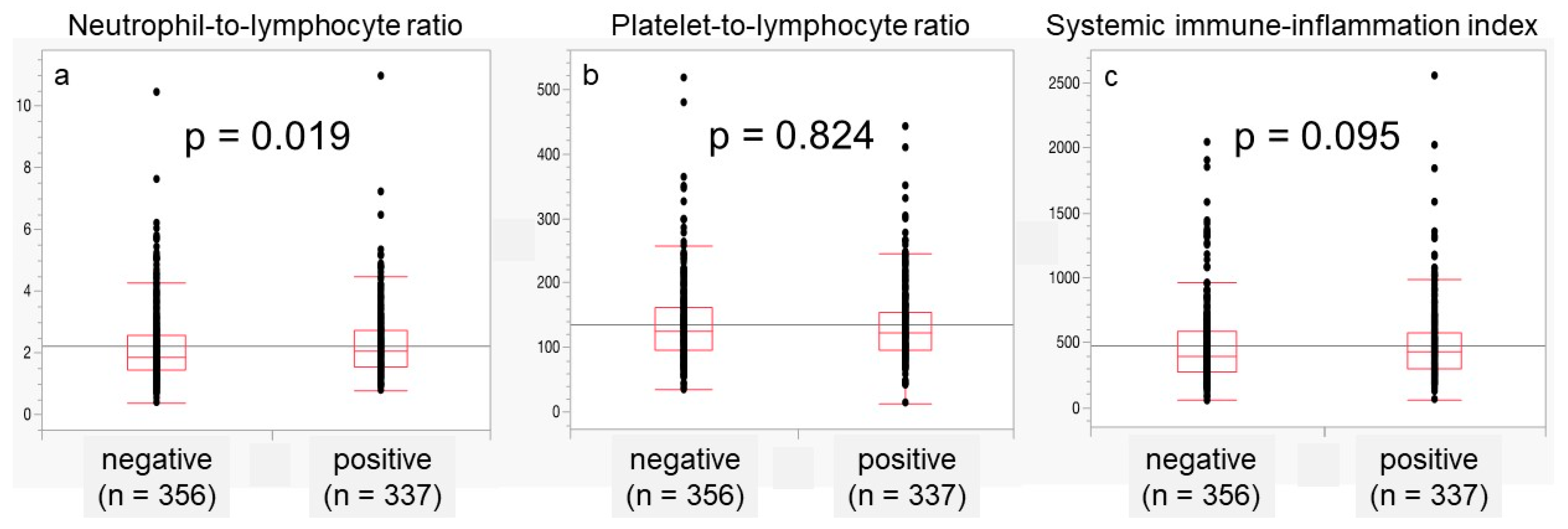
Subsequently, we examined the association of NLR, PLR, and SII with lymphovascular invasion (LVI) in patients with locally advanced PCa with ≥ pT3 (Figure 2). The median NLR was 2.069 in patients with positive LVI and 1.850 in those with negative LVI, which was significantly higher in patients with positive LVI (p = 0.019; Figure 2a). PLR and SII did not differ significantly between LVI-positive and LVI-negative patients.
Table 2 shows the patient demographics of the two groups of cases divided by a cutoff value of 2.01 NLR. Compared to the group with NLR ≥ 2.01, age, CRP, PLR, SII, and LVI were significantly lower in the group with NLR < 2.01. By contrast, BMI and BCR were significantly lower in patients with NLR ≥ 2.01 than in those with NLR < 2.01. In addition, radiographic recurrence and the rate of progression to CRPC tended to be lower in these patients.
A univariate analysis showed significant differences in the clinical T stage, biopsy GG, initial PSA, and NLR. In a multivariate analysis, the clinical T stage, biopsy GG, and initial PSA were significant predictors of locally advanced disease and LVI in patients with PCa undergoing RARP; however, the NLR was not significantly different in this study (Table 3).

4. Discussion

Several studies have shown that many types of malignant neoplasms play an important role in the carcinogenic process and survival with respect to systemic inflammation [6,7,8,9,10,11,12,13,14,15,16,24]. Tumor cells themselves release cytokines and chemokines, which are thought to circulate and induce a systemic inflammatory response [25], resulting in changes in neutrophils, lymphocytes, and platelets [26]. Neutrophils play distinct roles in regulating tumor cell proliferation and angiogenesis [27,28]. Lymphocytes play an important role in the regulation and integration of systemic immune responses [26]. Platelets have been shown to protect tumor cells from adhering to and being excluded from the vascular endothelium, thus contributing to their survival and spread [29,30]. In fact, inflammatory markers have been reported to be associated with tumor stage and grade, oncological outcome, and prognosis in various types of malignant neoplasms [5,6,7,8,9,10]. Because neutrophils, lymphocytes, and platelets have these functions with respect to tumor cells, the role of the inflammation-related parameters in PCa is of interest.
Inflammation-related parameters such as NLR, PLR, and SII have been focused on as potential useful markers for the assessment of systemic inflammation, and previous studies have suggested that these biomarkers may be one of the prognostic predictive parameters for PCa [12,26,31]. In a study of 291 patients with pathologically confirmed localized PCa who underwent RP, 114 (39.2%) developed BCR within a median follow-up of 48 months [12]. The incidence of BCR was significantly higher in the high NLR group (50.7%) and the high SII group (48.77%) than in the low NLR and SII groups [12]. A Kaplan–Meier analysis showed that patients with a high SII and NLR had a significantly poorer BRFS (p < 0.001 and p < 0.001, respectively) [12]. In the multivariate analysis, both a high SII ≥ 528.54 (HR, 4.251; 95%CI, 2.262–9.037; p < 0.001) and NLR ≥ 2.62 (HR, 4.787; 95%CI, 2.339–9.798; p < 0.001) were significant predictors of BCR after RP (p < 0.001 and p < 0.001, respectively) [12]. A high derived NLR (p = 0.044), high PLR (p = 0.028), and low prognostic nutrition index (PNI; p = 0.004) were associated with a poor BRFS in 380 patients who underwent RP for high-risk localized PCa without neoadjuvant androgen deprivation therapy [26]. In particular, the PNI is an independent predictive factor for BCR (HR, 0.56; 95%CI, 0.35–0.90; p = 0.016) [26]. The pooled results from 32 studies, including 21,949 patients, showed that higher pretreatment NLRs (HR, 1.55; 95%CI, 1.37–1.76), PLRs (HR, 1.72; 95%CI, 1.36–2.18), and neutrophil (HR, 1.10; 95%CI, 1.03–1.18) and monocyte counts (HR, 1.75; 95%CI, 1.36–2.25) were associated with poorer OS, while a higher pretreatment LMR correlated with better OS (HR, 2.27; 95%CI, 1.76–2.94) [30]. Furthermore, a higher NLR (HR, 1.62; 95%CI, 1.29–2.04) and monocyte count (HR, 1.75; 95%CI, 1.36–2.25) and a lower LMR (HR, 2.18; 95%CI, 1.58–3.02) predicted worse PFS [31]. These studies suggest that the pretreatment inflammation-related parameters may be useful for predicting oncological outcomes in patients with PCa after RP.
Conversely, several studies have reported no association between the inflammation-related parameters and oncological outcomes in patients with PCa after RP [13,15,16,32,33]. In a study of 668 patients with localized PCa who underwent RP, the NLR was not associated with the pathological status, including the GG (p = 0.159), lymph-node involvement (p = 0.15), or surgical margin status (p = 0.159) [13]. In addition, a high NLR was not statistically associated with MFS, CSS, OS, BRFS, or PCa grade [13]. In a study evaluating the NLR in 73 patients undergoing RP for PCa, there was no significant correlation between NLR and tumor grade, pathologic T stage, lymph-node metastasis, or surgical margin status [16]. Maeda et al. [16] found no significant correlation between NLR and tumor grade (p = 0.834), pathologic T stage (p = 0.082), lymph-node involvement (p = 0.062), or surgical margin status (p = 0.772). Based on an ROC curve analysis predicting biochemical recurrence after RP, a possible NLR cutoff point was determined to be 2.88 or 3.88; however, none of these cutoff points accurately predicted prognosis [16]. With respect to 237 patients with PCa who underwent RP, for the investigation of the relationship between pretreatment NLR and BCR, the NLR was not an independent predictor of BCR in a multivariate analysis, even though patients with a higher NLR had a significantly shorter BRFS than those with a lower NLR (p = 0.019) [32]. Additionally, the NLR was not statistically associated with upstaging, upgrading, or BCR (p = 0.368, p = 0.573, and p = 0.504, respectively) in a study of 217 patients with PCa [33]. In the present study, the NLR tended to be higher in patients with pathological T3 and LVI, although the multivariate analysis showed no significant difference. Therefore, further validation of the possibility that the NLR reflects the characteristics of PCa and the local immune microenvironment is needed because there are contradictory data on this issue.
This research has certain limitations. First, this study had a retrospective multicenter design and may have been susceptible to potential selection bias caused by diagnostic and surgical approach differences among the participating centers. Second, the relatively short follow-up period and varying intervals among each institution may have precluded the consideration of its true usefulness as a surrogate marker for BCR in analyzing the inflammation-related parameters. Finally, a prospective multicenter study is needed to evaluate the usefulness of the inflammation-related parameters in predicting the oncological outcomes of PCa.

5. Conclusions

The inflammation-related parameters, such as CRP, NLR, PLR, and SII, were not significantly associated with the incidence of BCR, at least among patients with PCa who underwent RARP. Further investigation into the inflammation-related parameters in RARP is warranted.

Author Contributions

Conceptualization, T.K., T.I., H.K., K.M. (Kazuhide Makiyama), K.S. (Kazutaka Saito), J.T., S.E., F.K., T.Y. and S.U.; methodology, K.M. (Kazumasa Murase), M.K. and T.K.; investigation, K.M. (Kazumasa Murase) and M.K.; resources, M.K., S.E., T.T., T.S., Y.I., A.N., M.T., T.Y. and K.S. (Kazushige Sakaguchi); data curation, K.M. (Kazumasa Murase), M.K. and T.K.; writing—original draft preparation, K.M. (Kazumasa Murase); writing—review and editing, T.K. All authors have read and agreed to the published version of the manuscript.

Funding

This study received no external funding.

Institutional Review Board Statement

The study protocol was approved by the Institutional Review Board of Gifu University (number: 2021-A050, approval date 4 August 2021). All procedures performed in studies involving human participants were in accordance with the ethical standards of the institutional and/or national research committee and with the 1964 Declaration of Helsinki and its later amendments or comparable ethical standards.

Informed Consent Statement

For this type of study, formal consent is not required. Pursuant to the provisions of the ethics committee and the ethics guidelines in Japan, written consent is not required in exchange for the public disclosure of study information in the case of retrospective and/or observational studies using a material such as the existing documentation. The study information is open for public consumption at https://www.med.gifu-u.ac.jp/visitors/disclosure/docs/2021-B039.pdf (accessed on 6 October 2023).

Data Availability Statement

The data presented in this study are available on request from the corresponding author. The data are not publicly available due to privacy and ethical reasons.

Conflicts of Interest

The authors have no conflicts of interest to declare and received no financial support for this survey.

References

  1. Zhou, M.; Liang, J.; Hui, J.; Xu, J. Inflammation-related indicators have a potential to increase overall quality of the prostate cancer management: A narrative review. Transl. Androl. Urol. 2023, 12, 809–822. [Google Scholar] [CrossRef]
  2. Kawase, M.; Goto, T.; Ebara, S.; Tatenuma, T.; Sasaki, T.; Ishihara, T.; Ikehata, Y.; Nakayama, A.; Toide, M.; Yoneda, T.; et al. Nomogram Predicting Locally Advanced Prostate Cancer in Patients with Clinically Organ-Confined Disease Who Underwent Robot-Assisted Radical Prostatectomy: A Retrospective Multicenter Cohort Study in Japan (The MSUG94 Group). Ann. Surg. Oncol. 2023, 30, 6925–6933. [Google Scholar] [CrossRef] [PubMed]
  3. Hashine, K.; Yamashita, N.; Tachou, T.; Kan, M.; Yanagaki, T.; Oka, A.; Takeda, H.; Shirato, A.; Watanabe, U.; Noriyoshi Miura, N.; et al. Radical prostatectomy trends between 2010 and 2020 in Ehime, Japan, identified using data from the Medical Investigation Cancer Network (MICAN) study. Int. J. Urol. 2023, 30, 547–553. [Google Scholar] [CrossRef] [PubMed]
  4. Kawase, M.; Ebara, S.; Tatenuma, T.; Sasaki, T.; Ikehata, Y.; Nakayama, A.; Toide, M.; Yoneda, T.; Sakaguchi, K.; Ishihara, T.; et al. A Nomogram for Predicting Prostate Cancer with Lymph Node Involvement in Robot-Assisted Radical Prostatectomy Era: A Retrospective Multicenter Cohort Study in Japan (The MSUG94 Group). Diagnostics 2022, 12, 2545. [Google Scholar] [CrossRef] [PubMed]
  5. Goto, Y.; Utsumi, T.; Maruo, M.; Kurozumi, A.; Noro, T.; Tanaka, S.; Sugawara, S.; Chiba, K.; Miyazaki, K.; Inoue, A.; et al. Development and validation of novel nomogram to identify the candidates for extended pelvic lymph node dissection for prostate cancer patients in the robotic era. Int. J. Urol. 2023, 30, 659–665. [Google Scholar] [CrossRef] [PubMed]
  6. Tomioka-Inagawa, R.; Nakane, K.; Enomoto, T.; Tomioka, M.; Taniguchi, T.; Ishida, T.; Ozawa, K.; Takagi, K.; Ito, H.; Takeuchi, S.; et al. The Impact of Neutrophil-to-Lymphocyte Ratio after Two Courses of Pembrolizumab for Oncological Outcomes in Patients with Metastatic Urothelial Carcinoma. Biomedicines 2022, 10, 1609. [Google Scholar] [CrossRef] [PubMed]
  7. Iinuma, K.; Enomoto, T.; Kawada, K.; Fujimoto, S.; Ishida, T.; Takagi, K.; Nagai, S.; Ito, H.; Kawase, M.; Nakai, C.; et al. Utility of Neutrophil-to-Lymphocyte Ratio, Platelet-to-Lymphocyte Ratio, and Systemic Immune Inflammation Index as Prognostic, Predictive Biomarkers in Patients with Metastatic Renal Cell Carcinoma Treated with Nivolumab and Ipilimumab. J. Clin. Med. 2021, 10, 5325. [Google Scholar] [CrossRef]
  8. Samejima, H.; Kojima, K.; Fujiwara, A.; Tokunaga, T.; Okishio, K.; Yoon, H. The combination of PD-L1 expression and the neutrophil-to-lymphocyte ratio as a prognostic factor of postoperative recurrence in non-small-cell lung cancer: A retrospective cohort study. BMC Cancer 2023, 23, 1107. [Google Scholar] [CrossRef]
  9. Tacconi, F.; Mangiameli, G.; Voulaz, E.; Patirelis, A.; Carlea, F.; Rocca, E.; Tamburrini, A.; Vanni, G.; Ambrogi, V. Blood-Derived Systemic Inflammation Markers and Risk of Nodal Failure in Stage Ia Non-Small Cell Lung Cancer: A Multicentric Study. J. Clin. Med. 2023, 12, 4912. [Google Scholar] [CrossRef]
  10. Jadoon, S.K.; Soomro, R.; Ahsan, M.N.; Ijaz Khan, R.M.; Iqbal, S.; Yasmin, F.; Najeeb, H.; Saleem, N.; Cho, N.; Resham; et al. Association of neutrophil-to-lymphocyte ratio with clinical, pathological, radiological, laboratory features and disease outcomes of invasive breast cancer patients: A retrospective observational cohort study. Medicine 2023, 102, e33811. [Google Scholar] [CrossRef]
  11. Nakane, K.; Watanabe, H.; Naiki, T.; Takahara, K.; Yasui, T.; Miyake, H.; Shiroki, R.; Koie, T. Trends in the Use of Second-Generation Androgen Receptor Axis Inhibitors for Metastatic Hormone-Sensitive Prostate Cancer and Clinical Factors Predicting Biological Recurrence. Diagnostics 2023, 13, 1661. [Google Scholar] [CrossRef] [PubMed]
  12. Wang, S.; Yang, X.; Yu, Z.; Du, P.; Cao, Y.; Ji, Y.; Ma, J.; Yang, Y. The values of systemic immune-inflammation index and neutrophil-lymphocyte ratio in predicting testicular germ cell tumors: A retrospective clinical study. Front. Oncol. 2022, 12, 893877. [Google Scholar] [CrossRef] [PubMed]
  13. Lu, Y.; Huang, H.H.; Lau, W.K.O. Evaluation of neutrophil-to-lymphocyte ratio as a prognostic indicator in a Singapore cohort of patients with clinically localized prostate cancer treated with prostatectomy. World J. Urol. 2020, 38, 103–109. [Google Scholar] [CrossRef] [PubMed]
  14. Wang, S.; Ji, Y.; Ma, J.; Du, P.; Cao, Y.; Yang, X.; Yu, Z.; Yang, Y. Role of inflammatory factors in prediction of Gleason score and its upgrading in localized prostate cancer patients after radical prostatectomy. Front. Oncol. 2023, 12, 1079622. [Google Scholar] [CrossRef] [PubMed]
  15. Cao, Z.; Ji, J.; Zhang, C.; Wang, F.; Xu, H.; Yu, Y.; Sun, Y. The preoperative neutrophil-to-lymphocyte ratio is not a marker of prostate cancer characteristics but is an independent predictor of biochemical recurrence in patients receiving radical prostatectomy. Cancer Med. 2019, 8, 1004–1012. [Google Scholar] [CrossRef] [PubMed]
  16. Maeda, Y.; Kawahara, T.; Koizumi, M.; Ito, H.; Kumano, Y.; Ohtaka, M.; Kondo, T.; Mochizuki, T.; Hattori, Y.; Teranishi, J.; et al. Lack of an Association between Neutrophil-to-Lymphocyte Ratio and PSA Failure of Prostate Cancer Patients Who Underwent Radical Prostatectomy. Biomed. Res. Int. 2016, 2016, 6197353. [Google Scholar] [CrossRef] [PubMed]
  17. Crusz, S.M.; Balkwill, F.R. Inflammation and cancer: Advances and new agents. Nat. Rev. Clin. Oncol. 2015, 12, 584–596. [Google Scholar] [CrossRef]
  18. Lu, G.; Cai, W.; Wang, X.; Huang, B.; Zhao, Y.; Shao, Y.; Wang, D. Identifying prognostic signatures in the microenvironment of prostate cancer. Transl. Androl. Urol. 2021, 10, 4206–4218. [Google Scholar] [CrossRef]
  19. Oken, M.M.; Creech, R.H.; Tormey, D.C.; Horton, J.; Davis, T.E.; McFadden, E.T.; Carbone, P.P. Toxicity and response criteria of the Eastern Cooperative Oncology Group. Am. J. Clin. Oncol. 1982, 5, 649–655. [Google Scholar] [CrossRef]
  20. Epstein, J.I.; Egevad, L.; Amin, M.B.; Delahunt, B.; Srigley, J.; Humphrey, P.A.; Committee, G. The 2014 International Society of Urological Pathology (ISUP) Consensus Conference on Gleason Grading of Prostatic Carcinoma: Definition of Grading Patterns and Proposal for a New Grading System. Am. J. Surg. Pathol. 2016, 40, 244–252. [Google Scholar] [CrossRef]
  21. Prostate Cancer. NCCN Guidelines®. 2022. Available online: https://www.nccn.org/guidelines/guidelines-process/transparency-process-and-recommendations/GetFileFromFileManager?fileManagerId=12813 (accessed on 6 November 2023).
  22. Buyyounouski, M.K.; Choyke, P.L.; McKenney, J.K.; Sartor, O.; Sandler, H.M.; Amin, M.B.; Kattan, M.W.; Lin, D.W. Prostate cancer—Major changes in the American Joint Committee on Cancer eighth edition cancer staging manual. CA Cancer J. Clin. 2017, 67, 245–253. [Google Scholar] [CrossRef] [PubMed]
  23. Perkins, N.J.; Schisterman, E.F. The inconsistency of “optimal” cutpoints obtained using two criteria based on the receiver operating characteristic curve. Am. J. Epidemiol. 2006, 163, 670–675. [Google Scholar] [CrossRef] [PubMed]
  24. Templeton, A.J.; McNamara, M.G.; Šeruga, B.; Vera-Badillo, F.E.; Aneja, P.; Ocaña, A.; Leibowitz-Amit, R.; Sonpavde, G.; Knox, J.J.; Tran, B.; et al. Prognostic role of neutrophil-to-lymphocyte ratio in solid tumors: A systematic review and meta-analysis. J. Natl. Cancer Inst. 2014, 106, dju124. [Google Scholar] [CrossRef] [PubMed]
  25. Salazar-Onfray, F.; López, M.N.; Mendoza-Naranjo, A. Paradoxical effects of cytokines in tumor immune surveillance and tumor immune escape. Cytokine Growth Factor Rev. 2007, 18, 171–182. [Google Scholar] [CrossRef] [PubMed]
  26. Shu, K.; Zheng, Y.; Chen, J.; Li, W.; Jiang, K. Prognostic value of selected preoperative inflammation-based scores in patients with high-risk localized prostate cancer who underwent radical prostatectomy. OncoTargets Ther. 2018, 11, 4551–4558. [Google Scholar] [CrossRef] [PubMed]
  27. Gregory, A.D.; Houghton, A.M. Tumor-associated neutrophils: New targets for cancer therapy. Cancer Res. 2011, 71, 2411–2416. [Google Scholar] [CrossRef] [PubMed]
  28. Cools-Lartigue, J.; Spicer, J.; McDonald, B.; Gowing, S.; Chow, S.; Giannias, B.; Bourdeau, F.; Kubes, P.; Ferri, L. Neutrophil extracellular traps sequester circulating tumor cells and promote metastasis. J. Clin. Investig. 2013, 123, 3446–3458. [Google Scholar] [CrossRef]
  29. Battinelli, E.M.; Markens, B.A.; Italiano, J.E., Jr. Release of angiogenesis regulatory proteins from platelet alpha granules: Modulation of physiologic and pathologic angiogenesis. Blood 2011, 118, 1359–1369. [Google Scholar] [CrossRef]
  30. Gay, L.J.; Felding-Habermann, B. Contribution of platelets to tumour metastasis. Nat. Rev. Cancer 2011, 11, 123–134. [Google Scholar] [CrossRef]
  31. Peng, H.; Luo, X. Prognostic significance of elevated pretreatment systemic inflammatory markers for patients with prostate cancer: A meta-analysis. Cancer Cell Int. 2019, 19, 70. [Google Scholar] [CrossRef]
  32. Zhang, G.M.; Zhu, Y.; Ma, X.C.; Qin, X.J.; Wan, F.N.; Dai, B.; Sun, L.J.; Ye, D.W. Pretreatment Neutrophil-to-Lymphocyte Ratio: A Predictor of Advanced Prostate Cancer and Biochemical Recurrence in Patients Receiving Radical Prostatectomy. Medicine 2015, 94, e1473. [Google Scholar] [CrossRef] [PubMed]
  33. Kwon, Y.S.; Han, C.S.; Yu, J.W.; Kim, S.; Modi, P.; Davis, R.; Park, J.H.; Lee, P.; Ha, Y.S.; Kim, W.J.; et al. Neutrophil and Lymphocyte Counts as Clinical Markers for Stratifying Low-Risk Prostate Cancer. Clin. Genitourin. Cancer 2016, 14, e1–e8. [Google Scholar] [CrossRef] [PubMed]
Figure 1. Kaplan–Meier estimates of biochemical recurrence-free survival when C-reactive protein (CRP) was stratified with a cutoff value of 0.07 mg/dL (a), neutrophil-to-lymphocyte ratio (NLR) was stratified with a cutoff value of 2.01 (b), platelet-to-lymphocyte ratio (PLR) was stratified with a cutoff value of 110 (c), and systemic immune inflammation index (SII) was stratified with a cutoff value of 426 (d). The 2-year BRFS rate was 91.5% for CRP < 0.07 mg/dL and 90.4% for CRP ≥ 0.07 mg/dL (p = 0.702 (a)), 89.7% for NLR < 2.01 and 91.9% for NLR ≥ 2.01 (p = 0.850 (b)), 89.2% for PLR < 110 and 92.5% for PLR ≥ 110 (p = 0.108 (c)), and 89.4% for SII < 426 and 92.2% for SII ≥ 426 (p = 0.741 (d)).
Jcm 12 07732 g001
Figure 2. The association of NLR, PLR, and SII with lymphovascular invasion (LVI) in locally advanced PCa above pT3. The median NLR was 2.069 for patients with positive LVI and 1.850 for those with negative LVI (a). The median PLR values of LVI-positive and -negative patients were 123 and 125, respectively (b). The median SII was 426 for patients with positive LVI and 392 for those with negative LVI (c).
Jcm 12 07732 g002
Table 1. Pre- and postoperative covariates for enrolled patients.
Age (year, median, IQR)68 (64–72)
Body mass index (kg/m2, median, IQR)23.5 (21.7–25.5)
ECOG-PS (number, %)
02356 (97.0)
173 (3.0)
Initial PSA (ng/mL, median, IQR)7.6 (5.6–11.3)
Prostate volume (mL, median, IQR)30 (23–40)
Biopsy Gleason grade group (number, %)
1529 (21.8)
2770 (31.7)
3511 (21.0)
4468 (19.3)
5151 (6.2)
Clinical T stage (number, %)
1494 (20.4)
21769 (72.9)
3164 (6.8)
Unknown2 (0.08)
CRP (mg/dL, median, IQR)0.07 (0.03–0.11)
NLR (median, IQR)2.01 (1.54–2.73)
PLR (median, IQR)129 (101–166)
SII (median, IQR)426 (306–587)
Pathological Gleason grade group (number, %)
1188 (7.8)
2982 (40.6)
3788 (32.6)
4260 (10.7)
5201 (8.3)
Unknown10 (4.1)
Pathological T stage (number, %)
03 (0.1)
21732 (71.3)
3688 (28.3)
45 (0.2)
Unknown1 (0.04)
Pelvic lymphadenectomy (number, %)
Not performed793 (32.6)
Performed1636 (67.4)
Unknown1 (0.04)
LVI (number, %)740 (30.5)
Follow-up period (months, median, IQR)25.1 (11.9–49.7)
IQR, interquartile range; ECOG-PS, Eastern Cooperative Oncology Group Performance Status; PSA, prostate-specific antigen; CRP, C-reactive protein; NLR, neutrophil-to-lymphocyte ratio; PLR, platelet-to-lymphocyte ratio; SII, systemic immune inflammation index; and LVI, lymphovascular invasion.
Table 2. Pre- and postoperative covariates for enrolled patients divided into two groups with a cutoff value of 2.01 for neutrophil-to-lymphocyte ratio.
NLR < 2.01
(n = 1211)
NLR ≥ 2.01
(n = 1218)
p-Value
Age (year, median, IQR)68 (64–72)69 (65–73)<0.001
Body mass index (kg/m2, median, IQR)23.8 (22.0–25.8)23.3 (21.5–25.3)<0.001
ECOG-PS (number, %) 0.420
01178 (97.3)1178 (96.7)
133 (2.7)40 (3.3)
Initial PSA (ng/mL, median, IQR)7.5 (5.5–11.2)7.7 (5.7–11.3)0.107
Prostate volume (mL, median, IQR)30 (22–39)30 (23–40)0.201
Biopsy Gleason grade group (number, %) 0.245
1251 (20.7)278 (22.8)
2397 (32.8)373 (30.6)
3240 (19.8)271 (22.2)
4247 (20.4)221 (18.1)
576 (6.3)75 (6.2)
Clinical T stage (number, %) 0.327
1232 (19.2)262 (21.6)
2894 (73.8)875 (72.0)
385 (7.0)79 (6.5)
Unknown0 (0)2 (0.2)
CRP (mg/dL, median, IQR)0.06 (0.03–0.10)0.07 (0.03–0.12)0.001
PLR (median, IQR)107 (86–132)153 (125 –93)< 0.001
SII (median, IQR)310 (241–382)582 (475–747)< 0.001
Pathological Gleason grade group (number, %) 0.294
194 (7.8)94(7.7)
2498 (41.3)484 (39.9)
3369 (30.6)419 (34.5)
4137 (11.4)123 (10.1)
5107 (8.9)94 (7.7)
Unknown6 (0.5)4 (0.3)
Pathological T stage (number, %) 0.375
01 (0.1)2 (0.2)
2846 (69.9)886 (72.7)
3360 (29.8)328 (26.9)
43 (0.3)2 (0.2)
Unknown1 (0.1)0
Pelvic lymphadenectomy (number, %) 0.349
Not performed386 (31.9)407 (33.4)
Performed822 (67.9)810 (66.5)
Unknown3 (0.2)1 (0.1)
LVI (number, %)343 (28.3)397 (32.6)0.022
BCR (number, %)148 (12.3)118 (9.7)0.037
Radiological recurrence (number, %)14 (1.2)8 (0.7)0.199
CRPC (number, %)7 (0.6)4 (0.3)0.365
Follow-up period (months, median, IQR)26.0 (11.9–49.9)24.2 (11.8–49.5)0.220
NLR, neutrophil-to-lymphocyte ratio; IQR, interquartile range; ECOG-PS, Eastern Cooperative Oncology Group Performance Status; PSA, prostate-specific antigen; CRP, C-reactive protein; PLR, platelet-to-lymphocyte ratio; SII, systemic immune inflammation index; LVI, lymphovascular invasion; BCR, biochemical recurrence; and CRPC, castration-resistant prostate cancer.
Table 3. Univariate and multivariate analyses for predicting prostate cancer with ≥ pathological T3 and lymphovascular invasion.
VariablesUnivariate Multivariate
OR (95%CI)p-ValueOR (95%CI)p-Value
Clinical T stage (continuous)2.644 (2.065–3.386)< 0.0011.745 (1.350–2.256)<0.001
Biopsy Gleason grade group (continuous)1.655 (1.501–1.826)< 0.0011.498 (1.351–1.660)<0.001
Initial PSA (continuous)1.062 (1.048–1.077)< 0.0011.049 (1.034–1.064)<0.001
NLR (continuous)1.224 (1.029–1.455)0.0221.204 (0.945–1.533)0.133
OR, odds ratio; CI, confidence interval; PSA, prostate-specific antigen; and NLR, neutrophil-to-lymphocyte ratio.
Disclaimer/Publisher’s Note: The statements, opinions and data contained in all publications are solely those of the individual author(s) and contributor(s) and not of MDPI and/or the editor(s). MDPI and/or the editor(s) disclaim responsibility for any injury to people or property resulting from any ideas, methods, instructions or products referred to in the content.