
Neuroimaging Studies of the Neural Correlates of Heart Rate Variability: A Systematic Review

 , , , and
Abstract
:1. Introduction
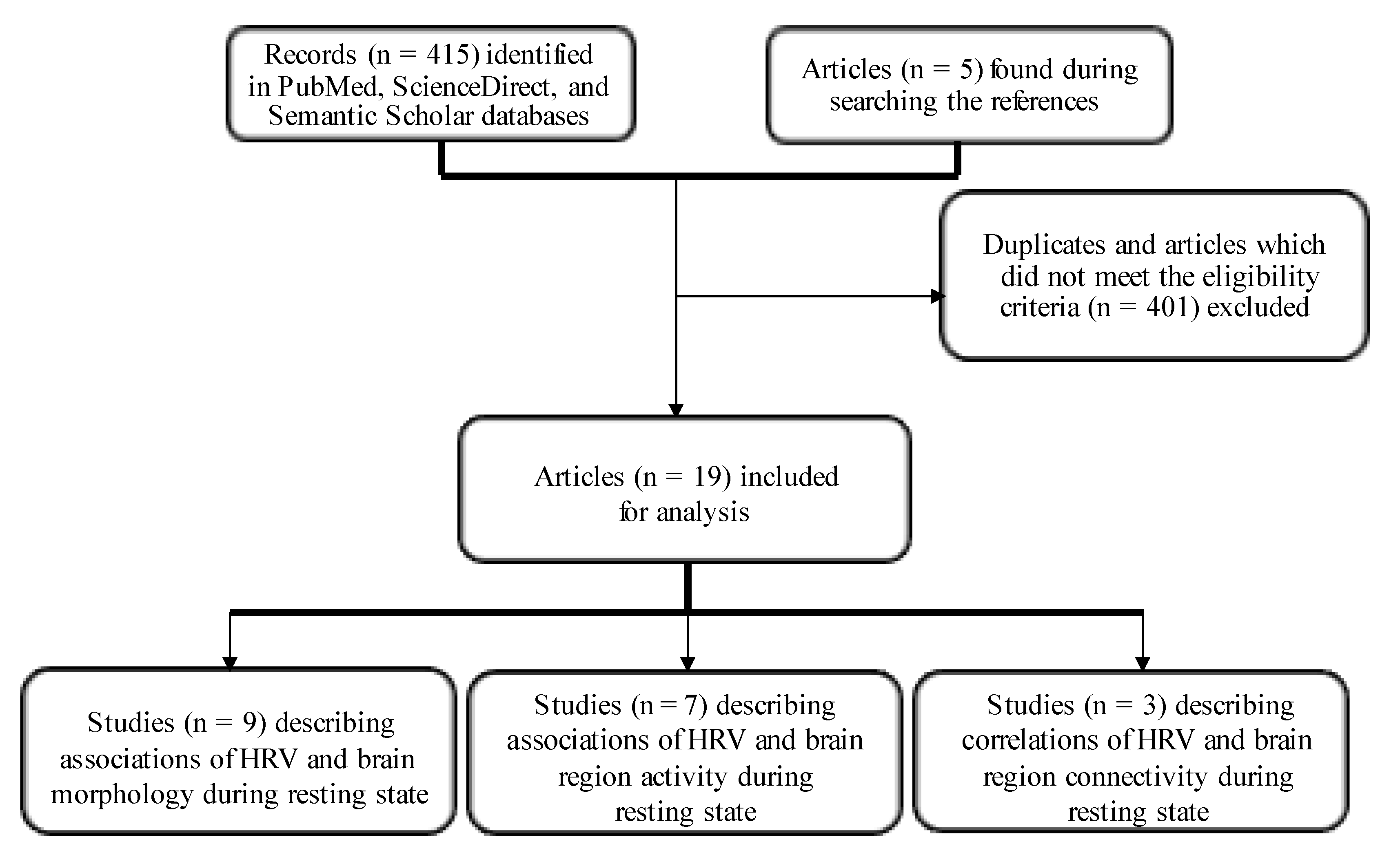
2. Materials and Methods
2.1. Information Sources and Search Strategy
2.2. Study Selection and Eligibility Criteria
2.3. Reporting of Findings
3. Results
3.1. Associations of HRV with Brain Morphology or Structural Covariates at Rest (Table 2)

| Study | Group | HRV Parameters and Methodology | MRI Methodology | Main Brain Regions Associated with HRV | Positive (+) or Negative (-) Correlations of HRV with Different Brain Regions | Studied (+) or Not (-) Age or Gender-Dependent Associations between HRV and Different Brain Regions |
|---|---|---|---|---|---|---|
| HF | 1.5T MRI | |||||
| Winkelmann et al., 2016 [42] | N = 30 young participants (8 female, mean age: 22.5 ± 3.9 years) | (3- leads ECG recorded in a sitting position during a 10-min resting phase, then five artifact-free minutes analyzed) | (thickness of cortical surfaces and volume of subcortical brain structures) | Positive correlations with mean cortical thickness of caudal ACC (R), LG (R), pars triangularis (R), precentral gyrus (R), rostral MFG (R), SFG (R), superior TG (R), transverse temporal cortex (R), caudal ACC (L), inferior TG (L) and SMG (L). Negative correlation with isthmus CC (L). | +/- | - |
| rMSSD | 3T MRI | |||||
| Wei et al., 2018 [44] | N = 185 from NKI-RS (95 female, mean age: 35.2 ± 14.0 years) | (IBI time series derived from PPG) | (GMV, the voxel-based morphometry analysis) | Structural correlations from amygdala (R) with dorsal mPFC (L)/dorsal ACC (R) extending into pre-SMA/SMA (R). | + | - |
| rMSSD | 3T MRI | |||||
| Wei et al., 2021 [43] | N = 114 from NKI-RS (47 female, age 36.1 ± 13.3 years) N = 108 from LEMON (31 female, mean age 41.4 ± 20.7 years) | (IBI time series derived from PPG) | (GMV, the voxel-based morphometry analysis) | Positive structural correlations from anterior insula (L) to bilateral OFC, dorsal ACC, mPFC (L), inferior lobe (R) and Precuneus (in the LEMON sample). Positive structural correlations from anterior insula (L) to OFC (R), mPFC (R), anterior insula (R) and dorsal ACC (L) (in the NKI sample). Positive structural correlation from anterior insula (L) to dorsal ACC on the conjunction maps. Positive structural correlations from anterior insula (L) to bilateral dorsal ACC and PCu and from anterior insula (R) to OFC (R) in the pooled data analysis. | + | - |
| HF, rMSSD, LF | 3T MRI | |||||
| Wei et al., 2018 [44] | N = 185 from NKI-RS (95 female, mean age: 35.2 ± 14.0 years) | (IBI time series derived from PPG) | (GMV, the voxel-based morphometry analysis) | HF: Negative correlations with GMV in putamen (R), caudate (R), amygdala (R), insula (R), superior temporal gyrus (R), temporal pole (R), para-hippocampal gyrus (R). rMSSD: Similar results obtained for HF power LF power No significant correlations | - | + |
| Mean IBI, rMSSD | 3T fMRI | |||||
| Yoo et al., 2018 [2] * | N = 19 older adults (9 female, age range: 62–78 years) N = 19 younger (7 female, age range:19–37 years) * | (IBIs derived from the ECG signal during a 3-min pre-scan | (cortical reconstruction and volumetric segmentation) | Mean IBI: Negative correlations with caudal ACC (L) in all subjects. rMSSD: Positive correlations with lateral OFC (L) (R) in the entire group and with lateral OFC (R) and pars orbitalis (R) in older subjects. Rostral ACC (R) trended to significance in the younger group. | +/- | + |
| rMSSD | 3T-fMRI | |||||
| Kumral et al., 2019 [46] | N = 388 (140 younger: 26.0 ± 4.2 years, 119 middle-aged: 46.3 ± 6.2 years, 129 older: 29 66.9 ± 4.7 years) | (ECG recordings, 10-s in LIFE and 4-min in LEMON study) | (GMV, the voxel-based morphometry analysis) | rMSSD: In the middle-aged group a significant rMSSD-related increase of GMV in the left cerebellum. No significant findings in the younger or older groups | + | + |
| SDNN, SD1, HF, total power | 3T-MRI | |||||
| Wood et al., 2017 [33] | N = 55 (21–73 years; 18 female) | (a standard three-lead ECG, 10-min recordings) | (a high-resolution T1-weighted structural volume was acquired with a 3D MPRAGE sequence) | SDNN, total spectral power: Cortical thickness correlated with SDNN and total spectral power over the right hemisphere, as well as the bilateral MPFC. SD1: correlated with cortical thickness at the left MPFC. HF power: correlated with the average cortical thickness in the right and left hemisphere, as well as the regions of interest, namely the bilateral MPFC and bilateral insula. Age influenced the relationship between cortical thickness and total power HF power and SD1. However, independent of age the thickness of the MPFC (L) was a dominant predictor of SDNN, total power and HF power (p = 0.05). | + | + |
| rMSSD | 3T-MRI | |||||
| Koenig et al., 2020 [47] | N = 1218 (50.5% female; mean age 36.7 [range: 12–87] years). | (both ECG and PPG recordings) | (cortical thickness of ROI in millimeters) | rMSSD: A decline in rMSSD, as well as cortical thickness with increasing age, especially in the OFC. After accounting for all potential confounds including: research group, age, sex and sex × age a significant relationship between cortical thickness of the lateral OFC (L,R), medial OFC (R), insula (R,L) and rMSSD. Exploratory analysis of all 34 ROIs in the right and left hemispheres revealed significant associations between rMSSD and cortical thickness in several regions. However, only the relationship between rMSSD and cortical thickness lateral OFC (L) remained significant after false discovery rate correction of p-value. | + | + |
| HF | 3T fMRI | |||||
| Fridman et al., 2020 [45] | N = 127 young women (mean age of 19.59 ± 0.49 years) | (30-min ECG recordings) | (cortical reconstruction and cortical thickness calculation) | None in the resting state. | None | - |
3.2. Association of HRV and Brain Region Activity at Rest (Table 3)
| Study | Group | HRV Parameters and Methodology | MRI Methodology | Main Brain Regions Associated with HRV | Positive (+) or Negative (-) Correlations of HRV with Different Brain Regions | Studied (+) or Not (-) Age or Gender-Dependent Associations between HRV and Different Brain Regions |
|---|---|---|---|---|---|---|
| Entropy analysis | 3T fMRI (BOLD) | |||||
| Valenza et al., 2020 [52] | N = 34 young healthy individuals (within the framework of the Human Connectome Project) | Inhomogeneous point-process approximate (ipApEn), sample entropy (ipSampEn), instantaneous dominant Lyapunov exponents (IDLE). Finger pulse oximeter placed on a digit used for the estimation of HRV. | Resting state data acquired in N = 4 runs ~15 min each) | ipSampEn: Negative correlations between BOLD signals and instantaneous changes in the temporal gyrus, planum temporale, frontal orbital cortex, opercular cortex, paracingulate gyri and cingulate gyri. ipApEn: The same areas as for ipSampEn, with the addition of the temporal fusiform. IDLE: Negative correlations with paracingulate gyri, cingulate gyri, temporal gyrus, superior and middle frontal gyri, lateral occipital cortex, angular gyrus, precuneus cortex, frontal pole, intra-calcarine, supra-calcarine cortices, para-hippocampal gyrus and hippocampus (L). | - | - |
| HF | 3T fMRI (BOLD) | |||||
| Valenza et al.,2019 [51] | N = 34 young healthy individuals (within the framework of the Human Connectome Project) | (Finger pulse oximeter used for the estimation of HRV) | (Resting state data acquired in N = 4 runs ~15 min each) | HF: Negative correlations with dorsal middle insula (R), paracentral lobule (R), Pop (R), posterior insula (L), bilateral anterior insula, bilateral medial dorsal and ventrolateral posterior thalamic nuclei, anterior MCC and posterior MCC/medial frontal gyrus/pre-SMA, primary motor cortex, superior TG, primary visual cortex, fusiform gyrus, lateral occipital gyrus and cerebellar lobule VIIIA. | - | - |
| LF, HF | 3T fMRI (BOLD) | |||||
| Valenza et al., 2017 [50] | N = 34 young healthy individuals (within the framework of the Human Connectome Project) | (Finger pulse oximeter used for the estimation of HRV) | (Resting state data acquired in N = 4 runs of ~ 15 min each) | LF: Negative correlations with caudate (R), insular cortex, superior, middle and IFG, LOC, PaCG and CG, precuneus cortex, thalamus, putamen, pallidum, brainstem, hippocampus and amygdala. HF: Negative correlations observed with caudate (R), pallidum (L), brainstem, hippocampus (L), amygdala (L), insular cortex, superior, middle and IFG, LOC, precentral and TG, precuneus cortex, TFC, FOC, thalamus and putamen. | - | - |
| HF, LF, LF/HF | 7T fMRI | |||||
| Duggento et al., 2016 [53] | N = 9 healthy volunteers (age 28 ± 3 years) | (Cardiac pulsation recorded by a piezoelectric finger pulse sensor) | (BOLD) | HF: Significant were transverse temporal gyri (R), lateral part of middle frontal gyrus (R), superior temporal pole (R), superior parietal lobule (R), amygdala (R), middle temporal gyrus (L), superior caudate nucleus (L), middle cingulate (L), brainstem, lobule III, IV, V of vermis, lobule IV, V of cerebellar hemisphere (R and L). LF: Significant were lobule IX of cerebellar hemisphere (R), posterior cingulate gyrus (L) and medial part of the superior frontal gyrus (L). LF/HF: Significant were lobule IV, V of cerebellar hemisphere (R), lobule X of vermis (nodulus), dorsolateral superior frontal gyrus (R), para=hippocampal gyrus (R), paracentral lobule (L), precuneus (L), hippocampus (L) and dorsolateral superior frontal gyrus (L), brainstem, lobule IV, V of vermis, lobule VI of cerebellar hemisphere (R and L), medial part of superior frontal gyrus (R and L). | Not studied (GCGC used to studying brain–heart networks) | - |
| LFa (0.06–0.1 Hz), LFb (0.1–0.14 Hz) | 3T MRI (BOLD) | |||||
| Pfurtscheller et al., 2018 [55] | N = 23 From 25 individuals (12 female, mean age 24 ± 3.2 years) two were excluded due to cardiac arrhythmia | (standard channels used for the positioning of the ECG electrodes) in two bands: | (Rest period followed by two movement sessions and a second rest period) | LFb: A significant correlation of neural BOLD between the precentral gyrus and the insula was discovered only for the LFb band in the right hemisphere (R). | + | - |
| LF | 3T MRI (BOLD) | |||||
| Pfurtscheller et al., 2017 [54] | N = 25 individuals (12 female, mean age 24 ± 3.2 years) | (Standard channels used for the positioning of the ECG electrodes) | (rest period followed by two movement sessions and a second rest period) | LF: Correlation between neural BOLD and LF was significant for the right hemisphere (R) during both rest periods and showed a trend for the left hemisphere (L) during movement period. Participants with neural BOLD and longer phase-locking episodes between precentral gyrus and insula displayed greater HRV values. | + | - |
| Mean IBI, SDNN, rMSSD, LF, HF and LF/HF ratio | 3T fMRI (TR = 0.645s, TR = 2.5 s) | For TR = 0.645 s | ||||
| Wu et al., 2016 [38] | N = 67 from NKI-RS (17 female, mean age: 50.6 ± 20.8 years) | (IBI data from PPG) | A key component of the BOLD signal, the hemodynamic response function (response height, time to peak and full width at half maximum) were studied. | Mean IBI: in midbrain, pons and surrounding areas (culmen, para-hippocampal gyrus, thalamus, insula, superior temporal gyrus and dorsal anterior cingulate) correlated with the full width at half maximum LF: in midbrain and cerebellum anterior lobe correlated with the full width at half maximum For TR = 2.5 s LF: in MCC correlated with response height (non-normalized) and cuneus, precuneus, inferior parietal lobule, angular, precentral gyrus, ACC, medial/superior frontal gyrus and superior parietal lobule correlated with response height (normalized) SDNN: in MCC correlated with response height (non-normalized) and in cuneus, precuneus, inferior parietal lobule, angular, precentral gyrus, ACC, medial/superior frontal gyrus and superior parietal lobule, hippocampus, para-hippocampal gyrus, caudate, middle/inferior/superior temporal gyrus, supramarginal gyrus, postcentral gyrus and inferior/middle frontal correlated with response height (normalized). | + | - |
3.3. Associations of HRV and Brain Region Functional Connectivity at Rest (Table 4)
| Study | Group | HRV Parameters and Methodology | MRI Methodology | Main Brain Regions Associated with HRV | Positive (+) or Negative (-) Correlations of HRV with Different Brain Regions | Studied (+) or Not (-) Age or Gender-Dependent Associations between HRV and Different Brain Regions |
|---|---|---|---|---|---|---|
| rMSSD, HF and LF | 3T fMRI | |||||
| Chang et al., 2013 [32] | N = 35 young, healthy male subjects | (the cardiac cycle monitored using a PPG placed on the right index finger) | (BOLD, scan duration of approx. 10 min) | HF: Connectivity between ROIs (amygdala (R), dACC) and thalamus and brainstem. LF: Connectivity between ROIs and parieto-occipital cortex. rMSSD: Connectivity between both ROIs and CC and basal ganglia (additionally, connectivity of dACC with the thalamus, amygdala (R) and midbrain and between amygdala (R) and the anterior insula and dlPFC) | + | - |
| rMSSD, HF, LF, HF (nu), LF (nu) | 3T fMRI | |||||
| Sakaki et al., 2016 [58] | N = 18 older adults (9 males, age range: 61–78 years) N = 17 younger adults (9 males, age range: 19–37 years) | (3-lead ECG activity recorded during the pre-scan of the mean duration lasted 3 min) | (BOLD, resting scan lasted 5.2 min) | rMSSD: connectivity between mPFC and amygdala (R), amygdala (R) and mPFC/ACC and amygdala (R) and vlPFC (similar, but weaker correlations for amygdala (L)) HF, HF (nu), LF (nu): Positive correlations with the mPFC-amygdala connectivity. LF: Trend towards significance in correlation with the mPFC-amygdala connectivity. Other regions, which connectivity with right amygdala correlated with HRV: HRV+ (across age): Superior Frontal Gyrus (L, R), Middle Frontal Gyrus (L, R). HRV− (across age): Inferior Parietal Lobe (R), Precentral Gyrus (R). HRV+: Young > Old: Globus Pallidus (R), Hypothalamus (R), Superior Temporal Gyrus (R, L), Para-hippocampal Gyrus (R, L), Inferior Frontal Gyrus (L), Insula (L), Cingulate Gyrus (R, L) HRV+: Old > Young: Superior Parietal Lobe (L), Cuneus (L), Precuneus (L). Other regions, which connectivity with left amygdala correlated with HRV: HRV+ (across age): No significant results. HRV− (across age): Cerebellum (R, L), Cuneus (R, L), Lingual Gyrus (L), Precuneus (L), Superior Parietal Lobe (L). HRV+: Young > Old: Para-hippocampal Gyrus (L, R), Superior Temporal Gyrus (L), Inferior Frontal Gyrus (L, R), Middle Temporal Gyrus (L), Inferior Temporal Gyrus (L), Putamen (R), Cingulate Gyrus (R). HRV+: Old > Young: Precentral Gyrus (L, R), Middle Frontal Gyrus (R), Medial Frontal Gyrus (R), Superior Frontal Gyrus (R), Inferior Parietal Lobe (L), Superior Temporal Gyrus (L), Supramarginal Gyrus (L), Inferior Parietal Lobe (L), Middle Temporal Gyrus (L). | +/- | + |
| HF | 3.0 T fMRI | |||||
| McIntosh et. al., 2020 [60] | N = 271 from NKI—RS (62.9% female; aged 18 to 85 years) | (ECG data collected by PPG; 5-min length segments extracted from each IBI series) | (10-min rest period of scan) | HF: Connectivity between the left dlPFC and right MFG associated with greater HF power. Connectivity between the right dlPFC and right SFG and bilateral MFG was associated with greater HF power. Only in women the associations of HF power with the connectivity between left dlPFC and right MFG and right dlPFC and right MFG remains. Analyses performed on a subsample of 232 healthy individuals were consistent with whole-sample findings. | + | + |
4. Discussion
4.1. Limitations
4.2. Future Directions
5. Conclusions
Author Contributions
Funding
Institutional Review Board Statement
Informed Consent Statement
Data Availability Statement
Acknowledgments
Conflicts of Interest
References
- Thayer, J.F.; Lane, R.D. Claude Bernard and the heart–brain connection: Further elaboration of a model of neurovisceral integration. Neurosci. Biobehav. Rev. 2009, 33, 81–88. [Google Scholar] [CrossRef]
- Yoo, H.J.; Thayer, J.F.; Greening, S.; Lee, T.-H.; Ponzio, A.; Min, J.; Sakaki, M.; Nga, L.; Mather, M.; Koenig, J. Brain structural concomitants of resting state heart rate variability in the young and old: Evidence from two independent samples. Anat. Embryol. 2018, 223, 727–737. [Google Scholar] [CrossRef] [PubMed] [Green Version]
- Stein, P.K.; Bosner, M.S.; E Kleiger, R.; Conger, B.M. Heart rate variability: A measure of cardiac autonomic tone. Am. Heart J. 1994, 127, 1376–1381. [Google Scholar] [CrossRef] [PubMed]
- Thayer, J.F.; Åhs, F.; Fredrikson, M.; Sollers, J.J., III; Wager, T.D. A meta-analysis of heart rate variability and neuroimaging studies: Implications for heart rate variability as a marker of stress and health. Neurosci. Biobehav. Rev. 2012, 36, 747–756. [Google Scholar] [CrossRef] [PubMed]
- Porges, S.W. The polyvagal theory: New insights into adaptive reactions of the autonomic nervous system. Clevel. Clin. J. Med. 2009, 76 (Suppl. S2), S86–S90. [Google Scholar] [CrossRef]
- Porges, S.W. The polyvagal perspective. Biol. Psychol. 2007, 74, 116–143. [Google Scholar] [CrossRef]
- Thayer, J.F.; Lane, R.D. A model of neurovisceral integration in emotion regulation and dysregulation. J. Affect. Disord. 2000, 61, 201–216. [Google Scholar] [CrossRef] [Green Version]
- Forte, G.; Favieri, F.; Casagrande, M. Heart Rate Variability and Cognitive Function: A Systematic Review. Front. Neurosci. 2019, 13, 710. [Google Scholar] [CrossRef] [Green Version]
- Matusik, P.S.; Bryll, A.; Matusik, P.T.; Pac, A.; Popiela, T.J. Electrocardiography and cardiac magnetic resonance imaging in the detection of left ventricular hypertrophy: The impact of indexing methods. Kardiol. Pol. 2020, 78, 889–898. [Google Scholar] [CrossRef]
- Matusik, P.S.; Matusik, P.T.; Stein, P.K. Heart rate variability in patients with systemic Lupus erythematosus: A systematic review and methodological considerations. Lupus 2018, 27, 1225–1239. [Google Scholar] [CrossRef]
- Domaradzki, D.; Lelakowski, J.; Konieczyńska, M.; Matusik, P.T. Continuous positive airway pressure treatment reduces ventricular arrhythmias in obstructive sleep apnea patients with nocturnal dominance of arrhythmias and in those with severe desaturations. Pol. Arch. Intern. Med. 2022, 132, 16236. [Google Scholar] [CrossRef] [PubMed]
- Hayıroğlu, M.; Çinier, G.; Yüksel, G.; Pay, L.; Durak, F.; Çınar, T.; Inan, D.; Parsova, K.E.; Vatanoğlu, E.G.; Şeker, M.; et al. Effect of a mobile application and smart devices on heart rate variability in diabetic patients with high cardiovascular risk: A sub-study of the LIGHT randomized clinical trial. Kardiol. Pol. 2021, 79, 1239–1244. [Google Scholar] [PubMed]
- Plaza-Florido, A.; Sacha, J.; Alcantara, J.M. Short-term heart rate variability in resting conditions: Methodological considerations. Kardiol. Pol. 2021, 79, 745–755. [Google Scholar] [CrossRef]
- Okólska, M.; Łach, J.; Matusik, P.T.; Pająk, J.; Mroczek, T.; Podolec, P.; Tomkiewicz-Pająk, L. Heart Rate Variability and Its Associations with Organ Complications in Adults after Fontan Operation. J. Clin. Med. 2021, 10, 4492. [Google Scholar] [CrossRef] [PubMed]
- Gitler, A.; Vanacker, L.; De Couck, M.; De Leeuw, I.; Gidron, Y. Neuromodulation Applied to Diseases: The Case of HRV Biofeedback. J. Clin. Med. 2022, 11, 5927. [Google Scholar] [CrossRef]
- Robles-Cabrera, A.; Torres-Arellano, J.M.; Fossion, R.; Lerma, C. Dependence of Heart Rate Variability Indices on the Mean Heart Rate in Women with Well-Controlled Type 2 Diabetes. J. Clin. Med. 2021, 10, 4386. [Google Scholar] [CrossRef] [PubMed]
- Stein, P.K.; Domitrovich, P.P.; Hui, N.; Rautaharju, P.; Gottdiener, J. Sometimes Higher Heart Rate Variability Is Not Better Heart Rate Variability: Results of Graphical and Nonlinear Analyses. J. Cardiovasc. Electrophysiol. 2005, 16, 954–959. [Google Scholar] [CrossRef] [PubMed]
- Matusik, P.S.; Matusik, P.T.; Stein, P.K. Heart rate variability and heart rate patterns measured from wearable and implanted devices in screening for atrial fibrillation: Potential clinical and population-wide applications. Eur. Heart J. 2022. Epub ahead of print. [Google Scholar] [CrossRef]
- Ozierański, K.; Kapłon-Cieślicka, A.; Peller, M.; Tymińska, A.; Balsam, P.; Galas, M.; Marchel, M.; Crespo-Leiro, M.; Maggioni, A.P.; Drożdż, J.; et al. Clinical characteristics and predictors of one-year outcome of heart failure patients with atrial fibrillation compared to heart failure patients in sinus rhythm. Kardiol. Pol. 2016, 74, 251–261. [Google Scholar] [CrossRef] [Green Version]
- Steinberg, B.A.; Kim, S.; Thomas, L.; Fonarow, G.C.; Gersh, B.J.; Holmqvist, F.; Hylek, E.; Kowey, P.R.; Mahaffey, K.W.; Naccarelli, G.; et al. Increased Heart Rate Is Associated With Higher Mortality in Patients With Atrial Fibrillation (AF): Results From the Outcomes Registry for Better Informed Treatment of AF (ORBIT-AF). J. Am. Heart Assoc. 2015, 4, e002031. [Google Scholar] [CrossRef]
- Hämmerle, P.; Eick, C.; Blum, S.; Schlageter, V.; Bauer, A.; Rizas, K.D.; Eken, C.; Coslovsky, M.; Aeschbacher, S.; Krisai, P.; et al. Heart Rate Variability Triangular Index as a Predictor of Cardiovascular Mortality in Patients With Atrial Fibrillation. J. Am. Hear. Assoc. 2020, 9, e016075. [Google Scholar] [CrossRef] [PubMed]
- Tymińska, A.; Ozierański, K.; Wawrzacz, M.; Balsam, P.; Maciejewski, C.; Kleszczewska, M.; Zawadzka, M.; Marchel, M.; Crespo-Leiro, M.G.; Maggioni, A.P.; et al. Heart rate control and its predictors in patients with heart failure and sinus rhythm. Data from the European Society of Cardiology Long-Term Registry. Cardiol. J. 2022. [Google Scholar] [CrossRef] [PubMed]
- Kapoor, J.R.; Heidenreich, P.A. Heart Rate Predicts Mortality in Patients With Heart Failure and Preserved Systolic Function. J. Card. Fail. 2010, 16, 806–811. [Google Scholar] [CrossRef] [PubMed]
- Shaffer, F.; Ginsberg, J.P. An Overview of Heart Rate Variability Metrics and Norms. Front. Public Health 2017, 5, 258. [Google Scholar] [CrossRef] [Green Version]
- Draghici, A.; Taylor, J.A. The physiological basis and measurement of heart rate variability in humans. J. Physiol. Anthr. 2016, 35, 22. [Google Scholar] [CrossRef] [Green Version]
- Chen, W.; Liu, Z.; Xiao, P.; Xu, Y.; Li, D.; Xiong, Q.; Zou, L.; Qin, F.; Tao, X.; Chen, J.; et al. Extracardiac Vagal Stimulation-Assisted Cardioneuroablation: Dynamically Evaluating the Impact of Sequential Ganglionated Plexus Ablation on Vagal Control of SAN and AVN in Patients with Sinoatrial Node Dysfunction. J. Cardiovasc. Dev. Dis. 2022, 9, 188. [Google Scholar] [CrossRef]
- Laborde, S.; Mosley, E.; Thayer, J.F. Heart Rate Variability and Cardiac Vagal Tone in Psychophysiological Research—Recommendations for Experiment Planning, Data Analysis, and Data Reporting. Front. Psychol. 2017, 8, 213. [Google Scholar] [CrossRef] [Green Version]
- Hayano, J.; Yuda, E. Assessment of autonomic function by long-term heart rate variability: Beyond the classical framework of LF and HF measurements. J. Physiol. Anthr. 2021, 40, 21. [Google Scholar] [CrossRef]
- Billman, G.E. The LF/HF ratio does not accurately measure cardiac sympatho-vagal balance. Front. Physiol. 2013, 4, 26. [Google Scholar] [CrossRef] [Green Version]
- Von Rosenberg, W.; Chanwimalueang, T.; Adjei, T.; Jaffer, U.; Goverdovsky, V.; Mandic, D.P. Resolving Ambiguities in the LF/HF Ratio: LF-HF Scatter Plots for the Categorization of Mental and Physical Stress from HRV. Front. Physiol. 2017, 8, 360. [Google Scholar] [CrossRef]
- Goldstein, D.S.; Bentho, O.; Park, M.-Y.; Sharabi, Y. Low-frequency power of heart rate variability is not a measure of cardiac sympathetic tone but may be a measure of modulation of cardiac autonomic outflows by baroreflexes. Exp. Physiol. 2011, 96, 1255–1261. [Google Scholar] [CrossRef]
- Chang, C.; Metzger, C.D.; Glover, G.H.; Duyn, J.H.; Heinze, H.-J.; Walter, M. Association between heart rate variability and fluctuations in resting-state functional connectivity. Neuroimage 2013, 68, 93–104. [Google Scholar] [CrossRef] [Green Version]
- Wood, K.N.; Badrov, M.B.; Speechley, M.R.; Shoemaker, J.K. Regional cerebral cortical thickness correlates with autonomic outflow. Auton. Neurosci. 2017, 207, 28–36. [Google Scholar]
- Hutton, C.; Draganski, B.; Ashburner, J.; Weiskopf, N. A comparison between voxel-based cortical thickness and voxel-based morphometry in normal aging. Neuroimage 2009, 48, 371–380. [Google Scholar]
- Hutton, C.; De Vita, E.; Ashburner, J.; Deichmann, R.; Turner, R. Voxel-based cortical thickness measurements in MRI. Neuroimage 2008, 40, 1701–1710. [Google Scholar] [CrossRef] [Green Version]
- Glover, G.H. Overview of Functional Magnetic Resonance Imaging. Neurosurg. Clin. N. Am. 2011, 22, 133–139. [Google Scholar]
- Krishnamurthy, V.; Krishnamurthy, L.; Drucker, J.H.; Kundu, S.; Ji, B.; Hortman, K.; Roberts, S.R.; Mammino, K.; Tran, S.M.; Gopinath, K.; et al. Correcting Task fMRI Signals for Variability in Baseline CBF Improves BOLD-Behavior Relationships: A Feasibility Study in an Aging Model. Front. Neurosci. 2020, 14, 336. [Google Scholar]
- Wu, G.-R.; Marinazzo, D. Sensitivity of the resting-state haemodynamic response function estimation to autonomic nervous system fluctuations. Philos. Trans. R. Soc. A Math. Phys. Eng. Sci. 2016, 374, 20150190. [Google Scholar]
- Jezzard, P.; A Chappell, M.; Okell, T.W. Arterial spin labeling for the measurement of cerebral perfusion and angiography. J. Cereb. Blood Flow Metab. 2017, 38, 603–626. [Google Scholar]
- Liberati, A.; Altman, D.G.; Tetzlaff, J.; Mulrow, C.; Gøtzsche, P.C.; Ioannidis, J.P.; Clarke, M.; Devereaux, P.J.; Kleijnen, J.; Moher, D. The PRISMA statement for reporting systematic reviews and meta-analyses of studies that evaluate health care interventions: Explanation and elaboration. PLoS Med. 2009, 6, e1000100. [Google Scholar] [CrossRef]
- Winkelmann, T.; Thayer, J.F.; Pohlack, S.; Nees, F.; Grimm, O.; Flor, H. Structural brain correlates of heart rate variability in a healthy young adult population. Anat. Embryol. 2016, 222, 1061–1068. [Google Scholar] [CrossRef]
- Wei, L.; Chen, H.; Wu, G.-R. Heart rate variability associated with grey matter volumes in striatal and limbic structures of the central autonomic network. Brain Res. 2018, 1681, 14–20. [Google Scholar] [CrossRef]
- Wei, L.; Wu, G.-R. Structural covariance of the salience network associated with heart rate variability. Brain Imaging Behav. 2021, 15, 896–905. [Google Scholar] [CrossRef]
- Wei, L.; Chen, H.; Wu, G.-R. Structural Covariance of the Prefrontal-Amygdala Pathways Associated with Heart Rate Variability. Front. Hum. Neurosci. 2018, 12, 2. [Google Scholar] [CrossRef] [Green Version]
- Fridman, A.J.; Yang, X.; Vilgis, V.; Keenan, K.E.; Hipwell, A.E.; Guyer, A.E.; Forbes, E.E.; Casement, M.D. Brain structure and parasympathetic function during rest and stress in young adult women. Anat. Embryol. 2021, 226, 1195–1207. [Google Scholar] [CrossRef]
- Kumral, D.; Schaare, H.L.; Beyer, F.; Reinelt, J.; Uhlig, M.; Liem, F.; Lampe, L.; Babayan, A.; Reiter, A.; Erbey, M.; et al. The age-dependent relationship between resting heart rate variability and functional brain connectivity. Neuroimage 2019, 185, 521–533. [Google Scholar] [CrossRef]
- Koenig, J.; Abler, B.; Agartz, I.; Åkerstedt, T.; Andreassen, O.A.; Anthony, M.; Bär, K.; Bertsch, K.; Brown, R.C.; Brunner, R.; et al. Cortical thickness and resting-state cardiac function across the lifespan: A cross-sectional pooled mega-analysis. Psychophysiology 2020, 58, e13688. [Google Scholar] [CrossRef]
- Desikan, R.S.; Ségonne, F.; Fischl, B.; Quinn, B.T.; Dickerson, B.C.; Blacker, D.; Buckner, R.L.; Dale, A.M.; Maguire, R.P.; Hyman, B.T.; et al. An automated labeling system for subdividing the human cerebral cortex on MRI scans into gyral based regions of interest. NeuroImage 2006, 31, 968–980. [Google Scholar] [CrossRef]
- Menon, V.; Uddin, L.Q. Saliency, switching, attention and control: A network model of insula function. Brain Struct. Funct. 2010, 214, 655–667. [Google Scholar]
- Valenza, G.; Duggento, A.; Passamonti, L.; Diciotti, S.; Tessa, C.; Barbieri, R.; Toschi, N. Resting-state brain correlates of instantaneous autonomic outflow. In Proceedings of the 2017 39th Annual International Conference of the IEEE Engineering in Medicine and Biology Society (EMBC), Jeju, Republic of Korea, 11–15 July 2017; 3325–3328. [Google Scholar]
- Valenza, G.; Sclocco, R.; Duggento, A.; Passamonti, L.; Napadow, V.; Barbieri, R.; Toschi, N. The central autonomic network at rest: Uncovering functional MRI correlates of time-varying autonomic outflow. Neuroimage 2019, 197, 383–390. [Google Scholar] [CrossRef]
- Valenza, G.; Passamonti, L.; Duggento, A.; Toschi, N.; Barbieri, R. Uncovering complex central autonomic networks at rest: A functional magnetic resonance imaging study on complex cardiovascular oscillations. J. R. Soc. Interface 2020, 17, 20190878. [Google Scholar] [CrossRef] [Green Version]
- Duggento, A.; Bianciardi, M.; Passamonti, L.; Wald, L.L.; Guerrisi, M.; Barbieri, R.; Toschi, N. Globally conditioned Granger causality in brain–brain and brain–heart interactions: A combined heart rate variability/ultra-high-field (7 T) functional magnetic resonance imaging study. Philos. Trans. R. Soc. A Math. Phys. Eng. Sci. 2016, 374, 20150185. [Google Scholar] [CrossRef] [PubMed] [Green Version]
- Pfurtscheller, G.; Schwerdtfeger, A.; Brunner, C.; Aigner, C.; Fink, D.; Brito, J.; Carmo, M.P.; Andrade, A. Distinction between Neural and Vascular BOLD Oscillations and Intertwined Heart Rate Oscillations at 0.1 Hz in the Resting State and during Movement. PLoS ONE 2017, 12, e0168097. [Google Scholar] [CrossRef] [PubMed] [Green Version]
- Pfurtscheller, G.; Schwerdtfeger, A.; Fink, D.; Brunner, C.; Aigner, C.H.; Brito, J.; Andrade, A. MRI-related anxiety in healthy individuals, intrinsic BOLD oscillations at 0.1 Hz in precentral gyrus and insula, and heart rate variability in low frequency bands. PLoS ONE 2018, 13, e0206675. [Google Scholar] [CrossRef] [Green Version]
- Allen, B.; Jennings, J.R.; Gianaros, P.J.; Thayer, J.F.; Manuck, S.B. Resting high-frequency heart rate variability is related to resting brain perfusion. Psychophysiology 2014, 52, 277–287. [Google Scholar] [CrossRef] [PubMed] [Green Version]
- Francesco, B.; Grazia, B.M.; Emanuele, G.; Valentina, F.; Sara, C.; Chiara, F.; Riccardo, M.; Francesco, F. Linear and Nonlinear Heart Rate Variability Indexes in Clinical Practice. Comput. Math. Methods Med. 2012, 2012, 219080. [Google Scholar] [CrossRef] [Green Version]
- Sakaki, M.; Yoo, H.J.; Nga, L.; Lee, T.-H.; Thayer, J.F.; Mather, M. Heart rate variability is associated with amygdala functional connectivity with MPFC across younger and older adults. Neuroimage 2016, 139, 44–52. [Google Scholar] [CrossRef] [PubMed]
- Jennings, J.R.; Sheu, L.K.; Kuan, D.C.-H.; Manuck, S.B.; Gianaros, P.J. Resting state connectivity of the medial prefrontal cortex covaries with individual differences in high-frequency heart rate variability. Psychophysiology 2016, 53, 444–454. [Google Scholar] [CrossRef] [PubMed] [Green Version]
- McIntosh, R.C.; Hoshi, R.; Nomi, J.S.; Di Bello, M.; Goodman, Z.T.; Kornfeld, S.; Uddin, L.Q.; Ottaviani, C. Neurovisceral integration in the executive control network: A resting state analysis. Biol. Psychol. 2020, 157, 107986. [Google Scholar] [CrossRef]
- Beissner, F.; Meissner, K.; Bär, K.-J.; Napadow, V. The Autonomic Brain: An Activation Likelihood Estimation Meta-Analysis for Central Processing of Autonomic Function. J. Neurosci. 2013, 33, 10503–10511. [Google Scholar] [CrossRef] [Green Version]
- Van Buuren, M.; Gladwin, T.E.; Zandbelt, B.B.; Van Den Heuvel, M.; Ramsey, N.F.; Kahn, R.S.; Vink, M. Cardiorespiratory effects on default-mode network activity as measured with fMRI. Hum. Brain Mapp. 2009, 30, 3031–3042. [Google Scholar] [CrossRef] [PubMed]
- Martin, P. The influence of the parasympathetic nervous system on atrioventricular conduction. Circ. Res. 1977, 41, 593–599. [Google Scholar] [CrossRef] [PubMed] [Green Version]
- Aksu, T.; Golcuk, E.; Yalin, K.; Guler, T.E.; Erden, I. Simplified Cardioneuroablation in the Treatment of Reflex Syncope, Functional AV Block, and Sinus Node Dysfunction. Pacing Clin. Electrophysiol. 2016, 39, 42–53. [Google Scholar] [CrossRef] [PubMed]
- Thayer, J.F.; Koenig, J. Resting Cerebral Blood Flow and Ethnic Differences in Heart Rate Variability: Links to Self-Reports of Affect and Affect Regulation. Neuroimage 2019, 202, 116154. [Google Scholar] [CrossRef] [PubMed]
- Antelmi, I.; De Paula, R.S.; Shinzato, A.R.; Peres, C.A.; Mansur, A.J.; Grupi, C.J. Influence of age, gender, body mass index, and functional capacity on heart rate variability in a cohort of subjects without heart disease. Am. J. Cardiol. 2004, 93, 381–385. [Google Scholar] [CrossRef] [PubMed]
- Stein, P.K.; Barzilay, J.I.; Chaves, P.H.M.; Domitrovich, P.P.; Gottdiener, J.S. Heart rate variability and its changes over 5 years in older adults. Age Ageing 2009, 38, 212–218. [Google Scholar] [CrossRef] [Green Version]
- Fjell, A.M.; Westlye, L.T.; Amlien, I.; Espeseth, T.; Reinvang, I.; Raz, N.; Agartz, I.; Salat, D.H.; Greve, D.N.; Fischl, B.; et al. Minute effects of sex on the aging brain: A multisample magnetic resonance imaging study of healthy aging and Alzheimer’s disease. J. Neurosci. 2009, 29, 8774–8783. [Google Scholar] [CrossRef]
- Fjell, A.M.; Walhovd, K.B.; Fennema-Notestine, C.; McEvoy, L.K.; Hagler, D.J.; Holland, D.; Blennow, K.; Brewer, J.B.; Dale, A.M. Alzheimer’s Disease Neuroimaging Initiative. Brain atrophy in healthy aging is related to CSF levels of Abeta1-42. Cereb. Cortex 2010, 20, 2069–2079. [Google Scholar] [CrossRef] [Green Version]
- Goldstein, J.M.; Jerram, M.; Poldrack, R.; Ahern, T.; Kennedy, D.N.; Seidman, L.J.; Makris, N. Hormonal Cycle Modulates Arousal Circuitry in Women Using Functional Magnetic Resonance Imaging. J. Neurosci. 2005, 25, 9309–9316. [Google Scholar] [CrossRef] [Green Version]
- Joffe, H.; Hall, J.E.; Gruber, S.; Sarmiento, I.A.; Cohen, L.S.; Yurgelun-Todd, D.; Martin, K.A. Estrogen therapy selectively enhances prefrontal cognitive processes: A randomized, double-blind, placebo-controlled study with functional magnetic resonance imaging in perimenopausal and recently postmenopausal women. Menopause 2006, 13, 411–422. [Google Scholar] [CrossRef]
- Sie, J.-H.; Chen, Y.-H.; Shiau, Y.-H.; Chu, W.-C. Gender- and Age-Specific Differences in Resting-State Functional Connectivity of the Central Autonomic Network in Adulthood. Front. Hum. Neurosci. 2019, 13, 369. [Google Scholar] [CrossRef] [Green Version]
- Stevens, M.C.; Clark, V.P.; Prestwood, K.M. Low-dose estradiol alters brain activity. Psychiatry Res. Neuroimaging 2005, 139, 199–217. [Google Scholar] [CrossRef]
- Liu, C.C.; Kuo, T.B.J.; Yang, C.C.H. Effects of estrogen on gender-related autonomic differences in humans. Am. J. Physiol. Circ. Physiol. 2003, 285, H2188–H2193. [Google Scholar] [CrossRef] [Green Version]
- Kuo, T.B.J.; Lin, T.; Yang, C.C.H.; Li, C.-L.; Chen, C.-F.; Chou, P. Effect of aging on gender differences in neural control of heart rate. Am. J. Physiol. Circ. Physiol. 1999, 277, H2233–H2239. [Google Scholar] [CrossRef]
- Quintana, D.S.; Heathers, J. Considerations in the assessment of heart rate variability in biobehavioral research. Front. Psychol. 2014, 5, 805. [Google Scholar] [CrossRef] [Green Version]
- McCraty, R.; Shaffer, F. Heart Rate Variability: New Perspectives on Physiological Mechanisms, Assessment of Self-regulatory Capacity, and Health risk. Glob. Adv. Health Med. 2015, 4, 46–61. [Google Scholar] [CrossRef] [Green Version]
- Chouchou, F.; Pichot, V.; Pépin, J.; Tamisier, R.; Celle, S.; Maudoux, D.; Garcin, A.; Lévy, P.; Barthélémy, J.; Roche, F.; et al. Sympathetic overactivity due to sleep fragmentation is associated with elevated diurnal systolic blood pressure in healthy elderly subjects: The PROOF-SYNAPSE study. Eur. Heart J. 2013, 34, 2122–2131. [Google Scholar] [CrossRef]
- Deus, L.A.; Sousa, C.V.; Rosa, T.S.; Filho, J.M.; Santos, P.A.; Barbosa, L.D.; Aguiar, S.S.; Souza, R.; Simões, L.H. Heart rate variability in middle-aged sprint and endurance athletes. Physiol. Behav. 2019, 205, 39–43. [Google Scholar] [CrossRef]
- Forte, G.; Troisi, G.; Pazzaglia, M.; De Pascalis, V.; Casagrande, M. Heart Rate Variability and Pain: A Systematic Review. Brain Sci. 2022, 12, 153. [Google Scholar] [CrossRef]
- Nikolaou, F.; Orphanidou, C.; Murphy, K.; Wise, R.G.; Mitsis, G.D. Investigation Of Interaction Between Physi logical Signals And fMRI Dynamic Functional Connectivity Using Independent Component Analysis. Annu. Int. Conf. IEEE Eng. Med. Biol. Soc. 2018, 2018, 1019–1023. [Google Scholar]
- Schumann, A.; de la Cruz, F.; Köhler, S.; Brotte, L.; Bär, K.J. The Influence of Heart Rate Variability Biofeedback on Cardiac Regulation and Functional Brain Connectivity. Front. Neurosci. 2021, 15, 691988. [Google Scholar] [CrossRef]
- Aziz, H.M.; Zarzecki, M.P.; Garcia-Zamora, S.; Kim, M.S.; Bijak, P.; Tse, G.; Won, H.H.; Matusik, P.T. Pathogenesis and Management of Brugada Syndrome: Recent Advances and Protocol for Umbrella Reviews of Meta-Analyses in Major Arrhythmic Events Risk Stratification. J. Clin. Med. 2022, 11, 1912. [Google Scholar] [CrossRef] [PubMed]

| HRV Variables | Definitions | Physiologic Meaning |
|---|---|---|
| NN interval (ms) | Time between two consecutive sinus beats. | Reflects both PNS and SNS control of heart rate. |
| Mean IBI (ms) | Mean inter-beat interval time series (NN interval), refers to the time interval between successive ECG R-wave occurrence times. | Reflects both PNS and SNS control of heart rate. |
| Time Domain HRV | ||
| SDNN (ms) | Standard deviation of NN intervals. | Reflects overall HRV for period of interest. |
| rMSSD (ms) (MSSD) | Root mean square of successive variances of NN intervals or the absolute value of the average change in interval between any two normal beats. | When rhythm is normal reflects PNS control of heart rate. |
| Frequency Domain HRV *# | ||
| LF (ms)2 | Low frequency power, represents HRV between 0.04 and 0.15 Hz. | Reflects baroreceptor-mediated SNS and PNS impact on heart rate. |
| HF (ms)2 | High frequency power, represents HRV between 0.15 and 0.4 Hz. | When rhythm is normal reflects PNS impact on heart rate. |
| LF/HF ratio | Low frequency power/High frequency power ratio. | Often interpreted as indicative of SNS to PNS balance, interpretation of this index is controversial [29,30]. |
| LF (nu) | Normalized low frequency power, represents the proportion of total HRV that occurs in the LF band. | Often interpreted as indicative of SNS activity, but interpretation of this index is controversial [29,31]. |
| HF (nu) | Normalized high frequency power, represents the proportion of total HRV that occurs in the HF band. | Reflects PNS activity. |
| Non-Linear Measurements | ||
| ipApEn | Inhomogeneous point-process approximate. | Measures the regularity and complexity of a time series. |
| ipSampEn | Sample entropy. | Measures the regularity and complexity of a time series. |
| IDLE | Instantaneous dominant Lyapunov exponents. | Measures a non-linear system’s sensitive dependence on starting conditions. |
| SD1 | Poincaré plot short axis of an ellipse fitted to plots. | Reflects short-term HRV, identical to rMSSD parameter. |
Disclaimer/Publisher’s Note: The statements, opinions and data contained in all publications are solely those of the individual author(s) and contributor(s) and not of MDPI and/or the editor(s). MDPI and/or the editor(s) disclaim responsibility for any injury to people or property resulting from any ideas, methods, instructions or products referred to in the content. |
© 2023 by the authors. Licensee MDPI, Basel, Switzerland. This article is an open access article distributed under the terms and conditions of the Creative Commons Attribution (CC BY) license (https://creativecommons.org/licenses/by/4.0/).
Share and Cite
Matusik, P.S.; Zhong, C.; Matusik, P.T.; Alomar, O.; Stein, P.K. Neuroimaging Studies of the Neural Correlates of Heart Rate Variability: A Systematic Review. J. Clin. Med. 2023, 12, 1016. https://doi.org/10.3390/jcm12031016
Matusik PS, Zhong C, Matusik PT, Alomar O, Stein PK. Neuroimaging Studies of the Neural Correlates of Heart Rate Variability: A Systematic Review. Journal of Clinical Medicine. 2023; 12(3):1016. https://doi.org/10.3390/jcm12031016
Chicago/Turabian StyleMatusik, Patrycja S., Chuwen Zhong, Paweł T. Matusik, Omar Alomar, and Phyllis K. Stein. 2023. "Neuroimaging Studies of the Neural Correlates of Heart Rate Variability: A Systematic Review" Journal of Clinical Medicine 12, no. 3: 1016. https://doi.org/10.3390/jcm12031016
APA StyleMatusik, P. S., Zhong, C., Matusik, P. T., Alomar, O., & Stein, P. K. (2023). Neuroimaging Studies of the Neural Correlates of Heart Rate Variability: A Systematic Review. Journal of Clinical Medicine, 12(3), 1016. https://doi.org/10.3390/jcm12031016