Case Report

Maxillary Antral Pseudocyst Drift after Osteotome Sinus Floor Elevation with Simultaneous Implant Placement: A Case Report and Literature Review

1
West China School of Stomatology, Sichuan University, Chengdu 610041, China
2
State Key Laboratory of Oral Diseases, National Center of Stomatology and General Dentistry, West China Hospital of Stomatology, Sichuan University, Chengdu 610041, China
*
Authors to whom correspondence should be addressed.
These authors contributed equally to this work.
J. Clin. Med. 2023, 12(3), 920; https://doi.org/10.3390/jcm12030920
Submission received: 22 November 2022 / Revised: 14 January 2023 / Accepted: 14 January 2023 / Published: 24 January 2023

Abstract

:
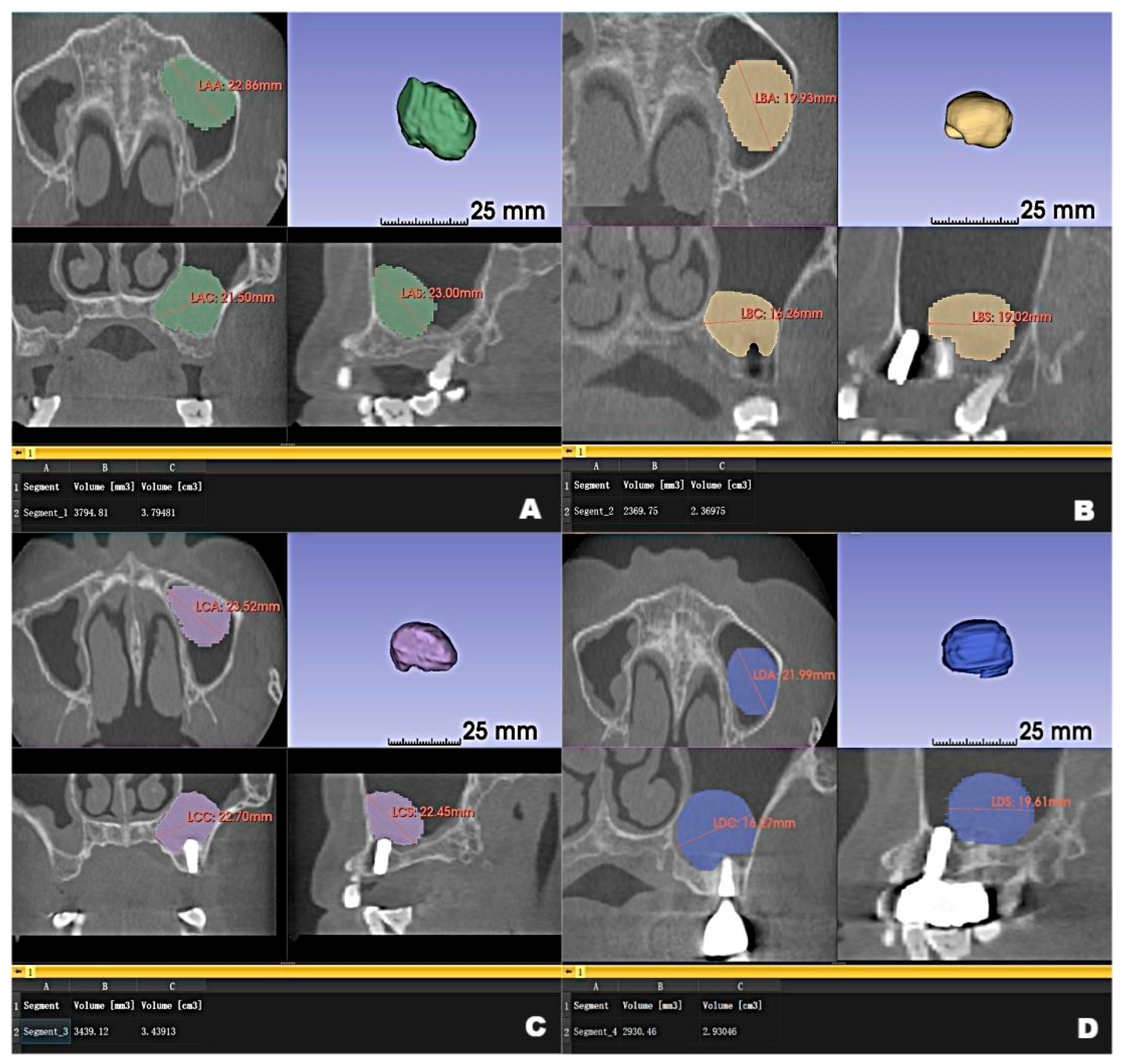
This report describes maxillary antral pseudocyst drift after maxillary sinus floor augmentation through osteotome sinus floor elevation with simultaneous implant placement. 3D Slicer was used to measure the pseudocyst and maxilla for the placement of the implants; follow-up visits were scheduled at 6, 12, and 22 months. No adverse effects were observed during or after surgery, and all implants exhibited osseointegration without mobility. At 6 months after surgery, the pseudocyst had moved posterolaterally from the preoperative position near the anterior medial maxillary sinus, then returned to its original position at 12 months. However, it had remigrated to the posterolateral position at 22 months. The preoperative volume of the pseudocyst was 3.795 mm3; it was 2.370, 3.439, and 2.930 mm3 at 6, 12, and 22 months after surgery, respectively. The changes in pseudocyst drift and volume did not have a substantial negative influence on the implants, presumably because of cystic attachment and the recurrence of multiple pseudocysts at different locations. The risks associated with changes in a pseudocyst can be avoided, if an appropriate treatment plan is selected.

1. Introduction

Anatomical variations and lesions of the maxillary sinus are common findings in cone beam computed tomography (CBCT) scans of the maxilla prior to placement of dental implants [1]. A clinical study of oral implant patients in a Chinese grade A stomatology hospital showed that the prevalence of maxillary antral cysts was 11.41%; many of these were pseudocysts, with dome-shaped soft tissue findings in radiographic assessment of the maxillary sinus floor [2,3].
In our clinical experience, relevant clinical symptoms are not present in most patients who exhibit a maxillary antral pseudocyst during radiographic assessment prior to implantation. Therefore, many patients prefer a conservative approach (i.e., pseudocyst preservation during implant surgery) because they have concerns about the risk of pseudocyst management surgery. Although no standard treatments and implant options have been established for patients with such antral lesions, the current literature suggests that the presence of a pseudocyst should not constitute an absolute contraindication for sinus grafting. Notably, surgery using a maxillary sinus lift procedure to resolve bone mass deficiency, which has achieved a generally high success rate in terms of guided bone augmentation and implant survival; the presence of a pseudocyst in the maxillary sinus may not influence the clinical effects of raising the sinus floor during healing [3,4,5].
A recent study showed that dental implant placement after antral augmentation in patients with pseudocysts is safe and results in a high survival rate, regardless of pseudocyst removal or preservation [6]. Although complete removal is the gold standard for the management of cystic lesions, this approach is limited by high rates of injury and complications [7,8]. Some studies have suggested that, unless clinically significant expansion is evident during radiographic assessment, or symptoms (e.g., headache) are present, a “wait and see” approach is an appropriate strategy for the management of retention cysts or pseudocysts [9,10,11,12,13].
The current literature contains multiple studies regarding the management of maxillary sinus pseudocysts associated with implantation, with a focus on the impacts of different management approaches; however, there are few reports of intraoperative and postoperative changes in these pseudocysts [3,6,8,9,14,15,16]. Notably, implant status is a key consideration during postoperative follow-up; however, because many patients refuse clinical management of asymptomatic pseudocysts, considerations regarding such pseudocysts should not be limited to preservation or surgical removal. Changes in pseudocyst size, location, and morphological characteristics after implantation should be closely monitored to guide subsequent treatment and facilitate risk assessment.
Since 1944, when Summer introduced his technique, osteotome sinus floor elevation using a trans-alveolar approach [17], the trans-alveolar approach for sinus floor elevation has been a recommended approach to address insufficient vertical bone height. This classical method can not only increase the density of the posterior maxillary, but also promote the initial stability of the implant [17,18]. Liu et al. [18] reported that immediate implant placement, combined with maxillary sinus floor elevation utilizing the trans-alveolar approach, revealed a satisfactory clinical effect in both submerged healing and non-submerged healing for the maxillary molar area.
Here, we describe implant placement with simultaneous maxillary sinus floor augmentation via osteotome sinus floor elevation in the presence of an antral pseudocyst. During the perioperative period and postoperative follow-up, the maxillary sinus pseudocyst exhibited recurrent drift on the implant side.

2. Case Report

A 53-year-old woman was referred to the Department of Oral Implantology, West China Stomatology Hospital of Sichuan University, for implant rehabilitation because of the bilateral loss of maxillary premolars and molars.

2.1. Preoperative Examinations

The gingiva surrounding the absent maxillary posterior teeth were in good condition, without ulceration or swelling. The patient consented to surgical treatment, with an initial focus on the left maxillary posterior region.
The anterior teeth of the patient were of shallow overjet and shallow overbite, and the occlusal relationship was relatively stable. The gingiva, with a normal width of attached gingiva, belonged to the biological type of thin gingiva. The patient’s temporomandibular joint was in a good condition, and the opening degree was greater than 3 fingers. The patient had no obvious symptoms of maxillary sinus blockage, runny nose, snoring, or and nasal congestion, did not exhibit any bad habits, such as night bruxism and one-sided chewing. No systemic diseases, such as diabetes, heart disease, or infectious disease, nor history of trauma or surgery, was reported.
CBCT examinations were performed on CBCT scanners (3D Accuitomo, J. Morita Mfg. Corp., Kyoto, Japan) using the following parameters: 85-kV tube voltage, 5.0-mA tube current, field of view of 100 mm × 50 mm, and slice thickness of 1.0 mm. Dental CBCT scans showed that the local alveolar ridge width was optimal (8.688 mm for the left maxillary second premolar and 10.130 mm for the first molar) (Figure 1B,D); however, sinus augmentation was needed because the height of the residual alveolar bone in the posterior maxillary area was insufficient for implant placement. The patient was observed to have type 3 bone quality in the posterior maxilla on both sides, and according to our observation, there was no obvious thickening of the nasal mucosa in the pre-operative CBCT image. Notably, the height of the crestal bone between the sinus floor and the alveolar ridges of the left maxillary second premolar and first molar were 8.688 mm and 4.385 mm, respectively (Figure 1A,C). CBCT also revealed a homogeneously opaque, dome-shaped, and well-delineated lesion (volume, 3.795 mm3) in the left maxillary sinus (Figure 1 and Figure 2A); the lesion did not exhibit an epithelial lining. Prior to attending our clinic, the patient had consulted an otolaryngologist and received confirmation that no mucous cyst was present (no aggressive lesion appearance, significant bone resorption, or invasion of adjacent structures observed on radiologic imaging). Because the patient did not exhibit relevant symptoms, the otolaryngologist suggested conservative management of the pseudocyst. Accordingly, the treatment plan comprised osteotome sinus floor elevation, with simultaneous implant placement on the left side, in the presence of an antral pseudocyst.

2.2. Surgical Procedures and Postoperative Management

Preoperative hematological tests revealed values within normal limits, and the patient did not show other systemic abnormalities. Thus, local anesthesia was induced with articaine hydrochloride plus 1:100,000 adrenalin (Primacaine, Merignac, France).
Osteotome sinus floor elevation was performed in the left posterior maxilla, followed by implantation of bovine bone xenograft (0.25 g; Bio Oss, Wolhusen, Switzerland). Two ITI implants (4.8 × 8 mm and 4.1 × 10 mm, ITI, Straumann, Basel, Switzerland) were inserted in the augmented sinus in a submerged mode. The torque values were 30 N·cm and 15 N·cm, respectively. During surgery, there was no pseudocyst rupture, pseudocyst fluid exudation, or maxillary sinus membrane perforation. Optimal implant orientation and spacing were achieved. Bone compression revealed acceptable initial stability; thus, healing abutments were simultaneously connected. The tissue flap was finely sutured, and no adverse effects were observed during or after surgery.
The patient was instructed to take the following medications after surgery: amoxicillin (Zhongnuo, Shijiazhuang, China), 1.5 g/day for 3 days; ornidazole dispersible (Meheco Topfond, Zhumadian, China), 1.0 g/day for 3 days; dexamethasone acetate (Xianju, Taizhou, China), 3 mg/day for 3 days; diclofenac sodium (Simcere, Nanjing, China), 0.1 g/day for 3 days; and compound gargle solution chlorhexidine gluconate (Chenpai Bond, Haimen, China), 3 times daily for 3 days. Routine follow-up visits were scheduled at 6, 12, and 22 months.

2.3. Postoperative Examinations

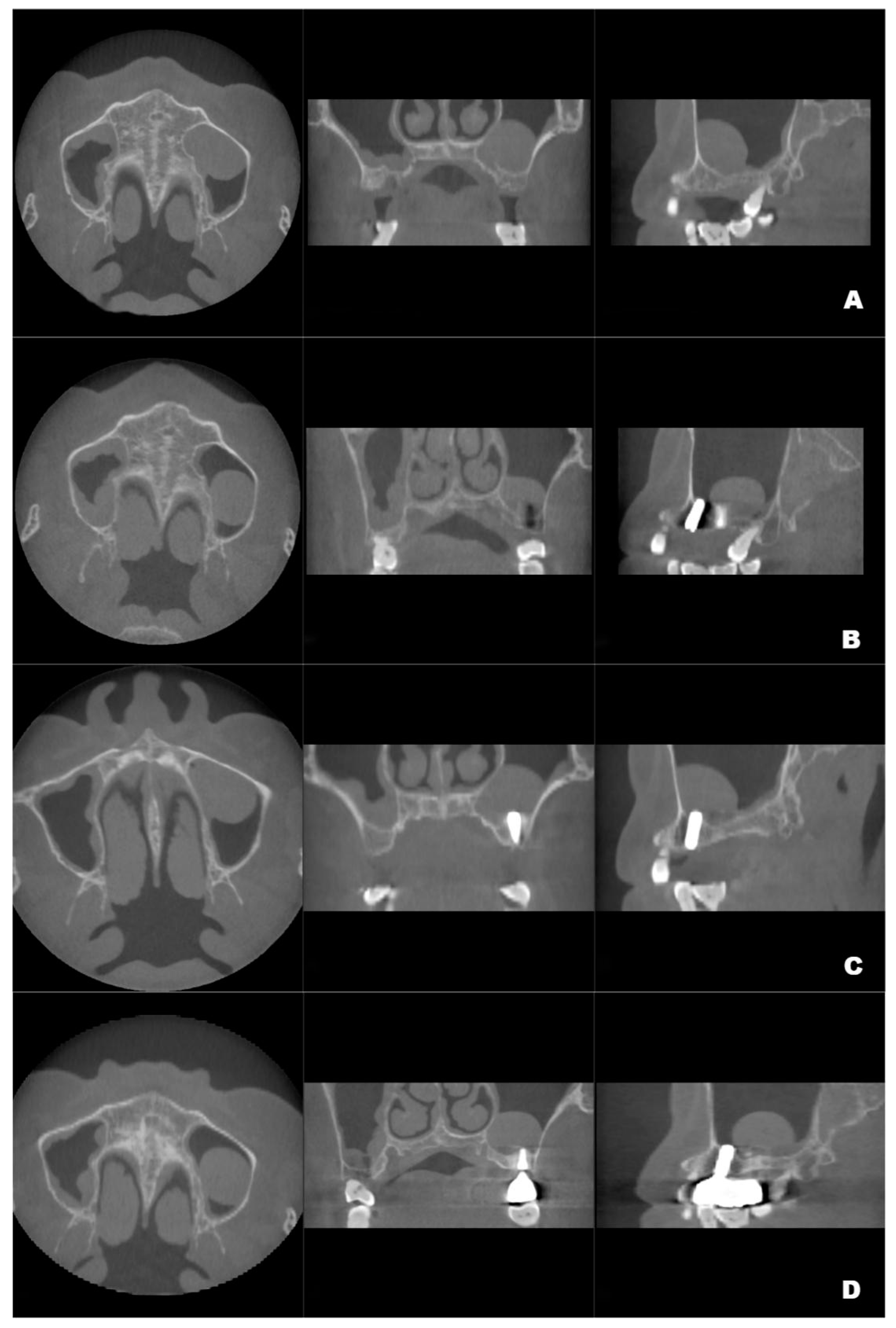
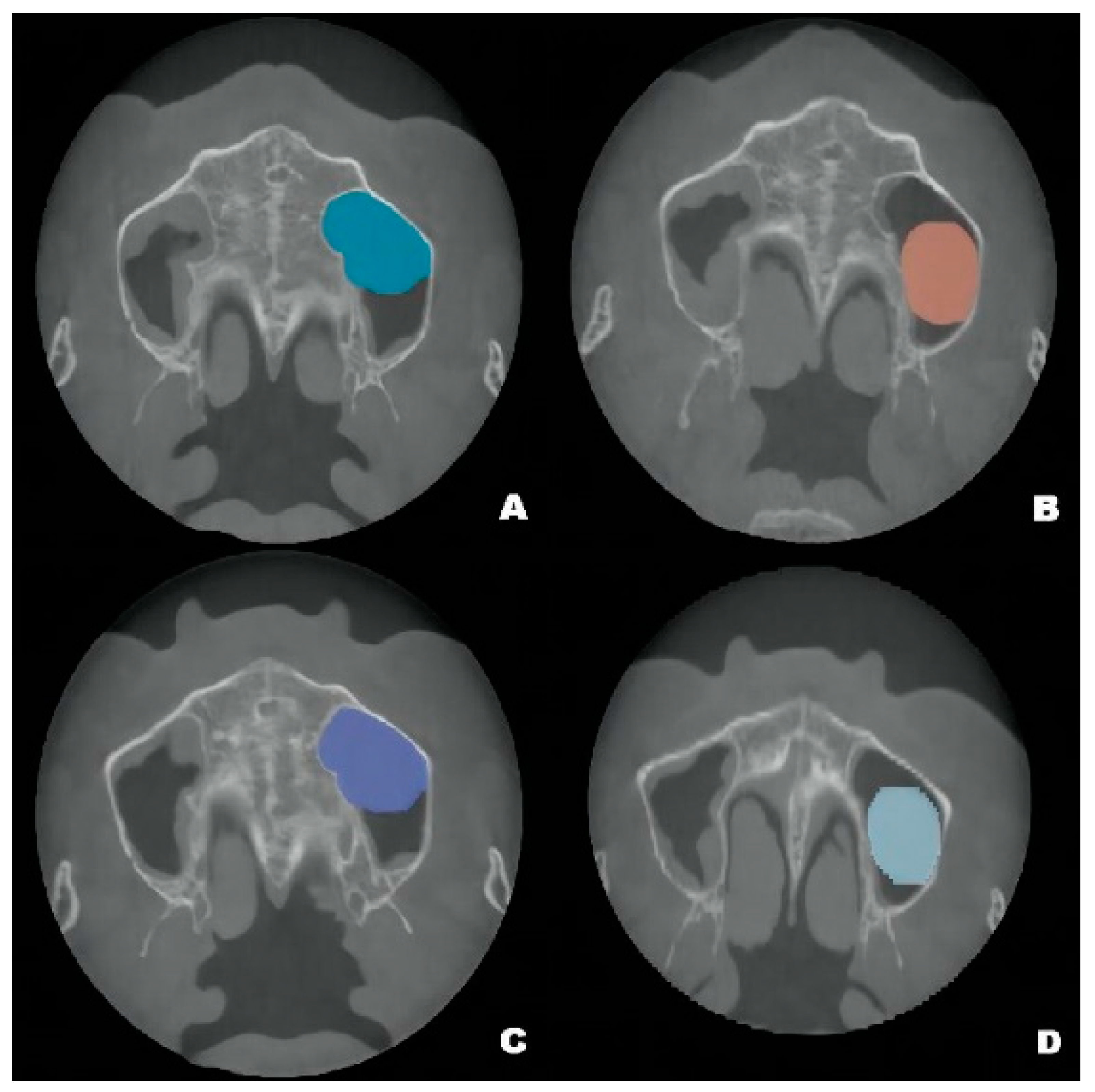
Although the final crown restoration (second procedure) was planned for 6 months after the first procedure, it was delayed until 10 months because of an outbreak of COVID-19. A CBCT scan at the 6-month follow-up showed that the implants had been correctly placed in the augmented sinus, resulting in osseointegration without mobility (Figure 3B). CBCT scans at 12 months and 22 months showed stable bone levels around the implants (Figure 3C,D). The patient reported no discomfort or other complaints in relation to the implants or the maxillary sinus, and no fluid leakage was evident on the incision line during follow-up. However, a comparison of the preoperative CBCT scans with the postoperative scans conducted at 6, 12, and 22 months revealed pseudocyst drift in the left maxillary sinus (Figure 4). At 6 months after surgery, the pseudocyst had moved posterolaterally from the preoperative position near the anterior medial maxillary sinus, then returned to its original position at 12 months. However, it had remigrated to the posterolateral position at 22 months.
The pseudocyst volume was manually segmented using 3D Slicer and corrected by a professional radiologist [19]. Measurements of the maxillary sinus pseudocyst revealed that its volume decreased from 3.795 mm3 before surgery to 2.370 mm3 at 6 months post-surgery; it gradually returned to the preoperative volume at 12 months (3.439 mm3), then decreased to 2.930 mm3 at 22 months (Figure 2).

3. Discussion

In this report, we have described the clinical treatment of a patient with a pseudocyst who underwent implant surgery, along with the postoperative findings that included an intriguing type of pseudocyst drift not reported in previous literature. Postoperative imaging showed that the presence of the pseudocyst, as well as its drift behavior, did not have a substantial adverse effect on the clinical outcomes of maxillary sinus augmentation and implantation. Successful osseointegration and stable bone levels around the implants were observed during follow-up.
Pseudocyst drift may result from the following factors. First, the pseudocyst may not be completely immobile on the maxillary sinus floor. The pseudocyst may be entirely detached from the sinus floor, or it may be solely attached to the anterior portion of the sinus floor. In either situation, various forces, including implant insertion, airflow pressure from breathing, and postural changes during radiographic assessment could influence pseudocyst location. Second, considering the changes in volume, multiple pseudocysts may be present; the sizes of the pseudocysts may increase and decrease over time.
The apparent reduction in pseudocyst volume after implantation may have resulted from the leakage of pseudocyst fluid during implantation; however, no obvious pseudocyst fluid leakage was observed during surgery because of difficulty distinguishing such fluid from saline solution. However, follow-up imaging revealed re-enlargement and re-contraction of the pseudocyst, indicating that the changes in volume were not solely related to surgery. We suspect that changes in the secretion and absorption of fluid from a single pseudocyst contributed to these imaging findings; alternatively, multiple pseudocysts were present in our patient. If multiple pseudocysts are present, the changes in volume may represent different pseudocysts and could explain the apparent pseudocyst drift behavior mentioned above.
Mucosal cysts of the maxillary sinus are common benign lesions that are generally harmless and asymptomatic; the incidence of such lesions ranges from 1.6% to 11.41% [2,14,15]. These lesions have three forms that are classified on the basis of their clinical features and biological characteristics: (1) true mucous cysts—thick-walled cystic lesions that can destroy bone and expand into adjacent structures, usually identified by their radiographic appearance; (2) retention cysts—small, opaque, mucus-filled lesions caused by the obstruction of mucosal glands, usually located near seromucinous glands around the ostium; (3) pseudocysts—lesions attached to the floor of the maxillary sinus, typically dome-shaped without an epithelial lining and occasionally filling the entire antral cavity, that either persist unchanged or disappear without an apparent reason [3,13,20] No medical or surgical treatment is needed for most mucosal cysts of the maxillary sinus if they exhibit spontaneous regression or maintain a consistent size [15].
Because there is no consensus regarding the indications and standard approach for sinus augmentation in patients with maxillary antral pseudocysts, multiple studies have investigated the clinical outcomes of available treatment options. We summarize the principal progress in recent research on this topic (Table 1).
Fu et al. [21] conducted a retrospective cohort study, dividing 26 patients into two groups (13 in the “removing the cyst group” and 13 in the “leaving the cyst alone” group). The results showed that antral pseudocyst removal before maxillary sinus floor augmentation and immediate implant placement after lateral sinus floor elevation could achieve higher bone graft volumetric stability. However, another retrospective cohort study revealed that the transcrestal raising of the sinus floor in the presence of antral pseudocysts may not have any influence on the clinical effects of raising the sinus floor during healing [5]. Sinus augmentation through a transcrestal window approach was described as a minimally invasive method for maxillary sinus augmentation [30]. Chiapasco and Palombo [24] combined pseudocyst enucleation with sinus grafting through a small bony window, which resulted in the survival of all 17 implants in their study. In addition to the discussion on whether to or not remove antral pseudocysts, researchers have also been focusing on improving the technology or combine the traditional method with some new technologies to reduce patient pain and various complications. Oh et al. [23] provided a technique combining the aspiration of the cysts during sinus floor elevation with a sinus augmentation procedure using a hydraulic sinus elevation system with simultaneous placement. After 6 months, a significant amount of bone formation around the implant at the sinus floor was observed, without evidence of cyst recurrence. Hu et al. [22] believed that the transoral endoscopic technique could be of great help in sinus floor augmentation, benefiting from its minimal invasion and optimal visualization. They successfully used the endoscopic-assisted intraoral surgical technique for sinus floor augmentation and the simultaneous removal of an antral pseudocyst. This method not only provided a clearer field of view for the complete removal of pseudocysts, but was also sensitive to small perforation, and only a small window was needed to observe the occurrence of mucosal perforation. Liu et al. [14] reported simultaneous implant placement with maxillary sinus elevation, along with pseudocyst treatment via cystic fluid extraction; all 14 patients were successfully treated without any discomfort, and all 28 implants were stable after 1 year of follow-up. Tang et al. [16] reported successful sinus augmentation without pseudocyst removal, along with simultaneous placement of two implants on the pseudocyst side; 1 year later, they observed a reduction in the dome-shaped opacity without bone graft resorption.
In patients with pseudocysts, the antral ostium may be blocked by obliteration of the sinus cavity, if bone graft overfilling occurs during sinus augmentation [4,6,7,8]. In addition to antral floor elevation using a lateral approach, the combination of an irregular graft surface with conventional bone graft volume may cause excessive pressure on the pseudocyst, leading to postoperative drainage of the pseudocyst fluid [3]. In our case, osteotome sinus floor elevation was chosen to minimize mucosal stimulation, antral sinus membrane perforation, and the loss of maxillary sinus space. This method involved using the concave edge of the internal lifting instrument to push the autologous bone in a manner that facilitated the formation of new bone in the sinus cavity. Moreover, the bone extrusion helped to improve the initial implant stability. The achievement of a satisfactory and stable clinical outcome, as well as the maintenance of pseudocyst integrity, indicated that our approach can reduce the risk of postoperative complications.
The cause of pseudocyst drift merits further investigation. However, our clinical findings may provide insights for other dentists. Notably, pseudocyst drift did not adversely affect the clinical outcomes of implantation or the patient’s postoperative quality of life. Thus, our findings suggest that the presence of maxillary sinus pseudocysts should not be regarded as an absolute indication for implant surgery; some risks associated with changes in maxillary pseudocysts can be minimized or avoided if an appropriate treatment plan is selected.

4. Conclusions

Changes in pseudocyst drift and volume did not have a substantial negative influence on the implants; this phenomenon may be related to the status of cystic attachment and the recurrence of multiple pseudocysts at different locations. The risks associated with changes in the pseudocysts themselves can be avoided if an appropriate treatment plan is selected.

Author Contributions

Conceptualization, P.W. and N.H.; methodology, B.H.; software, P.W.; validation, J.L., J.R. and P.G.; formal analysis, P.W.; investigation, N.H.; resources, B.H.; data curation, P.W.; writing—original draft preparation, P.W.; writing—review and editing, B.H and N.H; visualization, B.H.; supervision, P.G. and J.L.; project administration, B.H.; funding acquisition, B.H. All authors have read and agreed to the published version of the manuscript.

Funding

This research was funded by the National Outstanding Youth Science Fund Project of the National Natural Science Foundation of China (82001090), the Health Department of Sichuan Province (20PJ091), and the Sichuan University Post-Doctoral Interdisciplinary Innovation Startup Fund (20201105).

Institutional Review Board Statement

This study was conducted in accordance with the principles of the Declaration of Helsinki (2013). The study was approved by the Institutional Ethics Committee of West China Hospital of Stomatology, Sichuan University. Informed consent was obtained from the patient.

Informed Consent Statement

The authors obtained the written consent of the patient for the publication of the data and the images that appear in the article.

Data Availability Statement

The data are not publicly available to protect the information that contains the privacy of the participant. The data can be available from the author on reasonable request.

Acknowledgments

Written informed consent was obtained from the patient for the publication of this case report and accompanying images.

Conflicts of Interest

The authors declare no conflict of interest.

References

  1. Lana, J.P.; Carneiro, P.M.; Machado Vde, C.; de Souza, P.E.; Manzi, F.R.; Horta, M.C. Anatomic variations and lesions of the maxillary sinus detected in cone beam computed tomography for dental implants. Clin. Oral. Implants Res. 2012, 23, 1398–1403. [Google Scholar] [CrossRef] [PubMed]
  2. Xuran, W.; Yu, Z.; Ping, D.; Xi, J.; Ye, L. Anatomic variations and lesions of the maxillary sinus observed by CBCT before sinus augmentation in 666 sinuses of 589 patients. J. Pract. Stomatol. 2019, 35, 100–104. [Google Scholar]
  3. Kara, I.M.; Küçük, D.; Polat, S. Experience of maxillary sinus floor augmentation in the presence of antral pseudocysts. J. Oral. Maxillofac. Surg. 2010, 68, 1646–1650. [Google Scholar] [CrossRef] [PubMed]
  4. Carmeli, G.; Artzi, Z.; Kozlovsky, A.; Segev, Y.; Landsberg, R. Antral computerized tomography pre-operative evaluation: Relationship between mucosal thickening and maxillary sinus function. Clin. Oral. Implants Res. 2011, 22, 78–82. [Google Scholar] [CrossRef] [PubMed]
  5. Gong, T.; Hu, C.; Chen, Y.; Zhou, N.; Wu, H.; Man, Y. Raising the transcrestal sinus floor in the presence of antral pseudocysts, and in sinus floors with a normal Schneiderian membrane: A retrospective cohort study. Br. J. Oral. Maxillofac. Surg. 2019, 57, 466–472. [Google Scholar] [CrossRef]
  6. Anitua, E.; Alkhraisat, M.H.; Torre, A.; Eguia, A. Are mucous retention cysts and pseudocysts in the maxillary sinus a risk factor for dental implants? A systematic review. Med. Oral. Patol. Oral. Cir. Bucal. 2021, 26, e276–e283. [Google Scholar] [CrossRef]
  7. Beaumont, C.; Zafiropoulos, G.G.; Rohmann, K.; Tatakis, D.N. Prevalence of maxillary sinus disease and abnormalities in patients scheduled for sinus lift procedures. J. Periodontol. 2005, 76, 461–467. [Google Scholar] [CrossRef]
  8. Yu, H.; Qiu, L. Histological and clinical outcomes of lateral sinus floor elevation with simultaneous removal of a maxillary sinus pseudocyst. Clin. Implant. Dent. Relat. Res. 2019, 21, 94–100. [Google Scholar] [CrossRef] [Green Version]
  9. Mardinger, O.; Manor, I.; Mijiritsky, E.; Hirshberg, A. Maxillary sinus augmentation in the presence of antral pseudocyst: A clinical approach. Oral. Surg. Oral. Med. Oral. Pathol. Oral. Radiol. Endod. 2007, 103, 180–184. [Google Scholar] [CrossRef]
  10. Celebi, N.; Gonen, Z.B.; Kilic, E.; Etoz, O.; Alkan, A. Maxillary sinus floor augmentation in patients with maxillary sinus pseudocyst: Case report. Oral. Surg. Oral. Med. Oral. Pathol. Oral. Radiol. Endod. 2011, 112, e97–e102. [Google Scholar] [CrossRef]
  11. Gardner, D.G. Pseudocysts and retention cysts of the maxillary sinus. Oral. Surg. Oral. Med. Oral. Pathol. 1984, 58, 561–567. [Google Scholar] [CrossRef]
  12. Bhattacharyya, N. Do maxillary sinus retention cysts reflect obstructive sinus phenomena? Arch. Otolaryngol. Head Neck Surg. 2000, 126, 1369–1371. [Google Scholar] [CrossRef] [Green Version]
  13. Wang, J.H.; Jang, Y.J.; Lee, B.J. Natural course of retention cysts of the maxillary sinus: Long-term follow-up results. Laryngoscope 2007, 117, 341–344. [Google Scholar] [CrossRef] [PubMed]
  14. Liu, D.; Shi, L.; Dai, X.; Zhou, Q.; Yang, F.; Shen, M.; Yu, Y.; Wu, Y. Implants placed simultaneously with maxillary sinus floor augmentation in the presence of antral pseudocysts: Presentation of a case series. Quintessence Int. 2018, 49, 479–485. [Google Scholar] [PubMed]
  15. Giotakis, E.I.; Weber, R.K. Cysts of the maxillary sinus: A literature review. Int. Forum. Allergy Rhinol. 2013, 3, 766–771. [Google Scholar] [CrossRef] [PubMed]
  16. Tang, Z.H.; Wu, M.J.; Xu, W.H. Implants placed simultaneously with maxillary sinus floor augmentations in the presence of antral pseudocysts: A case report. Int. J. Oral. Maxillofac. Surg. 2011, 40, 998–1001. [Google Scholar] [CrossRef] [PubMed]
  17. Summers, R.B. A new concept in maxillary implant surgery: The osteotome technique. Compendium 1994, 15, 152, 154–156, 158 passim; quiz 162. [Google Scholar] [PubMed]
  18. Liu, H.; Liu, R.; Wang, M.; Yang, J. Immediate implant placement combined with maxillary sinus floor elevation utilizing the transalveolar approach and nonsubmerged healing for failing teeth in the maxillary molar area: A randomized controlled trial clinical study with one-year follow-up. Clin. Implant Dent. Relat. Res. 2019, 21, 462–472. [Google Scholar] [CrossRef]
  19. Fedorov, A.; Beichel, R.; Kalpathy-Cramer, J.; Finet, J.; Fillion-Robin, J.C.; Pujol, S.; Bauer, C.; Jennings, D.; Fennessy, F.; Sonka, M.; et al. 3D Slicer as an image computing platform for the Quantitative Imaging Network. Magn. Reson Imaging 2012, 30, 1323–1341. [Google Scholar] [CrossRef] [Green Version]
  20. Skoulakis, C.E.; Velegrakis, G.A.; Doxas, P.G.; Papadakis, C.E.; Bizakis, J.G.; Helidonis, E.S. Mucocele of the maxillary antrum in an eight-year-old boy. Int. J. Pediatr. Otorhinolaryngol. 1999, 47, 283–287. [Google Scholar] [CrossRef]
  21. Fu, M.; Jiang, Z.; Ren, L.; He, J.; Zhu, D.; Yang, G. Maxillary sinus floor augmentation comparing removing versus retaining antral pseudocyst: A retrospective cohort study. Clin. Oral. Implants Res. 2022, 33, 1098–1113. [Google Scholar] [CrossRef]
  22. Hu, Y.K.; Yang, C.; Qian, W.T. Endoscopic-Assisted Sinus Floor Augmentation Combined With Removal of an Antral Pseudocyst of the Ipsilateral Maxillary Sinus. J. Craniofac. Surg. 2017, 28, 1549–1551. [Google Scholar] [CrossRef]
  23. Oh, J.H.; An, X.; Jeong, S.M.; Choi, B.H. Crestal Sinus Augmentation in the Presence of an Antral Pseudocyst. Implant Dent. 2017, 26, 951–955. [Google Scholar] [CrossRef] [PubMed]
  24. Chiapasco, M.; Palombo, D. Sinus grafting and simultaneous removal of large antral pseudocysts of the maxillary sinus with a micro-invasive intraoral access. Int. J. Oral. Maxillofac. Surg. 2015, 44, 1499–1505. [Google Scholar] [CrossRef]
  25. Feng, Y.; Tang, Y.; Liu, Y.; Chen, F.; Li, D. Maxillary sinus floor elevation using the osteotome technique in the presence of antral pseudocysts: A retrospective study with an average follow-up of 27 months. Int. J. Oral. Maxillofac. Implants 2014, 29, 408–413. [Google Scholar] [CrossRef] [PubMed] [Green Version]
  26. Acocella, A.; Bertolai, R.; Nissan, J.; Ellis, E., 3rd; Sacco, R. Maxillary sinus lift using fresh frozen bone chips in presence of sinus cyst: Clinical and histological report. Cell Tissue Bank 2012, 13, 327–332. [Google Scholar] [CrossRef] [PubMed]
  27. Cortes, A.R.; Corrêa, L.; Arita, E.S. Evaluation of a maxillary sinus floor augmentation in the presence of a large antral pseudocyst. J. Craniofac. Surg. 2012, 23, e535–e537. [Google Scholar] [CrossRef]
  28. Kara, M.I.; Kirmali, O.; Ay, S. Clinical evaluation of lateral and osteotome techniques for sinus floor elevation in the presence of an antral pseudocyst. Int. J. Oral. Maxillofac. Implants 2012, 27, 1205–1210. [Google Scholar]
  29. Lin, Y.; Hu, X.; Metzmacher, A.R.; Luo, H.; Heberer, S.; Nelson, K. Maxillary sinus augmentation following removal of a maxillary sinus pseudocyst after a shortened healing period. J. Oral. Maxillofac. Surg. 2010, 68, 2856–2860. [Google Scholar] [CrossRef]
  30. Wang, M.; Yan, M.; Xia, H.; Zhao, Y. Sinus Elevation Through Transcrestal Window Approach and Delayed Implant Placement in 1- to 2-mm Residual Alveolar Bone: A Case Report. Implant Dent. 2016, 25, 866–869. [Google Scholar] [CrossRef]
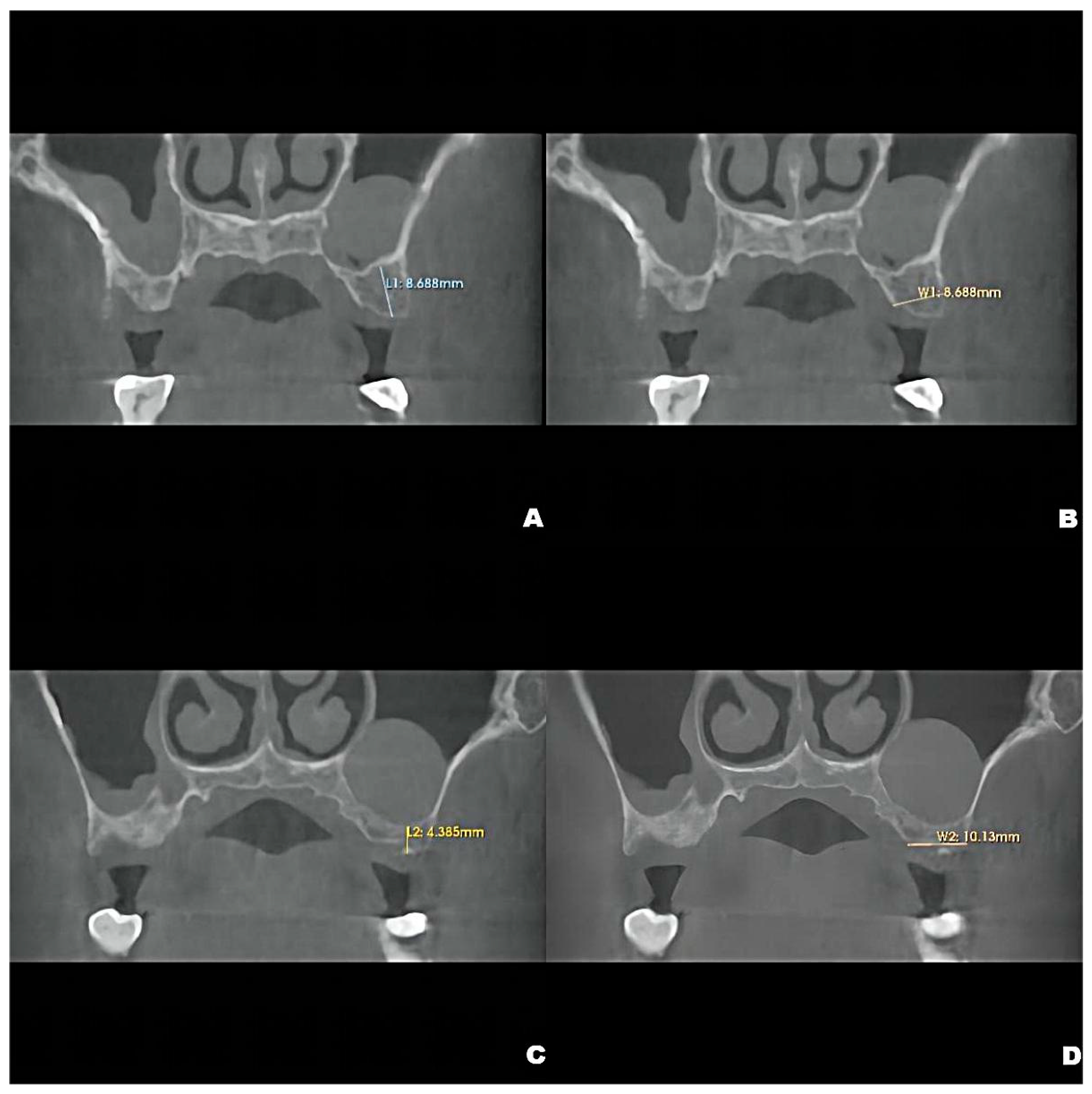
Figure 1. Preoperative CBCT showing the crestal height of L1 = 8.688 mm, (A), and local alveolar ridge width of W1 = 8.688 mm, (B), of the residual bone of the left maxillary second premolar and the crestal height of L2 = 4.285 mm, (C), and alveolar ridge width of W2 = 10.130 mm, (D), of the residual bone of the left maxillary first molar.
Jcm 12 00920 g001
Figure 2. The volume changes and maximum diameter changes in the axial, coronal and sagittal planes of the patient’s left maxillary antral pseudocyst in the preoperative CBCT image (A) and the follow-up CBCT examinations in 6 (B), 12 (C) and 22 months (D), respectively.
Jcm 12 00920 g002
Figure 3. Preoperative CBCT (A) and follow-up CBCT images (6 months (B), 12 months (C), and 22 months (D)) of the patient’s left maxillary antral pseudocyst in the axial, coronal, and sagittal planes.
Jcm 12 00920 g003
Figure 4. Pseudocyst drift behavior: the pseudocyst had moved posterolaterally from the preoperative position (A) at 6 months after surgery (B), then returned to its original position at 12 months after surgery (C). At 22 months, the pseudocyst had remigrated to the posterolateral position (D).
Jcm 12 00920 g004
Table 1. Recent studies on the treatment options and corresponding clinical outcomes of maxillary antral pseudocysts.
NOAuthor
(Year)
Number of CasesSurgical Method of Maxillary Antral Floor ElevationManagement of Antral PseudocystsImplant TimingFollow-UpOutcome
1Fu et al. [21]
(2022)
26LSFERemoved in 13 cases1-Stage12-month, ~2 to 5 yearsFailure in 1 case at 12 months, with higher bone graft volumetric stability after 6–12 months.
Preserved in 13 cases1-StageFailure in 2 cases at 12 months and 2~5-years follow-up, respectively.
2Gong et al. [5]17CSFEPreserved1-Stage4–6 monthsOsseointegration in all cases.
3(2019)
Yu and Qiu [8]
(2019)
15LSFEPreserved1-Stage6 monthsFailure in 1 implant before loading.
4Liu et al. [14]
(2018)
14LSFECystic fluid extraction1-Stage12 monthsOsseointegration in all cases.
5Hu et al. [22]
(2017)
1Endoscopic-Assisted LSFERemoved2-Stage12 monthsOsseointegration
6Oh et al. [23]
(2017)
2CSFECystic fluid extraction1-Stage6 monthsOsseointegration in all cases.
7Chiapasco and Palombo [24]
(2015)
12LSFERemoved1-Stage in 7 cases12–96 monthsOsseointegration in all cases.
2-Stage in 5 cases
8Feng et al. [25]
(2014)
21OSFE Preserved1-StageAverage of 27 monthsOsseointegration in all cases.
9Acocella et al. [26]
(2012)
1LSFERemoved2-Stage2 yearsOsseointegration
10Cortes et al. [27]
(2012)
1LSFERemoved1-Stage12 monthsOsseointegration
11Kara et al. [28]
(2012)
29LSFE in 17 casesPreserved1-StageAverage of 17 monthsOsseointegration in all cases.
OSFE in 12 cases2-Stage
12Celebi et al. [10]
(2011)
4LSFE in 2 casesPreserved1-Stage6–8 monthsOsseointegration in all cases.
CSFE in 2 cases2-Stage
13Tang et al. [16]
(2011)
1LSFEPreserved1-Stage12 monthsOsseointegration
14Lin et al. [29]
(2010)
11LSFERemoved2-StageAverage of 29.2 months after prosthetic loading Osseointegration
15Kara et al. [3]
(2010)
2LSFE in 1 casePreserved2-Stage16 monthsOsseointegration in all cases.
CSFE in 1 case13 months
16Mardinger et al. [9]
(2007)
8LSFEPreserved1-Stage in 7 casesAverage of 20 monthsOsseointegration in all cases.
2-Stage in 1 case
LSFE: lateral sinus floor elevation; CSFE: crestal sinus floor elevation; OSFE: osteotome sinus floor elevation.
Disclaimer/Publisher’s Note: The statements, opinions and data contained in all publications are solely those of the individual author(s) and contributor(s) and not of MDPI and/or the editor(s). MDPI and/or the editor(s) disclaim responsibility for any injury to people or property resulting from any ideas, methods, instructions or products referred to in the content.