Article

A Potential Therapy Using Antisense Oligonucleotides to Treat Autosomal Recessive Polycystic Kidney Disease

1
Department of Nephrology, Children’s Hospital of Nanjing Medical University, Nanjing 210008, China
2
Nanjing Key Laboratory of Pediatrics, Children’s Hospital of Nanjing Medical University, Nanjing 210008, China
3
Jiangsu Key Laboratory of Pediatrics, Nanjing Medical University, Nanjing 210008, China
*
Authors to whom correspondence should be addressed.
These authors contributed equally to this work.
J. Clin. Med. 2023, 12(4), 1428; https://doi.org/10.3390/jcm12041428
Submission received: 27 December 2022 / Revised: 1 February 2023 / Accepted: 8 February 2023 / Published: 10 February 2023

Abstract

:
(1) Background: Autosomal recessive polycystic kidney disease (ARPKD) is a rare ciliopathy characterized by progressively enlarged kidneys with fusiform dilatation of the collecting ducts. Loss-of-function mutations in the PKHD1 gene, which encodes fibrocystin/polyductin, cause ARPKD; however, an efficient treatment method and drug for ARPKD have yet to be found. Antisense oligonucleotides (ASOs) are short special oligonucleotides which function to regulate gene expression and alter mRNA splicing. Several ASOs have been approved by the FDA for the treatment of genetic disorders, and many are progressing at present. We designed ASOs to verify whether ASOs mediate the correction of splicing further to treat ARPKD arising from splicing defects and explored them as a potential treatment option. (2) Methods: We screened 38 children with polycystic kidney disease for gene detection using whole-exome sequencing (WES) and targeted next-generation sequencing. Their clinical information was investigated and followed up. The PKHD1 variants were summarized and analyzed, and association analysis was carried out to analyze the relationship between genotype and phenotype. Various bioinformatics tools were used to predict pathogenicity. Hybrid minigene analysis was performed as part of the functional splicing analysis. Moreover, the de novo protein synthesis inhibitor cycloheximide was selected to verify the degraded pathway of abnormal pre-mRNAs. ASOs were designed to rescue aberrant splicing, and this was verified. (3) Results: Of the 11 patients with PKHD1 variants, all of them exhibited variable levels of complications of the liver and kidneys. We found that patients with truncating variants and variants in certain regions had a more severe phenotype. Two splicing variants of the PKHD1 genotypes were studied via the hybrid minigene assay: variants c.2141-3T>C and c.11174+5G>A. These cause aberrant splicing, and their strong pathogenicity was confirmed. We demonstrated that the abnormal pre-mRNAs produced from the variants escaped from the NMD pathway with the use of the de novo protein synthesis inhibitor cycloheximide. Moreover, we found that the splicing defects were rescued by using ASOs, which efficiently induced the exclusion of pseudoexons. (4) Conclusion: Patients with truncating variants and variants in certain regions had a more severe phenotype. ASOs are a potential drug for treating ARPKD patients harboring splicing mutations of the PKHD1 gene by correcting the splicing defects and increasing the expression of the normal PKHD1 gene.

1. Introduction

Autosomal recessive polycystic kidney disease (ARPKD) is a rare ciliopathy caused by homozygous or compound heterozygous mutations in the polycystic kidney and hepatic disease 1 gene (PKHD1). The incidence of ARPKD is estimated to be 1/20,000 of live births. It features progressively growing multiple cysts in the collecting ducts of the kidney and further development to end-stage renal disease (ESRD), but also presents as congenital hepatic fibrosis or even portal hypertension [1,2,3]. ESRD typically occurs in the first 10 years of life and leads to the need for renal replacement therapy before the age of 20 [4] in approximately 50% of cases and progressive hepatobiliary disease requiring a liver transplant in 10% of cases. A variable age of onset has also been reported, ranging from 0.67 to 14 years. The PKHD1 gene is a large genomic segment that spans ~472 kb, consisting of 4074 amino acids on chromosome 6p12.2, encoding fibrocystin/polyductin (FPC) in the collecting tubules of the kidney. FPC is a ubiquitously expressed single transmembrane (TM)-spanning protein, although it is more abundant in the primary cilium or basal body complex of epithelial cells in the renal tubules and hepatic bile ducts [5,6]. PKHD1 mutations lead to the extension and dilation of the collecting ducts to form microcysts, further resulting in the progressive enlargement of the kidneys. To date, more than 800 PKHD1 variants have been reported (Human Gene Variant Database, HGMD, April 2022, date last accessed). The existing disease management mainly consists of symptomatic treatment, the use of RRT therapy and even transplantation at the late stage of the disease.
Gene therapy is increasingly being applied to a variety of diseases [7,8]. Missense or nonsense variants in the PKHD1 gene are the most common (69.7%). However, the large genomic size of PKHD1 and the highly intricate proteolytic processing of the FPC protein in the mechanism of the protein translation processes made the study of PKHD1/FPC particularly difficult. Among the PKHD1 gene variants, 7.7% are splicing variants, reminding us of the necessity of studying the pathogenic characteristics of and potential treatments for splicing variants in PKHD1. Nucleic acid medicines such as small interfering RNAs (siRNAs) and antisense oligonucleotides (ASOs) have been verified as promising potent drugs for the treatment of incurable genetic disorders and are emerging as some of the most pragmatic therapeutic modalities for the multimodal treatment of genetic and rare diseases [9,10]. Since the discovery of ASOs in 1978, research on small nucleic acid drugs has shown continuous progress [11]. ASOs are single-stranded oligonucleotides which specifically bind to the target mRNA and cause steric hindrance by cleaving the mRNA, by degrading the target’s transcript or by closing the key regions through complementary anti-parallel-based pairing [12,13], which in turn lead to altered mRNA processing and modulated gene expression. Clinical experiments of ASOs have been carried out since 1993, and ASOs have gradually become available and applied to clinical practice since 2016.
In recent years, ASO-based therapies for specific splicing variants in patients have been successfully developed [14]. In 2016, the FDA approved nusinersen, the first ever targeted intrathecal injection therapy using an antisense drug, which is used for spinal muscular atrophy (SMA). It stimulates the production of the functional SMN protein of the spinal column and CNS (including motor neurons) by inducing the retention of Exon 7 of the SMN2 gene, resulting in improvements in motor activity and survival [15,16,17]. ASOs are also useful for generating a partially functional truncated protein by causing exon skipping to restore in-frame reading of the dystrophin gene to achieve a treatment for Duchenne muscular dystrophy (DMD). Eteplirsen, which was approved by the FDA in September 2016, is designed to promote the in-frame skipping of dystrophin’s Exon 51 [18], whereas golodirsen (which was approved in December 2019) is designed to cause the skipping of Exon 53 [19,20], and casimersen causes the skipping of Exon 45 [21].
To date, more than 50 antisense drugs are now in clinical trials worldwide. ASOs’ huge potential is linked to their illness fighting ability. In this research, we attempted to ascertain novel exon-extending targets in PKHD1 by reporting the relationship between exons’ overexpressed patterns and the rescue of extended mRNA expression in cells. We showed that all ASOs targeting PKHD1′s Exon 21, to a great extent, restore the expression of the normal transcript in cells, indicating that these ASOs might be promising therapeutic agents in the treatment of ARPKD caused by Exon 21 splicing mutations of the PKHD1 gene.

2. Materials and Methods

2.1. Clinical and Laboratory Data

Our center carried out genetic sequencing screening in 38 children with PKD. We enrolled these children with cysts in their kidneys, and the clinical data, ultrasound findings and laboratory test results were collected in detail. The clinical data included age at diagnosis, ethnicity, phenotypic appearance (renal cysts, kidney enlargement, Caroli disease, hepatic cysts, splenomegaly, etc.), family history and relevant laboratory data. The patients’ parents or legal guardians gave informed consent. The institutional ethics committee of the Children’s Hospital of Nanjing Medical University approved our study before whole-exome sequencing was carried out.

2.2. Genetic Analysis

Genetic tests were performed as previously described. Genomic DNA was extracted using a DNA isolation kit (Tiangen, Beijing, China) following the manufacturer’s protocol. We amplified the sequences including the exonic and intron–exon boundaries of PKHD1 using a polymerase chain reaction (PCR) and designed primers using Primer Premier 5.0 (Palo Alto, CA, USA). The products of PCR were purified using the QIAquick PCR Purification Kit (Qiagen, Shanghai, China) and were then sequenced using the Big Dye Terminator (Applied Biosystems, Foster City, CA, USA) via an ABI 3130 genetic analyzer (Applied Biosystems). All variants were denoted on the basis of the NCBI reference sequence for PKHD1 (NM_138694.4). Genomic DNA was used for whole-exome sequencing (WES) and a targeted next-generation sequencing test (including 140 short related genes, which are shown in Supplementary Table S1). The test was performed on an Illumina HiSeq 2500 sequencing platform for 150 bp paired reads. The sequences were compared with the reference human genome (hg19 build), and the deletions (INDEL), inserts and single nucleotide variations (SNV) were filtered. The variants were annotated and those with allele frequencies of more than 1% were removed by the databases (dbSNP, an in-house MAF database; 1000 Genomes MAF (Chinese); Genome Aggregation Database (gnomAD) and ExAC). All candidate variants were selected via the criteria of the ACMG (American College of Medical Genetics and Genomics) and further verified via Sanger sequencing.

2.3. In Silico Analysis

The potential pathogenicity of novel missense variants of PKHD1 were predicted using bioinformatics tools such as SIFT, PolyPhen-2, Provean, ACMG, VariantTaster and Consurf conservative prediction. Splice Site software was used to predict the latent impact of the consensus 5′ and 3’ splice sites and to predict the generation and/or activation of new splicing sites. We used the MaxEntScan (http://hollywood.mit.edu/burgelab/maxent/Xmaxentscan_scoreseq.html, accessed on 29 March 2021), SpliceAI (https://spliceailookup.broadinstitute.org/, accessed on 29 March 2021) and Human Splicing Finder (http://www.umd.be/HSF3/HSF.shtml, accessed on 29 March 2021). The analysis of the variants with these bioinformatics tools was carried out simultaneously.

2.4. Construction of the Plasmid

The creation of hybrid minigene constructs was based on the pSPL3 vector, which contains a conventional expression system with two exons (SD6 and SA2) to further prompt the synthetic mRNA transcript analysis [22]. The sequence of upstream primer SD6 is (5′-3′) TCTGAGTCACCTGGACAACC and the downstream SA2 is (5′-3′) ATCTCAGTGGTATTTGTGAGC. The fragment containing PKHD1 target exon (22, 47 and 61) fragments where the variants were located and at least 200 bp of their intronic flanking regions with XhoI and BamHI restriction sites were amplified via PCR using specific oligonucleotide primers and the extracted DNA. The web-based source primer (https://www.ncbi.nlm.nih.gov/tools/primer-blast/index.cgi, accessed on 1 April 2021) was used to design the primers, which are shown in the Supplementary Table S2. PCR products were purified and sequenced, and then were cloned into the pSPL3 vector via restriction enzymes XhoI and BamHI and the wild-type and mutant minigene recombinants constructed. A hybrid minigene mainly produces two transcripts; one is a larger transcript located in the upper part of the figure (SD6, the inserted exon and SA2), another is a smaller-sized (263 bp) transcript located below (SD6 and SA2) (Figure 1). The obtained wild-type (WT) and mutant plasmids were verified using bi-directional sequencing.

2.5. In Vitro Minigene Assay

HEK293T, HeLa, A549 and HepG2 cells were seeded with 10% PAN serum (DMEM/10% FAN) in an environment of 5% CO2 at 37 °C until they were 75–80% confluent. The minigenes (wild-type and mutants) of PKHD1 were transiently transfected into the HEK293T, HeLa, A549 and HepG2 cell lines using Lipofectamine 2000 (Invitrogen, Carlsbad, CA, USA) or PolyJetTM DNA Transfection Reagent (Signagen, Shanghai, China), following the instruction manuals.
Total RNA was extracted after 48 h of transfection and was reverse-transcribed into cDNA. PCR amplification was performed by the primers SD6 and SA2. The size and transcripts of the products of PCR were analyzed after agarose gel electrophoresis and sequenced. The following formula was used to quantify the percentage of aberrant splicing: exclusion percentage (%) = (lower band/(lower band + upper band)) × 100. The error bar represents the SEM (n = 3, * p < 0.05, unpaired Student’s t-test).

2.6. Design of Antisense Oligonucleotides

Mfold software was used to analyze the structure of the RNA near the PKHD1 gene variant c.2141-3T>C and to confirm the open and closed regions. Subsequently, the appropriate sequences of ASOs were chosen, which included nucleotide lengths between 16 and 25, a GC content from 40 to 60% and a Tm higher than 48 °C. In addition, we also verified that the free energy value of the ASO was above −4, the free energy value of the dimer was above −14 and the difference in the energy values between the candidate region and the ASOs’ binding region was 21–28. The precise sequences were UUAAGAUGGUAGACUUGCUGUGUGG (ASO1), AGACUUGCUGUGUGGAAAAUCC (ASO2) and GUGACUUAAGAUGGUAGACUUGCU (ASO3) (Figure 2D). All ASOs with phosphorothioate chains and 2-methyl sugar modifications were synthesized by Tsingke (Tsingke Biological Technology, Beijing, China).

2.7. ASOs Rescue Assays

HEK293T cells were cultured and transfected with minigene plasmids. The ASOs were transfected at a concentration of 0.5 μM, 24 h after transfection. Total RNA was extracted after 48 h of transfection for transcriptional analysis via RT–PCR.

3. Results

3.1. Clinical Analysis

We identified 11 patients who carried homozygous or compound heterozygous PKHD1 gene variants out of the 38 individuals with kidney cysts. The age of onset of 11 patients (six females and five males) ranged from 0.67 to 14 years (median = 5 years). All patients were identified from the Pediatric Nephrology Department and had been assessed by an experienced sonographer, except for Patient 8 (who was born with cysts). Patients 2, 9 and 11 had a family history of renal cysts (Figure S1). Patients 1 and 6 also presented with inguinal hernia disease. Patients 2 and 9 suffered from multiple recurrent urinary tract infections, and Patient 9 presented with the renal complication of kidney stones. Patient 8 was diagnosed at birth because of their apparent symptoms and signs of peripheral facial paralysis, hypothyroid and atrial septal defects. Moreover, Patient 3, who had the severe hepatic complication of Caroli disease, manifested with hepatosplenomegaly, intrahepatic bile duct dilatation, liver fibrosis, severe portal systemic shunting and portal hypertension, which resulted in an emergency laparoscopic splenectomy in 2020; the patient was accepted for allogeneic kidney transplantation last year. Patient 10 was on a regimen of oral ACEI combined with diuretics, and had long-lasting hematuria, hypertension and mild proteinuria, but normal renal function. The eGFR for all patients was greater than 100 mL·min–1·m–2, although some had elevated serum creatinine (such as Patient 9 in this study). End-stage renal disease has developed in no patient so far. The clinical features of the 11 patients are shown in Table 1.

3.2. Analysis of PKHD1 Variants

We identified 17 PKHD1 variants (3 nonsense, 10 missense, 1 frameshift and 3 splice sites) in the 11 different pedigrees (Table 2, Figure S1). The missense variants c.325G>A(p.(Ala109Thr)), c.3500T>C(p.(Leu1167Pro)), c.5869G>A(p.(Asp1957Asn)), c.6245C>T(p.(Thr2082Ile)), c.10072G>A(P.(Asp3358Asn)) and c.11525G>A(p.(Arg3842Gln)) had not been reported in the genomic databases or the previous literature. The six novel missense variants p.Ala109Thr, p.Leu1167Pro, p.Asp1957Asn, p.Thr2082Ile, p.Asp3358Asn and p.Arg3842Gln were all predicted to be damaging and deleterious, and probably affected protein function, according to the in silico analysis (Table S3). At the same time, the variants p.Leu1167Pro, p.Asp1957Asn, p.Asp3358Asn and p.Arg3842Gln were predicted to be highly conservative.
A novel splicing variant, c.11174+5G>A, which is located at +5 bp near the splice donor site in Intron 61 (Figure 1E and Figure 3A), was detected in Patient 3 and his mother. Another novel variant, c.2141-3T>C, was a T/C transition located at −3 bp near the splice acceptor site in Intron 21 in Patient 1 (Figure 1D). The c.11174+5G>A variant should be classified as VUS (PM2 + PP3), and c.2141-3T>C should be classified as VUS (PM2 + PM5 + PP3) according to the ACMG’s standard. The SpliceAI analysis indicated that the variant c.2141-3T>C activated the cryptic splice acceptor site of the intron, whereas c.11174+5G>A may disturb the authentic splice donor site and activate an intronic potential donor site of Intron 61 (Table S4). Furthermore, four bioinformatic splicing tools (Human Splicing Finder, MaxEntScan, NNSplice and BDGP) predicted that c.2141-3T>C would activate the splice acceptor site and the variant c.11174+5G>A would disrupt the splice donor site (Table S4). Nephronophthisis (NPHP)-related genes or protein kinase D1 (PKD1) or PKD2 with pathogenic mutations were not detected.

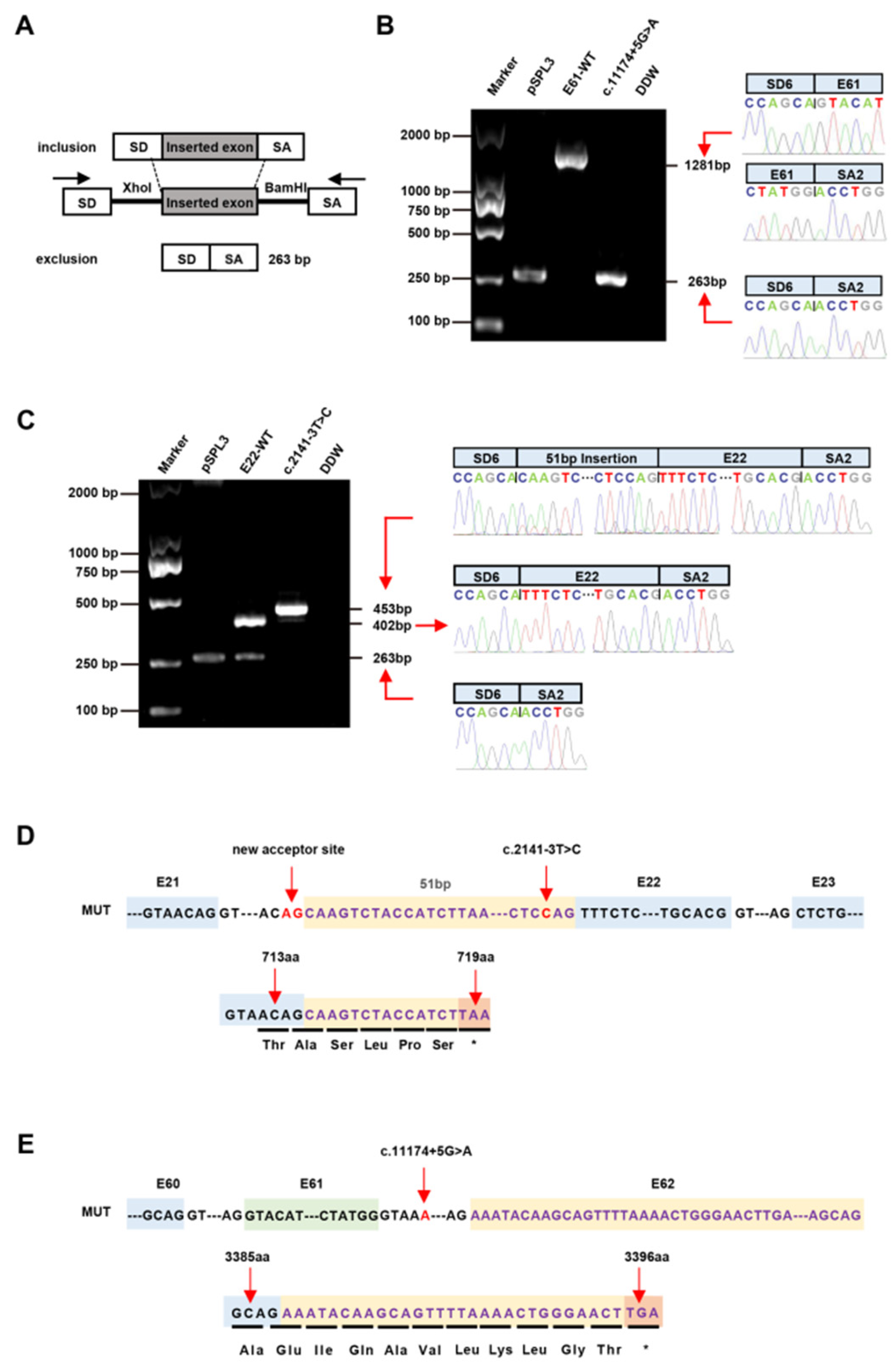
3.3. Splicing Analysis of the PKHD1 Minigene

Given the pathogenic potential of the nonclassical splicing variants and the fact that samples from patients are not always available, the functional tests of hybrid minigenes in splicing reporter plasmids, such as pSPL3, have become valuable tools to check the splicing profiles induced by a sequence variation [29,30]. We first cloned the classical donor site splice variant c.7351-2A>T from Patient 1 into the pSPL3 minigene vector. The RT-PCR results showed that the c.7351-2A>T variant induced aberrant splicing patterns (Exon 47 skipping) which confirmed that the pSPL3 minigene reporter was a dependable tool for functionally assaying the potential splice site variants (see Figure S2).
To further identify the pathogenicity of the c.2141-3T>C and c.11174+5G>A variants, wild-type (E22-WT and E61-WT) and mutant (c.2141-3T>C and c.11174+5G>A) minigene vectors were created and transfected to verify the splicing via agarose gel electrophoresis (Figure 1). In the pSPL3 context, the E22-WT construct produced two transcripts; one was a full-length transcript (SD6, E22 and SA2) (402 bp) and the other was a smaller (263 bp) transcript (SD6 and SA2). However, the c.2141-3T>C construct produced a larger transcript of 453 bp including SD6, E22, SA2 and the inserted 51 bp sequences. The E61-WT construct produced a transcript with the expected size (SD6, E61 and SA2) (1281 bp). However, the c.11174+5G>A construct produced a transcript of 263 bp (SD6 and SA2). Subsequent sequence analysis showed that the c.2141-3T>C mutation minigene led to aberrant splicing, as the 51 bp sequence downstream from Intron 21 was restricted (Figure 1). Sequence analysis of the c.11174+5G>A mutation minigene indicated that it lacked the entire Exon 61. HeLa, A549 and HepG2 cells were used to clarify whether this transcriptional change and proportional change in exon exclusion also happened in different cells. Likewise, the c.2141-3T>C mutant minigenes produced a 453 bp transcript in the three cell lines, similar to that in the HEK293 cells after sequencing (see Figure S3). The c.11174+5G>A mutant minigenes produced a transcript that skipped Exon 61.

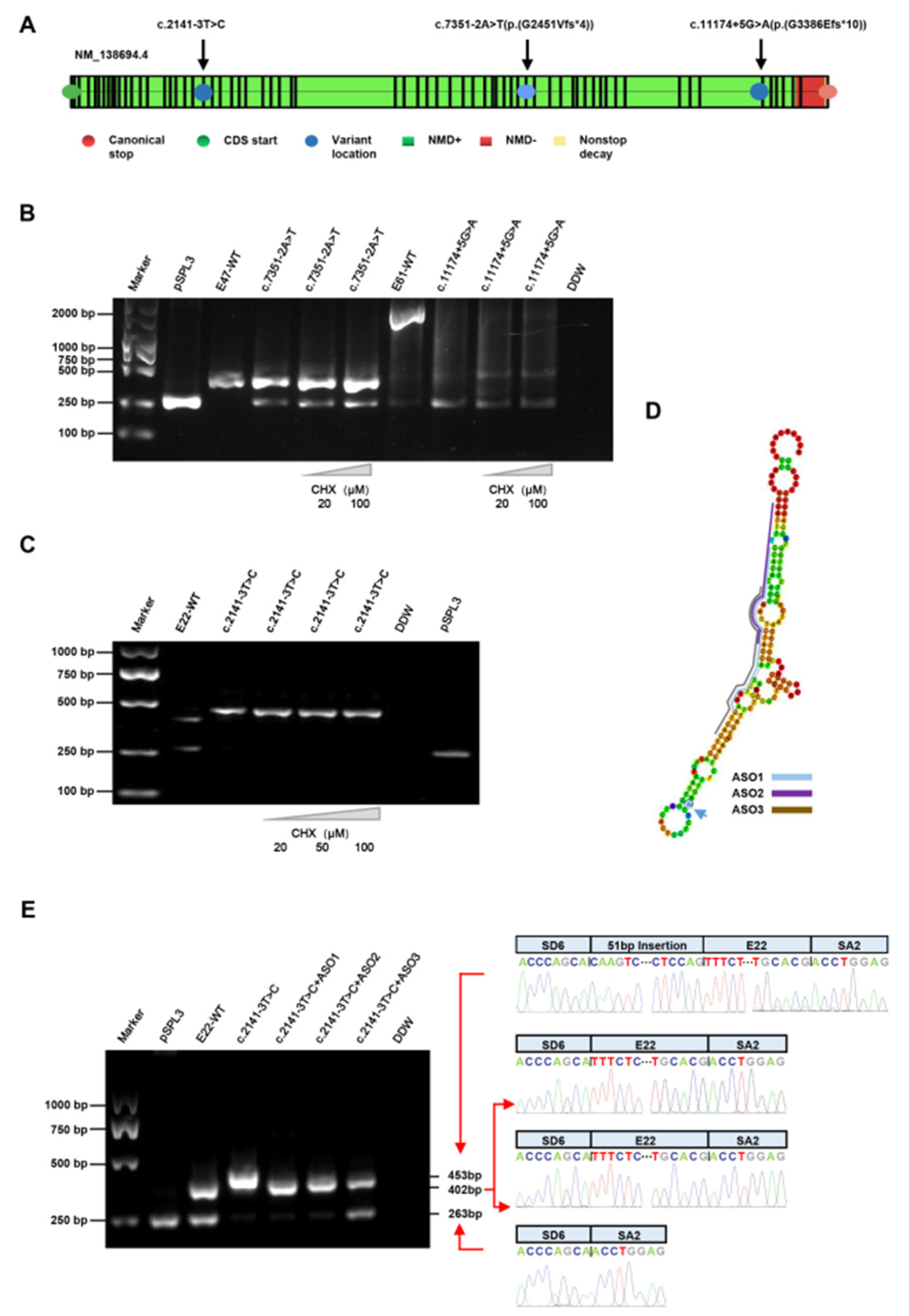
3.4. Treatment with CHX

The variants c.7351-2A>T and c.11174+5G>A were confirmed to have caused the abnormal transcripts, according to the results. The c.2141-3T>C mutant transcript had a 51 bp insert, adding more amino acids to the original length of 714 amino acids and introducing a premature termination codon (PTC) in 719 amino acids (p.V714Afs*5), according to computational prediction analysis (Figure 1). The c.11174+5G>A mutant transcript introduced a frameshift involving a change in the open reading frame with a PTC (p.G3386Efs*10) (Figure 1). The aberrant transcripts with premature termination codons (PTCs) were generated because the splicing and nonsense variants resulted in exon skipping, causing frameshifts in the coding sequence. The abnormal pre-mRNAs with PTCs may be rapidly degraded by the NMD pathway; this has made progress in the fight against many diseases [31,32]. According to the NMD prediction (https://nmdpredictions.shinyapps.io/shiny/, accessed on 17 September 2021) tool, no predictions were available for the non-frameshift variant of c.2141-3T>C; the c.7351-2A>T (p.G2451Vfs*4) and c.11174+5G>A (p.G3386Efs*10) variants were predicted to be subjected to degradation by the NMD pathway (Figure 2A). In fact, c.2141-3T>C was confirmed to have caused the 51 bp insertion, which was likely to lead to the production of PTC, reminding us of the importance of the next experiment.
To clarify whether the transcripts from the mutant minigenes were vulnerable to NMD, we used a de novo protein synthesis inhibitor cycloheximide (CHX), which has been shown to inhibit NMD [33]. Interestingly, after treatment with CHX, the three variants had no effect on the volume or size of these artificial transcripts, suggesting that the c.2141-3T>C, c.7351-2A>T and c.11174+5G>A splicing variants may escape from the control of NMD (Figure 2B,C).

3.5. Treatment with ASOs

A classical antisense therapeutic approach to mediate the skipping of the variant exons or the control of mRNA expression is to use ASOs, which are short fragments of single-stranded DNA sequences that target specific exons by recognizing the target’s sequence and binding the pre-mRNA and mRNA to directly alter pre-mRNA splicing, mRNA degradation and gene expression [34,35]. ASOs have been confirmed to specifically increase the expression of productive transcripts and reduce seizures and the incidence of SUDEP as a gene-specific treatment for Dravet syndrome and diastolic function in a murine heart failure model through the inhibition of RBM20 with ASOs in a recent study [36,37]. In vitro minigene analysis verified that exon inclusion had occurred, which may have caused the production of PTC via a frameshift. We supposed that the ASOs may block the pseudoexon insertion associated with variant c.2141-3T>C by modulating the splicing enhancer and modifying the pre-mRNA splicing pattern.
To verify whether ASOs blocked the pseudoexon insertion associated with c.2141-3T>C, two ASOs were designed to block the exonic splicing enhancer and exclude the pseudoexons. We then analyzed productive and nonproductive PKHD1 transcripts and evaluated the influence of ASOs on the level of RNA. We identified that these ASOs significantly increased the expression of their respective unspliced full-length PKHD1 transcripts in HEK293 cells, with a concomitant reduction in the E22-skipping transcript of the WT and c.2141-3T>C in ASO1 and ASO2, while the E22-skipping transcript was unaffected by ASO3 (see Figure S4). Moreover, these ASOs almost completely restored the correct splicing, to a certain extent (Figure 2E).

4. Discussion

Herein, we identified 11 patients carrying rare compound heterozygous variants of the PKHD1 gene. The 17 variants included 3 nonsense variants, 10 missense variants, 1 frameshift and 3 splice sites. The clinical features of our 11 patients were in accordance with the pathophysiology of ARPKD, including multiple cysts, hematuria, hypertension, proteinuria, urinary tract infections, kidney stones, inguinal hernia, hepatic fibrosis, portal hypertension, end-stage renal disease (ESRD) and hepatobiliary disease, etc. Using genetic analysis, all patients were diagnosed with ARPKD.
The clinical phenotypes of ARPKD are highly variable, ranging from severe early-onset antenatal multiple cysts to late-onset hepatic fibrosis, portal hypertension and ESRD [38], similar to the results of a previous report. In this study, Patient 3 had the most severe symptoms, and was admitted for abdominal pain [39], was diagnosed with Caroli’s syndrome and underwent hepatic transplantation because of severe portal systemic shunting and portal hypertension after the subsequent 3-year follow-up period.
The severity of protein dysfunction has been proposed to explain this phenomenon. The PKHD1 gene encodes fibrocystin, a single-pass membrane protein, which is mainly expressed in the primary cilium of the renal collecting tubules and hepatic bile ducts [40]. Pathogenic mutations in the PKHD1 gene underlie FPC dysfunction, resulting in a reduced capacity to bind PC2, which, in turn, causes the collecting ducts to elongate and dilate, the formation of microcysts and the diffuse enlargement of the kidney [41,42]. As a consequence, the affected individuals progress to ESRD and hepatobiliary disease in childhood.
We screened six novel missense variants, of which p.Ala109Thr and p.Leu1167Pro were located in part of six highly conserved immunoglobulin-like plexin transcription factor (IPT) domains. These variants mapped to the domains that may impair the function of FPC through their effects on integrins, transcription factors, the regulation of cell separation, motility and the invasion of the extracellular matrices [43,44]. The variants p.Asp1957Asn and p.Thr2082Ile are in the G8 domain and may affect extracellular ligand binding and catalytic activity. The missense variants located in the G8 domains of the human PKHD1 protein caused an unstable protein because of the incorrect folding of G8 domains, as has been reported by [45]. The variant p.Arg3842Gln is upstream of the ciliary targeting sequence (CST) which allows efficient PKHD1 trafficking to the cilium by controlling trafficking and affecting glycosylation [46,47]. In silico analysis predicted that these missense variants may affect the function of FPC.
The novel splicing variant c.11174+5G>A was predicted, causing the skipping of Exon 61 by altering the WT donor site, and may introduce a PTC 10 codons downstream, according to the in silico analysis. Our minigene splicing assays verified that c.11174+5G>A resulted in aberrant splicing. The aberrant mRNA transcripts were not degraded by the NMD pathway, as revealed by the administration of the specific de novo protein synthesis inhibitor CHX, indicating that a truncated protein without the C-terminal region including the CST and the nuclear localization signal (NLS) was produced. This reminds us that the variant c.11174+5G>A of ARPKD is likely to be highly pathogenic. Therefore, we upgraded the variant c.11174+5G>A to “likely pathogenic” (PM2 + PP3 + PS3).
The other novel splice site variant c.2141-3T>C may cause the skipping of Exon 22 through obtaining a new WT acceptor site, according to the in silico analysis. Surprisingly, the variant c.2141-3T>C was verified to have inserted an additional 51 bp. We considered that the c.2141-3T>C variant inserted the sequence “AG” to the 5′ original splice acceptor site, which may have deactivated the physiological GT-AG splice sites determining PKHD1′s Exon 22 and concurrently activated the cryptic splice sites located in the chimeric Intron 21, ultimately producing an alternative splice acceptor site that was superior to the original one.
The physiological GT-AG splicing site that determines PKHD1′s Exon 22 can be inactivated, while the crypto-splicing site within the chimeric Intron 21 can be activated to form an alternative splicing receptor site that is superior to the original receptor site during transcription.
Moreover, aberrant mRNA transcripts may be degraded through the NMD pathway, according to the NMD predictions. However, the small transcriptional products are stable, and the use of de novo protein synthesis inhibitors revealed that c.2141-3T>C was also likely to be highly pathogenic. Thus, we upgraded the variant c.2141-3T>C (p.V714Afs*5) to “likely pathogenic” (PM2 + PM5 + PP3 + PS3).
To date, an efficient disease-modifying therapy for ARPKD has not been approved. Tesevatinib (TSV), as a possible therapy for ARPKD, has yielded promising results in preclinical studies, has obtained approval and is undergoing clinical Phase I and II trials [48]. Renal transplantation is still the last resort for ARPKD with severe ESRD, although the shortage of organs is the most pressing concern. Renal gene therapy is already a suitable alternative to orthotopic renal transplantation for the treatment of inherited renal diseases, such as ASOs [49,50,51]. Splicing mutations cause exon skipping in the PKHD1 gene encoding the FPC protein, either causing or increasing the severity of ARPKD. Heavy intracellular accumulation of truncated FPC proteins may underlie the more severe symptoms of ARPKD. However, functional studies of FPC and the pathogenic mechanism of diseases have yet to be carried out, potentially limiting the therapeutic effect and viability of these approaches. In light of these findings, reducing the production of exon-free mRNA may provide a disease-modifying therapy for ARPKD. Antisense technology has advanced, and ASOs benefiting patients with various common and rare diseases are increasing [52]. RGLS4326, as part of the microRNA-17 (miR-17) family, was designed to directly bind or inhibit specific miRNAs, displacing miR-17 from translationally active polysomes, and to de-represses multiple miR-17 mRNA targets; this has led to clinical trials for the treatment of ADPKD [53]. Here, ASOs were designed to destroy the novel splicing site that causes inactivation, with no effect on the binding between the auxiliary splicing proteins and splicing enhancer motifs which are related to the function of original splicing. Our results suggested that ASOs may be a new option for ARPKD patients with aberrant splicing, as all of the ASOs could effectively rescue the splicing abnormalities caused by the c.2141-3T>C mutation of the PKHD1 gene in vitro. Surprisingly, we discovered that ASOs correct the production of abnormal mRNA in HEK293 cells overexpressing PKHD1. ASO therapy can potentially overcome some of the limitations of the current approaches by correcting the truncated FPC production by targeting PKHD1 RNA intracellularly. It is important to further determine the efficacy of ASOs in ARPKD in iPSC-derived ARPKD patient kidneys and ARPKD patients’ tissues, as well as ARPKD animal models.

5. Conclusions

Our study found eight novel variants, expanding the genotype of PKHD1, and verified that the c.2141-3T>C variant causes a 51 bp insertion upstream from Exon 22, and the c.11174+5G>A variant leads to the skipping of Exon 61 in the mRNA transcript of the PKHD1 gene. Aberrant transcripts were not degraded through the NMD pathway. This phenomenon means that a potentially pathogenic splicing variant is relevant to renal cysts and congenital hepatic fibrosis. Moreover, the aberrant transcript caused by the c.2141-3T>C variant was rescued by ASOs in vitro, introducing the ASOs as a promising strategy for treating ARPKD patients with intronic variants for the first time.

Supplementary Materials

The following supporting information can be downloaded at: https://www.mdpi.com/article/10.3390/jcm12041428/s1, Figure S1: Pedigrees of nine affected families with PKHD1 pathogenic variants and variants of the probands; Figure S2: Hybrid minigene assay reveled the aberrant mRNA splicing of the classic splicing variant of the PKHD1 gene in vitro in ARPKD; Figure S3: Effect of PKHD1 gene on three splicing variants via minigene assays in HEK293, HeLa, A549 and HepG2 cells; Figure S4: Antisense oligonucleotides (ASOs) rescue the c.2141-3T>C variant of the PKHD1 gene, which causes aberrant splicing in HEK293T cells; Table S1: Targeted next-generation sequencing of 180 short related genes; Table S2: PKHD1 primers for minigene constructs; Table S3: Prediction of nine missense variants of PKHD1; Table S4: Prediction of three splice site variants of PKHD1.

Author Contributions

Conceptualization and funding acquisition, F.Z. and G.D.; methodology, H.L. and C.W.; data curation and software, B.Z. and W.Z.; investigation, H.L. and R.C.; writing—original draft preparation, H.L., C.W. and R.C.; writing—review and editing, S.H., Z.J. and A.Z. All authors have read and agreed to the published version of the manuscript.

Funding

This research was funded by the National Natural Science Foundation of China (grant. no. 82070688, grant. no. 81974084 and grant. no. 82200744) and the Health Science and Technology Development Special Foundation of Nanjing (grant no. ZKX21047, grant no. ZKX19040 and grant no. YKK22159).

Institutional Review Board Statement

The study was conducted in accordance with the Declaration of Helsinki, and approved by the Institutional Ethics Committee of the Children’s Hospital of Nanjing Medical University (Nanjing, China). Approval number: 201705103-1.

Informed Consent Statement

Informed consent was obtained from all subjects involved in the study.

Data Availability Statement

Strains and plasmids are available upon request. The authors confirm that all of the data involved in the conclusion of the article are present in the article, figures and tables. The data presented in this study are available at Journal of Clinical Medicine online. Table 2 contains the genotypes for each individual.

Conflicts of Interest

The authors declare no conflict of interest.

References

  1. Goggolidou, P.; Richards, T. The genetics of Autosomal Recessive Polycystic Kidney Disease (ARPKD). Biochim. Biophys. Acta Mol. Basis Dis. 2022, 1868, 166348. [Google Scholar] [CrossRef] [PubMed]
  2. Besse, W.; Roosendaal, C.; Tuccillo, L.; Roy, S.G.; Gallagher, A.R.; Somlo, S. Adult Inactivation of the Recessive Polycystic Kidney Disease Gene Causes Polycystic Liver Disease. Kidney360 2020, 1, 1068–1076. [Google Scholar] [CrossRef] [PubMed]
  3. Yao, X.; Ao, W.; Fang, J.; Mao, G.; Chen, C.; Yu, L.; Cai, H.; Xu, C. Imaging manifestations of Caroli disease with autosomal recessive polycystic kidney disease: A case report and literature review. BMC Pregnancy Childbirth 2021, 21, 294. [Google Scholar] [CrossRef] [PubMed]
  4. Hoyer, P.F. Clinical manifestations of autosomal recessive polycystic kidney disease. Curr. Opin. Pediatr. 2015, 27, 186–192. [Google Scholar] [CrossRef]
  5. Ma, M. Cilia and polycystic kidney disease. Semin Cell Dev Biol. 2021, 110, 139–148. [Google Scholar] [CrossRef]
  6. Leebens-Mack, J.; Milligan, B. Pollination biology in hybridizing Baptisia (Fabaceae) populations. Am. J. Bot. 1998, 85, 500. [Google Scholar] [CrossRef]
  7. Tang, R.; Xu, Z. Gene therapy: A double-edged sword with great powers. Mol. Cell Biochem. 2020, 474, 73–81. [Google Scholar] [CrossRef]
  8. Zu, H.; Gao, D. Non-viral Vectors in Gene Therapy: Recent Development, Challenges, and Prospects. AAPS J. 2021, 23, 78. [Google Scholar] [CrossRef]
  9. Chen, C.; Yang, Z.; Tang, X. Chemical modifications of nucleic acid drugs and their delivery systems for gene-based therapy. Med. Res. Rev. 2018, 38, 829–869. [Google Scholar] [CrossRef]
  10. Alshaer, W.; Zureigat, H.; Al Karaki, A.; Al-Kadash, A.; Gharaibeh, L.; Hatmal, M.M.; Aljabali, A.A.A.; Awidi, A. siRNA: Mechanism of action, challenges, and therapeutic approaches. Eur. J. Pharmacol. 2021, 905, 174178. [Google Scholar] [CrossRef]
  11. Shadid, M.; Badawi, M.; Abulrob, A. Antisense oligonucleotides: Absorption, distribution, metabolism, and excretion. Expert Opin. Drug Metab. Toxicol. 2021, 17, 1281–1292. [Google Scholar] [CrossRef] [PubMed]
  12. Liu, J.; Huang, L.; Luo, M.; Xia, X. Bacterial translocation in acute pancreatitis. Crit. Rev. Microbiol. 2019, 45, 539–547. [Google Scholar] [CrossRef] [PubMed]
  13. Hagedorn, P.H.; Brown, J.M.; Easton, A.; Pierdomenico, M.; Jones, K.; Olson, R.E.; Mercer, S.E.; Li, D.; Loy, J.; Hog, A.M.; et al. Acute Neurotoxicity of Antisense Oligonucleotides After Intracerebroventricular Injection Into Mouse Brain Can Be Predicted from Sequence Features. Nucleic Acid Ther. 2022, 32, 151–162. [Google Scholar] [CrossRef]
  14. Fish, L.; Khoroshkin, M.; Navickas, A.; Garcia, K.; Culbertson, B.; Hanisch, B.; Zhang, S.; Nguyen, H.C.B.; Soto, L.M.; Dermit, M.; et al. A prometastatic splicing program regulated by SNRPA1 interactions with structured RNA elements. Science 2021, 372, 1–26. [Google Scholar] [CrossRef] [PubMed]
  15. Bennett, C.F. Therapeutic Antisense Oligonucleotides Are Coming of Age. Annu. Rev. Med. 2019, 70, 307–321. [Google Scholar] [CrossRef]
  16. Bennett, C.F.; Krainer, A.R.; Cleveland, D.W. Antisense Oligonucleotide Therapies for Neurodegenerative Diseases. Annu. Rev. Neurosci. 2019, 42, 385–406. [Google Scholar] [CrossRef]
  17. Finkel, R.S.; Mercuri, E.; Darras, B.T.; Connolly, A.M.; Kuntz, N.L.; Kirschner, J.; Chiriboga, C.A.; Saito, K.; Servais, L.; Tizzano, E.; et al. Nusinersen versus Sham Control in Infantile-Onset Spinal Muscular Atrophy. N. Engl. J. Med. 2017, 377, 1723–1732. [Google Scholar] [CrossRef] [PubMed]
  18. Charleston, J.S.; Schnell, F.J.; Dworzak, J.; Donoghue, C.; Lewis, S.; Chen, L.; Young, G.D.; Milici, A.J.; Voss, J.; DeAlwis, U.; et al. Eteplirsen treatment for Duchenne muscular dystrophy: Exon skipping and dystrophin production. Neurology 2018, 90, e2146–e2154. [Google Scholar] [CrossRef] [PubMed]
  19. Aartsma-Rus, A.; Corey, D.R. The 10th Oligonucleotide Therapy Approved: Golodirsen for Duchenne Muscular Dystrophy. Nucleic Acid Ther. 2020, 30, 67–70. [Google Scholar] [CrossRef]
  20. Frank, D.E.; Schnell, F.J.; Akana, C.; El-Husayni, S.H.; Desjardins, C.A.; Morgan, J.; Charleston, J.S.; Sardone, V.; Domingos, J.; Dickson, G.; et al. Increased dystrophin production with golodirsen in patients with Duchenne muscular dystrophy. Neurology 2020, 94, e2270–e2282. [Google Scholar] [CrossRef] [Green Version]
  21. Zakeri, S.E.; Pradeep, S.P.; Kasina, V.; Laddha, A.P.; Manautou, J.E.; Bahal, R. Casimersen for the treatment of Duchenne muscular dystrophy. Trends Pharmacol. Sci. 2022, 43, 607–608. [Google Scholar] [CrossRef] [PubMed]
  22. Wang, C.; Han, Y.; Zhou, J.; Zheng, B.; Zhou, W.; Bao, H.; Jia, Z.; Zhang, A.; Huang, S.; Ding, G.; et al. Splicing Characterization of CLCNKB Variants in Four Patients With Type III Bartter Syndrome. Front. Genet. 2020, 11, 81. [Google Scholar] [CrossRef] [PubMed]
  23. Krall, P.; Pineda, C.; Ruiz, P.; Ejarque, L.; Vendrell, T.; Camacho, J.A.; Mendizabal, S.; Oliver, A.; Ballarin, J.; Torra, R.; et al. Cost-effective PKHD1 genetic testing for autosomal recessive polycystic kidney disease. Pediatr. Nephrol. 2014, 29, 223–234. [Google Scholar] [CrossRef]
  24. Losekoot, M.; Haarloo, C.; Ruivenkamp, C.; White, S.J.; Breuning, M.H.; Peters, D.J. Analysis of missense variants in the PKHD1-gene in patients with autosomal recessive polycystic kidney disease (ARPKD). Hum. Genet. 2005, 118, 185–206. [Google Scholar] [CrossRef] [PubMed]
  25. Hao, X.; Liu, S.; Dong, Q.; Zhang, H.; Zhao, J.; Su, L. Whole exome sequencing identifies recessive PKHD1 mutations in a Chinese twin family with Caroli disease. PLoS ONE 2014, 9, e92661. [Google Scholar] [CrossRef] [PubMed]
  26. Melchionda, S.; Palladino, T.; Castellana, S.; Giordano, M.; Benetti, E.; De Bonis, P.; Zelante, L.; Bisceglia, L. Expanding the mutation spectrum in 130 probands with ARPKD: Identification of 62 novel PKHD1 mutations by sanger sequencing and MLPA analysis. J. Hum. Genet. 2016, 61, 811–821. [Google Scholar] [CrossRef]
  27. Sharp, A.M.; Messiaen, L.M.; Page, G.; Antignac, C.; Gubler, M.C.; Onuchic, L.F.; Somlo, S.; Germino, G.G.; Guay-Woodford, L.M. Comprehensive genomic analysis of PKHD1 mutations in ARPKD cohorts. J. Med. Genet. 2005, 42, 336–349. [Google Scholar] [CrossRef]
  28. Qiu, L.R.; Xu, R.R.; Tang, J.H.; Zhou, J.H. Possible PKHD1 Hot-spot Mutations Related to Early Kidney Function Failure or Hepatofibrosis in Chinese Children with ARPKD: A Retrospective Single Center Cohort Study and Literature Review. Curr. Med. Sci. 2020, 40, 835–844. [Google Scholar] [CrossRef]
  29. Yon, D.; Frith, C.D. Precision and the Bayesian brain. Curr. Biol. 2021, 31, R1026–R1032. [Google Scholar] [CrossRef]
  30. Song, M.H.; Cho, H.J.; Lee, H.K.; Kwon, T.J.; Lee, W.S.; Oh, S.; Bok, J.; Choi, J.Y.; Kim, U.K. CHD7 mutational analysis and clinical considerations for auditory rehabilitation in deaf patients with CHARGE syndrome. PLoS ONE 2011, 6, e24511. [Google Scholar] [CrossRef] [Green Version]
  31. Green, I.D.; Pinello, N.; Song, R.; Lee, Q.; Halstead, J.M.; Kwok, C.T.; Wong, A.C.H.; Nair, S.S.; Clark, S.J.; Roediger, B.; et al. Macrophage development and activation involve coordinated intron retention in key inflammatory regulators. Nucleic Acids Res. 2020, 48, 6513–6529. [Google Scholar] [CrossRef] [PubMed]
  32. Sergeeva, O.V.; Shcherbinina, E.Y.; Shomron, N.; Zatsepin, T.S. Modulation of RNA Splicing by Oligonucleotides: Mechanisms of Action and Therapeutic Implications. Nucleic Acid Ther. 2022, 32, 123–138. [Google Scholar] [CrossRef] [PubMed]
  33. Yabe, S.G.; Nishida, J.; Fukuda, S.; Takeda, F.; Nasiro, K.; Yasuda, K.; Iwasaki, N.; Okochi, H. Expression of mutant mRNA and protein in pancreatic cells derived from MODY3- iPS cells. PLoS ONE 2019, 14, e0217110. [Google Scholar] [CrossRef] [PubMed]
  34. Leier, A.; Moore, M.; Liu, H.; Daniel, M.; Hyde, A.M.; Messiaen, L.; Korf, B.R.; Selvakumaran, J.; Ciszewski, L.; Lambert, L.; et al. Targeted exon skipping of NF1 exon 17 as a therapeutic for neurofibromatosis type I. Mol. Ther. Nucleic Acids 2022, 28, 261–278. [Google Scholar] [CrossRef] [PubMed]
  35. Sait, H.; Srivastava, S.; Saxena, D. Integrated Management Strategies for Epidermolysis Bullosa: Current Insights. Int. J. Gen. Med. 2022, 15, 5133–5144. [Google Scholar] [CrossRef]
  36. Han, Z.; Chen, C.; Christiansen, A.; Ji, S.; Lin, Q.; Anumonwo, C.; Liu, C.; Leiser, S.C.; Meena; Aznarez, I.; et al. Antisense oligonucleotides increase Scn1a expression and reduce seizures and SUDEP incidence in a mouse model of Dravet syndrome. Sci. Transl. Med. 2020, 12, 1–14. [Google Scholar] [CrossRef]
  37. Radke, M.H.; Badillo-Lisakowski, V.; Britto-Borges, T.; Kubli, D.A.; Juttner, R.; Parakkat, P.; Carballo, J.L.; Huttemeister, J.; Liss, M.; Hansen, A.; et al. Therapeutic inhibition of RBM20 improves diastolic function in a murine heart failure model and human engineered heart tissue. Sci. Transl. Med. 2021, 13, eabe8952. [Google Scholar] [CrossRef]
  38. Adeva, M.; El-Youssef, M.; Rossetti, S.; Kamath, P.S.; Kubly, V.; Consugar, M.B.; Milliner, D.M.; King, B.F.; Torres, V.E.; Harris, P.C. Clinical and molecular characterization defines a broadened spectrum of autosomal recessive polycystic kidney disease (ARPKD). Medicine 2006, 85, 1–21. [Google Scholar] [CrossRef]
  39. Burgmaier, K.; Brinker, L.; Erger, F.; Beck, B.B.; Benz, M.R.; Bergmann, C.; Boyer, O.; Collard, L.; Dafinger, C.; Fila, M.; et al. Refining genotype-phenotype correlations in 304 patients with autosomal recessive polycystic kidney disease and PKHD1 gene variants. Kidney Int. 2021, 100, 650–659. [Google Scholar] [CrossRef]
  40. Outeda, P.; Menezes, L.; Hartung, E.A.; Bridges, S.; Zhou, F.; Zhu, X.; Xu, H.; Huang, Q.; Yao, Q.; Qian, F.; et al. A novel model of autosomal recessive polycystic kidney questions the role of the fibrocystin C-terminus in disease mechanism. Kidney Int. 2017, 92, 1130–1144. [Google Scholar] [CrossRef]
  41. Tsunoda, T.; Kakinuma, S.; Miyoshi, M.; Kamiya, A.; Kaneko, S.; Sato, A.; Tsuchiya, J.; Nitta, S.; Kawai-Kitahata, F.; Murakawa, M.; et al. Loss of fibrocystin promotes interleukin-8-dependent proliferation and CTGF production of biliary epithelium. J. Hepatol. 2019, 71, 143–152. [Google Scholar] [CrossRef] [PubMed]
  42. Society for Maternal-Fetal, M.; Swanson, K. Autosomal recessive polycystic kidney disease. Am. J. Obstet. Gynecol. 2021, 225, B7–B8. [Google Scholar] [CrossRef]
  43. Aravind, L.; Koonin, E.V. Gleaning non-trivial structural, functional and evolutionary information about proteins by iterative database searches. J. Mol. Biol. 1999, 287, 1023–1040. [Google Scholar] [CrossRef] [PubMed]
  44. Bork, P.; Doerks, T.; Springer, T.A.; Snel, B. Domains in plexins: Links to integrins and transcription factors. Trends Biochem. Sci. 1999, 24, 261–263. [Google Scholar] [CrossRef]
  45. He, Q.Y.; Liu, X.H.; Li, Q.; Studholme, D.J.; Li, X.W.; Liang, S.P. G8: A novel domain associated with polycystic kidney disease and non-syndromic hearing loss. Bioinformatics 2006, 22, 2189–2191. [Google Scholar] [CrossRef]
  46. Kim, I.; Fu, Y.; Hui, K.; Moeckel, G.; Mai, W.; Li, C.; Liang, D.; Zhao, P.; Ma, J.; Chen, X.Z.; et al. Fibrocystin/polyductin modulates renal tubular formation by regulating polycystin-2 expression and function. J. Am. Soc. Nephrol. 2008, 19, 455–468. [Google Scholar] [CrossRef]
  47. Nagano, J.; Kitamura, K.; Hujer, K.M.; Ward, C.J.; Bram, R.J.; Hopfer, U.; Tomita, K.; Huang, C.; Miller, R.T. Fibrocystin interacts with CAML, a protein involved in Ca2+ signaling. Biochem. Biophys. Res. Commun. 2005, 338, 880–889. [Google Scholar] [CrossRef]
  48. Sweeney, W.E.; Frost, P.; Avner, E.D. Tesevatinib ameliorates progression of polycystic kidney disease in rodent models of autosomal recessive polycystic kidney disease. World J. Nephrol. 2017, 6, 188–200. [Google Scholar] [CrossRef]
  49. Parisi, M.A. The molecular genetics of Joubert syndrome and related ciliopathies: The challenges of genetic and phenotypic heterogeneity. Transl. Sci. Rare Dis. 2019, 4, 25–49. [Google Scholar] [CrossRef]
  50. Imai, E.; Isaka, Y. Gene electrotransfer: Potential for gene therapy of renal diseases. Kidney Int. 2002, 61, S37–S41. [Google Scholar] [CrossRef] [Green Version]
  51. Ricker, J.L.; Mata, J.E.; Iversen, P.L.; Gattone, V.H. c-myc antisense oligonucleotide treatment ameliorates murine ARPKD. Kidney Int. 2002, 61, S125–S131. [Google Scholar] [CrossRef] [PubMed]
  52. Zhou, Y.; Liu, S.; Liu, G.; Ozturk, A.; Hicks, G.G. ALS-associated FUS mutations result in compromised FUS alternative splicing and autoregulation. PLoS Genet. 2013, 9, e1003895. [Google Scholar] [CrossRef] [PubMed]
  53. Lee, E.C.; Valencia, T.; Allerson, C.; Schairer, A.; Flaten, A.; Yheskel, M.; Kersjes, K.; Li, J.; Gatto, S.; Takhar, M.; et al. Discovery and preclinical evaluation of anti-miR-17 oligonucleotide RGLS4326 for the treatment of polycystic kidney disease. Nat. Commun. 2019, 10, 4148. [Google Scholar] [CrossRef] [PubMed] [Green Version]
Figure 1. The hybrid minigene assay showed the aberrant mRNA splicing of two splicing variants of the PKHD1 gene in vitro in ARPKD. (A) The products of hybrid minigene transcripts in HEK293 cells were amplified by RT-PCR. The transcripts produced by the hybrid minigene are schematically shown, and the arrows show the primers used for amplification (inset) (Wang et al., 2018b). (B) Agarose gel electrophoresis and directly sequences (right panel) of the RT–PCR products of the minigene transcripts in HEK293T cells of the variant c.11174+5G>A. cDNA sequencing of variant c.11174+5G>A showed the skipping of Exon 61. Lane 1: marker; Lane 2: pSPL3 (263 bp); Lane 3: E61-WT (1281 bp); Lane 4: c.11174+5G>A (1281 bp and 263 bp); Lane 5: DDW. (C) Agarose gel electrophoresis and direct sequences (right panel) of the RT–PCR products of the minigene transcripts in HEK293T cells of the variant c.2141-3T>C. Sequencing of the variant c.2141-3T>C cDNA showed an additional insert of 51 bp between Exon 21 and Exon 22. Lane 1: marker; Lane 2: pSPL3 (263 bp); Lane 3: E22-WT (402 bp and 263bp); Lane 4: c.2141-3T>C (453 bp); Lane 5: DDW. (D) Schematic representation of the abnormal splicing of the PKHD1 exons of variant c.2141-3T>C (numbered boxes). A pseudoexon with a 51 bp insert in the junction of Exon 21 and Exon 22 appears in the mutant transcript (upper panel). The predicted amino acid structures were translated from the aberrant transcripts of the c.2141-3T>C variant with a fragment of 51 bp within Intron 21 in PKHD1 (lower panel). (E) Schematic representation of the abnormal splicing of the PKHD1 exons of the variant c.11174+5G>A (numbered boxes). Exon 61 was skipped in the mutant transcript (upper panel). The predicted amino acid structures were translated from the aberrant transcripts of the c.11174+5G>A variant with a 1018 bp deletion of Exon 61 in PKHD1 (lower panel).
Jcm 12 01428 g001
Figure 2. The effect of the de novo protein synthesis inhibitor (cycloheximide) and antisense oligonucleotides (ASOs) on the c.2141-3T>C variant of the PKHD1 gene of aberrant splicing. (A) Schematic representation of the PKHD1 gene showing the localization of the truncating variants subjected to (NMD+) and escaping(NMD-) to nonsense-mediated RNA decay regions (https://nmdpredictions.shinyapps.io/shiny/). The loss-of-function variants p.G2451Vfs*4 and p.G3386Efs*10 detected in this study are shown in red. The region of PKHD1 where the truncating variants trigger NMD is indicated in green. The region that escaped NMD is represented in red. The nonstop decay region is indicated in yellow. (B,C) Treatment with cycloheximide (CHX) showed that the transcriptional products were stable. CHX did not degrade the abnormal pre-mRNAs of the splicing variants of the PKHD1 gene in HEK293T cells. The gray triangle represents the dose-dependent concentration of the reagents. CHX, cycloheximide. (D) The positions of three ASOs for the c.2141-3T>C variant in this study. (E) RT–PCR analysis of the splicing correction after the ASOs treatments. HEK293T cells were transfected with the wild-type (WT) minigene and the corresponding mutant (MU) minigene containing the variant c.2141-3T>C. With the exception of the nontreated lanes (NT), the remaining lanes show that the administration of three ASOs (ASO1, ASO2, and ASO3) efficiently rescued the retention of the intron caused by c.2141-3T>C. The ASOs rescued the c.2141-3T>C variant of the PKHD1 gene, which causes abnormal splicing in HEK293T cells.
Jcm 12 01428 g002
Figure 3. Identification of the PKHD1 variants in patients with cysts. (A) Sequence chromatograms of the PKHD1 variants as detected. (B) Distribution of the PKHD1 variants in corresponding domains. (C) The PKHD1 variants are located in the corresponding domains. Abbreviations: TM domain, transmembrane span; IPT/TIG domain, Ig-like, plexins and transcriptions factors; PA14 domain, Pfam Entry 14; G8 domain, eight conserved glycine residues; PbH1 domain, parallel beta-helix repeats; CST domain, ciliary targeting sequence; NLS domain, nuclear localization signal. The red arrow is points to the orthologous and paralogous protein alignments showing the high conservation of each amino acid altered by four missense variants from humans to gibbon.
Jcm 12 01428 g003
Table 1. Clinical features of 11 PKD patients with the PKHD1 variants.
PatientAge (Year)SexEthnicityFamily HistoryRenal CystsKidney EnlargementCaroli DiseaseHepatic CystsSplenomegalyRenal DysfunctionLiver Dysfunction
P17FemaleHanNoYesYesNoNoNoNoNo
P21.6FemaleHanNoYesNoNoNoNoNoNo
P39MaleHanYesYesYesYesYesYesNoElevated liver enzymes
P414.3FemaleHanNoYesNoNoNoNoNoNo
P50.7MaleHanNoYesNoNoNoNoNoNo
P61.6MaleHanNoYesNoNoNoNoNo, with urinary
occult blood
No
P71MaleHanNoYesYesNoNoNoNoNo
P80.8FemaleHanNoYesNoNoNoNoNoNo
P912FemaleHanYesYesNoNoNoNoNoNo
P101.1FemaleHanNoYesYesNoYesNoElevated blood urea and uric acidElevated liver enzymes
P115MaleHanYesYesNoNoNoNoNoNo
Table 2. Variants of the PKHD1 gene identified in this study.
PatientNucleotide ChangeAmino Acid ChangeHom/HetLocationDomainMutation TypeACMGReported
P1c.2141-3T>C-HetI21-SplicingVUS (PM2 + PP3 + PM5)No
P2, P9c.7942G>Ap.Gly2648SerHetE50ExtracellularMissenseVUS (PM2)[23]
P2c.7351-2A>T-HetI46-SplicingP (PVS1 + PM2 + PM5)[24]
P3c.3500T>Cp.Leu1167ProHetE30IPT/TIG 6;
atypical
MissenseVUS (PM2 + PP3)No
P3c.11174+5G>A-HetI61-SplicingVUS (PM2 + PP3)No
P4c.325G>Ap.Ala109ThrHetE5IPT/TIG 1;
atypical
MissenseLP (PS2 + PM2)No
P5c.6001G>Tp.Glu2001 *HetE37G8 1NonsenseP (PVS1 + PM2 + PM5)No
P1, P5, P6c.2507T>Cp.Val836AlaHetE24ExtracellularMissenseVUS (PM2 + PP3 + PM3)[25]
P6c.5869G>Ap.Asp1957AsnHetE36G8 1MissenseLP (PS2 + PM2 + PP3)No
P7c.6245C>Tp.Thr2082IleHetE38ExtracellularMissenseVUS (PM2 + PP3)No
P8c.6910C>Tp.Gln2304 *HetE43PbH1 3′NonsenseP (PVS1 + PM2 + PM3)[26]
P8c.4199C>Tp.Ser1400LeuHetE32IPT/TIG 9MissenseVUS (PM2 + PP3)[26]
P9c.11525G>Ap.Arg3842GlnHetE65ExtracellularMissenseVUS (PM2 + PP3)No
P10c.9780G>Ap.Trp3260 *HetE58ExtracellularNonsenseP (PVS1 + PM2 + PM3)No
P10c.8518C>Tp.Arg2840CysHetE54G8 2MissenseLP (PS1 + PM2 + PM3 + PP3)[27]
P11c.10756_10759delAACTp.Asn3586Serfs *22HetE61ExtracellularFrameshiftP (PVS1 + PM2 + PP3)[28]
P11c.10072G>Ap.Asp3358AsnHetE60ExtracellularMissenseLP (PS2 + PM2 + PM2 + PP3)No
Hom: homozygote. Het: heterozygote. *: premature termination codon.
Disclaimer/Publisher’s Note: The statements, opinions and data contained in all publications are solely those of the individual author(s) and contributor(s) and not of MDPI and/or the editor(s). MDPI and/or the editor(s) disclaim responsibility for any injury to people or property resulting from any ideas, methods, instructions or products referred to in the content.

