Article

High-Throughput Sequencing of Oral Microbiota in Candida Carriage Sjögren’s Syndrome Patients: A Pilot Cross-Sectional Study

1
Department of General Dentistry, National Center for Stomatology, National Clinical Research Center for Oral Diseases, National Engineering Research Center of Oral Biomaterials and Digital Medical Devices, Peking University School and Hospital of Stomatology, Beijing 100081, China
2
Department of Oral Medicine, National Center for Stomatology, National Clinical Research Center for Oral Diseases, National Engineering Research Center of Oral Biomaterials and Digital Medical Devices, Peking University School and Hospital of Stomatology, Beijing 100081, China
*
Author to whom correspondence should be addressed.
J. Clin. Med. 2023, 12(4), 1559; https://doi.org/10.3390/jcm12041559
Submission received: 17 December 2022 / Revised: 13 February 2023 / Accepted: 14 February 2023 / Published: 16 February 2023
(This article belongs to the Section Dentistry, Oral Surgery and Oral Medicine)

Abstract

:
Background: This study sought to characterize the saliva microbiota of Candida carriage Sjögren’s syndrome (SS) patients compared to oral candidiasis and healthy patients by high-throughput sequencing. Methods: Fifteen patients were included, with five Candida carriage SS patients (decayed, missing, and filled teeth (DMFT) score 22), five oral candidiasis patients (DMFT score 17), and five caries active healthy patients (DMFT score 14). Bacterial 16S rRNA was extracted from rinsed whole saliva. PCR amplification generated DNA amplicons of the V3–V4 hypervariable region, which were sequenced on an Illumina HiSeq 2500 sequencing platform and compared and aligned to the SILVA database. Taxonomy abundance and community structure diversity was analyzed using Mothur software v1.40.0. Results: A total of 1016/1298/1085 operational taxonomic units (OTUs) were obtained from SS patients/oral candidiasis patient/healthy patients. Treponema, Lactobacillus, Streptococcus, Selenomonas, and Veillonella were the primary genera in the three groups. The most abundant significantly mutative taxonomy (OTU001) was Veillonella parvula. Microbial diversity (alpha diversity and beta diversity) was significantly increased in SS patients. ANOSIM analyses revealed significantly different microbial compositional heterogeneity in SS patients compared to oral candidiasis and healthy patients. Conclusion: Microbial dysbiosis differs significantly in SS patients independent of oral Candida carriage and DMFT.

1. Introduction

Sjögren’s syndrome (SS) is a systemic autoimmune exocrinopathy characterized by mouth and eye dryness, fatigue, joint pain, and other symptoms [1]. As an effect of pathology, the oral manifestations of SS include xerostomia and hyposalivation, dental caries, oral candidiasis, dental erosion and/or abrasion, and others [2]. In 2015, untreated dental caries affected an estimated 2.6 billion adults globally. That year, oral conditions contributed to the declined health in 35 of 39 categories of cancer [3]. Among all adults with dental caries, patients with SS showed a significantly higher dental caries rate compared to non-SS patients [4]. SS patients are also more likely to have had teeth extractions [5] and have a higher rate of decayed, missing, and filled teeth (DMFT) [6].
The development of dental caries depends on the balance of pathological and protective factors. Pathological factors include acidogenic bacteria, inhibition of salivary function, and frequency of ingestion of carbohydrates [7]. Hyposalivation (defined as a whole salivary flow rate <0.1 mL/min without stimulation) is an important influence during the development of dental caries in SS [8]. Hyposalivation might delay the clearance of sugar and acid from the dental surface. The lack of buffers influences the survival and growth of oral bacteria and fungi [9], leading to dental caries, erosion, candidiasis, etc. [10]. For SS patients with hyposalivation, antibacterial methods might be a necessary intervention to manage dental caries [11].
Changes in the oral microbiota can involve many pathological and non-pathological aspects within the oral cavity [12]. Knowledge of the oral microbiota is important for the management of dental caries. However, whether the oral microbiota in SS patients differ from healthy individuals is unclear. Generally speaking, quantitative and qualitative changes in saliva might lead to specific changes in the salivary microbial composition, with increasing cariogenic colonization by Streptococcus mutans and Lactobacillus [13]. Colonization by cariogenic and acidophilic microorganisms in the microbiota elevates the risk of caries [4]. Microbial dysbiosis has been described in SS patients with normal salivary flow rates [14]. However, a comparable salivary microbiota was found in hyposalivation patients, compared with control patients having normal saliva flow rates and comparable DMFT [15,16]. Many other factors might contribute to the salivary microbiota in SS patients, making comparison of the findings of studies difficult [17].
Among these factors, oral health status might be influential, including the rate of dental caries and/or DMFT. Even without medical conditions affecting saliva, the mean prevalence of dental caries reported in one study was 140% higher in hyposalivation patients than the healthy control group [18]. Other authors reported that with comparable DMFT, the salivary microbiota in the SS patients was not significantly different from that of healthy controls, with the most abundant genera in both groups being Veillonella, Streptococcus, Prevotella, and Haemophilus [19]. With higher DMFT in SS patients, the relative abundance of Veillonella was also reportedly higher than healthy controls, while Neisseria, Actinomyces, Haemophilus, Rothia, Porphyromonas, and Peptostreptococcus were significantly lower [6]. The findings indicate that DMFT is related with the microbiota dysbiosis [20] and should be considered when researched the oral microbiota in SS patients.
The other potential aspect that oral health status impacts the oral microbiota is the carriage of oral Candida. In one study, 87.5% of hyposalivation patients were colonized by Candida, which was isolated from saliva, with the majority (80.6%) being C. albicans [21]. For SS patients lacking saliva-mediated prevention of Candida adhesion to the mucosa [22], oral signs of candidiasis were evident in 13.1% to 60% of SS patients [23,24], which was significantly higher than the rate of candidiasis in healthy controls [25]. During the past decade, there has been increased recognition of the synergistic or antagonistic interactions between oral Candida and the oral microbiota [26,27]. C. albicans are widely distributed within the architecture of caries biofilms and might influence the ecology of cross-kingdom microbiota in caries [28]. Therefore, the carriage of Candida should also be considered in studies of the oral microbiota in SS patients.
This study explored the differences in the salivary microbiota in SS patients positive for Candida, with DMFT comparable to a control group of non-SS patients. This comparison has not been explored previously to our knowledge. Next-generation sequencing and Illumina-based sequencing of bacterial 16S rRNA were used to analyze microbiota diversity using unstimulated whole saliva rinse samples from Candida carriage SS patients and age-, gender-, and DMFT-matched control individuals.

2. Materials and Methods

2.1. Participants

Because pSS has a strong female propensity, all participants in this study were female [29]. They were recruited at Peking University School and Hospital of Stomatology, Beijing, China, from January to August 2017 consecutively. The individuals in the Test group were diagnosed with primary SS according to the revised international classification criteria proposed by the American–European Consensus Group, 2002 [30]. They also met the 2016 ACR-EULAR Classification Criteria for primary Sjögren’s Syndrome, with a total score ≥4 [31]. All participants were interviewed. Those who had not received antibiotics in the preceding 3 months and had no systemic diseases (including hepatitis, tuberculosis, and diabetes) and no smoking or alcohol abuse were enrolled. All enrolled participants provided signed informed consent. This study was approved by the Institutional Review Board of Peking University School and Hospital of Stomatology (#PKUSSIRB-201947088).

2.2. Clinical Examinations

A comprehensive oral examination was performed by an experienced dentist through visual/tactile methods after air drying of the mouth. The total numbers of teeth present, decayed (D), missed (M), filled (F) teeth (T)/surfaces (S), incisal/cusp caries, and root surfaces caries were recorded separately. The dental examination was completed within 15 min, and the findings were recorded by an assistant. The examiner checked the record after the examination. Individuals with more than 20 teeth were enrolled. To compare the oral health status, the inclusion criteria for SS patients (Test group) were DMFS ≥15, teeth with active caries ≥5, and unstimulated whole salivary flow rate (UWSF) <0.1 mL/min. The respective values for oral candidiasis patients (positive control group, Group P) and healthy control group (Group N) were ≥10, ≥1, and ≥0.1 mL/min. Patients with severe chronic periodontitis (probing depth ≥ 6 mm) were excluded. Concentrated oral rinse fluid was cultured on Sabouraud dextrose agar. Candida counts >200 colony-forming units (CFU)/mL indicated Candida carriage for SS and candidiasis patients. Finally, the total of 15 patients included 5 Candida carriage SS patients in the Test group (A01 to A05), 5 patients in Group P (B01 to B05), and 5 caries active healthy patients in Group N (C01 to C05). Details of the original data from clinical examination was uploaded as Supplementary Materials.

2.3. Whole Saliva Collection

All included patients stopped oral hygiene 24 h prior to sample collection and did not eat on the morning of the examination day. The participants were instructed to rinse their mouths thoroughly for 1 min with 10 mL of sterile saline. The saline was completely spit into a sterile graduated test tube. Each tube was stored at −80 °C until analyzed.

2.4. DNA Isolation, PCR Amplification, and High-Throughput Sequencing

All samples were washed twice with TE buffer (10 mM Tris-HCl, 1M EDTA, pH 8.0). Bacterial DNA was isolated and purified using a Wizard Genomic DNA Purification Kit (Promega, Madison, WI, USA) according to the manufacturer’s instructions. The concentration of the purified DNA was determined using a NanoDrop 2000 spectrophotometer (Thermo Fisher Scientific, Waltham, MA, USA) at an optical density of 260 nm (OD260). DNA integrity was verified by 1% agarose gel electrophoresis. For each individual sample, a standard concentration of 10 ng/uL DNA was diluted for the subsequent PCR assay and then stored at −80 °C.
High-throughput sequencing of all samples was performed by Cnkingbio Lab (Beijing, China). The V3−V4 hypervariable regions of the bacterial 16S rRNA gene were amplified by PCR using a GeneAmp 9700 device (Applied Biosystems, Foster City, CA, USA) from the isolated DNA, with primers 515F (GTGCCAGCMGCCGCGGTAA) and 806R (GGACTACHVGGGTWTCTAAT) and a unique barcode. PCR involved 10 min at 95 °C, followed by 35 cycles of 10 s at 95 °C, 30 s at 55 °C, and 30 s at 72 °C, and a final 10 min extension at 72 °C. The amplified PCR products were purified and checked with 2% agarose gel electrophoresis. The final amplicon preparation products were pooled in an emulsion PCR performed using a GS Lib-L kit (Roche Diagnostics GmbH, Mannheim, Germany) at a molecules-per-bead ratio of 0.7 according to the manufacturer’s instructions. Sequencing was performed on a HiSeq 2500 system (Illumina, San Diego, CA, USA) using paired-end and dual-end sequencing for the V3−V4 region.

2.5. Data Processing and Statistical Analyses

The generated sequences from Illumina HiSeq 2500 system were annotated as unique target sequences (raw tags) by FLASH (V1.2.7, http://ccb.jhu.edu/software/FLASH accessed on 9 October 2017). Clean tags were generated by filter raw tags using QIIME quantity control process (V1.9.0, http://qiime.org/scripts/split_libraries_fastq.html, accessed on 9 October 2017) [32]. The quality filtering strategy was performed by tags truncation and tags length. The effective tags sequences were selected by redundancy removal to remove chimera and impurity sequences using Mothur software v1.35.1 (http://www.mothur.org/wiki/Download_mothur, accessed on 9 October 2017) [33]. The effective unique tag sequences pre-clustered using the single linkage pre-clustering approach with 97% similarity were defined as one operational taxonomic unit (OTU). Species annotation of OTUs were classified to study the species composition diversity of the samples. The representative sequences from each OTU were compared and aligned to the SILVA database (Version 1123, http://www.mothur.org/wiki/Silva_reference_files, Bremen, Germany, accessed on 9 October 2017).
Average age was compared by independent-samples t-test. Clinical data (DMFT, DMFS, incisal caries) were compared by the chi-square test in the three groups via SPSS 20.0 (IBM, Armonk, NY, USA). OTU differences across groups were analyzed by Metastat analysis using Mothur software v1.35.1 (http://www.mothur.org/wiki/Download_mothur, accessed on 9 October 2017). Alpha diversity was defined as the mean species diversity in sites (within-community) or habitats at a more local scale. Alpha diversity is the number of bacterial taxa and the proportional representation of each taxon per sample. The ACE, Chao, Shannon, and Simpson indices of alpha diversity were tested using the Wilcoxon rank-sum test. To examine the differences in the alpha diversity, data were analyzed using Bayesian methods by the limma package in R software (v2.2.0). Beta diversity was determined by principal component analysis (PCoA) based on weighted Unifrac matrix using R software (v2.2.0). Analysis of similarities (ANOSIM) determined the difference in bacterial composition and structure among the three groups.

3. Results

3.1. Patients Characteristics

The baseline characteristics of SS patients and controls are presented in Table 1. With 15 participants, significant differences were not evident for the mean age, DMFT, and DMFS (p > 0.05) among the three groups (Table 1). Incisal caries was examined only in the Test group of SS patients.

3.2. General Outline

An average of 132,153 (ranging from 13,112 to 298,785) total pairs reads was obtained. An average of 129,221 combined reads as effective tags was obtained after splicing using Flash software. A total of 3399 OTUs, comprising 1016 in the Test group, 1298 in Group P, and 1083 in Group N, were obtained from 15 samples. The samples comprised 10 phyla, 22 classes, 43 orders, 67 families, and 121 genera. The 121 genera comprised 85 genera in the Test group, 101 in Group P, and 95 in Group N. The ten most predominant genera constituting the sequences detected in saliva are presented in Figure 1. The five most predominant genera in the Test group/Group P/Group N were Treponema (10.2%/9.9%/10.2%), Lactobacillus (9.5%/10%/8.4%), Streptococcus (7.6%/7.9%/8.5%), Selenomonas (8.6%/7.1%/7.8%), and Veillonella (7.9%/7.1%/7.8%). The numbers and proportions of sequences identified at the genus level were comparable among the three groups (p > 0.05).
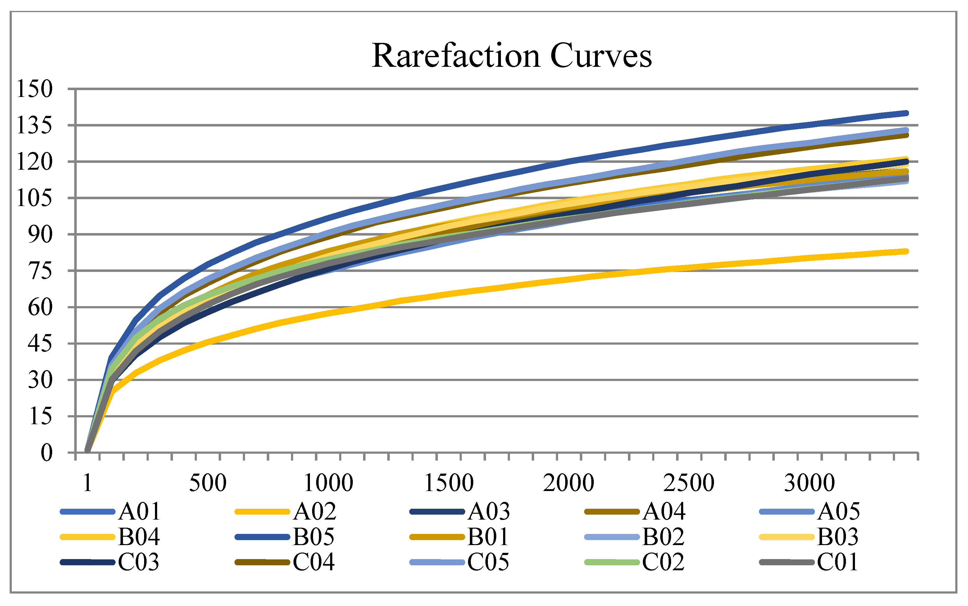
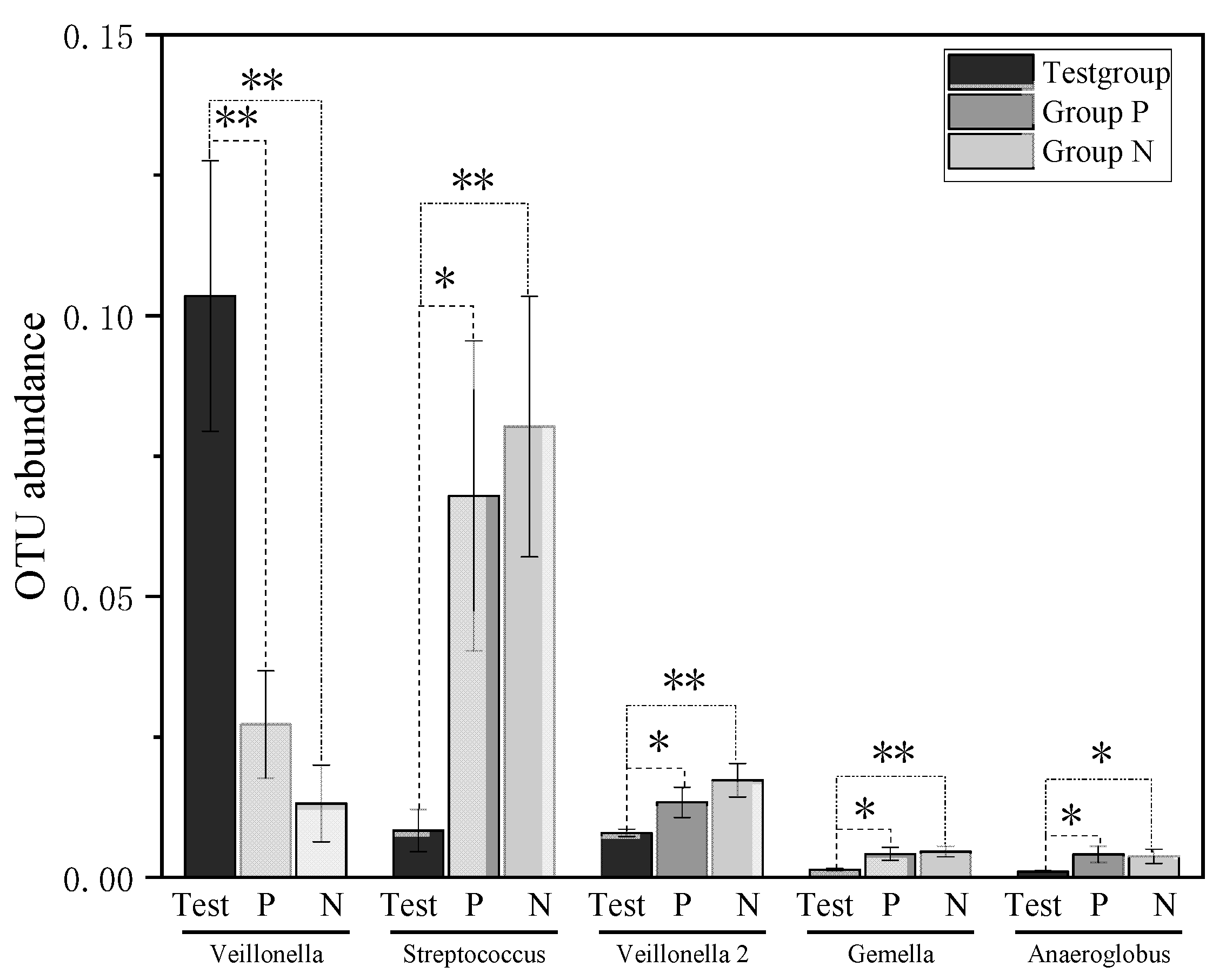
Rarefaction curves of samples are presented in Figure 2. Significant differences (p < 0.05) in the abundance of the taxa from saliva was estimated at the species level using Metastat analysis. Compared with Group P, 19 OTUs were significantly different in the Test group, with 7 OTUs increased and 12 OTUs decreased. Compared with Group N, 26 OTUs were significantly different in the Test group, with only 1 OTU (OTU0001, Veillonella parvula) increased and 25 OTUs decreased. Low taxonomy abundance is defined as an abundance less than 1% [34]. More details about taxonomy abundance greater than 0.001 are presented in Figure 3 and Table 2.

3.3. Biodiversity of the Salivary Microbiota

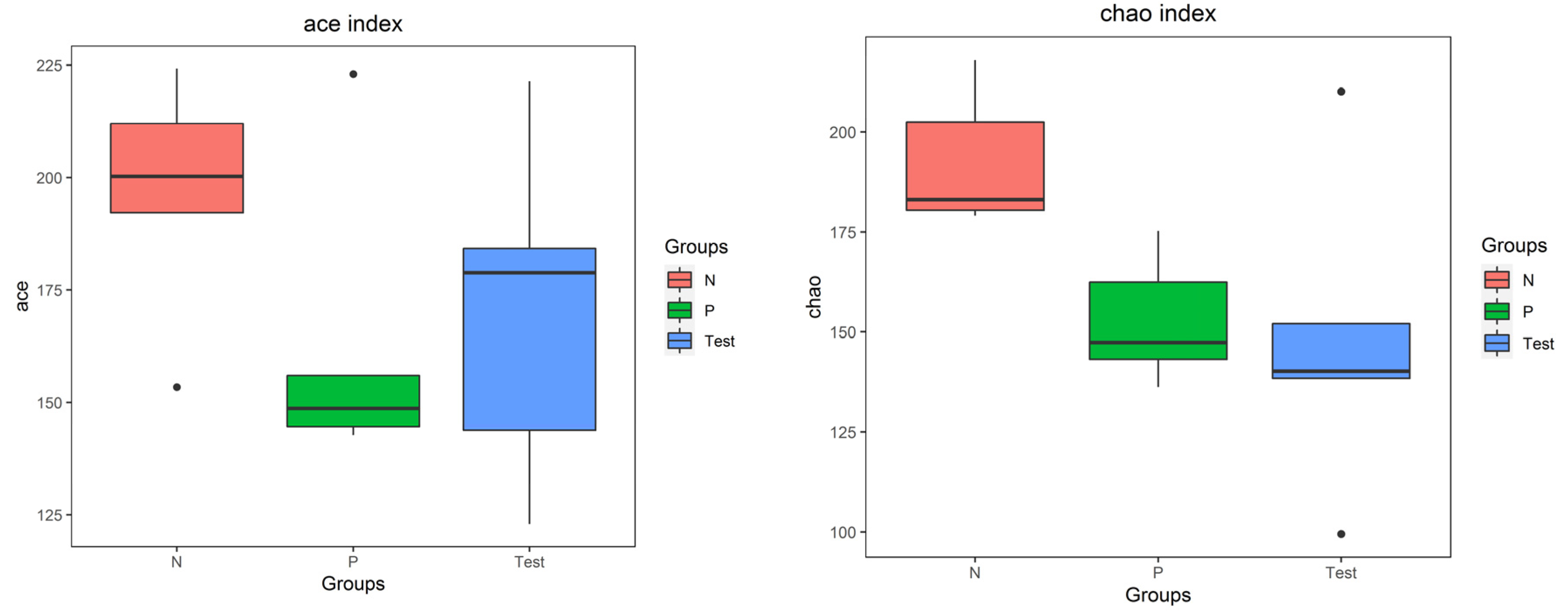
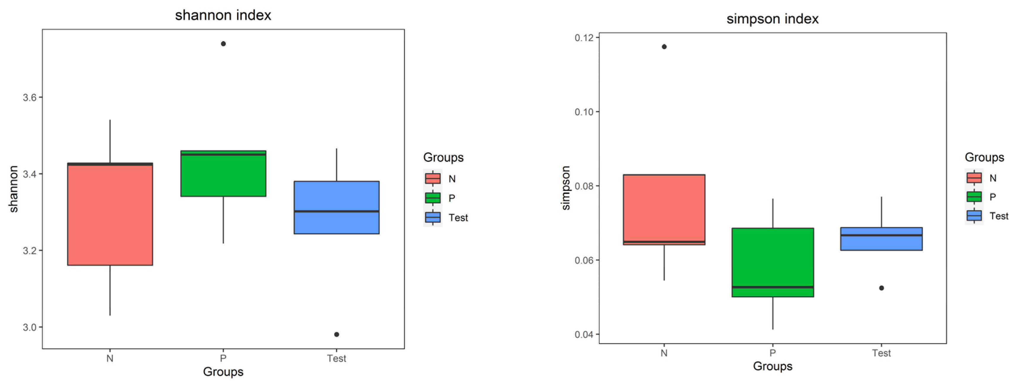
For alpha diversity, microbiota community richness was analyzed by the ACE and Chao indices, and the community diversity was analyzed by the Shannon and Simpson indices. For all four indices, significant differences were evident among the three groups (p < 0.05), as in Figure 4. Results of the beta diversity of the salivary microbiota determined by PCoA is shown in Figure 5. A clear separation of sample distribution was evident in the Test group, with roughly greater values than those from both Group P and Group N (principal components of 12.80% and 14.80%, respectively).
ANOSIM analysis also revealed significant differences in bacterial composition and structure among three groups (R = 0.304889, p = 0.005 < 0.01, respectively). Significant differences were evident in bacterial composition and structure between the Test group and Group P (R = 0.308, P = 0.043 < 0.05, respectively) and the Test group and Group N (R = 0.612, P = 0.003 < 0.01, respectively). No significant differences in bacterial composition and structure were evident between Group P and Group N (R = −0.032, p = 0.551 > 0.05, respectively).

4. Discussion

Our study findings strongly support the existence of distinct differences in the composition of the salivary microbiota in SS patients, independent of Candida carriage and DMFT. These findings further suggest that dysbiosis in oral microbiota might be a characteristic in SS patients.
Treponema, Lactobacillus, Streptococcus, Selenomonas, and Veillonella were the dominant bacterial genera in saliva. Treponema, especially T. denticola, is commonly associated with periodontal disease [35]. Lactobacillus, Streptococcus, Selenomonas, and Veillonella have been significantly associated with the progression of dental caries [20]. Lactobacillus and Streptococcus are well-known cariogenic bacteria. Yet, prior studies have reported discordant results. One study found a similar salivary abundance of Lactobacillus and Streptococcus compared to a healthy population [6]. In contrast, a higher abundance in SS patients than a healthy population was found for Lactobacillus in two studies [17], and a similar abundance for Streptococcus also in two studies [14] but low in another study [36]. The reported abundance of Selenomonas increased in three studies [6,19,37] but was low in another [36]. Another predominant bacterial genera, Prevotella and Veillonella, was enriched in salivary microbiota in two studies [6,14]. The abundance of Neisseria was decreased in two studies [16,38].
In this current study, we did not observed differences in the abundance of any of these taxa. There are two possible explanations for this discrepancy. One is dental caries severity caused by oral cariogenic bacteria, and the other is the interaction between oral Candida and oral bacteria. Dental caries is a bacterial disease caused by dysbiosis in the complex microbiota community [21]. SS patients exhibit higher DMFT and DMFS. Our results agreed with the prior observations that with comparable DMFT, the salivary microbiota with most abundant genera of Prevotella, Veillonella, and Streptococcus in SS patients did not differ significantly from healthy controls [18]. Moreover, the relative abundance of Veillonella in SS patients with higher DMFT was reported to be higher than the abundance in healthy controls [6]. In this current study, the nonsignificant differences in the abundance of these cariogenic bacterial genera among the groups could be attributed to the comparable DMFT, leading to similar microbiota community dysbiosis.
Candida is a major cause of the opportunistic disease. The higher prevalence of oral Candida has been associated with hyposalivation and adverse effects on oral health [39]. Increasing evidence is solidifying the interaction between oral Candida and oral cariogenic bacteria, especially C. albicans [40]. In early in vitro oral biofilms, the presence of C. albicans alters the bacterial microbiota, leading to the presence of strictly anaerobic bacteria when oxygen is abundant [26]. The presence of C. albicans would increase the abundance of Streptococcus (particularly S. mutans), certain Lactobacillus species, and salivary Veillonella and Prevotella [26,41] and also increase the cariogenic virulence of biofilms [42] and even serve as potential “keystone” components of oral biofilms [43]. Oral Candida are also correlated with the number of untreated DMFT [44]. It was reported that Candida-infected individuals had 2.3 times more dental caries than those without Candida in their oral cavity [45]. C. albicans promotes caries activity that is dependent on PHR2 in a mixed microbial consortium [46]. In this current study, with similar oral Candida carriage and comparable DMFT, the abundance of the salivary microbiota at the genus level was not significantly different between SS patients with controls.
At the species level, the significantly increasing abundance of V. parvula (OTU001) in SS patients than controls was observed in this present study. The presence of Veillonella in the salivary microbiota has been related to the occurrence of dental caries [47]. Veillonella is a gram-negative anaerobic coccus capable of fermenting organic acids such as malate and lactate. This may result in increasing abundance of the salivary microbiota in SS patients [48]. The survival of V. parvula can alter the physiology of S. mutans by changing the expression of genes coding for many proteins [49]. This could contribute to more prevalent carious lesions in SS subjects. In this current study, even with comparable DMFT, significantly more V. parvula was detected in SS patients. These findings demonstrate the connection between V. parvula and the autoimmune disease SS. Elevated dysbiosis of V. parvula can initiate the progression of SS [50]. Hence, V. parvula, as an immunomodulatory commensal bacterium, may serve as a unique microbial biomarker for SS. This suggestion is supported by the results in other studies [6,14,51]. Uniquely, this present study is the first confirmation of the dominant role of V. parvula. Detection of the abundance of V. parvula might benefit the diagnosis, treatment, and monitoring of SS.
Measurement of microbial community diversity can be critical for understanding the composition of the microbiota [52]. Different from previous studies [16,17,19,38], alpha diversity analyses of each sample revealed significantly higher diversity and more equal distribution of abundance in samples from SS patients than from control patients in this present study. Our results are supported by prior studies [6,14,53], which also reported higher alpha diversity in SS patients compared to healthy controls. Concerning beta diversity, the differentiation among sites or habitats contrasts with previous study findings [54]. In this present study, we observed more diverse bacterial composition of the saliva microbiota from SS patients. Similar results were previously reported in SS patients compared to a control group [36]. Normally, the difference in the oral microbial diversity of patients might be related to xerogenic medications, oral health status, saliva flow rates, salivary pH, severity of dental caries, and oral Candida carriage. Compared with oral Candida carriage and dental caries severity, the significant differences in microbial diversity might be a result of the effects caused by SS on the oral environment. ANOSIM analysis indicated SS itself might be associated with oral microbial dysbiosis. This was also a conclusion previously [14].
Our collective results suggest that SS patients with Candida carriage have different diversity and abundance of microbiota composition compared to patients with only Candida carriage and healthy patients, irrespective of DMFT. It remains unclear whether these changes observed in microbiota are the cause or consequence of SS. Interaction between the oral microbiota and autoimmune diseases is complex. For SS, activated CD4+ T cells and B cells infiltrate into the salivary glands [55]. Salivary gland destruction changes in saliva protein composition, which leads to microbiota dysbiosis in the oral cavity [50]. Changes in the oral microbiota may also be a factor for the onset of systemic autoimmune diseases, including SS [50]. Autoimmune diseases might be influenced by oral microbiota through the activation of Toll-like receptors, molecular mimicry, epitope spread, and antigen persistence [56]. The molecular mimicry theory posits that immune cells respond to self-antigens sharing epitopes with foreign antigens from the microbiota. Animal studies have demonstrated that peptides from oral bacteria could activate Ro60-reactive T cells [57], followed by the activation of B cells to plasma cells that produce SS antigen A [56]. Future studies should examine interactions between microbiota imbalances and autoimmune responses in the development and progression of SS, especially the role of V. parvula.
The diagnosis and management of SS represent a challenge for the clinician, especially the dentist. Accompanying various lesions and pseudoconditions of the oral mucosa, the differential diagnosis must be made [58]. This study found significant dysbiosis in the oral microbiota of SS patients through the marked change in salivary microbiota compared with controls. This study provided a potential way to help with diagnosis and management. There are some limitations of this study. First, the sample size is small, with only five in each group. This may have limited the power to detect significant associations. However, for the high-throughput sequencing based experiments, power and sample size calculations are not established. In some oral microbiome-related references, comparisons have involved 11 subjects divided into 2 groups [59] and 10 SS patients divided into 2 subgroups [60]. These two studies attained reasonable results and conclusions. Therefore, the five subjects for each group in this study were likely sufficient to prove the composition diversity and identify the most important bacteria in samples [61]. Second, 16S rRNA gene sequencing remains the gold standard of environmental microbiology. However, species level classification is not reliable with 200–400 bp reads of the 16S rRNA gene, and analyses have been confined to genus and higher taxonomies [62]. Finally, as one of the clinical characteristics, the UWSF in primary SS patients was −0.43 mL/min compared to controls [63]. It is difficult to conclude whether the microbial dysbiosis was caused by SS or hyposalivation. Future studies should involve many more patients (females and males) and controls to provide a more comprehensive view of microbiota composition. Quantitative real-time PCR should be used with standard strains to determine the absolute abundance of each taxon.

5. Conclusions

Oral saliva microbiota changes are documented for the first time in oral Candida carriage SS patients compared to control patients of comparable age and DMFT. The present findings extended the depth of previous findings. V. parvula might be a unique immunomodulatory commensal bacterium and perhaps a microbial biomarker for SS. The abundance of dominant bacteria did not differ significantly between the groups. However, the distinct community structure diversity identified microbial dysbiosis as a significant characteristic in SS patients, irrespective of oral Candida carriage and dental caries status.

Supplementary Materials

The following supporting information can be downloaded at: https://www.mdpi.com/article/10.3390/jcm12041559/s1. It presented STROBE Statement and details data from the clinical examination.

Author Contributions

Conceptualization, H.X., H.L. and J.P.; methodology, H.X., H.L. and J.P., investigation, H.X.; data curation, H.X.; writing—original draft preparation, H.X.; writing—review and editing, H.X., H.L. and J.P.; supervision, H.L. and J.P. All authors have read and agreed to the published version of the manuscript.

Funding

This research received no external funding.

Institutional Review Board Statement

This study was approved by the Institutional Review Board of Peking University School and Hospital of Stomatology (#PKUSSIRB-201947088).

Informed Consent Statement

Informed consent was obtained from all subjects involved in the study.

Data Availability Statement

The data presented in this study are available on request from the corresponding author.

Acknowledgments

Bacterial 16S rRNA gene sequences and analysis were performed by Beijing Cnkingbio Biotechnology Co., LTD, Beijing, China.

Conflicts of Interest

The authors declare no conflict of interest.

References

  1. Mariette, X.; Criswell, L.A. Primary Sjögren’s Syndrome. N. Engl. J. Med. 2018, 378, 931–939. [Google Scholar] [CrossRef] [PubMed]
  2. Zunt, S.L. Xerostomia/Salivary Gland Hypofunction: Diagnosis and Management. Compend. Contin. Educ. Dent. 2018, 39, 365. [Google Scholar] [PubMed]
  3. Kassebaum, N.J.; Smith, A.G.C.; Bernabé, E.; Fleming, T.D.; Reynolds, A.E.; Vos, T.; Murray, C.J.L.; Marcenes, W.; GBD 2015 Oral Health Collaborators. Global, Regional, and National Prevalence, Incidence, and Disability-Adjusted Life Years for Oral Conditions for 195 Countries, 1990–2015: A Systematic Analysis for the Global Burden of Diseases, Injuries, and Risk Factors. J. Dent. Res. 2017, 96, 380–387. [Google Scholar] [CrossRef] [PubMed]
  4. González, S.; Sung, H.; Sepúlveda, D.; González, M.; Molina, C. Oral manifestations and their treatment in Sjögren′s syndrome. Oral Dis. 2014, 20, 153–161. [Google Scholar] [CrossRef]
  5. Maarse, F.; Jager, D.J.; Forouzanfar, T.; Wolff, J.; Brand, H. Tooth loss in Sjogren’s syndrome patients compared to age and gender matched controls. Med. Oral Patol. Oral. Cir. Bucal. 2018, 23, e545–e551. [Google Scholar] [CrossRef]
  6. Zhou, Z.; Ling, G.; Ding, N.; Xun, Z.; Zhu, C.; Hua, H.; Chen, X. Molecular analysis of oral microflora in patients with primary Sjögren’s syndrome by using high-throughput sequencing. PeerJ 2018, 6, e5649. [Google Scholar] [CrossRef]
  7. Featherstone, J.D.B. The Continuum of Dental Caries—Evidence for a Dynamic Disease Process. J. Dent. Res. 2004, 83 (Suppl. 1), C39–C42. [Google Scholar] [CrossRef]
  8. Saleh, J.; Figueiredo, M.A.Z.; Cherubini, K.; Salum, F.G. Salivary hypofunction: An update on aetiology, diagnosis and therapeutics. Arch. Oral Biol. 2015, 60, 242–255. [Google Scholar] [CrossRef]
  9. Mosaddad, S.A.; Tahmasebi, E.; Yazdanian, A.; Rezvani, M.B.; Seifalian, A.; Yazdanian, M.; Tebyanian, H. Oral microbial biofilms: An update. Eur. J. Clin. Microbiol. Infect. Dis. 2019, 38, 2005–2019. [Google Scholar] [CrossRef]
  10. Dawes, C. Salivary flow patterns and the health of hard and soft oral tissues. J. Am. Dent. Assoc. 2008, 139, 18S–24S. [Google Scholar] [CrossRef]
  11. Twetman, S. Prevention of dental caries as a non-communicable disease. Eur. J. Oral Sci. 2018, 126 (Suppl. 1), 19–25. [Google Scholar] [CrossRef] [PubMed] [Green Version]
  12. Contaldo, M.; Lucchese, A.; Lajolo, C.; Rupe, C.; Di Stasio, D.; Romano, A.; Petruzzi, M.; Serpico, R. The Oral Microbiota Changes in Orthodontic Patients and Effects on Oral Health: An Overview. J. Clin. Med. 2021, 10, 780. [Google Scholar] [CrossRef] [PubMed]
  13. Müller, V.J.; Belibasakis, G.N.; Bosshard, P.P.; Wiedemeier, D.B.; Bichsel, D.; Rücker, M.; Stadlinger, B. Change of saliva composition with radiotherapy. Arch. Oral Biol. 2019, 106, 104480. [Google Scholar] [CrossRef] [PubMed]
  14. Siddiqui, H.; Chen, T.; Aliko, A.; Mydel, P.M.; Jonsson, R.; Olsen, I. Microbiological and bioinformatics analysis of primary Sjögren’s syndrome patients with normal salivation§. J. Oral Microbiol. 2016, 8, 31119. [Google Scholar] [CrossRef] [PubMed] [Green Version]
  15. Belstrøm, D.; Holmstrup, P.; Fiehn, N.-E.; Rosing, K.; Bardow, A.; Paster, B.; Pedersen, A.L. Bacterial composition in whole saliva from patients with severe hyposalivation—A case-control study. Oral Dis. 2016, 22, 330–337. [Google Scholar] [CrossRef]
  16. Sembler-Møller, M.L.; Belstrøm, D.; Locht, H.; Enevold, C.; Pedersen, A.M.L. Next-generation sequencing of whole saliva from patients with primary Sjögren’s syndrome and non-Sjögren’s sicca reveals comparable salivary microbiota. J. Oral Microbiol. 2019, 11, 1660566. [Google Scholar] [CrossRef]
  17. Sharma, D.; Sandhya, P.; Vellarikkal, S.K.; Surin, A.K.; Jayarajan, R.; Verma, A.; Kumar, A.; Ravi, R.; Danda, D.; Sivasubbu, S.; et al. Saliva microbiome in primary Sjögren’s syndrome reveals distinct set of disease-associated microbes. Oral Dis. 2020, 26, 295–301. [Google Scholar] [CrossRef]
  18. Cunha-Cruz, J.; Scott, J.; Rothen, M.; Mancl, L.; Lawhorn, T.; Brossel, K.; Berg, J. Salivary characteristics and dental caries: Evidence from general dental practices. J. Am. Dent. Assoc. 2013, 144, e31–e40. [Google Scholar] [CrossRef]
  19. Rusthen, S.; Kristoffersen, A.K.; Young, A.; Galtung, H.K.; Petrovski, B.; Palm, Ø.; Enersen, M.; Jensen, J.L. Dysbiotic salivary microbiota in dry mouth and primary Sjögren’s syndrome patients. PLoS ONE 2019, 14, e0218319. [Google Scholar] [CrossRef]
  20. Zhang, Y.; Wang, X.; Li, H.; Ni, C.; Du, Z.; Yan, F. Human oral microbiota and its modulation for oral health. Biomed. Pharmacother. 2018, 99, 883–893. [Google Scholar] [CrossRef]
  21. Tarapan, S.; Matangkasombut, O.; Trachootham, D.; Sattabanasuk, V.; Talungchit, S.; Paemuang, W.; Phonyiam, T.; Chokchaitam, O.; Mungkung, O.; Lam-Ubol, A. Oral Candida colonization in xerostomic postradiotherapy head and neck cancer patients. Oral Dis. 2019, 25, 1798–1808. [Google Scholar] [CrossRef] [PubMed]
  22. Nikou, S.-A.; Kichik, N.; Brown, R.; Ponde, N.; Ho, J.; Naglik, J.; Richardson, J. Candida albicans Interactions with Mucosal Surfaces during Health and Disease. Pathogens 2019, 8, 53. [Google Scholar] [CrossRef] [PubMed] [Green Version]
  23. Serrano, J.; López-Pintor, R.; Ramírez, L.; Fernández-Castro, M.; Sanz, M.; Melchor, S.; Peiteado, D.; Hernández, G. Risk factors related to oral candidiasis in patients with primary Sjögren’s syndrome. Med. Oral. Patol. Oral. Cir. Bucal. 2020, 25, e700–e705. [Google Scholar] [CrossRef] [PubMed]
  24. Xin, W.; Leung, K.C.M.; Lo, E.C.M.; Mok, M.Y.; Leung, M.H. Sicca Symptoms, Oral Health Conditions, Salivary Flow and Oral Candida in Sjögren’s Syndrome Patients. Int. J. Environ. Res. Public Health 2020, 17, 3625. [Google Scholar] [CrossRef]
  25. Ergun, S.; Cekici, A.; Topcuoglu, N.; Migliari, D.; Kulekci, G.; Tanyeri, H.; Isik, G. Oral status and Candida colonization in patients with Sjogren s Syndrome. Med. Oral. Patol. Oral. Cir. Bucal. 2010, 15, e310–e315. [Google Scholar] [CrossRef]
  26. Janus, M.M.; Crielaard, W.; Volgenant, C.M.C.; van der Veen, M.H.; Brandt, B.W.; Krom, B.P. Candida albicans alters the bacterial microbiome of early in vitro oral biofilms. J. Oral Microbiol. 2017, 9, 1270613. [Google Scholar] [CrossRef] [Green Version]
  27. Montelongo-Jauregui, D.; Lopez-Ribot, J.L. Candida Interactions with the Oral Bacterial Microbiota. J. Fungi 2018, 4, 122. [Google Scholar] [CrossRef] [Green Version]
  28. Dige, I.; Nyvad, B. Candida species in intact in vivo biofilm from carious lesions. Arch. Oral Biol. 2019, 101, 142–146. [Google Scholar] [CrossRef]
  29. Patel, R.; Shahane, A. The epidemiology of Sjögren’s syndrome. Clin. Epidemiol. 2014, 6, 247–255. [Google Scholar] [CrossRef] [Green Version]
  30. Vitali, C.; Bombardieri, S.; Jonsson, R.; Moutsopoulos, H.M.; Alexander, E.L.; Carsons, S.E.; Daniels, T.E.; Fox, P.C.; Fox, R.I.; Kassan, S.S.; et al. Classification criteria for Sjogren’s syndrome: A revised version of the European criteria proposed by the American-European Consensus Group. Ann. Rheum. Dis. 2002, 61, 554–558. [Google Scholar] [CrossRef] [Green Version]
  31. Shiboski, C.H.; Shiboski, S.C.; Seror, R.; Criswell, L.A.; Labetoulle, M.; Lietman, T.M.; Mariette, X. 2016 ACR-EULAR classification criteria for primary Sjögren’s syndrome: A consensus and data-driven methodology involving three international patient cohorts. Arthritis Rheumatol. 2017, 69, 35–45. [Google Scholar] [CrossRef]
  32. Caporaso, J.G.; Kuczynski, J.; Stombaugh, J.; Bittinger, K.; Bushman, F.D.; Costello, E.K.; Fierer, N.; Gonzalez Peña, A.; Goodrich, J.K.; Gordon, J.I.; et al. QIIME allows analysis of high-throughput community sequencing data. Nat. Methods 2010, 7, 335–336. [Google Scholar] [CrossRef] [PubMed] [Green Version]
  33. Schloss, P.D.; Westcott, S.L.; Ryabin, T.; Hall, J.R.; Hartmann, M.; Hollister, E.B.; Lesniewski, R.A.; Oakley, B.B.; Parks, D.H.; Robinson, C.J.; et al. Introducing mothur: Open-Source, Platform-Independent, Community-Supported Software for Describing and Comparing Microbial Communities. Appl. Environ. Microbiol. 2009, 75, 7537–7541. [Google Scholar] [CrossRef] [PubMed] [Green Version]
  34. Dyrhovden, R.; Rippin, M.; Øvrebø, K.K.; Nygaard, R.M.; Ulvestad, E.; Kommedal, Ø. Managing Contamination and Diverse Bacterial Loads in 16S rRNA Deep Sequencing of Clinical Samples: Implications of the Law of Small Numbers. mBio 2021, 12, e0059821. [Google Scholar] [CrossRef]
  35. Zeng, H.; Chan, Y.; Gao, W.; Leung, W.K.; Watt, R.M. Diversity of Treponema denticola and Other Oral Treponeme Lineages in Subjects with Periodontitis and Gingivitis. Microbiol. Spectr. 2021, 9, e0070121. [Google Scholar] [CrossRef] [PubMed]
  36. van der Meulen, T.A.; Harmsen, H.J.M.; Bootsma, H.; Liefers, S.C.; Vila, A.V.; Zhernakova, A.; Weersma, R.K.; Spijkervet, F.K.L.; Kroese, F.G.M.; Vissink, A. Reduced salivary secretion contributes more to changes in the oral microbiome of patients with primary Sjögren’s syndrome than underlying disease. Ann. Rheum. Dis. 2018, 77, 1542–1544. [Google Scholar] [CrossRef] [PubMed]
  37. Alam, J.; Lee, A.; Lee, J.; Kwon, D.I.; Park, H.K.; Park, J.-H.; Jeon, S.; Baek, K.; Lee, J.; Park, S.-H.; et al. Dysbiotic oral microbiota and infected salivary glands in Sjögren’s syndrome. PLoS ONE 2020, 15, e0230667. [Google Scholar] [CrossRef] [PubMed]
  38. Zhou, S.; Cai, Y.; Wang, M.; Yang, W.; Duan, N. Oral microbial flora of patients with Sicca syndrome. Mol. Med. Rep. 2018, 18, 4895–4903. [Google Scholar] [CrossRef]
  39. Buranarom, N.; Komin, O.; Matangkasombut, O. Hyposalivation, oral health, and Candida colonization in independent dentate elders. PLoS ONE 2020, 15, e0242832. [Google Scholar] [CrossRef]
  40. Ponde, N.O.; Lortal, L.; Ramage, G.; Naglik, J.R.; Richardson, J.P. Candida albicans biofilms and polymicrobial interactions. Crit. Rev. Microbiol. 2021, 47, 91–111. [Google Scholar] [CrossRef]
  41. Xiao, J.; Grier, A.; Faustoferri, R.; Alzoubi, S.; Gill, A.; Feng, C.; Liu, Y.; Quivey, R.; Kopycka-Kedzierawski, D.; Koo, H.; et al. Association between Oral Candida and Bacteriome in Children with Severe ECC. J. Dent. Res. 2018, 97, 1468–1476. [Google Scholar] [CrossRef] [PubMed]
  42. Fujinami, W.; Nishikawa, K.; Ozawa, S.; Hasegawa, Y.; Takebe, J. Correlation between the relative abundance of oral bacteria and Candida albicans in denture and dental plaques. J. Oral Biosci. 2021, 63, 175–183. [Google Scholar] [CrossRef] [PubMed]
  43. Young, T.; Alshanta, O.-A.; Kean, R.; Bradshaw, D.; Pratten, J.; Williams, C.; Woodall, C.; Ramage, G.; Brown, J.L. Candida albicans as an Essential “Keystone” Component within Polymicrobial Oral Biofilm Models? Microorganisms 2020, 9, 59. [Google Scholar] [CrossRef] [PubMed]
  44. Nishimaki, F.; Yamada, S.-I.; Kawamoto, M.; Sakurai, A.; Hayashi, K.; Kurita, H. Relationship Between the Quantity of Oral Candida and Systemic Condition/Diseases of the Host: Oral Candida Increases with Advancing Age and Anemia. Mycopathologia 2019, 184, 251–260. [Google Scholar] [CrossRef] [PubMed]
  45. Eidt, G.; Waltermann, E.D.M.; Hilgert, J.B.; Arthur, R.A. Candida and dental caries in children, adolescents and adults: A systematic review and meta-analysis. Arch. Oral Biol. 2020, 119, 104876. [Google Scholar] [CrossRef] [PubMed]
  46. Du, Q.; Ren, B.; He, J.; Peng, X.; Guo, Q.; Zheng, L.; Li, J.; Dai, H.; Chen, V.; Zhang, L.; et al. Candida albicans promotes tooth decay by inducing oral microbial dysbiosis. ISME J. 2020, 15, 894–908. [Google Scholar] [CrossRef]
  47. Dame-Teixeira, N.; de Lima, A.K.A.; Do, T.; Stefani, C.M. Meta-Analysis Using NGS Data: The Veillonella Species in Dental Caries. Front. Oral Health 2021, 2, 770917. [Google Scholar] [CrossRef]
  48. Periasamy, S.; Kolenbrander, P.E.; Zeng, L.; Das, S.; Burne, R.A. Central Role of the Early Colonizer Veillonella sp. in Establishing Multispecies Biofilm Communities with Initial, Middle, and Late Colonizers of Enamel. J. Bacteriol. 2010, 192, 2965–2972. [Google Scholar] [CrossRef] [Green Version]
  49. Luppens SBI, K.D.; Bandounas, L.; Jonker, M.J.; Wittink, F.R.A.; Bruning, O.; Breit, T.M.; ten Cate, J.M.; Crielaard, W. Effect of Veillonella parvula on the antimicrobial resistance and gene expression of Streptococcus mutans grown in a dual-species biofilm. Oral Microbiol. Immunol. 2008, 23, 183–189. [Google Scholar] [CrossRef]
  50. Nikitakis, N.G.; Papaioannou, W.; I Sakkas, L.; Kousvelari, E. The autoimmunity-oral microbiome connection. Oral Dis. 2016, 23, 828–839. [Google Scholar] [CrossRef]
  51. Singh, M.; Teles, F.; Uzel, N.G.; Papas, A. Characterizing Microbiota from Sjogren’s Syndrome Patients. JDR Clin. Trans. Res. 2020, 6, 324–332. [Google Scholar] [CrossRef] [PubMed]
  52. Lozupone, C.A.; Hamady, M.; Kelley, S.T.; Knight, R. Quantitative and Qualitative β Diversity Measures Lead to Different Insights into Factors That Structure Microbial Communities. Appl. Environ. Microbiol. 2007, 73, 1576–1585. [Google Scholar] [CrossRef] [PubMed] [Green Version]
  53. Palmer, R.J.; Cotton, S.L.; Kokaras, A.S.; Gardner, P.; Grisius, M.; Pelayo, E.; Warner, B.; Paster, B.J.; Alevizos, I. Analysis of oral bacterial communities: Comparison of HOMINGS with a tree-based approach implemented in QIIME. J. Oral Microbiol. 2019, 11, 1586413. [Google Scholar] [CrossRef] [PubMed] [Green Version]
  54. de Paiva, C.S.; Jones, D.B.; Stern, M.E.; Bian, F.; Moore, Q.L.; Corbiere, S.; Streckfus, C.F.; Hutchinson, D.S.; Ajami, N.J.; Petrosino, J.F.; et al. Altered Mucosal Microbiome Diversity and Disease Severity in Sjögren Syndrome. Sci. Rep. 2016, 6, 23561. [Google Scholar] [CrossRef] [Green Version]
  55. Shen, L.; He, J.; Kramer, J.M.; Bunya, V.Y. Sjögren’s Syndrome: Animal Models, Etiology, Pathogenesis, Clinical Subtypes, and Diagnosis. J. Immunol. Res. 2019, 2019, 8101503. [Google Scholar] [CrossRef]
  56. Zorba, M.; Melidou, A.; Patsatsi, A.; Ioannou, E.; Kolokotronis, A. The possible role of oral microbiome in autoimmunity. Int. J. Women’s Dermatol. 2020, 6, 357–364. [Google Scholar] [CrossRef]
  57. Szymula, A.; Rosenthal, J.; Szczerba, B.M.; Bagavant, H.; Fu, S.M.; Deshmukh, U.S. T cell epitope mimicry between Sjögren’s syndrome Antigen A (SSA)/Ro60 and oral, gut, skin and vaginal bacteria. Clin. Immunol. 2014, 152, 1–9. [Google Scholar] [CrossRef] [Green Version]
  58. della Vella, F.; Lauritano, D.; Lajolo, C.; Lucchese, A.; Di Stasio, D.; Contaldo, M.; Serpico, R.; Petruzzi, M. The pseudolesions of the oral mucosa: Differential diagnosis and related systemic conditions. Appl. Sci. 2019, 9, 2412. [Google Scholar] [CrossRef] [Green Version]
  59. Ames, N.J.; Sulima, P.; Ngo, T.; Barb, J.; Munson, P.J.; Paster, B.J.; Hart, T.C. A Characterization of the Oral Microbiome in Allogeneic Stem Cell Transplant Patients. PLoS ONE 2012, 7, e47628. [Google Scholar] [CrossRef]
  60. Li, M.; Zou, Y.; Jiang, Q.; Jiang, L.; Yu, Q.; Ding, X.; Yu, Y. A preliminary study of the oral microbiota in Chinese patients with Sjögren’s syndrome. Arch. Oral Biol. 2016, 70, 143–148. [Google Scholar] [CrossRef] [Green Version]
  61. Di Bella, J.M.; Bao, Y.; Gloor, G.B.; Burton, J.P.; Reid, G. High throughput sequencing methods and analysis for microbiome research. J. Microbiol. Methods 2013, 95, 401–414. [Google Scholar] [CrossRef]
  62. Sanschagrin, S.; Yergeau, E. Next-generation sequencing of 16S ribosomal RNA gene amplicons. J. Vis. Exp. 2014. [Google Scholar] [CrossRef]
  63. Martínez-Ceballos, M.A.; Aguilera, N.; Garzón-González, K.C.; Cajamarca-Baron, J.; Alzate-Granados, J.P.; Rojas-Villarraga, A. Unstimulated whole salivary flow in Sjögren’s Syndrome: Systematic literature review and meta-analysis. Hortic. Bras. 2021, 61, 12. [Google Scholar] [CrossRef]
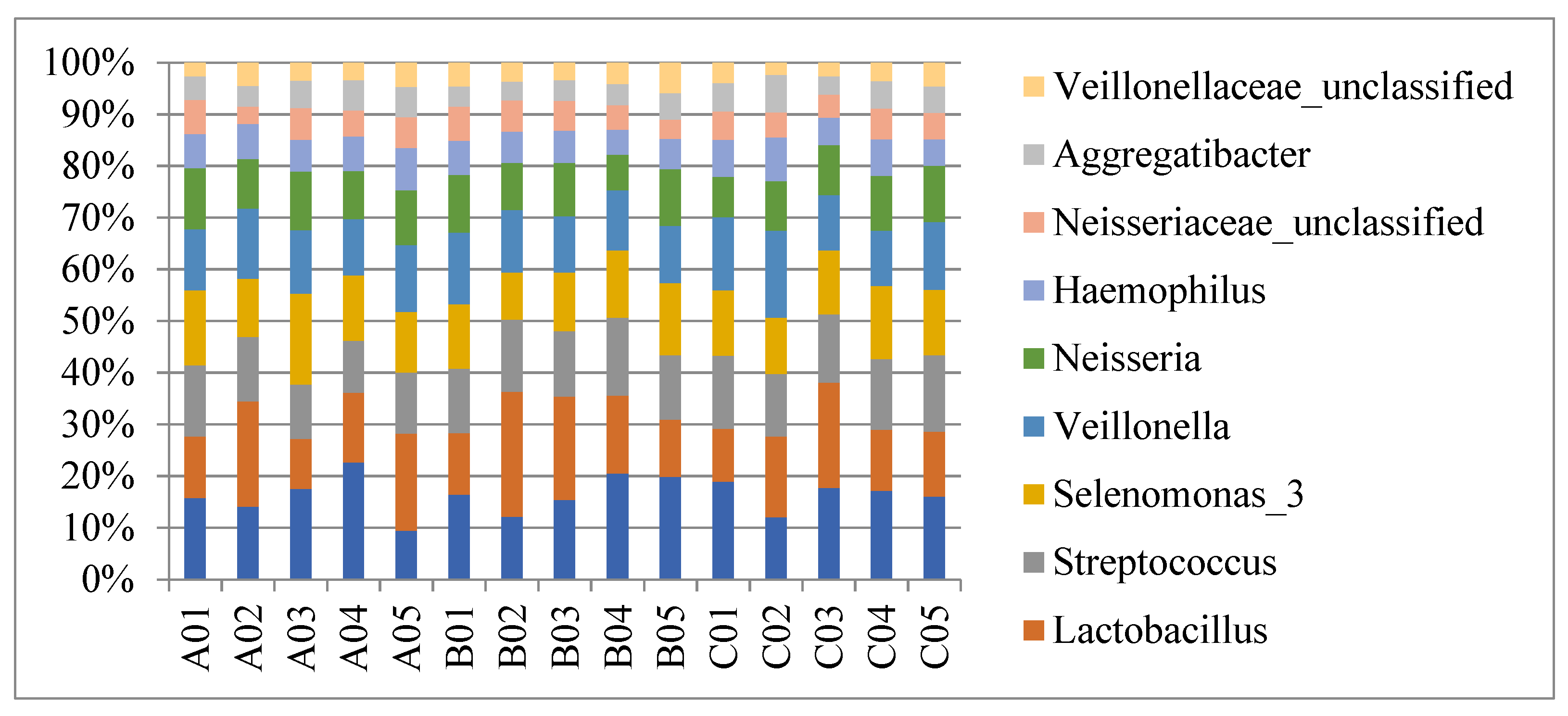
Figure 1. Species profiling histogram showing the 10 most predominant genera of saliva in Test group (A01–A05)/Group P (B01–B05)/Group N (C01–C05). The abundance of the total sequences of the five most predominant genera in Test group/Group P/Group N were Treponema (10.2%/9.9%/10.2%), Lactobacillus (9.5%/10%/8.4%), Streptococcus (7.6%/7.9%/8.5%), Selenomonas (8.6%/7.1%/7.8%), and Veillonella (7.9%/7.1%/7.8%). The numbers and proportions of sequences identified at the genus level were comparable among the three groups (p > 0.05).
Jcm 12 01559 g001
Figure 2. Rarefaction curves showing that sequencing reached saturation.
Jcm 12 01559 g002
Figure 3. Five OTUs with mean abundance of taxonomy >0.001, displayed significant changes among all three groups: OTU001, genera Veillonella; OTU011, genera Streptococcus; OTU021, genera Veillonella; OTU045, genera Gemella; and OTU047, genera Anaeroglobus. * significantly different (p < 0.05); ** extremely significantly different (p < 0.01).
Jcm 12 01559 g003
Figure 4. Alpha diversity (Ace, Chao, Shannon, and Simpson indices) of the three groups at the genus level. Microbiota community richness was analyzed by the ACE and Chao indices. Community diversity was analyzed by the Shannon and Simpson indices. Box plots indicate medium, minimum, and maximum values. Significant differences among three groups were evident for the four indices (p < 0.05).
Jcm 12 01559 g004aJcm 12 01559 g004b
Figure 5. Beta diversity evident in principal coordinate analysis (PCoA) based on weighted Unifrac matrix. Similarity in bacterial composition is indicated by the distance between dots, with smaller distance denoting increased similarity. The spatial distribution of plots from Group P and N subjects overlapped, suggesting a similar set of genera. Plots from the Test group of subjects formed a cluster, having roughly greater values than the plot clusters from Group P and N subjects. Test group samples displayed a trend of dispersion along the PCoA2 axis. Group P and N samples displayed a trend of dispersion along the PCoA1 axis.
Jcm 12 01559 g005
Table 1. Demographic characteristics of the three groups of patients.
Test GroupGroup PGroup Np-Value
Number of cases555NA
Mean age (years) #59.8 ± 7.557.4 ± 15.245.0 ± 9.40.122
Women (n, %)5 (100%)5 (100%)5 (100%)NA
Smoker (n)000NA
Autoimmune diseases other than pSS (n)000NA
Received DMARDs in last 3 months (n)300NA
Duration of clinically apparent xerostomia (median months, range)24 (6–36)0 (0–0)0 (0–0)NA
Ocular dryness (n)300NA
Positivity of anti-SSA (n)300NA
LSG biopsy focus score ≥ 1(n)200NA
Candida carriage (cfu/mL)>200>2000NA
Wearing removable denture (n)110NA
DMFT #21.6 ± 6.717.4 ± 6.513.6 ± 3.80.137
DMFS #67.6 ± 12.958.0 ± 27.934.0 ± 19.70.069
Total incisal caries
UWSF (mL/min)
3
0
0
>0.1
0
>0.1
0.148
0.047 *
Test group: SS patients group; Group P: oral candidiasis patients (positive control group) and Group N: healthy control patients. # Values are presented as mean ± standard deviation. * A p < 0.05 was considered statistically significant. DMARDs disease modifying anti-rheumatic drugs, LSG labial salivary gland, DMFT/DMFS total numbers of teeth present decayed (D), missed (M), filled (F) teeth (T)/surfaces (S), UWSF unstimulated whole salivary flow rate, NA not applicable.
Table 2. (a) Different taxonomy between Test group and Group P, with mean abundance > 0.001. (b) Different taxonomy between Test group and group N, with mean abundance > 0.001.
(a)
OTUTaxonomy
(Genera)
Mean ± SD
Test Group
Mean ± SD
Group P
Metastat
p-Value
Increase in SS
OTU001Veillonella0.103472 ± 0.0240920.027183 ± 0.009530.007 **
OTU038Gemella0.01297 ± 0.0021960.006591 ± 0.0005990.008 **
OTU042Neisseria0.019715 ± 0.0082020.002831 ± 0.0006350.039 *
OTU066Lactobacillus0.005269 ± 0.0013210.00035 ± 0.00030.002 **
OTU102Selenomonas0.001091 ± 0.0004220.000218 ± 0.0000960.042 *
Decrease in SS
OTU008Neisseria0.017735 ± 0.0070670.053774 ± 0.0151880.030 *
OTU011Streptococcus0.008269 ± 0.0037570.067866 ± 0.0276170.032 *
OTU013Firmicutes0.003479 ± 0.0018230.021472 ± 0.006270.009 **
OTU021Veillonella0.007866 ± 0.0006520.013299 ± 0.0026850.049 *
OTU045Gemella0.001301 ± 0.0002110.004119 ± 0.0011530.019 *
OTU047Anaeroglobus0.000994 ± 0.000150.004037 ± 0.0014390.035 *
OTU060Cardiobacterium0.000349 ± 0.0000870.005424 ± 0.0023080.029 *
(b)
OTUTaxonomy (Genera)Mean ± SD
Test Group
Mean ± SD
Group N
Metastat
p-Value
Increase in SS
OTU001Veillonella0.103472 ± 0.0240920.013111 ± 0.0068310.002 **
Decrease In SS
OTU002Streptococcus0.082284 ± 0.0063970.182442 ± 0.0283160.003 **
OTU011Streptococcus0.008269 ± 0.0037570.080205 ± 0.0232120.006 **
OTU012Veillonella0.001973 ± 0.0004530.006733 ± 0.0017540.014 *
OTU015Neisseriaceae_unclassified0.011237 ± 0.0028640.03873 ± 0.0101310.015 *
OTU021Veillonella0.007866 ± 0.0006520.017235 ± 0.0029760.006 **
OTU045Gemella0.001301 ± 0.0002110.004557 ± 0.000960.004 **
OTU047Anaeroglobus0.000994 ± 0.000150.003697 ± 0.0012580.037 *
OTU050Megasphaera0.002128 ± 0.000470.007184 ± 0.0024690.048 *
OTU058Selenomonas0.000647 ± 0.0001790.01557 ± 0.0059490.018 *
OTU061Veillonella0.001773 ± 0.0007450.008027 ± 0.002440.020 *
OTU086Veillonella0.000267 ± 0.000110.003788 ± 0.0012470.010 **
OTU, operational taxonomic unit; SS, Sjögren’s syndrome. * significantly different (p < 0.05); ** extremely significantly different (p < 0.01).
Disclaimer/Publisher’s Note: The statements, opinions and data contained in all publications are solely those of the individual author(s) and contributor(s) and not of MDPI and/or the editor(s). MDPI and/or the editor(s) disclaim responsibility for any injury to people or property resulting from any ideas, methods, instructions or products referred to in the content.