
Hand and Wrist Involvement in Seropositive Rheumatoid Arthritis, Seronegative Rheumatoid Arthritis, and Psoriatic Arthritis—The Value of Classic Radiography


Abstract
1. Introduction
2. Aim of the Study
3. Materials and Methods

| Seropositive RA | Seronegative RA | PsA | |
|---|---|---|---|
| n = 180 | n = 36 | n = 154 | |
| Female | 151 (83.9%) | 32 (88.9%) | 109 (70.8%) |
| Male | 29 (16.1%) | 4 (11.1%) | 45 (29.2%) |
| Mean age (SD) | 53.4 (12.6) | 53.1 (17.7) | 48.1 (12.4) |
4. Statistical Analysis
5. Results
5.1. Overview of the Symmetry, Distribution, and Radiographic Lesions in the Three Compared Diseases
5.2. Differentiating Seropositive RA from PsA
5.3. Differentiating Seropositive RA from Seronegative RA
5.4. Differentiating Seronegative RA from PsA
6. Discussion
7. Limitations
8. Conclusions
Author Contributions
Funding
Institutional Review Board Statement
Informed Consent Statement
Data Availability Statement
Conflicts of Interest
References
- Davies, A.M.; Grainger, A.J.; James, S.J. Imaging of the Hand and Wrist: Techniques and Applications, 1st ed.; Springer: Berlin/Heidelberg, Germany, 2013. [Google Scholar] [CrossRef]
- Sudoł-Szopińska, I.; Teh, J.; Cotten, A. Rheumatoid Hand and Other Hand-deforming Rheumatic Conditions. Semin. Musculoskelet. Radiol. 2021, 25, 232–245. [Google Scholar] [CrossRef]
- Scott, D.L.; Coulton, B.L.; Symmons, D.P.M. Longterm outcome of treating rheumatoid arthritis: Results after 20 years. Lancet 1987, 1, 1108–1111. [Google Scholar] [CrossRef] [PubMed]
- Plant, M.J.; Jones, P.W.; Saklatvala, J.; Ollier, W.; Dawes, P.T. Patterns of radiological progression in early rheumatoid arthritis: Results of an 8 year prospective study. J. Rheumatol. 1998, 25, 417–426. [Google Scholar] [PubMed]
- Gladman, D.D.; Stafford-Brady, F.; Chang, C.H.; Lewandowski, K.; Russell, M.L. Longitudinal study of clinical and radiological progression in psoriatic arthritis. J. Rheumatol. 1990, 17, 809–812. [Google Scholar] [PubMed]
- Salaffi, F.; Carotti, M.; Beci, G.; Di Carlo, M.; Giovagnoni, A. Radiographic scoring methods in rheumatoid arthritis and psoriatic arthritis. Radiol. Med. 2019, 124, 1071–1086. [Google Scholar] [CrossRef] [PubMed]
- Braum, L.S.; McGonagle, D.; Bruns, A.; Philipp, S.; Hermann, S.; Aupperle, K.; Tan, A.L.; Diekhoff, T.; Hamm, B.; Hermann, K.-G.A. Characterisation of hand small joints arthropathy using high-resolution MRI-limited discrimination between osteoarthritis and psoriatic arthritis. Eur. Radiol. 2013, 23, 1686–1693. [Google Scholar] [CrossRef] [PubMed]
- Colebatch, A.N.; Edwards, C.J.; Østergaard, M.; Van Der Heijde, D.; Bálint, P.V.; D’Agostino, M.-A.; Forslind, K.; Grassi, W.; Haavardsholm, E.A.; Haugeberg, G.; et al. EULAR recommendations for the use of imaging of the joints in the clinical management of rheumatoid arthritis. Ann. Rheum. Dis. 2013, 72, 804–814. [Google Scholar] [CrossRef]
- Garlaschi, G.; Silvestri, E.; Satragno, L.; Cimmino, M.A. The Rheumatoid Hand. Diagnostic Imaging, 1st ed.; Springer: Milano, Italy, 2002. [Google Scholar] [CrossRef]
- Aletaha, D.; Neogi, T.; Silman, A.J.; Funovits, J.; Felson, D.T.; Bingham, C.O., 3rd.; Birnbaum, N.S.; Burmester, G.R.; Bykerk, V.P.; Cohen, M.D.; et al. 2010 Rheumatoid arthritis classification criteria: An American College of Rheumatology/European League against rheumatism collaborative initiative. Arthritis Rheum. 2010, 62, 2569–2581. [Google Scholar] [CrossRef]
- Kurowska, W.; Kuca-Warnawin, E.H.; Radzikowska, A.; Maśliński, W. The role of anti-citrullinated protein antibodies (ACPA) in the pathogenesis of rheumatoid arthritis. Cent. Eur. J. Immunol. 2017, 42, 390–398. [Google Scholar] [CrossRef]
- Merola, J.F.; Espinoza, L.R.; Fleischmann, R. Distinguishing rheumatoid arthritis from psoriatic arthritis. RMD Open 2018, 4, e000656. [Google Scholar] [CrossRef]
- Brown, A.K. How to interpret plain radiographs in clinical practice. Best Pract. Res. Clin. Rheumatol. 2013, 27, 249–269. [Google Scholar] [CrossRef]
- Carotti, M.; Salaffi, F.; Di Carlo, M.; Sessa, F.; Giovagnoni, A. Magnetic resonance imaging of the craniovertebral junction in early rheumatoid arthritis. Skelet. Radiol. 2019, 48, 553–561. [Google Scholar] [CrossRef]
- Sudoł-Szopińska, I.; Matuszewska, G.; Pracoń, G. Radiographic Atlas of Inflammatory Rheumatic Diseases, 1st ed.; Medisfera: Warsaw, Poland, 2022. [Google Scholar]
- Gadeholt, O.; Hausotter, K.; Eberle, H.; Klink, T.; Pfeil, A. Differing X-ray patterns in seronegative and seropositive rheumatoid arthritis. Clin. Rheumatol. 2019, 38, 2403–2410. [Google Scholar] [CrossRef] [PubMed]
- Moll, J.M.; Wright, V. Psoriatic arthritis. Semin. Arthritis Rheum. 1973, 3, 55–78. [Google Scholar] [CrossRef] [PubMed]
- Tillett, W.; Costa, L.; Jadon, D.; Wallis, D.; Cavill, C.; McHugh, J.; Korendowych, E.; McHugh, N. The ClASsification for Psoriatic ARthritis (CASPAR) Criteria—A Retrospective Feasibility, Sensitivity, and Specificity Study. J. Rheumatol. 2012, 39, 154–156. [Google Scholar] [CrossRef] [PubMed]
- Zahran, E.; Youssof, A.; Shehata, W.; Bahgat, A.; Elshebiny, E. Predictive role of serum rheumatoid factor in different disease pattern of psoriasis and psoriatic arthritis. Egypt J. Intern. Med. 2021, 33, 49. [Google Scholar] [CrossRef]
- Gruber, C.; Skare, T.; Campos, A.P.B.; Simioni, J.; Maestri, V.; Nisihara, R. Assessment of serum levels of anti-cyclic citrullinated peptide antibodies in patients with psoriatic arthritis: A cross-sectional study in a Brazilian cohort. Biomed. Rep. 2020, 13, 36. [Google Scholar] [CrossRef] [PubMed]
- Salaffi, F.; Carotti, M.; Di Donato, E.; Di Carlo, M.; Luchetti, M.M.; Ceccarelli, L.; Giovagnoni, A. Preliminary validation of the Simplified Psoriatic Arthritis Radiographic Score (SPARS). Skelet. Radiol. 2019, 48, 1033–1041. [Google Scholar] [CrossRef]
- Lefèvre, G.; Meyer, A.; Launay, D.; Machelart, I.; DeBandt, M.; Michaud, J.; Tournadre, A.; Godmer, P.; Kahn, J.E.; Behra-Marsac, A.; et al. Seronegative polyarthritis revealing antisynthetase syndrome: A multicentre study of 40 patients. Rheumatology 2015, 54, 927–932. [Google Scholar] [CrossRef]
- Paalanen, K.; Rannio, K.; Rannio, T.; Asikainen, J.; Hannonen, P.; Sokka, T. Does early seronegative arthritis develop into rheumatoid arthritis? A 10-year observational study. Clin. Exp. Rheumatol. 2019, 37, 37–43. [Google Scholar]
- Gadeholt, O. Rheumatoid Arthritis is not a single disease. Clin. Exp. Rheumatol. 2017, 104, 20–21. [Google Scholar]
- Taljanovic, M.S.; Melville, D.M.; Gimber, L.H.; Scalcione, L.R.; Miller, M.D.; Kwoh, C.K.; Klauser, A.S. High-Resolution US of Rheumatologic Diseases. Radiographics 2015, 35, 2026–2048. [Google Scholar] [CrossRef] [PubMed]
- Kirchgesner, T.; Stoenoiu, M.; Durez, P.; Michoux, N.; Berg, B.V. MRI of Hands with Early Rheumatoid Arthritis: Usefulness of Three-Point Dixon Sequences to Quantitatively Assess Disease Activity. J. Belg. Soc. Radiol. 2022, 106, 1. [Google Scholar] [CrossRef] [PubMed]
- Kay, J.; Upchurch, K.S. ACR/EULAR 2010 rheumatoid arthritis classification criteria. Rheumatology 2012, 51 (Suppl. S6), vi5–vi9. [Google Scholar] [CrossRef] [PubMed]


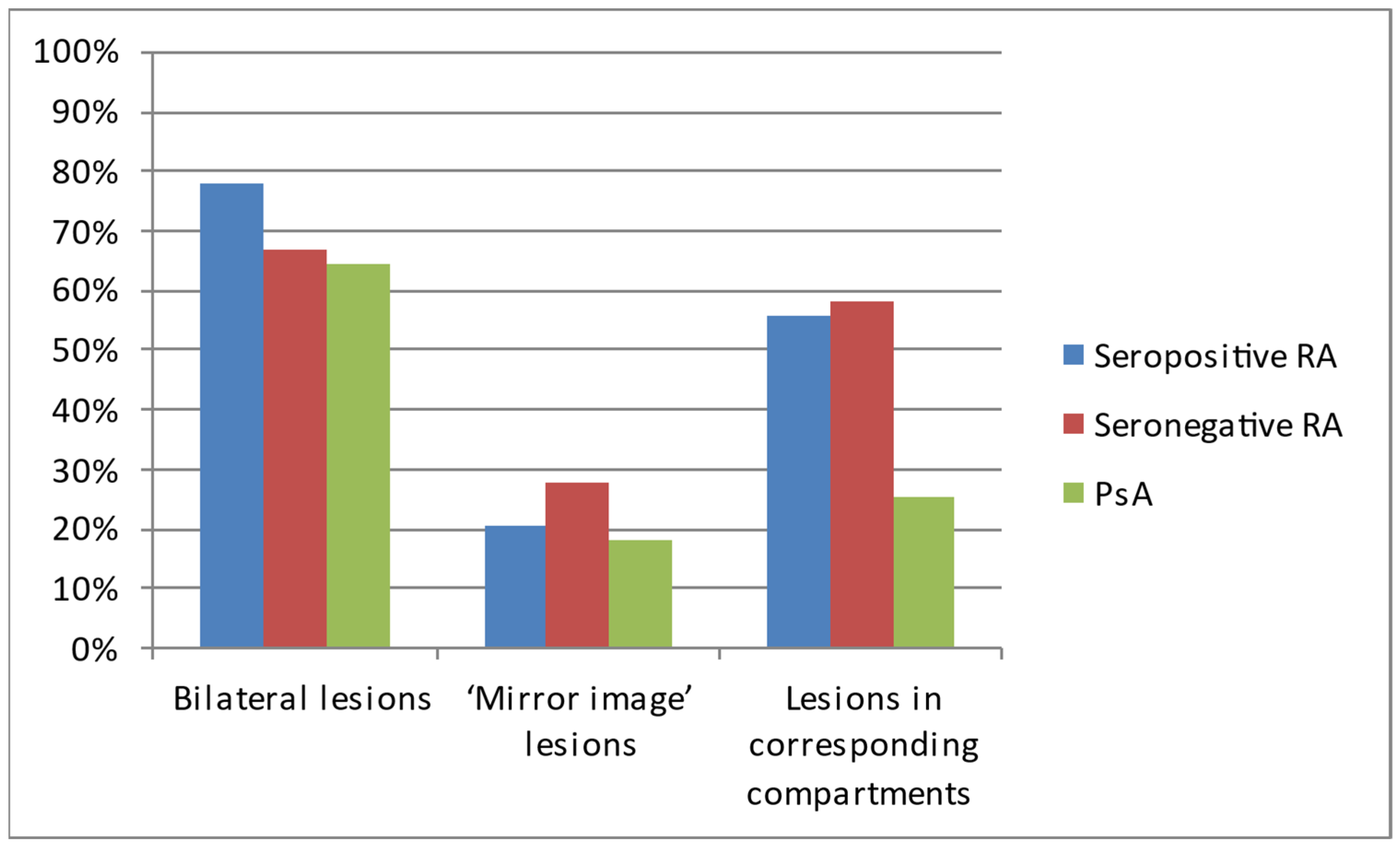
| Type of Symmetry | Seropositive RA n = 180 | Seronegative RA n = 36 | PsA n = 154 |
|---|---|---|---|
| Bilateral lesions | 140 (77.8%) | 24 (66.7%) | 99 (64.3%) |
| ‘Mirror-image’ lesions | 37 (20.6%) | 10 (27.8%) | 28 (18.2%) |
| Lesions in corresponding compartments | 100 (55.6%) | 21 (58.3%) | 39 (25.3%) |
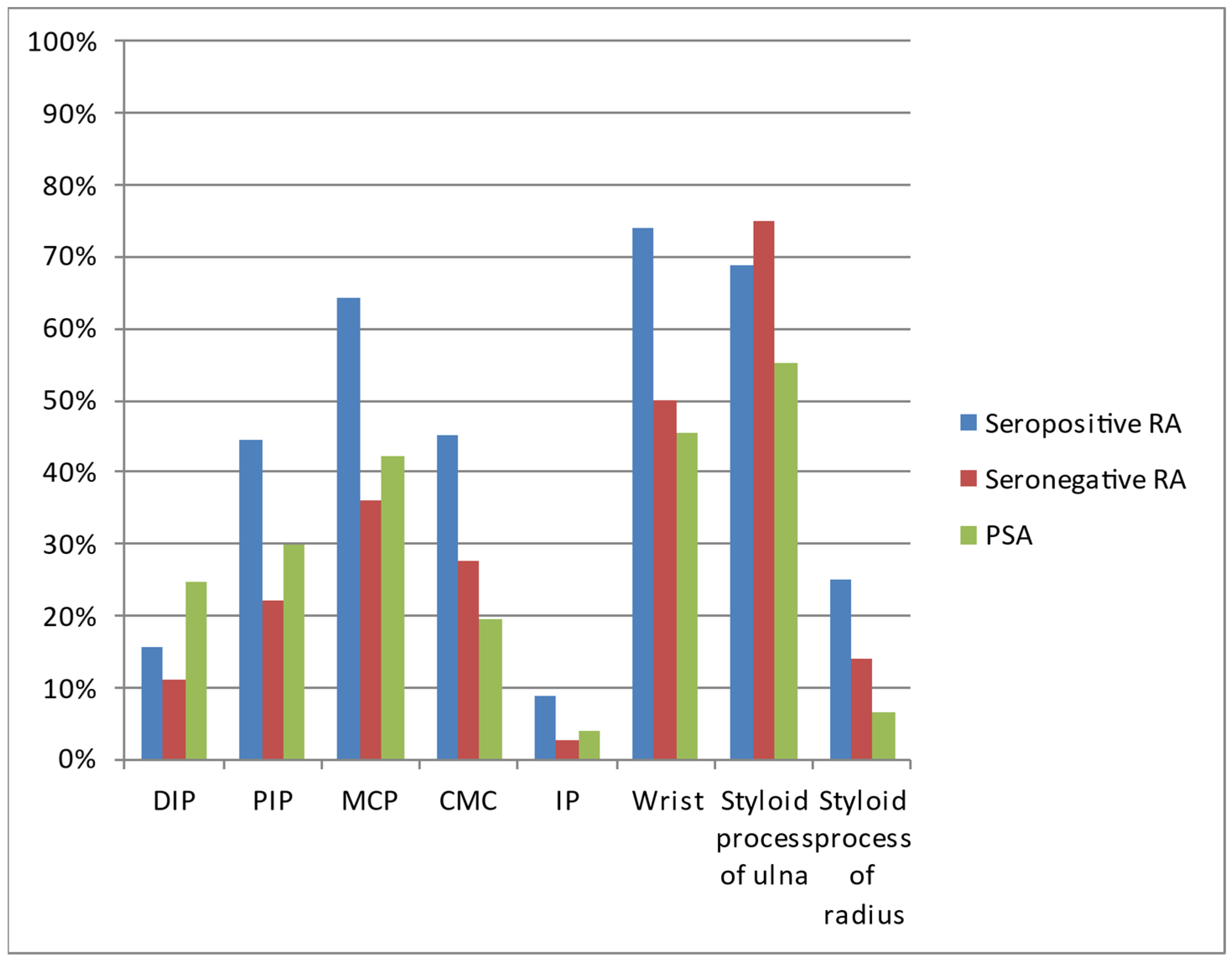
| Location | Seropositive RA n = 180 | Seronegative RA n = 36 | PSA n = 154 |
|---|---|---|---|
| Distal interphalangeal joints (DIP) | 28 (15.6%) | 4 (11.1%) | 38 (24.7%) |
| Proximal interphalangeal joints (PIP) | 80 (44.4%) | 8 (22.2%) | 46 (29.9%) |
| Metacarpophalangeal joints (MCP) | 116 (64.4%) | 13 (36.1%) | 65 (42.2%) |
| Carpometacarpal joints (CMC) | 81 (45.0%) | 10 (27.8%) | 30 (19.5%) |
| Interphalangeal joint of the thumb (IP) | 16 (8.9%) | 1 (2.8%) | 6 (3.9%) |
| Wrist | 133 (73.9%) | 18 (50.0%) | 70 (45.5%) |
| Styloid process of the ulna | 124 (68.9%) | 27 (75.0%) | 85 (55.2%) |
| Styloid process of the radius | 45 (25.0%) | 5 (13.9%) | 10 (6.5%) |
| Lesion | Seropositive RA n = 180 | Seronegative RA n = 36 | PsA n = 154 |
|---|---|---|---|
| Bone cysts | 166 (92.2%) | 36 (100.0%) | 132 (85.7%) |
| Erosions | 99 (55.0%) | 9 (25.0%) | 49 (31.8%) |
| Advanced destructive changes | 49 (27.2%) | 2 (5.6%) | 14 (9.1%) |
| Subluxations/dislocations | 64 (35.6%) | 5 (13.9%) | 13 (8.4%) |
| Proliferative bone changes | 6 (3.3%) | 0 (0.0%) | 18 (11.7%) |
| Juxta-articular osteoporosis | 112 (62.2%) | 16 (44.4%) | 54 (35.1%) |
| Joint space narrowing | 114 (63.3%) | 15 (41.7%) | 53 (34.4%) |
| Ankylosis | 25 (13.9%) | 3 (8.3%) | 8 (5.2%) |
| Contractures | 61 (33.9%) | 7 (19.4%) | 37 (24.0%) |
| Acroosteolysis | 0 (0.0%) | 0 (0.0%) | 5 (3.3%) |
| Ulnarisation | 21 (11.7%) | 2 (5.6%) | 0 (0.0%) |
| Factor | Univariate Analysis | Multivariate Analysis | ||
|---|---|---|---|---|
| OR (95% CI) | p | OR (95% CI) | p | |
| Female sex | 2.15 (1.27–3.64) | 0.005 | 1.88 (1.06–3.31) | 0.030 |
| Age (increase by 1 year) | 1.03 (1.02–1.05) | <0.001 | 1.03 (1.01–1.05) | 0.001 |
| Bilateral lesions | 1.94 (1.20–3.15) | 0.007 | 0.87 (0.48–0.57) | 0.638 |
| ‘Mirror-image’ lesions | 1.16 (0.67–2.01) | 0.585 | - | - |
| Lesions in corresponding compartments | 3.67 (2.31–5.88) | <0.001 | 3.73 (2.13–6.53) | <0.001 |
| Factor | Univariate Analysis | Multivariate Analysis | ||
|---|---|---|---|---|
| OR (95% CI) | p | OR (95% CI) | p | |
| Female sex | 2.15 (1.27–3.64) | 0.005 | 1.81 (1.00–3.28) | 0.049 |
| Age (increase by 1 year) | 1.03 (1.02–1.05) | <0.001 | 1.03 (1.01–1.05) | 0.015 |
| DIP | 0.56 (0.41–0.97) | 0.038 | 0.38 (0.20–0.74) | 0.005 |
| PIP | 1.88 (1.19–2.96) | 0.006 | 2.20 (1.27–3.81) | 0.005 |
| MCP | 2.48 (1.60–3.86) | <0.001 | 1.68 (0.98–2.89) | 0.061 |
| CMC | 3.38 (2.06–5.55) | <0.001 | 1.46 (0.80–2.66) | 0.223 |
| IP | 2.41 (0.92–6.31) | 0.074 | - | - |
| Wrist | 3.40 (2.15–5.38) | <0.001 | 1.94 (1.12–3.38) | 0.024 |
| Styloid process of the ulna | 1.80 (1.15–2.81) | 0.010 | 1.47 (0.88–2.46) | 0.141 |
| Styloid process of the radius | 4.80 (2.33–9.90) | <0.001 | 2.58 (1.13–5.89) | 0.015 |
| Factor | Univariate Analysis | Multivariate Analysis | ||
|---|---|---|---|---|
| OR (95% CI) | p | OR (95% CI) | p | |
| Female sex | 2.14 (1.33–3.45) | 0.002 | - | - |
| Age (increase by 1 year) | 1.04 (1.02–1.05) | <0.001 | 1.03 (1.01–1.05) | 0.004 |
| Bone cysts | 1.98 (0.97–4.01) | 0.059 | - | - |
| Erosions | 2.62 (1.67–4.10) | <0.001 | 1.40 (0.72–2.75) | 0.324 |
| Advanced destructive changes | 3.74 (1.97–7.09) | <0.001 | 1.03 (0.41–2.56) | 0.953 |
| Subluxations/dislocations | 5.98 (3.14–11.40) | <0.001 | 2.83 (1.29–6.21) | 0.010 |
| Proliferative bone changes | 0.26 (0.10–0.67) | 0.006 | 0.21 (0.07–0.62) | 0.005 |
| Juxta-articular osteoporosis | 3.05 (1.95–4.77) | <0.001 | 1.96 (1.13–3.40) | 0.017 |
| Joint space narrowing | 3.29 (2.10–5.16) | <0.001 | 1.91 (1.04–3.52) | 0.038 |
| Ankylosis | 2.94 (1.29–6.73) | 0.003 | 1.24 (0.43–3.61) | 0.690 |
| Contractures | 1.62 (1.00–2.62) | 0.049 | 1.23 (0.70–2.16) | 0.476 |
| Acroosteolysis | - | 0.998 | - | - |
| Ulnarisation | - | 0.997 | - | - |
| Factor | Univariate Analysis | Multivariate Analysis | ||
|---|---|---|---|---|
| OR (95% CI) | p | OR (95% CI) | p | |
| Female sex | 0.65 (0.21–1.98) | 0.449 | - | - |
| Age (increase by 1 year) | 1.00 (0.98–1.03) | 0.903 | - | - |
| Bilateral lesions | 1.75 (0.81–3.81) | 0.158 | - | - |
| ‘Mirror-image’ lesions | 0.67 (0.30–1.52) | 0.340 | - | - |
| Lesions in corresponding compartments | 0.89 (0.43–1.84) | 0.759 | - | - |
| Factor | Univariate Analysis | Multivariate Analysis | ||
|---|---|---|---|---|
| OR (95% CI) | p | OR (95% CI) | p | |
| Female sex | 0.65 (0.21–1.98) | 0.449 | - | - |
| Age (increase by 1 year) | 1.00 (0.98–1.03) | 0.903 | - | - |
| DIP | 1.47 (0.48–4.49) | 0.319 | - | - |
| PIP | 2.80 (1.21–6.48) | 0.016 | 2.29 (0.96–5.48) | 0.062 |
| MCP | 3.21 (1.52–6.76) | 0.002 | 2.01 (0.86–4.72) | 0.107 |
| CMC | 2.13 (0.97–4.67) | 0.060 | - | |
| IP | 3.42 (0.44–26.61) | 0.241 | - | - |
| Wrist | 2.83 (1.36–5.89) | 0.005 | 1.96 (0.85–4.49) | 0.113 |
| Styloid process of the ulna | 0.74 (0.33–1.67) | 0.467 | - | - |
| Styloid process of the radius | 2.07 (0.76–5.64) | 0.156 | - | - |
| Factor | Univariate Analysis | Multivariate Analysis | ||
|---|---|---|---|---|
| OR (95% CI) | p | OR (95% CI) | p | |
| Female sex | 0.65 (0.21–1.98) | 0.449 | - | - |
| Age (increase by 1 year) | 1.00 (0.98–1.03) | 0.903 | - | - |
| Bone cysts | - | - | - | - |
| Erosions | 3.67 (1.63–8.24) | 0.002 | 2.01 (0.76–5.33) | 0.160 |
| Advanced destructive changes | 6.34 (1.47–27.47) | 0.013 | 2.76 (0.52–14.71) | 0.236 |
| Subluxations/dislocations | 3.42 (1.27–9.23) | 0.015 | 1.64 (0.53–5.12) | 0.391 |
| Proliferative bone changes | - | - | - | - |
| Juxta-articular osteoporosis | 2.06 (1.00–4.24) | 0.050 | - | |
| Joint space narrowing | 2.42 (1.17–5.01) | 0.018 | 1.17 (0.50–2.73) | 0.722 |
| Ankylosis | 1.77 (0.51–6.22) | 0.371 | - | - |
| Contractures | 2.12 (0.88–5.13) | 0.094 | - | - |
| Acroosteolysis | - | - | - | - |
| Ulnarisation | 2.25 (0.50–10.03) | 0.290 | - | - |
| Factor | Univariate Analysis | Multivariate Analysis | ||
|---|---|---|---|---|
| OR (95% CI) | p | OR (95% CI) | p | |
| Female sex | 3.30 (1.10–9.88) | 0.033 | 2.55 (0.83–7.85) | 0.104 |
| Age (increase by 1 year) | 1.03 (1.00–1.06) | 0.050 | - | - |
| Bilateral lesions | 1.11 (0.52–2.39) | 0.788 | - | - |
| ‘Mirror-image’ lesions | 1.73 (0.75–3.99) | 0.199 | - | - |
| Lesions in corresponding compartments | 4.13 (1.94–8.79) | <0.001 | 3.50 (1.62–7.57) | 0.001 |
| Factor | Univariate Analysis | Multivariate Analysis | ||
|---|---|---|---|---|
| OR (95% CI) | p | OR (95% CI) | p | |
| Female sex | 3.30 (1.10–9.88) | 0.033 | 3.32 (1.10–10.00) | 0.033 |
| Age (increase by 1 year) | 1.03 (1.00–1.06) | 0.050 | - | - |
| DIP | 0.38 (0.13–1.15) | 0.087 | - | - |
| PIP | 0.67 (0.28–1.58) | 0.362 | - | |
| MCP | 0.77 (0.37–1.64) | 0.504 | - | - |
| CMC | 1.59 (0.69–3.65) | 0.274 | - | |
| IP | 0.71 (0.08–6.04) | 0.750 | - | - |
| Wrist | 1.20 (0.58–2.48) | 0.623 | - | - |
| Styloid process of the ulna | 2.44 (1.07–5.52) | 0.033 | 2.44 (1.07–5.59) | 0.034 |
| Styloid process of the radius | 2.32 (0.74–7.27) | 0.148 | - | - |
| Factor | Univariate Analysis | Multivariate Analysis | ||
|---|---|---|---|---|
| OR (95% CI) | p | OR (95% CI) | p | |
| Female sex | 3.30 (1.10–9.88) | 0.033 | - | - |
| Age (increase by 1 year) | 1.03 (1.00–1.06) | 0.050 | - | - |
| Bone cysts | - | - | - | - |
| Erosions | 0.71 (0.31–1.63) | 0.425 | - | |
| Advanced destructive changes | 0.59 (0.13–2.71) | 0.496 | - | - |
| Subluxations/dislocations | 1.75 (0.58–5.27) | 0.320 | - | - |
| Proliferative bone changes | - | - | - | - |
| Juxta-articular osteoporosis | 1.48 (0.71–3.09) | 0.295 | - | |
| Joint space narrowing | 1.36 (0.65–2.86) | 0.455 | - | - |
| Ankylosis | 1.66 (0.42–6.59) | 0.472 | - | - |
| Contractures | 0.76 (0.31–1.89) | 0.558 | - | - |
| Acroosteolysis | - | - | - | - |
| Ulnarisation | - | - | - | - |
Disclaimer/Publisher’s Note: The statements, opinions and data contained in all publications are solely those of the individual author(s) and contributor(s) and not of MDPI and/or the editor(s). MDPI and/or the editor(s) disclaim responsibility for any injury to people or property resulting from any ideas, methods, instructions or products referred to in the content. |
© 2023 by the authors. Licensee MDPI, Basel, Switzerland. This article is an open access article distributed under the terms and conditions of the Creative Commons Attribution (CC BY) license (https://creativecommons.org/licenses/by/4.0/).
Share and Cite
Żelnio, E.; Taljanovic, M.; Mańczak, M.; Sudoł-Szopińska, I. Hand and Wrist Involvement in Seropositive Rheumatoid Arthritis, Seronegative Rheumatoid Arthritis, and Psoriatic Arthritis—The Value of Classic Radiography. J. Clin. Med. 2023, 12, 2622. https://doi.org/10.3390/jcm12072622
Żelnio E, Taljanovic M, Mańczak M, Sudoł-Szopińska I. Hand and Wrist Involvement in Seropositive Rheumatoid Arthritis, Seronegative Rheumatoid Arthritis, and Psoriatic Arthritis—The Value of Classic Radiography. Journal of Clinical Medicine. 2023; 12(7):2622. https://doi.org/10.3390/jcm12072622
Chicago/Turabian StyleŻelnio, Ewa, Mihra Taljanovic, Małgorzata Mańczak, and Iwona Sudoł-Szopińska. 2023. "Hand and Wrist Involvement in Seropositive Rheumatoid Arthritis, Seronegative Rheumatoid Arthritis, and Psoriatic Arthritis—The Value of Classic Radiography" Journal of Clinical Medicine 12, no. 7: 2622. https://doi.org/10.3390/jcm12072622
APA StyleŻelnio, E., Taljanovic, M., Mańczak, M., & Sudoł-Szopińska, I. (2023). Hand and Wrist Involvement in Seropositive Rheumatoid Arthritis, Seronegative Rheumatoid Arthritis, and Psoriatic Arthritis—The Value of Classic Radiography. Journal of Clinical Medicine, 12(7), 2622. https://doi.org/10.3390/jcm12072622