Article

Surrogates of Muscle Mass on Cardiac MRI Correlate with Exercise Capacity in Patients with Fontan Circulation

1
Department of Pediatrics, UPMC Children’s Hospital of Pittsburgh, University of Pittsburgh School of Medicine, Pittsburgh, PA 15224, USA
2
Department of Radiology, UPMC Children’s Hospital of Pittsburgh, University of Pittsburgh School of Medicine, Pittsburgh, PA 15224, USA
3
Department of Pediatric Cardiology, UPMC Children’s Hospital of Pittsburgh, University of Pittsburgh School of Medicine, Pittsburgh, PA 15224, USA
*
Author to whom correspondence should be addressed.
J. Clin. Med. 2023, 12(7), 2689; https://doi.org/10.3390/jcm12072689
Submission received: 3 March 2023 / Revised: 29 March 2023 / Accepted: 31 March 2023 / Published: 4 April 2023
(This article belongs to the Section Cardiology)

Abstract

:
Background: Sarcopenia is an increasingly recognized marker of frailty in cardiac patients. Patients with a history of congenital heart disease and Fontan procedure have a higher risk of developing progressive muscle wasting. Our objective was to determine if we could use routine cardiac MRI (CMR) for the surveillance of muscle wasting. Methods: A retrospective study of all Fontan patients (n = 75) was conducted at our institution, with CMR performed from 2010 to 2022 and exercise stress testing performed within 12 months (4.3 ± 4.2 months). The skeletal muscle area (SMA) for the posterior paraspinal and anterior thoracic muscles were traced and indexed for body surface area (BSA). Patients were stratified by percentile into the upper and lower quartiles, and the two groups were compared. Multivariable regression was performed to control for sex and age. Results: There was a significant positive association of both anterior (r = 0.34, p = 0.039) and paraspinal (r = 0.43, p = 0.007) SMA to peak VO2. Similarly, paraspinal but not anterior SMA was negatively associated with the VE/VCO2 (r = –0.45, p = 0.006). The upper quartile group had significantly more males (18/19 vs. 8/20; p = 0.0003) and demonstrated a significantly higher peak VO2 (32.2 ± 8.5 vs. 23.8 ± 4.7, p = 0.009), a higher peak RER (1.2 ± 0.1 vs. 1.1 ± 0.04, p = 0.007), and a significantly lower VE/VCO2 (32.9 ± 3.6 vs. 40.2 ± 6.2, p = 0.006) compared to the lowest quartile. The association of SMA to VO2 peak and VE/VCO2 was redemonstrated after controlling for sex and age. Conclusion: Thoracic skeletal muscle area may be an effective surrogate of muscle mass and is correlated to several measures of cardiorespiratory fitness post-Fontan. CMR would be an effective tool for the surveillance of sarcopenia in post-Fontan patients given its accessibility and routine use in these patients.

1. Introduction

The Fontan operation is the final palliative procedure for single-ventricle hearts, and the procedure redirects systemic venous return to pulmonary arteries without a sub-pulmonary pumping chamber [1]. Despite the improvement in outcomes, patients may still face numerous complications such as heart failure stemming from systolic or diastolic ventricular dysfunction [2,3], protein-losing enteropathy [4,5], arrhythmias [2], muscle wasting [3,6], and impaired exercise capacity [7,8]. Fontan patients are less physically active compared to their healthy counterparts because of various cardiopulmonary factors [9]. Physiological changes, including but not limited to increased dead space ventilation, reduced vital capacity, high pulmonary arterial wedge pressure, and an inability to maintain stroke volume during exercise, contribute to hypercapnia and subsequent inefficient cardiorespiratory function during exercise [10,11]. Exercise intolerance serves as a prognostic marker in congenital heart disease patients; thus, a better understanding of exercise stress testing and its relationship to disease progression can be a valuable tool in disease surveillance [12,13,14,15].
In adult patients with Fontan circulation, muscle wasting is a common complication [16,17] that is associated with adverse events in other chronic medical conditions such as cancer [18,19] and heart failure [20,21]. In children, the Fontan operation has been associated with abnormalities in body composition, bone strength, and growth [6,22,23]. Assessment of the abdominal skeletal muscle at the third lumbar vertebra (L3) has been common practice to define sarcopenia in non-Fontan patients [24,25]. Sarcopenia has also been assessed by measuring abdominal muscle mass in Fontan patients on abdominal magnetic resonance imaging (MRI) performed initially to evaluate for liver disease [26]. Lower skeletal muscle mass seen on abdominal MRI has been associated with decreased cardiorespiratory fitness; however, abdominal MRI is not routinely performed in Fontan patients [26]. Cardiac MRIs (CMR), on the other hand, are routinely used post-Fontan and provide important diagnostic information. The objective of this study is to assess the feasibility and reproducibility of determining skeletal muscle mass via thoracic muscle groups seen on routine CMR and assess the correlation of muscle mass with exercise capacity, specifically peak VO2, VE/VCO2, and respiratory exchange ratio (RER).

2. Materials and Methods

2.1. Study Population

This was a retrospective study conducted at the University of Pittsburgh Medical Center, Children’s Hospital of Pittsburgh. All patients with a history of Fontan surgery who had a CMR were included in this study. Patients who did not have adequate CMR images to measure the thoracic muscle mass or sufficient exercise stress testing data were excluded. A total of 10 patients was excluded based on this criterion, making the total study population 75 patients.

2.2. Data Collection

Data for baseline and demographic characteristics, imaging, and cardiopulmonary exercise testing were collected from patient charts. The data dictionary and definitions from the FORCE registry (Fontan Outcome Registry using CMR Examinations) were used [27]. Research Electronic Data Capture (REDCap) was used to store the data for this study. This study was approved by the University of Pittsburgh Institutional Review Board and was conducted in compliance with the Health Insurance Portability and Accountability Act. The requirement for informed consent was waived due to the retrospective nature of the study.

2.3. CMR Data Analysis

CMR studies were performed by using 1.5 Tesla scanners (GE Medical Systems, Milwaukee, WI, USA or Siemens AG, Munich, Germany). Briefly, ventricular assessment was performed via an electrocardiographically gated, balanced steady-state free precession (bSSFP) cine CMR in vertical and horizontal ventricular long-axis planes and a stack of slices in a ventricular short-axis plane encompassing the atrioventricular junction through the cardiac apex. Retrospectively cardiac-gated, through-plane phase-contrast flow measurements were obtained in the branch pulmonary arteries and venae cavae. If a patient had multiple CMR studies, the most recent study was used for analysis. The mean time period from Fontan operation and CMR was 14.1 ± 7.7 years. Ventricular volumes and function were measured by manually tracing the endocardial and epicardial borders on each short-axis bSSFP cine slice at end-diastole (maximal volume) and end-systole (minimal volume). Analysis was performed by using commercially available software (Cvi-42, Circle Cardiovascular Imaging Inc., Calgary, AB, Canada) and IBM SSPS Statistics (Version 27, Armonk, NY, USA: IBM Corp). All CMR exams were reanalyzed by a single pediatric cardiologist with clinical experience interpreting CMR studies in the single ventricle population. Ventricular contours were redrawn [28].

2.4. Muscle Measurements

Muscle measurements were obtained by analyzing each patient’s most recent CMR. The skeletal muscle area (SMA) of the patients was assessed in the axial view on bright blood bSSFP static images at the level of Carina with offline analysis software (Cvi-42, Circle Cardiovascular Imaging Inc., Calgary, AB, Canada). Measurements of muscle area were conducted in images in static SSFP axial stack. The pectoralis major and minor muscles were traced for the assessment of anterior muscle area, while the paraspinal muscles were traced for posterior muscle area at the level of T4 (Figure 1) [29]. Due to the large variation in height and body habitus of our cohort, SMA, which is reported in cm2, was indexed by body surface area (BSA) by using the Mosteller calculation [30]. BSA-indexed SMA was reported as cm2/m2. Due to a lack of standardized values that matched our baseline characteristics in a non-Fontan population, we divided patients into high- and low-muscle groups, with high being the upper 25th percentile and low being the lower 25th percentile for BSA-indexed SMA.

2.5. Cardiopulmonary Exercise Testing

Patients underwent CPET by using a treadmill, as per the Bruce protocol. Gas exchange was analyzed at rest, during exercise, and during recovery to determine measures of oxygen uptake (VO2). Since peak VO2 is influenced by age, sex, and body weight, the percent of predicted peak VO2 value (% predicted VO2) was used due to the wide age range in this study [31]. Cardiopulmonary exercise stress testing data were collected within 12 months (4.3 ± 4.2 months) of the most recent CMR.

2.6. Statistical Analyses

Categorical variables were reported as counts and percentages, while continuous variables were expressed as mean ± standard deviation. Comparison between two continuous variables was performed by using t-testing or Mann–Whitney U for group sizes less than 30 subjects, while Chi-square tests were used for comparing categorical variables. Comparison between multiple continuous variables was performed by using Kruskal–Wallis testing, accounting for uneven distribution. Correlational testing was performed by using the Pearson correlation test. Multivariate analysis to adjust for sex and age was expressed as a parameter estimate ± standard deviation. Due to the sample size, muscle mass measurements were adjusted for one covariate at a time, and the results were reported. Cohen’s d and Hedges’ g tests were used to calculate effect sizes. Cohen’s d test was used for groups with sample sizes > 20 or groups with similar sample sizes. Hedges’ g test was used for sample sizes < 20 or groups that differed greatly in size. Cohen’s d test is reported as d, and Hedges’ g test is reported as g. To account for observer variability in muscle area tracing, there was retracing of ten random scans by another observer, and a comparison between SMA measurements of anterior and posterior muscle area was performed. A p-value of <0.05 was considered statistically significant. Interclass correlation was reported. Statistical analysis was conducted by using JMP Pro, Version 16.2.0.

3. Results

Our cohort consisted of seventy-five patients with Fontan circulation. The mean age at CMR was 19.1 ± 8.6 years and 49 (65%) males. In total, 1 patient had an atriopulmonary Fontan (1.3%), 49 patients (65%) had an extracardiac conduit Fontan, 23 (31%) had a lateral tunnel Fontan, and 2 (2.6%) were unknown. No reported deaths or transplant. Additional baseline characteristics are listed in Table 1 and Table 2.

3.1. Anterior and Paraspinal Muscle Index to BSA in the Fontan Population

The mean BSA-indexed anterior muscle and paraspinal muscle area was 20.8 ± 5.7 cm2/m2 and 6.5 ± 1.7 cm2/m2, respectively. Following exclusion of ineligible patients, our study cohort consisted of 75 patients with CMR (Table 1). In the rest of the patients, there was artifact at the level of the carina precluding accurate measurements. Combined BSA-indexed muscle area was 27.3 ± 6.6 cm2/m2. BSA-indexed anterior muscle, paraspinal muscle, and combined muscle areas were all significantly larger in male patients than female patients (BSA-indexed anterior muscle area: 22.4 ± 6.0 cm2/m2 vs. 17.8 ± 3.3 cm2/m2, p = 0.0004; BSA-indexed paraspinal muscle area: 6.9 ± 1.8 cm2/m2 vs. 5.9 ± 1.3 cm2/m2, p = 0.0105; BSA-indexed combined muscle area: 29.3 ± 7.0 cm2/m2 vs. 23.6 ± 3.8 cm2/m2, p = 0.0002). The differences between males and females are summarized in Table 3.

3.2. Intra-Observer and Inter-Observer Reliability:

There was good intra-observer reliability for measurements of anterior and paraspinal SMA on CMR of 0.97 (95% CI, 0.89–0.99) and 0.93 (95% CI, 0.79–0.98). In Inter-observer reliability, there was good intra-class correlation for repeated measurements of anterior and paraspinal SMA on CMR of 0.98 (95% CI, 0.94–0.99) and 0.96 (95% CI, 0.90–0.99), respectively. The Bland Altman plot for measurements can be seen in Figure 2.

3.3. Comparison between High- and Low-Muscle Groups

Between low- and high-muscle groups, both cohorts were similar in age (17.2 ± 7.0 vs. 17.8 ± 7.1, p = 0.499, d = 0.10) with a significant difference in BMI (low: 25.1 ± 5.3 vs. high: 21.3 ± 4.9, p = 0.027, d = −0.76) and sex distribution (Low-muscle group: Males 40%, Females 60% vs. High-muscle group: Males 95%, Females 5%, p = 0.0003). Further characteristics are shown in Table 4.

3.4. Correlation of Muscle Mass with Exercise Capacity and Other Clinical Parameters

BSA-indexed paraspinal and anterior muscle have a significant positive correlation with peak VO2 (r = 0.43, p = 0.0070 and r = 0.34, p = 0.0389). BSA-indexed paraspinal muscle area is negatively associated with VE/VCO2 (r = −0.45, p = 0.0060). Further characteristics are summarized on Figure 3 and Table 4.
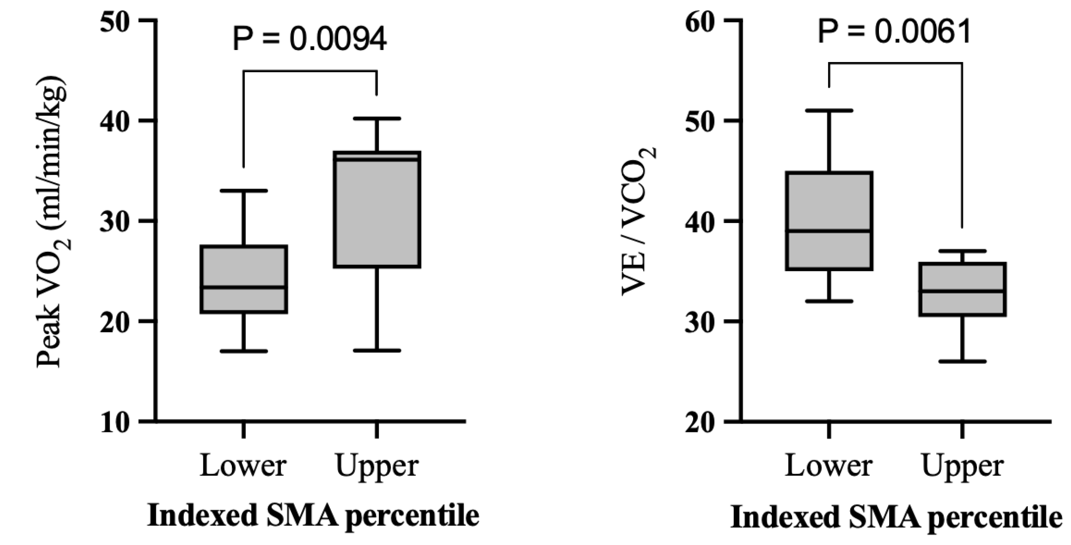
When conducting analysis based on high- and low-muscle groups, the high-muscle group had significantly higher peak VO2 (32.2 ± 8.5 vs. 23.8 ± 4.7, p = 0.009, g = 1.22), a higher peak RER (1.2 ± 0.1 vs. 1.1 ± 0.04, p = 0.007, g = 1.47) and lower VE/VCO2 (32.9 ± 3.6 vs. 40.2 ± 6.2, p = 0.006, g = −1.34) (Table 5 and Figure 4). When ventricular morphology was analyzed, the systemic right, left, or mixed ventricle did not significantly differ in any exercise parameters including peak VO2, RER, and VE/VCO2 (Supplemental Table S1).
Sex-specific analysis showed males had significantly higher % predicted peak VO2 (74.9 ± 9.9 vs. 60.3 ± 14.1, p = 0.002)
In multivariate analysis, BSA-indexed paraspinal muscle area was significantly associated with peak VO2 when adjusted for age (parameter estimate ± standard error, 1.7 ± 0.6 per 1cm2/m2, p = 0.008) and sex (1.5 ± 0.6 per 1cm2/m2, p = 0.019). Similarly, BSA-indexed anterior muscle area remained significant for peak VO2 when adjusted for age (0.36 ± 0.2 per 1cm2/m2, p = 0.043) and sex (0.4 ± 0.2 per 1cm2/m2, p = 0.013). Paraspinal muscle area was also significantly associated with VE/VCO2 when adjusted for sex (−1.4 ± 0.6, p = 0.0191) and age (−1.6 ± 0.5, p = 0.004) (Table 6).

4. Discussion

In this retrospective study, we analyzed anterior and posterior muscle areas via MRI-based cross-sectional imaging in post-Fontan patients. It was feasible to perform the measurements with high reproducibility via CMR. Muscle mass measurements via CMR correlated well with peak VO2 and with VE/VCO2 via CPET. Larger indexed anterior and posterior SMA were associated with higher peak VO2, while larger paraspinal muscle area alone was associated with lower VE/VCO2.
In comparison with previous studies that analyzed indexed SMA in a healthy cohort, our population had similar results, with males having significantly higher SMA compared to their female counterparts [32]. These findings were also seen in studies that established an association between larger pectoralis muscle area with younger age and male sex in patients with acute and chronic illnesses. Refs [33,34] Posterior muscle areas at the thoracic level have not been frequently studied; therefore, it was difficult to compare our cohort to a population of healthy patients. With that said, studies that assessed posterior muscle areas at L3 did show larger SMA in males compared to females, which matched our results [32]. In this study, we established a significant relationship with increased thoracic muscle area with exercise capacity.

4.1. Indexed Skeletal Muscle Area and Cardiorespiratory Function

Better exercise capacity is linked to improved survival in Fontan patients [35]. In a previous study analyzing Fontan patients, peak VO2 was negatively associated with age, and some of the cardiorespiratory decline was attributed to natural degradation over time in Fontan physiology [36]. As expected, this trend is also seen in normal heart patients as well. In our population, age was not significantly different between high- and low-muscle groups, and the significant association of SMA to the cardiorespiratory function remained after adjusting for age. BMI was significantly lower in our high-muscle group, while the opposite was seen in our low-muscle group. This trend was likely due to a higher activity level in patients with higher muscle mass and, consequently, lower BMI. Our study supports the notion that skeletal muscle mass significantly affects exercise capacity in Fontan patients. Exercise training may increase muscle mass and improve exercise capacity [37,38,39].
We also found a significant negative association between increased posterior SMA and decreased VE/VCO2. Higher VE/VCO2 reflects ventilatory inefficiency and has been associated with worse prognosis in heart failure patients [40,41,42]. Similarly, Fernandes et al. showed an association with worse risk of mortality in Fontan patients [43]. Lower muscle mass may be synonymous with poor conditioning, resulting in a higher VE/VCO2, as seen in our study.

4.2. Limitations

There are several limitations to our study. Due to the lack of standardized values for the indexed SMA of anterior and posterior muscles assessed at the level of T4, we were unable to define which patients in our cohort met the clinical definition of sarcopenia. A larger cohort study with matched subjects could help address this issue and provide insight into how we can use these measurements from CMR to monitor the health of Fontan patients. In addition, when splitting our population into high- and low-muscle groups, the high-muscle group category heavily comprised males, and the low-muscle group largely comprised females. To mitigate this limitation, we analyzed SMA as a continuous variable, and the associations remained significant after adjusting for sex and age. In addition, questions concerning lifestyle habits such as daily exercise, years of exercise history, diet, and other lifestyle factors were not available in this retrospective analysis; therefore, we are unable to address other key factors contributing to body composition in this study.

5. Conclusions

Our study found a significant association between increased thoracic skeletal muscle mass and markers of cardiorespiratory fitness in Fontan circulation patients. This study found that skeletal muscle area, a reproducible and easily obtained measure via CMR, is positively associated with higher peak VO2 and negatively associated with VE/VCO2, supporting previous findings that muscle development is an important factor for exercise capacity in this population. These findings support the idea that supervised exercise activity or physical therapy could be beneficial to improving fitness in Fontan patients. In addition, with CMR muscle mass correlating well with markers of cardiorespiratory fitness, it is possible that some patients may be able to skip CPET and alternate surveillance with imaging. Moreover, imaging could potentially be used to assess response to exercise programs. More research will be needed to support this idea.

Supplementary Materials

The following supporting information can be downloaded at: https://www.mdpi.com/article/10.3390/jcm12072689/s1, Table S1: Ventricular Morphology Exercise Parameters.

Author Contributions

Conceptualization, K.L.S. and T.A.; methodology, K.L.S., E.B.G. and T.A.; formal analysis, K.L.S., E.B.G. and T.A.; investigation, K.L.S., E.B.G. and T.A.; resources, M.E.G. and T.A.; data curation, K.L.S., E.B.G., M.E.G., A.C., L.J.O., T.H., A.P.S., J.K., B.F. and T.A.; writing—original draft, K.L.S., E.B.G. and T.A.; writing—review and editing, K.L.S., E.B.G., M.E.G., A.C., L.J.O., S.S.T., T.H., A.P.S., J.K., B.F. and T.A.; visualization, K.L.S. and E.B.G.; supervision, T.A.; funding acquisition, T.A. All authors have read and agreed to the published version of the manuscript.

Funding

There are no funding sources to declare.

Institutional Review Board Statement

The study was conducted in accordance with the Declaration of Helsinki, and approved by the Institutional Review Board of University of Pittsburgh IRB STUDY21070169 on 24 August 2022.

Informed Consent Statement

Patient consent was waived due to retrospective nature of the study as approved by IRBS.

Data Availability Statement

The data that support the findings of this study are available from the corresponding author upon reasonable request.

Conflicts of Interest

The authors have no conflicts of interest to disclose.

References

  1. Fontan, F.; Baudet, E. Surgical repair of tricuspid atresia. Thorax 1971, 26, 240–248. [Google Scholar] [CrossRef] [Green Version]
  2. Budts, W.; Ravekes, W.J.; Danford, D.A.; Kutty, S. Diastolic Heart Failure in Patients With the Fontan Circulation: A Review. JAMA Cardiol. 2020, 5, 590. [Google Scholar] [CrossRef]
  3. Stout, K.K.; Broberg, C.S.; Book, W.M.; Cecchin, F.; Chen, J.M.; Dimopoulos, K.; Everitt, M.D.; Gatzoulis, M.; Harris, L.; Hsu, D.T.; et al. Chronic Heart Failure in Congenital Heart Disease: A Scientific Statement From the American Heart Association. Circulation 2016, 133, 770–801. [Google Scholar] [CrossRef] [Green Version]
  4. Mertens, L.; Hagler, D.J.; Sauer, U.; Somerville, J.; Gewillig, M. Protein-losing enteropathy after the Fontan operation: An international multicenter study. J. Thorac. Cardiovasc. Surg. 1998, 115, 1063–1073. [Google Scholar] [CrossRef] [Green Version]
  5. Ostrow, A.M.; Freeze, H.; Rychik, J. Protein-Losing Enteropathy After Fontan Operation: Investigations Into Possible Pathophysiologic Mechanisms. Ann. Thorac. Surg. 2006, 82, 695–700. [Google Scholar] [CrossRef]
  6. Cordina, R.; O’Meagher, S.; Gould, H.; Rae, C.; Kemp, G.; Pasco, J.A.; Celermajer, D.S. Skeletal muscle abnormalities and exercise capacity in adults with a Fontan circulation. Heart 2013, 99, 1530–1534. [Google Scholar] [CrossRef]
  7. Ohuchi, H.; Negishi, J.; Miike, H.; Toyoshima, Y.; Morimoto, H.; Fukuyama, M.; Iwasa, T.; Sakaguchi, H.; Miyazaki, A.; Shiraishi, I.; et al. Positive pediatric exercise capacity trajectory predicts better adult Fontan physiology rationale for early establishment of exercise habits. Int. J. Cardiol. 2019, 274, 80–87. [Google Scholar] [CrossRef]
  8. Dulfer, K.; Bossers, S.S.M.; Utens, E.M.W.J.; Duppen, N.; Kuipers, I.M.; Kapusta, L.; van Iperen, G.; Schokking, M.; ten Harkel, A.D.J.; Takken, T.; et al. Does functional health status predict health-related quality of life in children after Fontan operation? Cardiol. Young 2016, 26, 459–468. [Google Scholar] [CrossRef]
  9. McCrindle, B.W.; Williams, R.V.; Mital, S.; Clark, B.J.; Russell, J.L.; Klein, G.; Eisenmann, J.C. Physical activity levels in children and adolescents are reduced after the Fontan procedure, independent of exercise capacity, and are associated with lower perceived general health. Arch. Dis. Child. 2007, 92, 509–514. [Google Scholar] [CrossRef] [Green Version]
  10. Ohuchi, H.; Ohashi, H.; Takasugi, H.; Yamada, O.; Yagihara, T.; Echigo, S. Restrictive ventilatory impairment and arterial oxygenation characterize rest and exercise ventilation in patients after fontan operation. Pediatr. Cardiol. 2004, 25, 513–521. [Google Scholar] [CrossRef]
  11. Zajac, A.; Tomkiewicz, L.; Podolec, P.; Tracz, W.; Malec, E. Cardiorespiratory response to exercise in children after modified fontan operation. Scand. Cardiovasc. J. SCJ 2002, 36, 80–85. [Google Scholar] [CrossRef]
  12. Diller, G.-P.; Dimopoulos, K.; Okonko, D.; Li, W.; Babu-Narayan, S.V.; Broberg, C.S.; Johansson, B.; Bouzas, B.; Mullen, M.J.; Poole-Wilson, P.A.; et al. Exercise intolerance in adult congenital heart disease: Comparative severity, correlates, and prognostic implication. Circulation 2005, 112, 828–835. [Google Scholar] [CrossRef] [PubMed] [Green Version]
  13. Diller, G.-P.; Dimopoulos, K.; Okonko, D.; Uebing, A.; Broberg, C.S.; Babu-Narayan, S.; Bayne, S.; Poole-Wilson, P.A.; Sutton, R.; Francis, D.P.; et al. Heart rate response during exercise predicts survival in adults with congenital heart disease. J. Am. Coll. Cardiol. 2006, 48, 1250–1256. [Google Scholar] [CrossRef] [Green Version]
  14. Dimopoulos, K.; Okonko, D.O.; Diller, G.-P.; Broberg, C.S.; Salukhe, T.V.; Babu-Narayan, S.V.; Li, W.; Uebing, A.; Bayne, S.; Wensel, R.; et al. Abnormal ventilatory response to exercise in adults with congenital heart disease relates to cyanosis and predicts survival. Circulation 2006, 113, 2796–2802. [Google Scholar] [CrossRef] [Green Version]
  15. Albouaini, K.; Egred, M.; Alahmar, A.; Wright, D.J. Cardiopulmonary exercise testing and its application. Postgrad. Med. J. 2007, 83, 675–682. [Google Scholar] [CrossRef] [Green Version]
  16. Tran, D.; D’Ambrosio, P.; Verrall, C.E.; Attard, C.; Briody, J.; D’Souza, M.; Fiatarone Singh, M.; Ayer, J.; d’Udekem, Y.; Twigg, S.; et al. Body Composition in Young Adults Living With a Fontan Circulation: The Myopenic Profile. J. Am. Heart Assoc. 2020, 9, e015639. [Google Scholar] [CrossRef] [PubMed]
  17. Rychik, J.; Atz, A.M.; Celermajer, D.S.; Deal, B.J.; Gatzoulis, M.A.; Gewillig, M.H.; Hsia, T.-Y.; Hsu, D.T.; Kovacs, A.H.; McCrindle, B.W.; et al. Evaluation and Management of the Child and Adult With Fontan Circulation: A Scientific Statement From the American Heart Association. Circulation 2019, 140, e234–e284. [Google Scholar] [CrossRef]
  18. Deng, H.-Y.; Hou, L.; Zha, P.; Huang, K.-L.; Peng, L. Sarcopenia is an independent unfavorable prognostic factor of non-small cell lung cancer after surgical resection: A comprehensive systematic review and meta-analysis. Eur. J. Surg. Oncol. 2019, 45, 728–735. [Google Scholar] [CrossRef] [PubMed]
  19. Ligibel, J.A.; Schmitz, K.H.; Berger, N.A. Sarcopenia in aging, obesity, and cancer. Transl. Cancer Res. 2020, 9, 5760–5771. [Google Scholar] [CrossRef] [PubMed]
  20. Sze, S.; Pellicori, P.; Zhang, J.; Weston, J.; Clark, A.L. Identification of Frailty in Chronic Heart Failure. JACC Heart Fail. 2019, 7, 291–302. [Google Scholar] [CrossRef]
  21. von Haehling, S.; Ebner, N.; dos Santos, M.R.; Springer, J.; Anker, S.D. Muscle wasting and cachexia in heart failure: Mechanisms and therapies. Nat. Rev. Cardiol. 2017, 14, 323–341. [Google Scholar] [CrossRef] [PubMed]
  22. Avitabile, C.M.; Goldberg, D.J.; Zemel, B.S.; Brodsky, J.L.; Dodds, K.; Hayden-Rush, C.; Whitehead, K.K.; Goldmuntz, E.; Rychik, J.; Leonard, M.B. Deficits in bone density and structure in children and young adults following Fontan palliation. Bone 2015, 77, 12–16. [Google Scholar] [CrossRef] [PubMed] [Green Version]
  23. Avitabile, C.M.; Leonard, M.B.; Zemel, B.S.; Brodsky, J.L.; Lee, D.; Dodds, K.; Hayden-Rush, C.; Whitehead, K.K.; Goldmuntz, E.; Paridon, S.M.; et al. Lean mass deficits, vitamin D status and exercise capacity in children and young adults after Fontan palliation. Heart 2014, 100, 1702–1707. [Google Scholar] [CrossRef] [Green Version]
  24. Prado, C.M.; Lieffers, J.R.; McCargar, L.J.; Reiman, T.; Sawyer, M.B.; Martin, L.; Baracos, V.E. Prevalence and clinical implications of sarcopenic obesity in patients with solid tumours of the respiratory and gastrointestinal tracts: A population-based study. Lancet Oncol. 2008, 9, 629–635. [Google Scholar] [CrossRef] [PubMed]
  25. Reisinger, K.W.; Bosmans, J.W.A.M.; Uittenbogaart, M.; Alsoumali, A.; Poeze, M.; Sosef, M.N.; Derikx, J.P.M. Loss of Skeletal Muscle Mass During Neoadjuvant Chemoradiotherapy Predicts Postoperative Mortality in Esophageal Cancer Surgery. Ann. Surg. Oncol. 2015, 22, 4445–4452. [Google Scholar] [CrossRef] [Green Version]
  26. Possner, M.; Alsaied, T.; Siddiqui, S.; Morales, D.; Trout, A.T.; Veldtman, G. Abdominal Skeletal Muscle Index as a Potential Novel Biomarker in Adult Fontan Patients. CJC Open 2020, 2, 55–61. [Google Scholar] [CrossRef] [Green Version]
  27. Alsaied, T.; Allen, K.Y.; Anderson, J.B.; Anixt, J.S.; Brown, D.W.; Cetta, F.; Cordina, R.; D’udekem, Y.; Didier, M.; Ginde, S.; et al. The Fontan outcomes network: First steps towards building a lifespan registry for individuals with Fontan circulation in the United States. Cardiol. Young 2020, 30, 1070–1075. [Google Scholar] [CrossRef]
  28. Gunsaulus, M.; Wang, L.; Haack, L.; Christopher, A.; Feingold, B.; Squires, J.; Horslen, S.; Hoskoppal, A.; Rose-Felker, K.; West, S.; et al. Cardiac MRI-Derived Inferior Vena Cava Cross-Sectional Area Correlates with Measures of Fontan-Associated Liver Disease. Pediatr. Cardiol. 2022. Available online: https://link.springer.com/10.1007/s00246-022-03054-0 (accessed on 20 December 2022). [CrossRef]
  29. Mathur, S.; Rodrigues, N.; Mendes, P.; Rozenberg, D.; Singer, L.G. Computed Tomography–Derived Thoracic Muscle Size as an Indicator of Sarcopenia in People With Advanced Lung Disease. Cardiopulm. Phys. Ther. J. 2017, 28, 99–105. [Google Scholar] [CrossRef]
  30. Mosteller, R. Simplified Calculation of Body-Surface Area. N. Engl. J. Med. 1987, 317, 1098. [Google Scholar] [CrossRef]
  31. Myers, J.; Kaminsky, L.A.; Lima, R.; Christle, J.W.; Ashley, E.; Arena, R. A Reference Equation for Normal Standards for VO2 Max: Analysis from the Fitness Registry and the Importance of Exercise National Database (FRIEND Registry). Prog. Cardiovasc. Dis. 2017, 60, 21–29. [Google Scholar] [CrossRef] [PubMed]
  32. Derstine, B.A.; Holcombe, S.A.; Ross, B.E.; Wang, N.C.; Su, G.L.; Wang, S.C. Skeletal muscle cutoff values for sarcopenia diagnosis using T10 to L5 measurements in a healthy US population. Sci. Rep. 2018, 8, 11369. [Google Scholar] [CrossRef] [Green Version]
  33. McDonald, M.-L.N.; Diaz, A.A.; Ross, J.C.; San Jose Estepar, R.; Zhou, L.; Regan, E.A.; Eckbo, E.; Muralidhar, N.; Come, C.E.; Cho, M.H.; et al. Quantitative computed tomography measures of pectoralis muscle area and disease severity in chronic obstructive pulmonary disease. A cross-sectional study. Ann. Am. Thorac. Soc. 2014, 11, 326–334. [Google Scholar] [CrossRef] [PubMed] [Green Version]
  34. Ufuk, F.; Demirci, M.; Sagtas, E.; Akbudak, I.H.; Ugurlu, E.; Sari, T. The prognostic value of pneumonia severity score and pectoralis muscle Area on chest CT in adult COVID-19 patients. Eur. J. Radiol. 2020, 131, 109271. [Google Scholar] [CrossRef] [PubMed]
  35. Ohuchi, H.; Negishi, J.; Noritake, K.; Hayama, Y.; Sakaguchi, H.; Miyazaki, A.; Kagisaki, K.; Yamada, O. Prognostic Value of Exercise Variables in 335 Patients after the Fontan Operation: A 23-year Single-center Experience of Cardiopulmonary Exercise Testing: Prognostic Variables in Fontan. Congenit. Heart Dis. 2015, 10, 105–116. [Google Scholar] [CrossRef]
  36. Weinreb, S.J.; Dodds, K.M.; Burstein, D.S.; Huang, J.; Rand, E.B.; Mancilla, E.; Heimall, J.R.; McBride, M.G.; Paridon, S.M.; Goldberg, D.J.; et al. End-Organ Function and Exercise Performance in Patients With Fontan Circulation: What Characterizes the High Performers? J. Am. Heart Assoc. 2020, 9, e016850. [Google Scholar] [CrossRef]
  37. Cordina, R.L.; O’Meagher, S.; Karmali, A.; Rae, C.L.; Liess, C.; Kemp, G.J.; Puranik, R.; Singh, N.; Celermajer, D.S. Resistance training improves cardiac output, exercise capacity and tolerance to positive airway pressure in Fontan physiology. Int. J. Cardiol. 2013, 168, 780–788. [Google Scholar] [CrossRef]
  38. Duppen, N.; Etnel, J.R.; Spaans, L.; Takken, T.; van den Berg-Emons, R.J.; Boersma, E.; Schokking, M.; Dulfer, K.; Utens, E.M.; Helbing, W.; et al. Does exercise training improve cardiopulmonary fitness and daily physical activity in children and young adults with corrected tetralogy of Fallot or Fontan circulation? A randomized controlled trial. Am. Heart J. 2015, 170, 606–614. [Google Scholar] [CrossRef] [PubMed]
  39. O’Byrne, M.L.; Desai, S.; Lane, M.; McBride, M.; Paridon, S.; Goldmuntz, E. Relationship Between Habitual Exercise and Performance on Cardiopulmonary Exercise Testing Differs Between Children With Single and Biventricular Circulations. Pediatr. Cardiol. 2017, 38, 472–483. [Google Scholar] [CrossRef] [Green Version]
  40. Shen, Y.; Zhang, X.; Ma, W.; Song, H.; Gong, Z.; Wang, Q.; Che, L.; Xu, W.; Jiang, J.; Xu, J.; et al. VE/VCO2 slope and its prognostic value in patients with chronic heart failure. Exp. Ther. Med. 2015, 9, 1407–1412. [Google Scholar] [CrossRef] [Green Version]
  41. Arena, R.; Myers, J.; Aslam, S.S.; Varughese, E.B.; Peberdy, M.A. Peak VO2 and VE/VCO2 slope in patients with heart failure: A prognostic comparison. Am. Heart J. 2004, 147, 354–360. [Google Scholar] [CrossRef] [PubMed]
  42. Cicoira, M.; Zanolla, L.; Franceschini, L.; Rossi, A.; Golia, G.; Zamboni, M.; Tosoni, P.; Zardini, P. Skeletal muscle mass independently predicts peak oxygen consumption and ventilatory response during exercise in noncachectic patients with chronic heart failure. J. Am. Coll. Cardiol. 2001, 37, 2080–2085. [Google Scholar] [CrossRef] [PubMed] [Green Version]
  43. Fernandes, S.M.; Alexander, M.E.; Graham, D.A.; Khairy, P.; Clair, M.; Rodriguez, E.; Pearson, D.D.; Landzberg, M.J.; Rhodes, J. Exercise testing identifies patients at increased risk for morbidity and mortality following Fontan surgery. Congenit. Heart Dis. 2011, 6, 294–303. [Google Scholar] [CrossRef] [PubMed]
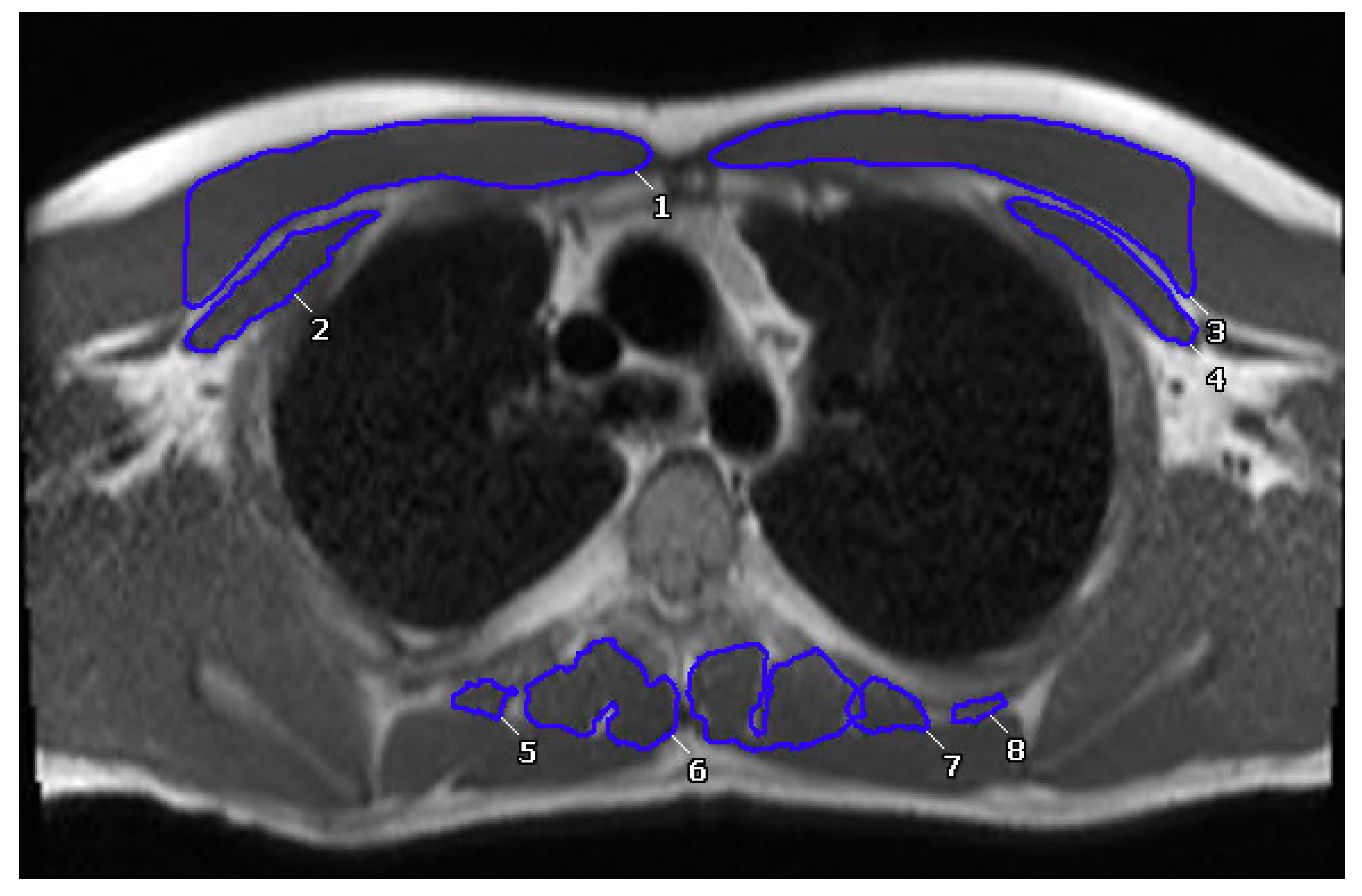
Figure 1. Segmentation of anterior and posterior paraspinal muscles at the level of the carina. Numbers seen on scan signify different hand traced muscles to quantify total muscle area of anterior and posterior muscles.
Jcm 12 02689 g001
Figure 2. Bland Altman plots for inter-observer measurements of (A) anterior and (B) paraspinal muscle areas and intra-observer measurements of (C) anterior and (D) paraspinal muscle areas. Central solid line is representative of the bias, with dotted lines being representative of 95% limits of agreement (A) 1.081 (95% CI, −3.4–5.6), (B) 0.07 (95% CI, −1.6–1.7), (C) 0.40 (95% CI, −3.5–4.4), and (D) 0.10 (95% CI, −1.6–1.7), respectively.
Jcm 12 02689 g002
Figure 3. Correlation between BSA-indexed anterior SMA with (A) peak VO2, (C) VE/VCO2 and BSA-indexed paraspinal SMA with (B) peak VO2 and (D) VE/VCO2. VO2—oxygen consumption; VE/VCO2—minute ventilation/carbon dioxide production; BSA—body surface area; SMA—skeletal muscle area. Pearson correlation testing was performed.
Jcm 12 02689 g003
Figure 4. Significant differences in average peak VO2 and VE/VCO2 in low- and high-muscle groups. Data presented in box and whisker plots. Bars represent upper and lower quartiles. Low-muscle peak VO2 mean and SD is 23.8 ± 4.7 mL/kg/min (1st quartile: 20.7, median: 23.4, 3rd quartile: 27.6). High-muscle peak VO2 mean and SD is 32.2 ± 8.5 mL/kg/min (1st quartile: 25.2, median: 36.1, 3rd quartile: 37). Low-muscle VE/VCO2 mean and SD is 40.2 ± 6.2 (1st quartile: 35, median: 39, 3rd quartile: 45). High-muscle VE/VCO2 mean and SD is 32.9 ± 3.6 (1st quartile: 30.5, median: 33, 3rd quartile 36).
Jcm 12 02689 g004
Table 1. General characteristics.
VariablesNResults
Age (y), mean ± SD7519.1 ± 8.6
Male sex, n (%)7549 (65%)
Muscle characteristics (N = 75)
BSA-indexed anterior muscle (cm2/m2), mean ± SD 20.8 ± 5.7
BSA-indexed paraspinal muscle (cm2/m2), mean ± SD 6.5 ± 1.7
BSA-indexed total skeletal muscle (cm2/m2), mean ± SD 27.3 ± 6.6
Fontan type (N = 75)
Atriopulmonary and other, n (%) 1 (1.3%)
Lateral tunnel, n (%) 23 (31%)
Extracardiac, n (%) 49 (65%)
Unkown, n (%) 2 (2.6%)
Ventricular Morphology (N = 75)
Left, n (%) 35 (47%)
Right, n (%) 28 (37%)
Both, n (%) 12 (16%)
BSA—body surface area.
Table 2. General exercise and CMR data.
VariablesNResults
Exercise parameters
Peak VO2 (mL/kg/min)3827.1 ± 6.6
Peak O2 pulse (mL/beat), mean ± SD199.6 ± 2.7
Peak HR (beats/min), mean ± SD51165.7 ± 25.5
% Predicted peak HR4583.1 ± 12.2
Peak RER431.2 ± 0.1
VE/VCO2 4236.5 ± 6.0
CMR Ventricular Volumetry
EF (%), mean ± SD7549.4 ± 9.6
BSA-indexed SVEDV (mL/m2), mean ± SD75100.6 ± 41.0
BSA-indexed SVESV (mL/m2), mean ± SD7550.7 ± 21.7
BSA-indexed ventricle mass, mean ± SD4143.8 ± 17.2
VO2—oxygen consumption; BSA—body surface area; RER—respiratory exchange ratio; VE—minute ventilation; VCO2—carbon dioxide production; EF—ejection fraction; SVEDV—end diastolic volume; SVESV—end systolic volume.
Table 3. Sex-specific characteristics. T-testing was used for sample sizes >30, for those <30, Mann–Whitney U was used. Effect sizes were calculated by using Cohen’s d test for groups with similar sample sizes or sample sizes > 20. Hedges’ g test was used to calculate effect size for groups with largely different sample sizes or sizes < 20.
VariablesFemaleMalep-Value
Effect Size (95% CI)
Mean ± SD (N)
[1st Quartile, Median, 3rd Quartile]
Mean ± SD (N)
[1st Quartile, Median, 3rd Quartile]
Age (years)17.2 ± 7.0 (20)
[13.3, 15, 21.5]
17.8 ± 7.1 (19)
[12, 17, 22]
0.499
0.085 (−0.39, 0.56)
Sex (N, %)Male: 8 (40%)
Female: 12 (60%)
Male: 18 (95%)
Female: 1 (5%)
0.0003 *
Height (cm)158.5 ± 17.0 (20)
[155.5, 162.9, 168]
154.6 ± 26.1 (19)
[130, 165, 174.4]
0.704
0.28 (−0.20, 0.75)
Weight (kg)65.0 ± 20.0 (20)
[54.4, 68.8, 77.8]
54.7 ± 25.4 (19)
[28, 59.1, 70.4]
0.136
0.24 (−0.24, 0.72)
BMI (kg/m2)25.1 ± 5.3 (20)
[21.1, 25.4, 28]
21.3 ± 4.9 (19)
[17.1, 20.8, 24.9]
0.027 *
0.32 (−0.44, 0.51)
Anterior Wall Muscle Area (cm2)25.8 ± 7.0 (20)
[23, 25.6, 31.4]
41.4 ± 15.5 (19)
[25.2, 42.8, 52.8]
0.002 *
0.86 (0.36, 1.35)
Paraspinal Wall Muscle Area (cm2)8.7 ± 2.0 (20)
[7.7, 9.3, 9.8]
12.1 ± 3.8 (19)
[9.6, 11.9, 14.3]
0.002 *
0.77 (0.28, 1.26)
Peak VO2 (mL/kg/min)23.8 ± 4.7 (12)
[20.7, 23.4, 27.6]
32.2 ± 8.5 (9)
[25.2, 36.1, 37]
0.009 *
0.15 (−0.48, 0.78)
Peak O2 Pulse (mL/beat)8.1 ± 0.9 (5)
[7.3, 8, 8.9]
11.1 ± 3.6 (5)
[8, 12.2, 13.7]
0.144
0.83 (−0.10, 1.82)
Peak HR (beats/min)167.3 ± 21.4 (14)
[154.5, 171, 181]
173.7 ± 12.0 (10)
[163.5, 173, 185.3]
0.519
0.07 (−0.51, 0.65)
% Predicted Peak HR (%)82.8 ± 8.1 (13)
[78.5, 84, 87.5]
88.6 ± 5.2 (8)
[83.5, 88, 94]
0.103
0.02 (−0.61, 0.66)
Peak RER1.1 ± 0.04 (12)
[1.1, 1.1, 1.2]
1.2 ± 0.1 (9)
[1.2, 1.2, 1.3]
0.006 *
0.51 (−0.14, 1.16)
VE/VCO2 40.2 ± 6.2 (11)
[35, 39, 45]
32.9 ± 3.6 (9)
[30.5, 33, 36]
0.006 *
−0.55 (−1.21, 0.12)
BSA—body surface area; VO2—oxygen consumption; VE—minute ventilation; VCO2—carbon dioxide production. * Denotes p-value < 0.05.
Table 4. Anterior and paraspinal muscle correlations.
VariableNIndexed Anterior MuscleIndexed Paraspinal MuscleIndexed Total Muscle
rp-Valuerp-Valuerp-Value
Peak VO2 (mL/kg/min)380.33640.039 *0.43030.007 *0.40230.012 *
Peak O2 Pulse (mL/beat)190.13390.1230.15150.5360.36920.120
Peak HR (beats/min)450.12560.4110.04780.7550.12100.429
% Predicted Peak HR (%)390.19690.230−0.02970.8580.16340.320
Peak RER370.12970.4440.28360.0890.18230.280
VE/VCO2 36−0.20130.239−0.44500.006 *−0.28650.090
EF (%)75−0.08990.443−0.02120.857−0.08270.481
Ventricle Mass (g/m2)410.07430.644−0.19360.2250.01810.911
BSA-Indexed Ventricle Mass410.06230.699−0.17330.2790.00230.988
VO2—oxygen consumption; BSA—body surface area; RER—respiratory exchange ratio; VE—minute ventilation; VCO2—carbon dioxide production; EF—ejection fraction. * Denotes p-value < 0.05.
Table 5. Low and high muscle mass characteristics. Mann–Whitney U was used due to sample sizes < 30. Effect sizes were calculated by using Cohen’s d test for groups with similar sample sizes or sample sizes > 20. Hedges’ g test was used to calculate effect size for groups with largely different sample sizes or sizes < 20.
VariableLow Muscle (N = 20)High Muscle (N = 19)p-Value
Effect Size (95% CI)
Mean ± SD (N)
[1st Quartile, Median, 3rd Quartile]
Mean ± SD (N)
[1st Quartile, Median, 3rd Quartile]
Age (years)17.2 ± 7.0 (20)
[13.3, 15, 21.5]
17.8 ± 7.1 (19)
[12, 17, 22]
0.499
0.10 (−0.53, 0.73)
Sex (N, %)Male: 8 (40%)
Female: 12 (60%)
Male: 18 (95%)
Female: 1 (5%)
0.0003 *
Height (cm)158.5 ± 17.0 (20)
[155.5, 162.9, 168]
154.6 ± 26.1 (19)
[130, 165, 174.4]
0.704
−0.18 (−0.81, 0.45)
Weight (kg)65.0 ± 20.0 (20)
[54.4, 68.8, 77.8]
54.7 ± 25.4 (19)
[28, 59.1, 70.4]
0.136
−0.45 (−1.08, 0.19)
BMI (kg/m2)25.1 ± 5.3 (20)
[21.1, 25.4, 28]
21.3 ± 4.9 (19)
[17.1, 20.8, 24.9]
0.027 *
−0.76 (−1.41, −0.10)
Anterior Wall Muscle Area (cm2)25.8 ± 7.0 (20)
[23, 25.6, 31.4]
41.4 ± 15.5 (19)
[25.2, 42.8, 52.8]
0.002 *
1.31 (0.61, 2.0)
Paraspinal Wall Muscle Area (cm2)8.7 ± 2.0 (20)
[7.7, 9.3, 9.8]
12.1 ± 3.8 (19)
[9.6, 11.9, 14.3]
0.002 *
1.14 (0.46, 1.82)
BSA-Indexed Anterior Muscle (cm2/m2)15.4 ± 2.2 (20)
[13.6, 15.7, 16.2]
27.5 ± 5.8 (19)
[24.2, 26.2, 28.4]
<0.0001 *
2.77 (1.87, 3.65)
BSA-Indexed Paraspinal Muscle (cm2/m2)5.2 ± 0.7 (20)
[5, 5.2, 5.7]
8.2 ± 1.8 (19)
[7, 7.8, 8.7]
<0.0001 *
2.18 (1.37, 2.97)
Exercise parameters:
Peak VO2 (mL/kg/min)23.8 ± 4.7 (12)
[20.7, 23.4, 27.6]
32.2 ± 8.5 (9)
[25.2, 36.1, 37]
0.009 *
1.22 (0.30, 2.13)
Peak O2 Pulse (mL/beat)8.1 ± 0.9 (5)
[7.3, 8, 8.9]
11.1 ± 3.6 (5)
[8, 12.2, 13.7]
0.144
1.03 (−0.22, 2.23)
Peak HR (beats/min)167.3 ± 21.4 (14)
[154.5, 171, 181]
173.7 ± 12.0 (10)
[163.5, 173, 185.3]
0.519
0.34 (−0.45, 1.13)
% Predicted Peak HR (%)82.8 ± 8.1 (13)
[78.5, 84, 87.5]
88.6 ± 5.2 (8)
[83.5, 88, 94]
0.103
0.78 (−0.11, 1.66)
Peak RER1.1 ± 0.04 (12)
[1.1, 1.1, 1.2]
1.2 ± 0.1 (9)
[1.2, 1.2, 1.3]
0.006 *
1.47 (0.50, 2.40)
VE/VCO2 40.2 ± 6.2 (11)
[35, 39, 45]
32.9 ± 3.6 (9)
[30.5, 33, 36]
0.006 *
−1.34 (−2.27, −0.37)
BSA—body surface area; VO2—oxygen consumption; RER—respiratory exchange ratio; VE—minute ventilation; VCO2—carbon dioxide production. * Denotes p-value < 0.05.
Table 6. Multivariate analysis using BSA-indexed paraspinal or anterior muscle area as one of the independent variables. Each multivariable analysis included two variables as independent variables to adjust for sex then age.
VariablesIndexed Paraspinal MuscleIndexed Anterior MuscleIndexed Total Muscle
Parameter Estimate ± SD
(p-Value)
Parameter Estimate ± SD
(p-Value)
Parameter Estimate ± SD
(p-Value)
Age-adjustedPeak VO21.7 ± 0.6 (0.008) *0.4 ± 0.2 (0.043) *0.4 ± 0.2 (0.012) *
VE/VCO2−1.4 ± 0.6 (0.019) *−0.1 ± 0.2 (0.522)−0.1 ± 0.2 (0.522)
Sex-adjustedPeak VO21.5 ± 0.6 (0.012) *0.4 ± 0.2 (0.013) *0.4 ± 0.1 (0.005) *
VE/VCO2−1.6 ± 0.5 (0.004) *−0.06 ± 0.1 (0.616)−0.2 ± 0.1 (0.115)
VO2—oxygen consumption; VE—minute ventilation; VCO2—carbon dioxide production. * Denotes p-value < 0.05.
Disclaimer/Publisher’s Note: The statements, opinions and data contained in all publications are solely those of the individual author(s) and contributor(s) and not of MDPI and/or the editor(s). MDPI and/or the editor(s) disclaim responsibility for any injury to people or property resulting from any ideas, methods, instructions or products referred to in the content.