

Gender Differences in Neuromuscular Control during the Preparation Phase of Single-Leg Landing Task in Badminton

Abstract
:
1. Introduction
2. Materials and Methods
2.1. Subjects
2.2. Prepare for Testing
2.3. Test Procedure
2.4. Data Processing and Analysis
3. Results
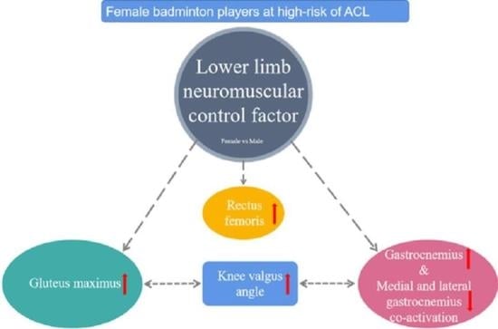
4. Discussion
5. Conclusions
Author Contributions
Funding
Institutional Review Board Statement
Informed Consent Statement
Data Availability Statement
Conflicts of Interest
References
- Fahlstrom, M.; Soderman, K. Decreased shoulder function and pain common in recreational badminton players. Scand. J. Med. Sci. Sports 2007, 17, 246–251. [Google Scholar] [CrossRef] [PubMed]
- Sasaki, S.; Nagano, Y.; Ichikawa, H. Differences in high trunk acceleration during single-leg landing after an overhead stroke between junior and adolescent badminton athletes. Sports Biomech. 2022, 21, 1160–1175. [Google Scholar] [CrossRef] [PubMed]
- Kimura, Y.; Ishibashi, Y.; Tsuda, E.; Yamamoto, Y.; Tsukada, H.; Toh, S. Mechanisms for anterior cruciate ligament injuries in badminton. Br. J. Sports Med. 2010, 44, 1124–1127. [Google Scholar] [CrossRef] [PubMed]
- Takahashi, S.; Okuwaki, T. Epidemiological survey of anterior cruciate ligament injury in Japanese junior high school and high school athletes: Cross-sectional study. Res. Sports Med. 2017, 25, 266–276. [Google Scholar] [CrossRef] [PubMed]
- Webster, K.E.; Hewett, T.E. Anterior Cruciate Ligament Injury and Knee Osteoarthritis: An Umbrella Systematic Review and Meta-analysis. Clin. J. Sport Med. 2022, 32, 145–152. [Google Scholar] [CrossRef]
- Renstrom, P.; Ljungqvist, A.; Arendt, E.; Beynnon, B.; Fukubayashi, T.; Garrett, W.; Georgoulis, T.; Hewett, T.E.; Johnson, R.; Krosshaug, T.; et al. Non-contact ACL injuries in female athletes: An International Olympic Committee current concepts statement. Br. J. Sports Med. 2008, 42, 394–412. [Google Scholar] [CrossRef] [PubMed]
- Cronstrom, A.; Tengman, E.; Hager, C.K. Risk Factors for Contra-Lateral Secondary Anterior Cruciate Ligament Injury: A Systematic Review with Meta-Analysis. Sports Med. 2021, 51, 1419–1438. [Google Scholar] [CrossRef]
- Cesar, G.M.; Pereira, V.S.; Santiago, P.R.P.; Benze, B.G.; da Costa, P.H.L.; Amorim, C.F.; Serrao, F.V. Variations in dynamic knee valgus and gluteus medius onset timing in non-athletic females related to hormonal changes during the menstrual cycle. Knee 2011, 18, 224–230. [Google Scholar] [CrossRef]
- Navacchia, A.; Ueno, R.; Ford, K.R.; DiCesare, C.A.; Myer, G.D.; Hewett, T.E. EMG-Informed Musculoskeletal Modeling to Estimate Realistic Knee Anterior Shear Force During Drop Vertical Jump in Female Athletes. Ann. Biomed. Eng. 2019, 47, 2416–2430. [Google Scholar] [CrossRef]
- Nasseri, A.; Lloyd, D.G.; Bryant, A.L.; Headrick, J.; Sayer, T.A.; Saxby, D.J. Mechanism of Anterior Cruciate Ligament Loading during Dynamic Motor Tasks. Med. Sci. Sports Exerc. 2021, 53, 1235–1244. [Google Scholar] [CrossRef]
- Maniar, N.; Cole, M.H.; Bryant, A.L.; Opar, D.A. Muscle Force Contributions to Anterior Cruciate Ligament Loading. Sports Med. 2022, 52, 1737–1750. [Google Scholar] [CrossRef] [PubMed]
- Beaulieu, M.L.; Lamontagne, M.; Xu, L.Y. Lower limb muscle activity and kinematics of an unanticipated cutting manoeuvre: A gender comparison. Knee Surg. Sports Traumatol. Arthrosc. 2009, 17, 968–976. [Google Scholar] [CrossRef] [PubMed]
- Bencke, J.; Zebis, M.K. The influence of gender on neuromuscular pre-activity during side-cutting. J. Electromyogr. Kinesiol. 2011, 21, 371–375. [Google Scholar] [CrossRef]
- Dwyer, M.K.; Boudreau, S.N.; Mattacola, C.G.; Uhl, T.L.; Lattermann, C. Comparison of Lower Extremity Kinematics and Hip Muscle Activation during Rehabilitation Tasks between Sexes. J. Athl. Train. 2010, 45, 181–190. [Google Scholar] [CrossRef]
- Landry, S.C.; McKean, K.A.; Hubley-Kozey, C.L.; Stanish, W.D.; Deluzio, K.J. Neuromuscular and lower limb biomechanical differences exist between male and female elite adolescent soccer players during an unanticipated side-cut maneuver. Am. J. Sports Med. 2007, 35, 1888–1900. [Google Scholar] [CrossRef] [PubMed]
- Llurda-Almuzara, L.; Perez-Bellmunt, A.; Labata-Lezaun, N.; Lopez-de-Celis, C.; Canet-Vintro, M.; Cadellans-Arroniz, A.; Moure-Romero, L.; Aiguade-Aiguade, R. Relationship between lower limb EMG activity and knee frontal plane projection angle during a single-legged drop jump. Phys. Ther. Sport 2021, 52, 13–20. [Google Scholar] [CrossRef]
- Zahradnik, D.; Jandacka, D.; Farana, R.; Uchytil, J.; Hamill, J. Identification of types of landings after blocking in volleyball associated with risk of ACL injury. Eur. J. Sport Sci. 2017, 17, 241–248. [Google Scholar] [CrossRef]
- Kimura, Y.; Ishibashi, Y.; Tsuda, E.; Yamamoto, Y.; Hayashi, Y.; Sato, S. Increased knee valgus alignment and moment during single-leg landing after overhead stroke as a potential risk factor of anterior cruciate ligament injury in badminton. Br. J. Sports Med. 2012, 46, 207–213. [Google Scholar] [CrossRef]
- Leardini, A.; Sawacha, Z.; Paolini, G.; Ingrosso, S.; Nativo, R.; Benedetti, M.G. A new anatomically based protocol for gait analysis in children. Gait Posture 2007, 26, 560–571. [Google Scholar] [CrossRef]
- Portinaro, N.; Leardini, A.; Panou, A.; Monzani, V.; Caravaggi, P. Modifying the Rizzoli foot model to improve the diagnosis of pes-planus: Application to kinematics of feet in teenagers. J. Foot Ankle Res. 2014, 7, 754. [Google Scholar] [CrossRef]
- Barbero, M.; Merletti, R.; Rainoldi, A. Atlas of Muscle Innervation Zones: Understanding Surface Electromyography and Its Applications; Springer: Milan, Italy; New York, NY, USA, 2012. [Google Scholar]
- de Britto, M.A.; Carpes, F.P.; Koutras, G.; Pappas, E. Quadriceps and hamstrings prelanding myoelectric activity during landing from different heights among male and female athletes. J. Electromyogr. Kinesiol. 2014, 24, 508–512. [Google Scholar] [CrossRef] [PubMed]
- Hu, Z.; Kim, Y.; Zhang, Y.A.; Zhang, Y.X.; Li, J.Y.; Tang, X.; Sohn, J.; Kim, S. Correlation of Lower Limb Muscle Activity with Knee Joint Kinematics and Kinetics during Badminton Landing Tasks. Int. J. Environ. Res. Public Health 2022, 19, 16587. [Google Scholar] [CrossRef] [PubMed]
- Ford, K.R.; Myer, G.D.; Schmitt, L.C.; Uhl, T.L.; Hewett, T.E. Preferential Quadriceps Activation in Female Athletes with Incremental Increases in Landing Intensity. J. Appl. Biomech. 2011, 27, 215–222. [Google Scholar] [CrossRef]
- Kipp, K.; Pfeiffer, R.; Sabick, M.; Harris, C.; Sutter, J.; Kuhlman, S.; Shea, K. Muscle Synergies During a Single-Leg Drop-Landing in Boys and Girls. J. Appl. Biomech. 2014, 30, 262–268. [Google Scholar] [CrossRef] [PubMed]
- Boyer, A.; Hug, F.; Avrillon, S.; Lacourpaille, L. Individual differences in the distribution of activation among the hamstring muscle heads during stiff-leg Deadlift and Nordic hamstring exercises. J. Sports Sci. 2021, 39, 1830–1837. [Google Scholar] [CrossRef]
- Neamatallah, Z.; Herrington, L.; Jones, R. An investigation into the role of gluteal muscle strength and EMG activity in controlling HIP and knee motion during landing tasks. Phys. Ther. Sport 2020, 43, 230–235. [Google Scholar] [CrossRef]
- Dix, J.; Marsh, S.; Dingenen, B.; Malliaras, P. The relationship between hip muscle strength and dynamic knee valgus in asymptomatic females: A systematic review. Phys. Ther. Sport 2019, 37, 197–209. [Google Scholar] [CrossRef]
- Hollman, J.H.; Hohl, J.M.; Kraft, J.L.; Strauss, J.D.; Traver, K.J. Modulation of Frontal-Plane Knee Kinematics by Hip-Extensor Strength and Gluteus Maximus Recruitment During a Jump-Landing Task in Healthy Women. J. Sport Rehabil. 2013, 22, 184–190. [Google Scholar] [CrossRef]
- Khayambashi, K.; Ghoddosi, N.; Straub, R.K.; Powers, C.M. Hip Muscle Strength Predicts Noncontact Anterior Cruciate Ligament Injury in Male and Female Athletes: A Prospective Study. Am. J. Sports Med. 2016, 44, 355–361. [Google Scholar] [CrossRef]
- Hogg, J.A.; Ackerman, T.; Nguyen, A.D.; Ross, S.E.; Schmitz, R.J.; Vanrenterghem, J.; Shultz, S.J. The Effects of Gluteal Strength and Activation on the Relationship Between Femoral Alignment and Functional Valgus Collapse During a Single-Leg Landing. J. Sport Rehabil. 2021, 30, 942–951. [Google Scholar] [CrossRef]
- Nguyen, A.D.; Shultz, S.J.; Schmitz, R.J.; Luecht, R.M.; Perrin, D.E. A Preliminary Multifactorial Approach Describing the Relationships Among Lower Extremity Alignment, Hip Muscle Activation, and Lower Extremity Joint Excursion. J. Athl. Train. 2011, 46, 246–256. [Google Scholar] [CrossRef] [PubMed]
- Hollman, J.H.; Galardi, C.M.; Lin, I.H.; Voth, B.C.; Whitmarsh, C.L. Frontal and transverse plane hip kinematics and gluteus maximus recruitment correlate with frontal plane knee kinematics during single-leg squat tests in women. Clin. Biomech. 2014, 29, 468–474. [Google Scholar] [CrossRef] [PubMed]
- Benjaminse, A.; Webster, K.E.; Kimp, A.; Meijer, M.; Gokeler, A. Revised Approach to the Role of Fatigue in Anterior Cruciate Ligament Injury Prevention: A Systematic Review with Meta-Analyses. Sports Med. 2019, 49, 565–586. [Google Scholar] [CrossRef] [PubMed]
- Nunley, R.M.; Wright, D.; Renner, J.B.; Yu, B.; Garrett, W.E. Gender Comparison of Patellar Tendon Tibial Shaft Angle with Weight Bearing. Res. Sports Med. 2006, 11, 173–185. [Google Scholar] [CrossRef]
- Li, G.; Defrate, L.E.; Rubash, H.E.; Gill, T.J. In vivo kinematics of the ACL during weight-bearing knee flexion. J. Orthop. Res. 2005, 23, 340–344. [Google Scholar] [CrossRef]
- Chappell, J.D.; Creighton, R.A.; Giuliani, C.; Yu, B.; Garrett, W.E. Kinematics and electromyography of landing preparation in vertical stop-jump: Risks for noncontact anterior cruciate ligament injury. Am. J. Sports Med. 2007, 35, 235–241. [Google Scholar] [CrossRef]
- Zazulak, B.T.; Ponce, P.L.; Straub, S.J.; Medvecky, M.J.; Avedisian, L.; Hewett, T.E. Gender comparison of hip muscle activity during single-leg landing. J. Orthop. Sports Phys. Ther. 2005, 35, 292–299. [Google Scholar] [CrossRef]
- Teng, H.L.; Powers, C.M. Hip-Extensor Strength, Trunk Posture, and Use of the Knee-Extensor Muscles During Running. J. Athl. Train. 2016, 51, 519–524. [Google Scholar] [CrossRef]
- Malfait, B.; Dingenen, B.; Smeets, A.; Staes, F.; Pataky, T.; Robinson, M.A.; Vanrenterghem, J.; Verschueren, S. Knee and Hip Joint Kinematics Predict Quadriceps and Hamstrings Neuromuscular Activation Patterns in Drop Jump Landings. PLoS ONE 2016, 11, e0153737. [Google Scholar] [CrossRef]
- Nagano, Y.; Ida, H.; Akai, M.; Fukubayashi, T. Gender differences in knee kinematics and muscle activity during single limb drop landing. Knee 2007, 14, 218–223. [Google Scholar] [CrossRef]
- Adouni, M.; Shirazi-Adl, A.; Marouane, H. Role of gastrocnemius activation in knee joint biomechanics: Gastrocnemius acts as an ACL antagonist. Comput. Methods Biomech. Biomed. Eng. 2016, 19, 376–385. [Google Scholar] [CrossRef] [PubMed]
- Teng, P.S.P.; Leong, K.F.; Phua, P.Y.X.; Kong, P.W. An exploratory study of the use of ultrasound in the measurement of anterior tibial translation under gastrocnemius muscle stimulation. Res. Sports Med. 2021, 29, 103–115. [Google Scholar] [CrossRef] [PubMed]
- Ali, N.; Andersen, M.S.; Rasmussen, J.; Robertson, D.G.E.; Rouhi, G. The application of musculoskeletal modeling to investigate gender bias in non-contact ACL injury rate during single-leg landings. Comput. Methods Biomech. Biomed. Eng. 2014, 17, 1602–1616. [Google Scholar] [CrossRef] [PubMed]
- Morgan, K.D.; Donnelly, C.J.; Reinbolt, J.A. Elevated gastrocnemius forces compensate for decreased hamstrings forces during the weight-acceptance phase of single-leg jump landing: Implications for anterior cruciate ligament injury risk. J. Biomech. 2014, 47, 3295–3302. [Google Scholar] [CrossRef]
- Buchanan, T.S.; Lloyd, D.G. Muscle activation at the human knee during isometric flexion-extension and varus-valgus loads. J. Orthop. Res. 1997, 15, 11–17. [Google Scholar] [CrossRef]
- Besier, T.F.; Lloyd, D.G.; Ackland, T.R. Muscle activation strategies at the knee during running and cutting maneuvers. Med. Sci. Sports Exerc. 2003, 35, 119–127. [Google Scholar] [CrossRef]
- Fonseca, M.; Gasparutto, X.; Leboeuf, F.; Dumas, R.; Armand, S. Impact of knee marker misplacement on gait kinematics of children with cerebral palsy using the Conventional Gait Model—A sensitivity study. PLoS ONE 2020, 15, e0232064. [Google Scholar] [CrossRef]
- Rota, S.; Rogowski, I.; Champely, S.; Hautier, C. Reliability of EMG normalisation methods for upper-limb muscles. J. Sports Sci. 2013, 31, 1696–1704. [Google Scholar] [CrossRef]


| Muscle | Electrode Site | Position | MVC Test Maneuver |
|---|---|---|---|
| Gluteus maxim (GMAX) | Lateral 80% of the line between the midpoint of the sacrum and the greater trochanter of the femur. | Prone | The stretch strap is fixed to the posterior side of the distal thigh and hip extension is performed with the knee in 90 degrees of flexion. |
| Gluteus medius (GMED) | Upper 20% of the line between the greater trochanter of the femur and the highest point of the iliac spine. | Lateral prone | The stretch strap is fixed to the lateral side of the distal thigh and hip abduction is performed with the knee flexed at 90 degrees. |
| Rectus femoris (RF) | Upper 40% of the line between the superior patella and the anterior superior iliac spine. | Sitting | Knee flexion at 90 degrees, the stretch strap is fixed on the anterior side of the distal lower leg, perform knee extension. |
| Medial hamstrings (MH) | Lower 80% of the line of the ischial tuberosity with the medial popliteal crease. | Prone | Knee flexion 45 degrees, the stretch strap is fixed on the back of the distal lower leg, internal rotation of the lower leg and perform knee flexion. |
| Lateral hamstrings (LH) | Lower 80% of the line of the ischial tuberosity to the lateral popliteal crease. | Prone | Knee flexion 45 degrees, the stretch strap is fixed on the back of the distal lower leg, external rotation of the lower leg and perform knee flexion. |
| Medial gastrocnemius (MG) | Upper 85% of the medial Achilles tendon in line with the medial popliteal crease | Prone | Knee extension, stretch strap fixed on the forefoot, internal rotation of the lower leg and perform plantarflexion. |
| Lateral gastrocnemius (LG) | Upper 85% of the line connecting the lateral Achilles tendon to the lateral popliteal crease | Prone | Knee extension, stretch strap fixed on the forefoot, external rotation of the lower leg and perform plantarflexion. |
| Variables | Male | Female | F | p-Value |
|---|---|---|---|---|
| Hip | ||||
| Flexion (+)/extension (−) | 11.26 ± 8.31 | 11.83 ± 8.60 | 0.018 | 0.8951 |
| Abduction (+)/adduction (−) | 36.33 ± 4.37 | 39.86 ± 5.59 | 1.981 | 0.1811 |
| External (+)/internal rotation (−) | 15.84 ± 5.32 | 17.94 ± 13.87 | 0.160 | 0.6950 |
| Knee | ||||
| Flexion (+)//extension (−) | 19.47 ± 6.42 | 18.08 ± 7.33 | 0.163 | 0.6924 |
| Valgus (+)//varus (−) | 1.72 ± 3.20 | 6.27 ± 2.75 | 9.284 | 0.0087 * |
| External (+)/internal rotation (−) | −4.39 ± 4.67 | 0.15 ± 4.29 | 4.105 | 0.0622 |
| Ankle | ||||
| Dorsiflexion (+)/plantar flexion (−) | −39.92 ± 9.25 | −33.39 ± 6.45 | 2.675 | 0.1242 |
| Eversion (+)/inversion (−) | 0.47 ± 3.45 | −0.28 ± 4.32 | 0.058 | 0.8141 |
| Abduction (+)/adduction (−) | 16.98 ± 4.63 | 23.16 ± 6.75 | 4.395 | 0.0562 |
Disclaimer/Publisher’s Note: The statements, opinions and data contained in all publications are solely those of the individual author(s) and contributor(s) and not of MDPI and/or the editor(s). MDPI and/or the editor(s) disclaim responsibility for any injury to people or property resulting from any ideas, methods, instructions or products referred to in the content. |
© 2023 by the authors. Licensee MDPI, Basel, Switzerland. This article is an open access article distributed under the terms and conditions of the Creative Commons Attribution (CC BY) license (https://creativecommons.org/licenses/by/4.0/).
Share and Cite
Hu, Z.; Zhang, Y.; Dong, T.; Dong, M.; Kim, S.; Kim, Y. Gender Differences in Neuromuscular Control during the Preparation Phase of Single-Leg Landing Task in Badminton. J. Clin. Med. 2023, 12, 3296. https://doi.org/10.3390/jcm12093296
Hu Z, Zhang Y, Dong T, Dong M, Kim S, Kim Y. Gender Differences in Neuromuscular Control during the Preparation Phase of Single-Leg Landing Task in Badminton. Journal of Clinical Medicine. 2023; 12(9):3296. https://doi.org/10.3390/jcm12093296
Chicago/Turabian StyleHu, Zhe, Yanan Zhang, Tengfei Dong, Maolin Dong, Sukwon Kim, and Youngsuk Kim. 2023. "Gender Differences in Neuromuscular Control during the Preparation Phase of Single-Leg Landing Task in Badminton" Journal of Clinical Medicine 12, no. 9: 3296. https://doi.org/10.3390/jcm12093296